COMPARATIVE EVALUATION OF EFFECT OF TOOTH
BRUSHING ON THE SURFACE ROUGHNESS AND COLOUR
STABILITY OF POLY(METHYL METHACRYLATE) HEAT
CURE DENTURE BASE RESIN WITH THE APPLICATION
OF DENTURE SURFACE SEALANT - AN IN VITRO STUDY
Dissertation Submitted to
THE TAMILNADU Dr. M.G.R. MEDICAL UNIVERSITY
In partial fulfillment for the Degree of
MASTER OF DENTAL SURGERY
BRANCH I
PROSTHODONTICS AND CROWN & BRIDGE
ACKNOWLEDGEMENT
Its my gratitude that gives fulfillment for my study to thank experts and my well wishers who supported throughout my thesis work. I would like to express my respect and gratitude to Professor Dr. N.S. Azhagarasan M.D.S., Principal, Professor and Head, Department of Prosthodontics & Crown and Bridge for encouraging me to do study on this topic and his continuous support and effort for my completion by letting me to make use of amenities available in the college.
I would like to pay my indebted gratefulness to my Guide
Dr. Jayakrishna Kumar M.D.S., Professor, Department of Prosthodontics & Crown and Bridge who is very supportive in bringing out the best in me. I am very thankful to him to complete this study under his guidance. He gave inspiration, immense support and motivation to learn new things every day and enlighted me with his suggestions.
My note of thanks will be incomplete without mentioning my
inspiration, my beloved teachers, Dr. Chitra Shankar M.D.S., Dr. R. Hariharan, M.D.S., Dr. M. Saravana Kumar, Dr. R. Hariharan
I would like to thank Dr. Ravanan, Principal, Presidency College
for his support in statistical works. I would like to thank Mr. Mohan Raj, Technician, CIPET, Guindy for his help in carrying out my study in cipet regarding spectrophotometer. I would like to thank Dr. Kamalan Kirubakaran, Satyabama university for his help in carrying out my study regarding Profilometer. I would also like to thank welder Mr. Balu for customizing the mold.
My work would not be possible without the support of my family. I would like to thank my parents, my father Mr. Mohmmad Hallajmoghaddam for being my back bone and my mother Mrs. Maryam Torshizi for her dedication and constant support. Most special thanks
and gratitude to my brothers, Mr. Soroush Hallajmoghaddam and Mr. Samer Hallajmoghaddam for their constant source of
encouragement and love. They have been selfless in giving me the best of everything.
My heartfelt thanks to my batchmates, Dr. Asish, Dr. Arjun, Dr.Jensy, Dr. Maniamuthu, Dr.Manimala for their support in my work, and also during these three years , I deeply appreciate it.
I am indebted to my Seniors and Juniors Dr. Priyadharshini,
Dr. Janani, Dr. Ashwini Sukanya, Dr. Gayathree, Dr. Abinaya, Dr. Sethu Raman, Dr. Surabhi, Dr. Kavya, Dr. Shivasankari,
Dr.Vaishali, Dr. Tejaswi, Dr. Reji, and Dr. Rachel for their immense help.
My heartfelt thanks to my friends Dr. Iyyappa Raj, Dr. Ali, Dr. Manoj, Dr. Maryam, and Mr. Jothi for their constant support
which made my work easier throughout three years.
I extend my thanks to the non teaching staffs, Mrs Sivamani, Mrs Gayathri, for their timely help during the tenure.
CONTENTS
S.NO
TITLE
PAGE NO
1
INTRODUCTION
1
2
REVIEW OF LITERATURE
8
3
MATERIALS AND METHODS
28
4
RESULTS
43
5
DISCUSSION
60
6
CONCLUSION
68
7
SUMMARY
71
LIST OF TABLES
S. NO TITLE PAGE
NO
1 Basic data for evaluation of surface roughness of the uncoated PMMA Heat cure denture base resin samples before brushing – Group I
46
2 Basic data for evaluation of colour stability of the uncoated PMMA Heat cure denture base resin samples before brushing – Group I
47
3 Basic data for evaluation of surface roughness of the PMMA Heat cure denture base samples coated with Denture surface sealant before brushing – Group II
48
4 Basic data for evaluation of colour stability of the PMMA Heat cure denture base resin samples coated with Denture surface sealant before brushing – Group II
49
5 Basic data for evaluation of surface roughness of the uncoated PMMA Heat cure denture base resin samples samples after brushing – Group I
50
6 Basic data for evaluation of colour stability of the uncoated PMMA Heat cure denture base resin samples after brushing – Group I
51
7 Basic data for evaluation of surface roughness of the PMMA Heat cure denture base resin samples coated with Denture surface sealant after brushing – Group II
8 Basic data for evaluation of colour stability of the PMMA Heat cure denture base resin samples coated with Denture surface sealant after brushing – Group II
53
9 Comparative evaluation of surface roughness of uncoated (Group I) and coated (Group II) PMMA Heat cure denture base resin samples before brushing using Independent ‘t’ test
54
10 Comparative evaluation of surface roughness of uncoated (Group I) and coated (Group II) PMMA Heat cure denture base resin samples after brushing using Independent ‘t’ test
55
11 Comparative evaluation of surface roughness of uncoated PMMA Heat cure denture base resin samples before and after brushing using Paired ‘t’ test.- Group I
56
12 Comparative evaluation of surface roughness of coated PMMA Heat cure denture base resin samples before and after brushing using Paired ‘t’ test.- Group II
57
13 Comparative evaluation of colour stability of uncoated (Group I) and coated (Group II) PMMA Heat cure
denture base resin samples before and after brushing
58
14 Statistical comparison of colour stability of uncoated (Group I) and coated (Group II) PMMA Heat cure denture base resin samples before and after brushing using Independent ‘t’ test
ANNEXURE I
METHODOLOGY OVERVIEW
ANNEXURE II
LIST OF FIGURES
Fig. No.
TITLE
Fig.1
Modelling wax
Fig.2
Plaster of Paris
Fig.3
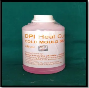
Cold mould seal
Fig. 4
Polyethylene sheets
Fig. 5
PMMA Heat cure denture base resin
Fig.6
Sand paper
Fig.7
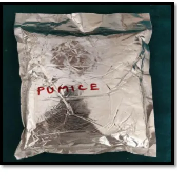
Pumice
Fig.8
Denture surface sealant
Fig.9
Metal scale
Fig.10
Wax carver
Fig.11
Wax knife
Fig. 12
Kidney tray
Fig.13
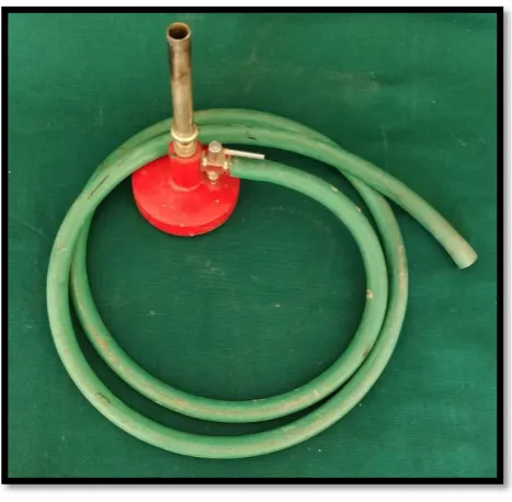
Bunsen burner
Fig 15
Graduated cylinder
Fig.16
Weighing balance
Fig.17
Camel hair brush
Fig.18
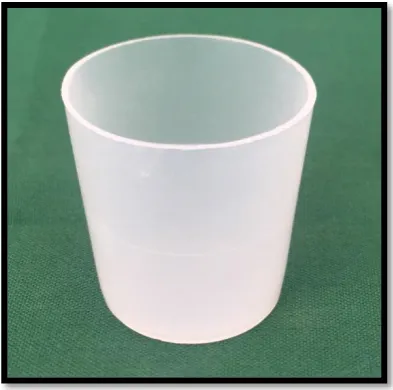
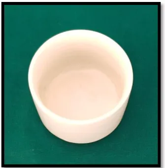
Porcelain mixing jar
Fig.19
Flask and clamp
Fig.20
Acrylic polishing kit
Fig. 21
Sand paper mandrel
Fig 22
Rag wheel
Fig 23
Mixing bowl and spatula
Fig 24
Applicator stick
Fig.25
Vacuum mixer
Fig.26
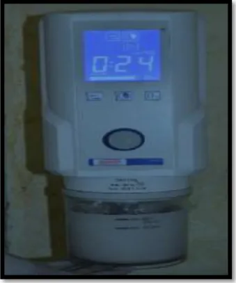
Acrliser
Fig.27
Laboratory lathe
Fig.28
Laboratory micromotor and handpiece
Fig.29
Hair dryer
Fig.30
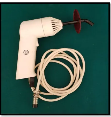
Light curing unit
Fig.31
Customised tooth brush holder
Fig.32
Electronic tooth brush
Fig.34
Spectrophotometer
Fig.35
Fabrication of custom made disc shaped metal
mold
Fig.36
Preparation of wax pattern
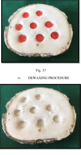
Fig.37
Investment of wax pattern
Fig.38
Dewaxing procedure
Fig.39
Acrylic samples
Fig.40
Finishing and polishing of samples
Fig.41
a)
Application of denture surface sealant
b)
Curing of sealant
Fig.42
Grouping of sample
Fig.43
Evaluation of surface roughness before brushing
(Group I and Group II)
Fig.44
Evaluation of colour stability before brushing
(Group I and Group II)
Fig.45
Brushing procedure for test samples
Fig.46
Evaluation of surface roughness after brushing
(Group I and Group II)
Fig.47
Evaluation of colour stability after brushing
ANNEXURE III
LIST OF GRAPHS
GRAPH
NO.
TITLE
1 Basic data for evaluation of surface roughness of the uncoated PMMA Heat cure denture base resin samples before brushing – Group I
2 Basic data for evaluation of colour stability of the uncoated PMMA Heat cure denture base resin samples before brushing – Group I
3 Basic data for evaluation of surface roughness of the PMMA Heat cure denture base samples coated with Denture surface sealant before brushing – Group II
4 Basic data for evaluation of colour stability of the PMMA Heat cure denture base resin samples coated with Denture surface sealant before brushing – Group II
5 Basic data for evaluation of surface roughness of the uncoated PMMA Heat cure denture base resin samples samples after brushing – Group I
6 Basic data for evaluation of colour stability of the uncoated PMMA Heat cure denture base resin samples after brushing – Group I
8 Basic data for evaluation of colour stability of the PMMA Heat cure denture base resin samples coated with Denture surface sealant after brushing – Group II
9 Comparative evaluation of surface roughness of uncoated (Group I) and coated (Group II) PMMA Heat cure denture base resin samples before brushing using Independent ‘t’ test
10 Comparative evaluation of surface roughness of uncoated (Group I) and coated (Group II) PMMA Heat cure denture base resin samples after brushing using Independent ‘t’ test
11 Comparative evaluation of surface roughness of uncoated PMMA Heat cure denture base resin samples before and after brushing using Paired ‘t’ test.- Group I
12 Comparative evaluation of surface roughness of coated PMMA Heat cure denture base resin samples before and after brushing using Paired ‘t’ test.- Group II
13 Comparative evaluation of colour stability of uncoated (Group I) and coated (Group II) PMMA Heat cure denture base resin samples before and after brushing 14 Statistical comparison of colour stability of uncoated
ANNEXURE IV
1
INTRODUCTION
Poly (methyl methacrylate) denture base resins have been considered traditional material of choice in the fabrication of removable denture prosthesis over decades mainly due to its ease of manipulation, low cost and acceptable aesthetics. However, it’s major limitation such as presence of residual monomer content, poor mechanical properties and dimensional
instability have prompted researchers to seek for newer denture base materials. 8,28,69
Alternatives to PMMA denture base resins have been introduced such as polyamide and butadiene PMMA co-polymer which have lower flexural strength and flexural modulus making it semi flexible.84 Main advantage of polyamide denture base material is its optical properties, which reflects the colour of the base tissue mimicking the natural appearance.54,84 Lower melting temperature makes polyamide, difficult to finish and polish, producing higher surface roughness when compared to PMMA, both before and after polishing.1 Irrespective of the denture base resins used, the presence of surface irregularities and microscopic voids on the restorative material/ restoration significantly affects the properties of the material and thereby limiting its durability. The surfaces of the acrylic dentures, generally acts as a nidus for the biofilm formation and colonisation of microorganisms.3,48,68
Surface roughness and surface free energy are important prognostic
2
Rough external surface of dental restoration facilitates plaque adhesion and subsequent aggregation of microorganism and debris, leading to tooth loss due to caries, periodontal disease and also denture stomatitis.11,44,64,68,73
Denture hygiene instructions are usually provided to the patients which involves both mechanical and chemical methods.13,55 The commonly used method is the use of a denture cleansing brush and an abrasive slurry, but this method is found to be ineffective due to patient’s lack of motivation. 4,59,66,85 Many a times, the use of brush and abrasive paste leaves scratch marks on the surface of the denture which makes it further prone for collection of debris, stains and tartar.4,18
The parameter that is mostly used to quantify surface topography is surface roughness average ( Ra ). An in vivo study by Bollen et al, have
established that increase in Ra value beyond 0.2μm significantly leads to
biofilm formation.10,11,50,64,69 Many studies have shown that denture brushing causes increase in surface roughness in acrylic resins.
3
Colour stability is an important factor for successful clinical service of a restoration.6,14,47 The discolouration of dental polymeric materials is directly related to several intrinsic and extrinsic factors such as polymerisation methods, chemical specification, residual monomer content, water sorption, surface roughness and beverages like tea and coffee.15,23,79 Plaque formation and discolouration can be reduced by polishing the denture surface and the teeth.
The finishing and polishing of dental prosthesis are essential steps in removable prosthodontics that enhances aesthetics and the clinical durability of dentures. It is normally carried out in the laboratory with Al2O3 paste and
liquid. Mechanical polishing is considered to be superior, but it involves sequential steps and is time consuming.4,44
To overcome the limitation of mechanical polishing, an alternative polishing technique was suggested wherein, the prosthesis is immersed in a heated methyl-methacrylate monomer bath ( 75º c) for 10s.4,12,66 The superiority of this technique is that it eliminates the conventional polishing technique.
4
roughness and colour stability.15,22,68 However, coating of surface sealant agents can lead to situations like low resistance to abrasion, weak bonding to the underlying resin material and poor wetting of the surface due to its high viscous nature.
Quantitative analysis of colour coordinates using CIE lab colour system with spectrophotometer have been universally employed in the dental fraternity and is regarded as standard in evaluating the colour parameters.15,22,37,68,69 Different ΔE values are used to compare differences in colour. ΔE values ≤ 1is considered undetectable by the human eye. Various studies have used different ΔE values as clinically acceptable. Perceptibility threshold was set at Δ E ≤ 1.30 units and clinical acceptability threshold was set at ΔE ≤ 2.25 units.
Roughness parameters can be analysed using either 2D /3D surface profilometer.15,41,68,69 Various measurements of surface roughness can be obtained with Ra, which is the arithmetic mean height measurement which is
commonly used universal roughness parameter. Profilometers can be used to compare the same surface in different times and different levels of wear and are classified as contact or non-contact. In the present study, 2D contact surface profilometry was employed to evaluate the surface topography of PMMA denture base resin. The instrument contains a stylus which physically contacts the surface of the samples, while recording its texture.
5
modelling required. Contacting the surface is often an advantage in contaminated surfaces where non-contact methods (optical methods) can end up measuring surface contaminants instead of the surface itself. Because the stylus is in contact with the surface, this method is not sensitive to surface reflectance or colour.9
Earlier studies have evaluated the sealing efficiency of several sealant agents on different restorative materials.15,68,69 Studies evaluating the surface topography and optical properties of PMMA denture base resins following application of sealant agents are available. Studies pertaining to the effect of surface sealant on acrylic resins following mechanical denture cleansing are very few.72
Since, the surface topography and colour stability are important requisites with regards to patient’s expectation, the introduction of surface glazing agents have proved to be beneficial in optimising the surface texture and colour stability. However, in vitro studies comparing the effect of surface sealant agent over PMMA denture base resins are very limited.
6
THE OBJECTIVES OF THIS PRESENT STUDY ARE:
1. To evaluate the surface roughness of uncoated PMMA Heat cure denture base resin samples before brushing using 2D surface profilometer.
2. To evaluate the colour stability of uncoated PMMA Heat cure denture base resin samples before brushing using spectrophotometer.
3. To evaluate the surface roughness of PMMA Heat cure denture base resin samples coated with Denture surface sealant before brushing using 2D surface profilometer.
4. To evaluate the colour stability of PMMA Heat cure denture base resin samples coated with Denture surface sealant before brushing using spectrophotometer.
5. To evaluate the surface roughness of uncoated PMMA Heat cure denture base resin samples after brushing using 2D surface profilometer.
6. To evaluate the colour stability of uncoated PMMA Heat cure denture base resin samples after brushing using spectrophotometer.
7. To evaluate the surface roughness of PMMA Heat cure denture base resin samples coated with Denture surface sealant after brushing using 2D surface profilometer.
7
9. To compare and evaluate the mean surface roughness of uncoated and coated PMMA Heat cure denture base resin samples before brushing. 10.To compare and evaluate the mean surface roughness of uncoated
coated PMMA Heat cure denture base resin samples after brushing. 11.To compare and evaluate the mean surface roughness of uncoated
PMMA Heat cure denture base resin samples before and after brushing.
12.To compare and evaluate the mean surface roughness of coated PMMA Heat cure denture base resin samples before and after brushing 13.To evaluate the colour difference () of uncoated and coated PMMA
Heat cure denture base resin samples before and after brushing.
8
REVIEW OF LITERATURE
Kuhar and Funduk44 (2005) compared the effects of 4 chairside
polishing kits and 2 conventional laboratory techniques used for polishing 3 different acrylic denture base resins. 54 specimens per acrylic material was
studied before and after cutting with a tungsten carbide bur and during and after chair side polishing with 4 polishing kits. Polished acrylic resin surfaces were evaluated under scanning electron microscope (SEM). They concluded that conventional lathe laboratory polishing and chair side silicon polishing kits produced a smoother surface of acrylic resin.
Oliveira L V et al 58 (2006) studied the effect of denture cleansers on hardness of resilient liner materials. Twenty specimens of three resilient liners, measuring 25X15X3mm and two denture cleanser materials were prepared.
Two denture cleansing approaches were used: 1) alkaline hypochlorite, for 20 minutes; 2) alkaline peroxide, for 30 minutes. This procedure was repeated
8 times a day, during 90 days. The specimens were evaluated before and after 360 and 720 cycles, to simulate 1 and 2 years of clinical cleaning procedures. The Shore A hardness was evaluated in a durometer and they concluded that denture cleansers had no effect on hardness of the resilient denture liners evaluated after 2 years of in vivo simulated conditions of hygiene.
9
concentration of FMA substituted into monomer and determined for surface roughness and flexural strength. They concluded that addition of FMA into denture base resin may lower the flexural strength and modulus of elasticity, regardless of tested concentration.
M.Q.Al-Rifaiy4 (2009) studied the effect of mechanical and chemical polishing techniques on the surface roughness of denture base acrylic resins (heat cure and auto cure). A total of sixty acrylic resin specimens were made, of which thirty received mechanical conventional lathe polishing and other thirty received chemical polishing by immersing in methyl-methacrylate monomer and analysed for surface roughness. No significant difference between heat and auto cured acrylic resin on mechanical or chemical polishing was established.
Cruz PC et al19 (2010) evaluated the effectiveness of chemical denture cleansers and ultrasonic device in biofilm removal from complete dentures. Eighty complete denture wearers were participated in the experiment and were divided into four groups. The Kruskal- Wallis test and Dunn multiple comparison test were used to compare the effectiveness between the groups and was concluded that all experimental methods were equally effective regarding the ability to remove biofilm and were superior than brushing with water.
Machado AL et al 51 (2010) analysed the changes in the roughness of
10
and divided into three groups. Roughness measurements were made after polymerization and after 1, 3, and 28 days and analysed by student’s t-test. It was concluded that microwave disinfection caused severe alterations on the surface of denture base and reline materials.
Abuzar M et al 1 (2010) compared the surface roughness of polyamide
denture base material with poly methyl methacrylate denture base material. A total of twenty samples were fabricated of polyamide and poly methyl methcrylate material, in which half was polished by conventional method (lathe with pumice) and other half of test samples were left unpolished. A profilometer was used to measure surface roughness along 3 tracks on each surface before and after polishing. Two way ANOVA was used to compare between two materials and was concluded that polyamide produces a clinically acceptable smoothness after conventional polishing.
H.E. Lee et al 49 (2011) evaluated the effect of different denture cleaning methods to remove candida albicans from acrylic resin denture base material. One hundred and forty identical acrylic resin specimen were fabricated and soaked in different suspension of candida albicans. Six cleaning methods were evaluated and stated that there was no significant difference among the effectiveness levels of cleaning. All denture cleaning techniques were equally efficient in reducing candida.
11
groups, with and without relining. Specimens were subjected to sodium hypochlorite and microwave disinfection and evaluated that specimens that underwent microwave disinfection showed changes in the dimensional stability. Significant shrinkage in the denture bases were observed.
Pahuja RK et al60 (2013) evaluated the effect of denture cleansers on surface hardness of resilient denture liners at various time intervals. 120 cylindrical specimens were made in a custom made metal mold and were stored in artificial saliva throughout the study and was divided into three groups. Forty specimens were cleansed daily in 0.5% sodium hypochlorite solution; forty were cleansed in sodium perborate and remaining forty specimens were daily rinsed in water. Testing was done at 1 week, 1 month, 3 months and 6 months for surface hardness using a Shore A Durometer. It was concluded that surface hardness of all the samples was significantly higher after a period of 6 months irrespective of the cleansing treatment and Silicone-based soft denture liners performed significantly better in all cleansing treatments than acrylic-based soft denture liners.
12
analyses revealed that the coating remained adhered to the PMMA, although signs of partial detachment were noticed in the Pumice Group. The Parylene coating resulted in a reduction of surface roughness of PMMA after brushing procedures.
Zilinskas J et al 87 (2013) studied the effect of cleaning substances on the surface of denture base material. Meliodent Heat Cure heat-polymerized acrylic resin was used to produce plates with all the characteristics of removable denture bases. Oral-B Complete toothbrushes of various brush head types were fixed to a device that imitated tooth brushing movements; table salt and baking soda (frequently used by patients to improve tooth brushing results), toothpaste, and water were also applied. Changes in plate surfaces were monitored by measuring surface reflection alterations on spectrometry. Measurements were conducted before the cleaning and at 2 and 6 hours after cleaning. It was concluded that all substances used by the patients caused surface abrasion of the denture base material, which reduced the reflection; a hard toothbrush with toothpaste had the greatest abrasive effect, while soft toothbrushes inflicted the least damage.
13
by SPSS using t-test and repeated measures ANOVA. It was concluded that injection moulding acrylic resins exhibited higher dimensional accuracy compared to conventional compression moulded technique.
Moon A et al 53 (2014) evaluated the color stability of denture teeth and acrylic base resin, subjected daily to various consumer cleansers. Two brands of denture teeth (Trubyte Portrait IPN, TP; SR Vivodent DCL, SR) in shades A1, B1, and C1 and three acrylic base resins (Lucitone, LU; Paragon, PA; Valplast, VA) prepared to manufacturer’s specifications, were exposed 10 hours daily to four cleansers (Clorox Bleach, CB; Polident 3-minute, PO3; Efferdent, EF; and Kleenite, KL) and distilled water (DW) control, approximating consumer overnight use. Color differences at 48 weeks were measured and after 48 weeks of daily simulation, it was concluded that TP teeth were more color-stable than SR in all cleansers except EF. Base resin Valplast was less color-stable than Lucitone and Paragon. Cleanser Kleenite resulted in the lowest color changes.
Salloum A.M70 (2014) studied the effect of 5.25% sodium hypochlorite
on color stability of acrylic and silicone based soft liners and denture base acrylic resin. Ten specimens from each type of materials tested were made and all were immersed in sodium hypochlorite. Color changes were evaluated and was concluded that color changes in denture base acrylic resin and soft denture liners tended to increase with longer immersion times.
14
Three types of denture base resin, which are conventional heat polymerized acrylic resin, thermoplastic polyamide resin, thermoplastic acrylic resin were used. One hundred and five specimens were fabricated and were immersed in coffee and green tea for 8 weeks. Color changes was measured in spectrometer and water sorption was tested immersing the samples in water. It was concluded that thermoplastic acrylic resin for non-metal clasp denture showed acceptable color stability, water sorption and cytotoxicity.
Jeyapalan et al 38 (2015) evaluated the effect of denture cleansers on the surface topography of denture base materials. Three chemically different denture cleansers (sodium perborate, sodium hypochlorite, chlorhexidine) were used on two denture base materials and changes were evaluated at 3 intervals. Changes in surface roughness was recorded using surface profilometer. It was noted that there was no significant changes on both the denture bases but as duration of immersion increases, changes may be noted.
15
Koroglu A et al 43 (2015) studied the effect of different surface treatment methods on the surface roughness and color stability of interim prosthodontics materials. Total of 160 specimens were fabricated from 2 poly methyl methacrylate and 2 bis-acryl interim crown materials and divided into 4 groups according to different surface treatment procedures. Surface roughness was measured with profilometer and color parameters were measured using spectrophotometer. It was noted that all specimens had surface roughness higher than plaque accumulation threshold and non perceivable color changes were seen.
Pavan Kumar Bohra et al 61 (2015) evaluated the color stability of heat and cold cure acrylic resins. Three different brands of heat cure acrylic resin and two rapid cure auto polymerizing acrylic resin were used and evaluated for discoloration and color variation on subjecting it to food colorants such as erythrosine, tartarizine, and sunset yellow. Samples were evaluated under UV visible spectrometer and was concluded that self cure acrylic resin showed higher colour variation compared to heat cure.
16
results and polishing with universal polishing paste is better. Pumice and gold rouge produced greater roughness value.
S.G.Jain, et al 35 (2015) reviewed about denture cleansers. It was a literature review about the uses and advantages of denture cleansers. In spite of various cleansing products and dental awareness, poor response in care of complete dentures made to review about denture cleansers.
Sahin O et al 68 (2015) studied about the influence of surface sealant agents on the surface roughness and color stability of artificial teeth. Eighty disk shaped specimens were prepared for each type of denture tooth material and assigned into four groups according to surface treatment used. The surface roughness values of thermocycled and non thermocycled specimens were measured with profilometer and color parameters with spectrophotometer. It was concluded that surfaces changes and color changes are evident with thermal cycling and conventional polishing technique.
Shah, et al 75 (2015) evaluated the flexural strength and color stability of different denture base materials including flexible material using different
denture cleaners. Total 120 specimens, 40 specimens of each material, ( valplast, meliodent compressionand injection moulding) were immersed in
17
safely for both nylon and acrylic resin denture base materials as far as color stability and flexural strength is concerned.
Catelan A et al 15 (2016) studied the influence of surface sealing on color stability and roughness of composite submitted to UV accelerated aging. Thirty two specimens of composite were prepared and specimens were polished after 24 hours and divided into four groups. Color stability is measured through spectrophotometer and surface roughness by profilometer. It was concluded from the study that sealed composite showed lower color change after UV aging, but all groups showed clinically acceptable color change and liquid polishing decreased roughness.
Dede D O et al 22 (2016) evaluated the effect of sealant agents on the color stability and surface roughness of nanohybrid composite resins. Forty disks were fabricated for each nanohybrid composite resin material and divided into 4 surface treatment groups; 1 conventional polishing, 3 different sealant agent and specimen were thermocycled. Surface roughness was obtained by profilometer and SEM images were also obtained and color measurements were obtained from spectrophotometer. It was concluded that all surface sealants produced no or less discoloration on nanohybrids compared to conventional polishing and palaseal produced smoother surface
18
universal testing machine and concluded that denture cleansers have an effect on denture surface roughness and hardness and also said that it concurrenty relates to patient’s oral condition.
Sahin O et al69 (2016) evaluated theeffect of surface sealant agents on the surface roughness and color stability of denture base materials. A total of 120 specimens were fabricated from 2 poly(methyl methacrylate) (PMMA) and 1 polyamide denture base materials and divided into 4 groups (n=10 in each group) according to the applied surface treatment procedure: conventional polishing (control) and 3 surface sealant coupling methods. Surface roughness average (Ra) values were measured using a profilometer. Color parameters were measured using a spectrophotometer before and after being stained with coffee. Color differences (CIEDE 2000 [DE00]) were calculated. Data were statistically analyzed using 2-way ANOVA and Tukey honest significant difference test. It was concluded that all specimens had a surface roughness value higher than the plaque accumulation threshold (0.20 mm). The color changes observed were clinically unacceptable, except for conventionally polished and one type of surface sealant applied microwave polymerized PMMA denture base material.
Swathi and Sruthi Keerthi Sudhapalli, 82 (2016) evaluated the
19
these were divided into four groups: first control group– all liners were kept in artificial saliva for entire period of study. Second group- liners were immersed daily in cleanser for 1 hour and then transferred to artificial saliva for rest of the day. Similarly samples of third and fourth groups were immersed in cleanser for 4 and 8 hours respectively and transferred to artificial saliva. Sorption and solubility tests were conducted and statistical analysis done with One-way ANOVA followed by Post-hoc Tukey’s test. It was concluded that silicones performed better than acrylics. Long term silicone was most stable. Short term acrylic was most unstable. The 8 hour immersion in denture cleanser caused significantly high sorption and solubility.
Al-Fauzan AF et al 3 (2017) studied the adherence of candida to complete denture surfaces and compared its effect on conventional and CAD-CAM dentures. A total of twenty disks were fabricated, of which, 10 by conventional heat cure acrylic resin by flasking and 10 by CAD-CAM processing. Candida colonization was done on all the samples and adherence of candida on denture bases were calculated using colony forming units and flouroscence microscopy. It was noted that CAD-CAM denture bases are smoother compared to conventional dentures and adherence of candida on CAD-CAM denture bases is minimal when compared to conventional denture bases.
20
PMMA denture base material was successfully evaluated using x-ray photoelectron spectroscopy and static air contact angle was measured. The anti adhesive effects to bacterial plaque were evaluated using streptococcus mutans biofilm formation assay and mechanical and chemical durability of photoreactive polymer were examined using soaking and immersion test. This resulted in inhibition of plaque formation on the treated surface compared to untreated PMMA surfaces. In addition, it was concluded that water soaking didn’t decrease the ability of treated PMMA to inhibit biofilm formation.
Hamedi Rad F et al 30(2017) evaluated the color stability of methyl methacrylate and nylon base polymers. Total of 20 samples were fabricated, of which, 10 is made of heat cure acrylic resin and 10 of nylon based polymers and evaluated for color stability using UV spectrophotometry at 100h and 200h and also after thermocycling. It was noted that after 100h of UV irradiation and thermocycling, both materials showed tangiable change in color and after 200h of UV irradiation and thermocycling, there is decrease in glaze of both the materials.
Heimer S et al 32 (2017) studied the discolouration of PMMA,
21
analysed. It was concluded that PEEK material shows least discolouration compared to PMMA material and composites.
Hayran Y et al 31 (2017) determined the effectiveness of anticandidal concentration of denture cleanser tablets on denture base resins. The surface-roughness of Acron-hi™, QC-20™ and Deflex™ (n=45 per resin) resins was standardized by using a profilometer and their contact angle or surface free energy was calculated. C. albicans biofilm was formed on all three resins and were treated with Polident 3 min™, Corega™ and Fittydent™ cleanser solutions at various concentrations and both resin-biofilm and cleanser-biofilm interest were determined by using a MTT protocol according to the European Committee on Antimicrobial Susceptibility Testing’s antifungal susceptibility testing (AFST-EUCAST). Scanning electron microscopy was used to compare the efficacy of different resin materials against C. albicans biofilm. Anticandidal activity and surface free energy statistical parameters were calculated by using 3-way and 1-way ANOVA, respectively. It was concluded that The polarity of the resins, the concentrations of tablets and the chemical content of the cleanser may directly affect C. albicans biofilm formations. Polident 3 min™ and Corega™ tablets should be suggested for patients who use any denture resin types, whereas the Fittydent™ tablet should only be proposed for those who use Deflex™, when two tablets are dropped into 150 mL water.
22
recording method. The purpose of this study is to present the workflow involved in fabricating a CD with a traditional clinical recording method and CAD/CAM technology and to summarize the advantages to the dental practitioner and the patient. It was concluded that polymerisation shrinkage occurring in traditional dentures are eliminated in CAD/CAM dentures and clinical appointments are reduced but trial procedure in CAD/CAM dentures are not possible to check in patient’s mouth.
23
L. G. Nguyen et al 54 (2017) studied the water sorption and solubility ofpolyamide denture base materials. Ten discs (40.0mm diameter, 2.0mm thick) of each material (PA: Valplast and Breflex; PMMA: SR Ivocap HIP) were prepared according to manufacturers’ recommendations. The specimens were tested for water sorption and water solubility, according to a modification of ISO 20795-1:2008. Released substances were analysed by gas chromatography/mass spectrometry (GC/MS). It was concluded that the Polyamide denture materials show differences in water sorption and solubility, but within the limits of the standard requirements. The polyamide showed a net increase in weight after long-term water sorption but its clinical implications are not yet elucidated.
24
subjected to repeated measure two-way ANOVA test and Tukey’s test. It was concluded that there was no significant difference in microhardness when the teeth were subjected to chemical disinfection but three cycles of microwave disinfection produced decrease in the microhardness of different types of artificial teeth.
Porwal A et al 63 (2017) studied the effect of denture cleansers on colour stability, surface roughness and hardness of different denture base resins. Three denture base resin materials (conventional heat cure resin, high impact resin, and polyamide denture base resin) were immersed for 180 days in commercially available two denture cleansers (sodium perborate and sodium hypochlorite). Color, surface roughness, and hardness were measured for each sample before and after immersion procedure. It was concluded that colour changes of all denture base resins were within the clinically accepted range for colour difference. Surface roughness change of conventional heat cure resin was not within the clinically accepted range of surface roughness. The choice of denture cleanser for different denture base resins should be based on the chemistry of resin and cleanser, denture cleanser concentration, and duration of immersion.
Sandhya Gopalakrishnan et al 71(2017) compared the efficacy of two
25
Stained samples were subjected to immersion in denture cleansers. Optical density values were measured using spectrophotometer at prestain, poststain, postcleansing 15minutes, postcleansing 8 hours and postcleansing brushing. Datas were analysed statistically and were concluded that, cleansers were effective in removing stains efficiently.
Sharma P et al 76 (2017) studied the effect of Denture Cleansers on Surface Roughness and Flexural Strength of Heat Cure Denture Base Resin. A total of 40 rectangular specimens (65 mm x 10 mm x 2.5 mm) and 32 disc shaped specimens (10 mm x 2 mm) were fabricated from heat cure denture base resin for evaluation of flexural strength and surface roughness respectively. The specimens not subjected to cleansing served as control whereas other specimens were subjected to daily cleansing with one of the three cleansers daily and stored in distilled water for three months. Surface analyser was used to evaluate change in surface roughness of the specimens (ΔRa) before and after cleansing. Also, flexural strength (S) of specimens after cleansing was evaluated by subjecting the specimens to load of 50 kgf at a crosshead speed of 5 mm/min in universal testing machine (INSTRON). It was concluded that immersion in 1% sodium hypochlorite solutions for three months influences the surface roughness and flexural strength of heat cure denture base resin; hence it should be used with caution when used for long period.
26
denture base resins. A total of 40 rectangular shaped polymerized CAD CAM resin samples were subjected to 40.000 and 60.000 brushing strokes under a 200-gram vertical load simulating three years of tooth brushing strokes using commercially available denture cleaning dentifrice. Data were analyzed by SPSS version 20, using descriptive statistics and ANOVA. It was concluded that CAD CAM resin displayed a homogenous surface initially with low surface roughness that was significantly affected following simulating three years of manual brushing, but despite the significant weight loss, the findings are within the clinically acceptable limits.
T. Sekita et al 78 (2017) verified a measuring system for 3-D movements (attitude angle) of the denture during function. For the calibration test, the sensor was fixed at the center of the rotary table. Operation and stopping of the rotary table were repeated 8 times, and the direction of rotation was reversed in the middle. The amount of rotation was 1.2 and 2.4. As a pilot clinical trial, the attitude angles of three upper complete dentures during tapping were measured by this system. It was analysed that the measurement accuracy of this system was equivalent to that of 3-D motion capture system by four infrared TV cameras. The measuring system using the IMU is reliable and easy to analyse the attitude angle of the denture during function. It may serve a diagnostic appliance to evaluate the quality of the denture.
27
CAD/CAM denture base resin specimens from five different manufacturers (AvaDent, Baltic Denture System, Vita VIONIC, Whole You Nexteeth, and Wieland Digital Dentures). A heat-polymerising resin and an autopolymerising resin served as the control groups. The breaking load, fracture toughness, and the elastic modulus were assessed. Additionally, the fracture surface roughness and texture were investigated. Only one CAD/CAM resin showed a significantly increased breaking load. Two CAD/CAM resins had a significantly higher fracture toughness than the control groups, and all CAD/CAM resins had higher elastic moduli than the controls. Our results indicate that CAD/CAM denture base resins do not generally have better mechanical properties than manually processed resins. Therefore, the lower minimum denture base thicknesses should be regarded with some caution.
28
MATERIALS AND METHODS
This in vitro study was conducted to comparatively evaluate, the effect of brushing on the surface roughness and colour stability of Poly(methyl methacrylate) Heat cure denture base resin following application of denture surface sealant.
The following materials, instruments and equipment were used in the present study:
MATERIALS USED:
1. Modelling wax (Hindustan Modelling Wax, Hyderabad) ( Fig 1) 2. Plaster of Paris (Dental Grade) ( Fig 2)
3. Cold mould seal (DPI, India) ( Fig 3) 4. Polyethylene sheets (DPI), India) (Fig 4)
5. PMMA Heat Cure Denture Base resin (DPI, India)). ( Fig 5) 6. Sand paper (Dentex store, India) (Fig 6)
7. Pumice (Dentex store, India) ( Fig 7)
8. Denture Surface sealant agent (Pala Seal, Kulzer) (Fig 8)
INSTRUMENTS USED:
1. Metal scale (Delta , India) ( Fig 9) 2. Wax carver (Delta, India) ( Fig 10) 3. Wax knife (Delta India) ( Fig 11)
29
5. Bunsen burner (Sivadent, India) (Fig 13) 6. Wax calliper (Sivadent, India) (Fig 14)
7. Graduated cylinder (Dentex store, India) (Fig 15) 8. Weighing balance (Contech, India) ( Fig 16) 9. Camel hair brush (Sivadent, India) (Fig 17)
10.Porcelain mixing jar (Dentex store, India) (Fig 18) 11.Flask and clamp (Dentex, India) (Fig 19)
12.Acrylic polishing kit : tungsten carbide bur, acrylic burs – coarse, medium, fine (Sivadent, India) (Fig 20)
13.Sandpaper mandrel (Dentex, India) (Fig 21) 14.Rag wheel (Sivadent, India) (Fig 22)
15.Mixing bowl and spatula (Classic, India) (Fig 23) 16.Applicator sticks (Medline, India) (Fig 24)
EQUIPMENTS USED:
1. Vacuum mixer (The Continental, Whip Mix, Kentucky,USA)(Fig 25) 2. Acrylizer c-73 (Confident dental equipment limited ) (Fig 26)
3. Laboratory lathe (Suguna motors, India) (Fig 27)
4. Laboratory micromotor and handpiece (Marathon M3) (Fig 28) 5. Hair dryer (Vega) (Fig 29)
6. Light curing unit (3M ESPE) (Fig 30) 7. Customised tooth brush holder (Fig 31)
8. Electronic tooth brush (PRO 600, Oral - B) (Fig 32)
30
DISCRIPTION OF 2D SURFACE PROFILOMETER (Fig 33)
Stylus Profilometer featuring four angstrom repeatability was used to evaluate the surface roughness of the PMMA Heat cure denture base resin samples before and after brushing.
This stylus profiler implements a single-arch design, a true-colour HD optical camera, and harnesses 64-bit parallel processing architecture.
Vision 64 operation and analysis software was used to analyse the data.
SPECIFICATIONS
Measurement Technique Stylus profilometry (contact measurement) Measurement Capability Two-dimensional surface profile measurements Sample Viewing Digital magnification, 0.275 to 2.2 mm vertical FOV Stylus Sensor Low Inertia Sensor (LIS 3)
Stylus Force 1 to 15 mg with LIS 3 sensor
Stylus Options Stylus radius options from 50 nm to 25 μm; High Aspect Ratio (HAR) tips 200 μm x 20 μm Sample X/Y Stage Manual 100 mm (4 in.) X/Y, manual levelling;
Motorized 150 mm (6 in.) X/Y, manual levelling Sample R-Theta Stage continuous 360 degrees;
Computer System 64 – bit multi-core parallel processor Software Vision 64 Operation and Analysis software Scan length range 55 mm; 200mm with scan stitching capability Data points per scan 120,000 maximum
Vertical range 1 nm
31
DESCRIPTION OF SPECTROPHOTOMETER: ( Fig 34)
The colour of the disc shaped PMMA Heat cure acrylic denture base resin samples over, white background before and after brushing , were analysed using CM-3600d spectrophotometer. JAYPAK 4808 software was used to analyse the data. CIE Illuminant D 65 was used in all colour measurements. CIELAB (1976) colour space was used for the colour measurements.
SPECIFICATIONS
Model CM3600d
Illumination / viewing system Reflectance : d/8 ( diffused illumination, 8 degree viewing) Light receiving element Silicon photo diode array (dual 40
elements )
Spectral separating device Diffraction grating
Wavelength range 360 nm – 740 nm
Wavelength pitch 10 nm
Reflectance range 0 to 200 %
Resolution 0.01%
Light source Pulsed xenon lamps
Measurement time Approximately 1.5 seconds
Power 100-200v; 50 – 60 Hz; 25W AC
32
by the specimen measuring optical system and the diffused light in the integrating chamber received by the illumination-monitoring optical fibre are guided to the sensor. In the sensor , the light in the wavelength range of 360-740 nm is divided into 10 nm-pitch components and is projected onto the sensory array sections, which convert the light into proportional current and direct the current to the analogue processing circuit. The spectrophotometer is connected to a computer which has a software installed JAYPAK 4808 to analyse the data.
LINE DIAGRAM OF SPECTROPHOTOMETER
METHODOLOGY
33
The methodology adopted in the present study is described under the following sections:
1. Fabrication of PMMA Heat cure denture base resin test samples i. Fabrication of custom metal mold (Fig 35) ii. Preparation of wax patterns (Fig 36)
iii. Investment of wax patterns (Fig 37) iv. Dewaxing procedure (Fig 38) v. Acrylization of the samples (Fig39)
vi. Finishing and polishing of the samples (Fig 40) 2. Application of Denture surface sealant ( Fig 41 a, 41 b) 3. Grouping of the samples ( Fig 42)
4. Evaluation for surface roughness of Group I and Group II PMMA Heat cure resin samples before brushing ( Fig 43)
5. Evaluation for colour stability of Group I and Group II PMMA Heat cure resin samples before brushing ( Fig 44)
6. Brushing procedure for the test samples (Fig 31, 45)
7. Evaluation for surface roughness of Group I and Group II PMMA Heat cure resin samples after brushing ( Fig 46)
8. Evaluation for colour stability of Group I and Group II PMMA Heat cure resin samples after brushing ( Fig 47)
9. Data tabulation and analysis
34
1. FABRICATION OF PMMA HEAT CURE DENTURE BASE RESIN
SAMPLES
Fabrication of custom metallic mold ( Fig 35)
A custom made metallic mold was made to obtain the disc shaped wax
patterns of size 10x2mm.
The mold was a two piece unit square in shape.
Upper piece had a funnel shaped aperture.
The lower piece had a depression measuring 10mm in diameter and 2mm in depth.
i. Preparation of wax patterns ( Fig 36)
Modelling wax (Fig. 1) was melted and poured into the mold (Fig.35) and was allowed to set. The set wax of size 10x2mm was then removed from the mold. The process was repeated to prepare 20 wax patterns. All the prepared wax patterns were confirmed to be of standard size by measuring using a wax calliper.(Fig.14)
ii. Investment of wax patterns (Fig 37)
35
was done, by mixing the plaster in water as already mentioned, and was allowed to set.
iii. Dewaxing procedure ( Fig 38)
Upon completion of the setting process, after 30 minutes, the denture flask was immersed in boiling water for 4 minutes. The flask was then removed from the water, and appropriate segments were separated. The residual wax sticking on to the mold cavity was carefully removed using hot water. The mold cavity was then cleaned with mild detergent solution and rinsed with boiling water.
iv. Acrylization of PMMA Heat cure denture base resin (Fig 39)
36
polyethylene sheet. The flask was maintained under pressure until bench curing was complete. The flask were then placed in acrylizer (Fig.26) in a constant temperature water bath maintained at 74 degree Celsius for 8 hours. Following the completion of polymerization cycle, the flasks were cooled slowly to room temperature
vi. Finishing and polishing of the samples (Fig 40)
The segments of the completely cooled flask were separated and the samples were retrieved from the mold cavity. Tungsten carbide bur (Fig.20) mounted on a laboratory lathe (Fig. 27) was used for bulk reduction after which acrylic burs (Fig,21) and sand paper (Fig. 6,21) were used for contouring. Rubber point was used to remove the scratches. Wet pumice (Fig. 7,23) applied to rag wheel was used for final polishing of the samples. The polished samples were segregated and stored in labelled boxes containing water.
2. APPLCATION OF DENTURE SURFACE SEALANT (Fig 41 a, 41 b)
37
3. GROUPING OF PMMA HEAT CURE TEST SAMPLES (Fig 42)
20 PMMA heat cure denture base material samples were then divided into two groups
GROUP I - PMMA heat cure denture base material – uncoated ( 10 no)
GROUP II - PMMA heat cure denture base material – coated with denture surface sealant agent (10 no)
4. EVALUATION OF SURFACE ROUGHNESS OF UNCOATED AND
COATED PMMA HEAT CURE DENTURE BASE ACRYLIC SAMPLES
BEFORE BRUSHING (Fig 43)
38
5. EVALUATION OF COLOUR STABILITY OF UNCOATED AND
COATED PMMA HEAT CURE ACRYLIC DENTURE BASE RESIN
SAMPLES BEFORE BRUSHING (Fig 44)
CM-3600d Spectrophotometer (Fig.34) was used to measure the Commission International de I’Eclairage (CIE) colour parameters L*, a*, b* of each sample in wavelength 360-740 nm using D 65 Illuminant and observer function at 10◦. The samples were dried using a hair dryer and held against the aperture using the sample holder.
6. BRUSHING PROCEDURE FOR THE TEST SAMPLES (Fig 31,45)
39
7. EVALUATION FOR SURFACE ROUGHNESS OF UNCOATED AND
COATED PMMA HEAT CURE ACRYLC DENTURE BASE RESIN
SAMPLES AFTER BRUSHING (Fig 46)
Surface roughness, Ra values of the samples were evaluated using 2D surface profilometer. The samples were dried using a hair dryer and placed on the specimen platform. A pick-up with a stylus was traversed over each sample at a constant velocity. The tip, attached to a cantilever, was drawn across each sample in the X direction. For each sample 3 measurements were made and the arithmetic mean value was calculated for statistical analysis. The measurement range and scan duration selected for thickness measurement were 2mm and 20s, respectively. Vertical movements of the cantilever were registered in a digital signal, and a profile of the surface of each sample was recorded.
8. EVALUATION FOR COLOUR STABILITY OF UNCOATED
AND COATED PMMA HEAT CURE ACRYLIC DENTURE BASE
RESIN SAMPLES AFTER BRUSHING (Fig 47)
CM-3600d Spectrophotometer was used to measure the Commission International de I’Eclairage (CIE) colour parameters L*, a*, b* of each sample in wavelength 360-740 nm using D 65 Illuminant and observer function at 10◦.
40
9. DATA TABULATION AND STATISTICAL ANALYSIS.
The results obtained were tabulated and the data was subjected to statistical analysis using the SPSS-16 software version 20.0, using Independent T test and Paired T test.
10. EVALUATION OF COLOUR STABILITY USING CIELAB
SYSTEM
The CIE system of colour specification provides a common means of analysing and presenting colour measurement data. To specify a particular object’s colour, the spectral distribution of light reflected from the object must be known and that spectral reflectance must be averaged by three weighing functions called the colour matching functions and they characterise the colour matching properties of an average observer with normal colour vision, known as the 1931 CIE standard observer. They are weighed by relative spectral power distribution of CIE standard Illuminants. The resultants Tristimulus X, Y, Z are then the standard response of the eye to the red, green and blue stimuli from the object.
Knowing the tristimulus values for a particular specimen is beneficial in terms of labelling its colour. In discussing the colour difference between objects, CIELAB system can be employed. The magnitude and direction or shift of the difference between two colour stimuli can be identified.
L* defines lightness,
41 b* the yellow/blue value.
The colours of each opponent pair are indicated by the positive and negative values of a* and b*.
Calculation of (CIELAB) L ,a and b:
From the tristimulus values the CIE L *, a* and b* values were calculated by using CIE 1976 CIELAB equation.
The L*, a*and b* values are derived from the tristimulus values X, Y and Z as follows
L*=116 (Y/Yn)1/3-16
a*=500[(X-Xn)1/3-(Y/Yn)1/3]
b*=200[(Y-Yn)1/3-(Z/Zn)1/3]
where, Xn, Yn and Zn are the tristimulus values of illuminant. For D 65 illumination at 2◦ observer.
Along the a*, axis colour measurement movement in the +a direction depicts a shift toward red and along the b* axis, +b movement represents a shift toward yellow.
The centre L* axis shows L=0 (black or total absorption) at the bottom.
At the centre of this plane it is neutral or grey.
42
ΔΕ*ab = [(ΔL*) 2 + (Δa*) 2 + (Δb*) 2] 1/2 ∆L difference in lightness
∆a difference in a coordinates
∆b difference in b coordinates
∆E –total colour difference
The expressions for these colour differences are ΔL* Δa* Δb* (Δ symbolizes “delta,” which indicates difference).
CM-3600d spectrophotometer was used to analyse the Colour difference. The system was connected to the computer. The target mask was selected based on the specimen and application. The system was calibrated with a white calibration plate and zero calibration plate. The specimen was held against the aperture with the help of a sample holder and the L a b values were displayed in the system. The colour difference was measured by the following formula :
ΔE = [(ΔL) 2
ANNEXURE I
METHODOLOGY – OVERVIEW
GROUP I : UNCOATED
PMMA HEAT CURE DENTURE BASE RESIN SAMPLE (n-10)
GROUP II : COATED
PMMA HEAT CURE DENTURE BASE RESIN SAMPLES (n-10)
SAMPLE PREPARATION (10X2mm DISC SHAPED SAMPLES MADE BY COMPRESSION MOLDING)
SAMPLE PREPARATION (10X2mm DISC SHAPED SAMPLES MADE BY COMPRESSION MOLDING)
SEALANT APPLICATION
(SINGLE LAYER, LIGHT CURED FOR 90SEC)
SURFACE ROUGHNESS (2D SURFACE PROFILOMETER) COLOUR STABILITY (SPECTROPHOTOMETER)
BRUSHING CYCLE – one year duration (48,000rpm / 200g weight / 20 minutes per sample)
SURFACE ROUGHNESS (2D SURFACE PROFILOMETER) COLOUR STABILITY (SPECTROPHOTOMETER)
ANNEXURE II
FIGURES
MATERIALS USED
[image:61.595.205.396.181.336.2][image:61.595.222.377.567.720.2]
Fig 1 : MODELLING WAX
Fig 2 : PLASTER OF PARIS
[image:62.595.205.403.73.244.2]
Fig 4 : POLYETHYLENE SHEETS
Fig 5: PMMA HEAT CURE DENTURE BASE RESIN
[image:62.595.174.420.286.515.2]
Fig 7: PUMICE
[image:63.595.210.383.335.522.2]INSTRUMENTS USED
Fig 9: METAL SCALE
Fig 10 : WAX CARVER
Fig 12 : KIDNEY TRAY
Fig 13 : BUNSEN BURNER
Fig 15: GRADUATED CYLINDER
[image:66.595.198.395.74.269.2]
Fig 16 : WEIGHING BALANCE
[image:66.595.194.416.313.536.2]Fig 18 : PORCELAIN MIXING JAR
Fig 19 : FLSK AND CLAMP
Fig 20: ACRYLIC POLISHING KIT
[image:67.595.218.376.316.462.2]Fig 21: SAND PAPER MANDREL
Fig 22: RAG WHEEL
Fig 24 : APPLICATOR STICK
[image:69.595.212.382.318.522.2]EQUIPMENTS USED
Fig 25 : VACUUM MIXER
Fig 27 : LABORATORY LATHE
Fig 28 : LABORATORY MICROMOTOR AND HANDPIECE
[image:70.595.216.378.489.655.2]Fig 30 : LIGHT CURING UNIT
[image:71.595.182.411.69.311.2]
Fig 32: ELECTRONIC TOOTH BRUSH
Fig 33 : 2D SURFACE PROFILOMETER
METHODOLOGY
1.
FABRICATION OF PMMA HEAT CURE DENTURE BASE
RESIN SAMPLES
i. CUSTOM METALLIC MOLD
Fig 35
ii. PREPERATION OF WAX PATTERNS
iii. INVESTMENT OF WAX PATTERNS
Fig. 37
[image:74.595.158.435.102.633.2]iv. DEWAXING PROCEDURE
v. ACRYLIC RESIN SAMPLES
Fig.39
[image:75.595.184.408.109.320.2]vi. FINISHING AND POLISHING OF SAMPLES
2. APPLICATION OF DENTURE SURFACE SEALANT
Fig. 41 a - application of sealant Fig 42 b - light curing
3. GROUPING OF THE SAMPLES
4. EVALUATION OF SURFACE ROUGHNESS BEFORE BRUSHING ( GROUP I AND GROUP II)
Fig 43
5. EVALUATION OF COLOUR STABILITY BEFORE BRUSHING ( GROUP I AND GROUP II)
[image:77.595.134.459.437.666.2]6. BRUSHING PROCEDURE FOR THE TEST SAMPLES
Fig 45 Test samples are subjected for brushing
7. EVALUATION OF SURFACE ROUGHNESS AFTER BRUSHING ( GROUP I AND GROUP II)
[image:78.595.166.474.446.654.2]
8. EVALUATION OF COLOUR STABILITY AFTER BRUSHING ( GROUP I AND GROUP II)
43
RESULTS
The present in vitro study was conducted to comparatively evaluate, the effect of brushing on the surface roughness and colour stability of Poly (methyl methacrylate) Heat cure denture base resin after application of denture surface sealant.
A total of 20 poly (methyl methacrylate) Heat cure denture base resin samples of diameter 10mm and thickness of 2 mm, were fabricated. Each sample was finished and polished to obtain a smooth surface. The samples were divided into 2 groups, each containing 10 samples. Group I had uncoated samples (n-10) and Group II had samples (n-10) that were coated with Denture surface sealant.
Surface roughness of each sample in both the groups was evaluated using a 2D Surface profilometer, before brushing. The sample were evaluated at 3 points quantitatively and the average (Ra) was calculated. Mean Ra value was calculated for both the groups.
44
Samples in both the groups were subjected to brushing using an electronic toothbrush with 48,000rpm, and a weight measuring 200g suspended from the shank of the brush, held against each sample for 20 minutes, to simulate the brushing effect of one year duration. The brush and the sample was positioned using a customized brush holder.
On completion of the brushing cycle, surface roughness of the samples in Group I and Group II were evaluated using a 2D Surface profilometer. Each sample was evaluated at 3 points and the average (Ra) was calculated. Mean Ra value was calculated for both Group I and Group II.
Colour stability of the sample in Group I and Group II were evaluated using a spectrophotometer, after brushing. The L*a*b* parameters were measured according to Commision Internationale de I’Eclairage (CIE) using D65 Illuminant and observer function at 10◦ with CM-3600d spectrophotometer at wavelength 360-740 nm.
The basic data for evaluation of surface roughness with mean Ra value and the colour stability with mean L*, a* and b* values of the uncoated PMMA Heat cure denture base resin samples (Group I) before brushing is given in tables 1 and 2 respectively.
45
The basic data for evaluation of surface roughness with mean Ra value and the colour stability with mean L*, a* and b* values of the uncoated samples (Group I) after brushing is given in tables 5 and 6 respectively.
The basic data for evaluation of surface roughness with mean Ra value and the colour stability with mean L*, a* and b* values of the coated samples (Group II) after brushing is given in tables 7 and 8 respectively.
Comparative evaluation of surface roughness of uncoated (Group I) and coated (Group II) samples before brushing done using Independent T test is given in table 9