A COMPARATIVE STUDY BETWEEN
BISAP AND APACHE II SCORE IN ASSESSING
THE SEVERITY OF ACUTE PANCREATITIS
A DISSERTATION SUBMITTED TO
THE TAMILNADU DR.M.G.R MEDICAL UNIVERSITY
In partial fulfillment of the regulations for the award of the
Degree of M.S., (GENERAL SURGERY) BRANCH – I
DEPARTMENT OF GENERAL SURGERY STANLEY MEDICAL COLLEGE AND HOSPITAL THE TAMILNADU DR.M.G.R MEDICAL UNIVERSITY
CHENNAI
CERTIFICATE
This is to certify that the dissertation entitled “A COMPARATIVE STUDY BETWEEN BISAP AND APACHE II SCORE IN
ASSESSING THE SEVERITY OF ACUTE PANCREATITIS” is the bonafide work done by Dr. M. SIVAKUMAR, Post Graduate student (2010 – 2013) in the Department of General Surgery, Government Stanley Medical College and Hospital, Chennai under my direct guidance and supervision, in partial fulfillment of the regulations of The Tamil Nadu Dr. M.G.R Medical University, Chennai for the award of M.S., Degree (General Surgery) Branch - I, Examination to be held in April 2013.
Prof. A. RAJENDRAN, M.S., Prof. P. DARWIN M.S.,
Professor of Surgery, Professor and Head of surgery, Dept. of General Surgery, Dept. of General Surgery, Stanley Medical College, Stanley Medical College,
Chennai-600001. Chennai-600001.
PROF. S. GEETHA LAKSHMI, M.D., PhD,
The Dean,
Stanley Medical College,
DECLARATION
I, DR. M. SIVAKUMAR solemnly declare that this dissertation titled “A COMPARATIVE STUDY BETWEEN BISAP AND APACHE II SCORE IN ASSESSING THE SEVERITY OF ACUTE PANCREATITIS” is a bonafide work done by me in the Department of General Surgery, Government Stanley Medical College and Hospital, Chennai under the guidance and
supervision of my unit
chief Prof. A. RAJENDRAN, M.S., and my Head of the Department Prof. P. DARWIN, M.S.
This dissertation is submitted to The Tamilnadu Dr. M.G.R. Medical University, Chennai in partial fulfillment of the university regulations for the award of M.S., Degree (General Surgery) Branch - I, Examination to be held in April 2013.
Place: Chennai.
ACKNOWLEDGEMENT
I am highly indebted to my guide Prof. A. Rajendran, Professor of Surgery for his constant help, inspiration and valuable advice in preparing this dissertation.
I express my deepest sense of thankfulness to my Assistant Professors Dr. P. Balaji, Dr. G. V. Manoharan, Dr. G. Venkatesh and Dr. Vignesh for their valuable inputs and constant encouragement without which this dissertation could not have been completed.
I consider it a privilege to have done this study under the supervision of my beloved Professor and Head of the Department Prof. P. Darwin, who has been a source of constant inspiration and encouragement to accomplish this work.
I express my sincere gratitude to my mentor Prof. S. Deivanayagam, former Head of Department of General Surgery. I thank him for the constant support, able guidance, inspiring words and valuable help he rendered to me during my course.
I am grateful to the Dean Prof. S. Geethalakshmi for permitting me to conduct the study and use the resources of the College.
It is my earnest duty to thank my parents without whom accomplishing this task would have been impossible.
CONTENTS
S. NO. CHAPTER PAGE NO
1. INTRODUCTION 1
2. REVIEW OF LITERATURE 3
3. AIMS AND ODJECTIVES 72
4. MATERIALS & METHODS 73
5. OBSERVATION AND RESULTS 76
6. DISCUSSION 93
7. CONCLUSION & SUMMARY 99
8. BIBLIOGRAPHY 9. ANNEXURE
(i) PROFORMA
(ii) INSTITUTIONAL ETHICAL COMMITTEE APPROVAL
CERTIFICATE (iii) MASTER CHART
(iv) TURNITIN SCREEN SHOT
INTRODUCTION
Acute pancreatitis is a common disorder with substantial burden on the healthcare system1. Acute pancreatitis includes wide spectrum of disease varying from mild self-limiting symptoms to fulminant multi organ failure and high mortality. The overall mortality rate is 3-10%, wherein 11-30% of cases are with severe disease manifested as pancreatic necrosis.
`1n 1889, Reginald Fitz described the classic clinico-pathological features of acute pancreatitis and opined about the ineffectiveness and hazards of early operative intervention.
The rationale behind the assessment of severity is mainly for practical purpose, where mild pancreatitis responds to supportive measures well but severe pancreatitis requires intensive monitoring of various parameters, specific therapeutic interventions and it has guarded prognosis.
Current methods of stratification of risk factors in acute pancreatitis have much important limitations. The Ranson’s and Modified Glasgow score (IMRIE’s) contains data which are not routinely collected during hospitalization. Both these study require 48 hrs. to complete, thereby minimizing the most precious early therapeutic window period.
The most commonly used APACHE II scoring system however was originally formulated as an intensive care instrument, which required a large number of parameters to be collected, some of them may not be relevant to prognosis1.
REVIEW OF LITERATURE
HISTORY OF THE PANCREAS
The pancreas was generally ignored in antiquity, both as an organ and as a seat of disease.
The pancreas was first discovered by Herophilus, a Greek anatomist cum surgeon, born in 336 BC on the Asiatic side of the Bosporus in Chalcedon2.
The word pancreas first mentioned in the writings of Eristratos (310-250 B.C.). The Four hundred years later, Rufus,(1st or 2nd Century AD), an anatomist cum surgeon of Ephesus, gave the name “pancreas”. Written in Greek language, the word meant “pan: all, kreas: flesh”2
.
Galen (Claudius Galenus 138-201 AD), “Physician to the Gladiators” of Rome& the Roman Emperor, taught that the pancreas serves as a cushion to protect the large blood vessels lying behind it2.
second part of duodenum, were first described by Vater in 1720. Santorini, in 1734, described the accessory duct that bears his name.
In 1869, Paul Langerhans (“Junior”), a student of the famous Berlin Institute of Pathology, headed by the eminent Professor Rudolph Virchow, described the islets of the pancreas that was subsequently known as the “islets of Langerhans”, an endocrine system which lies within the pancreas2. This was the first good histologic description of the pancreas.
In 1893, Laguesse suggested that the islet cells produce a hormone. In 1909 Jean de Meyer suggested the name 'insulin' for this hormone.
Eugene Lindsay Opie (1873-1971) was able to show the association between diabetes and failure of the islet cells and in 1901, proposed his "common channel" hypothesis3.
In 1908, Julius Wohlgemuth, of Berlin, devised a method for measuring the concentration of serum amylase (“diastase”), which was found to be most useful for diagnosing the acute pancreatitis prior to laparotomy or autopsy2.
American missionaries in Persia, was recognized as the “Father of Pancreatic Surgery” for his successful single stage surgery in pancreatic head tumors2
.
In 1963, the first Marseilles Symposium favored the development of classification system for pancreatitis. This was revised in 1984; at the second Marseilles Symposium.
Finally, at the Atlanta Symposium, in 1992, clinically oriented classification system was established for acute pancreatitis.
In the upcoming years, we may expect further refinements in classification systems with the availability of MRI and other newer innovative technologies.
Gross Anatomy
The pancreas perhaps considered as the most unforgivable organ in the human body, which threatens most surgeons to even palpating it unnecessarily6. This is a retroperitoneal organ which lies obliquely from the C-loop of the duodenum to the hilum of spleen7.
The pancreas lies posterior to the stomach, roughly in the Trans pyloric plane. The gland weighs approximately 80 g, varying from 75 – 125g and measures 15 to 22 cm length in adults7.
The pancreas has four parts7, 8:
The head (which includes the uncinate process), The neck,
The head lies in the C- loop of the duodenum overlying the body of second lumbar vertebra and the inferior vena cava, with the aorta beneath the neck of the gland, more medially and lies posterior to transverse mesocolon. The right renal artery and both renal veins lie posterior to the head. Coming off the side of the pancreatic head and passing to the left and behind the superior mesenteric vein is the pancreatic uncinate process.
The neck of pancreas lies directly anterior to the portal vein. Behind the neck of the pancreas, near its upper border, the superior mesenteric vein joins the splenic vein and continues toward the portahepatis as the portal vein. The inferior mesenteric vein often drains into the splenic vein near its confluence with portal vein. Sometimes, the inferior mesenteric vein drains into the superior mesenteric vein or merges with the superior mesenteric portal venous junction and forms a trifurcation. The common bile duct lies within a groove in head of the gland or embedded within it, until joining the main pancreatic duct at ampulla of Vater and opens into the 2nd part of the duodenum.
and just superior to the vein along the postero superior edge of the body and tail of pancreas. The splenic artery often has tortuous course. The peritoneum covers the anterior surface of the pancreatic body. Once the gastrocolic omentum was divided, the body and tail of pancreas can be visualized along the floor of the lesser sac, just posterior to the stomach. Pancreatic pseudocysts commonly develop in this area, and the posterior aspect of the stomach can form the anterior wall of the pseudocyst, allowing drainage into the stomach. The transverse mesocolon base attaches to the inferior margin of the body and tail of pancreas.
The body of pancreas overlies the aorta at origin of the superior mesenteric artery. The neck of the pancreas overlies the vertebral body of L1 and L2, and blunt antero-posterior trauma can compress the neck of the pancreas against the spine, causing parenchymal and ductal injury. The neck divides the pancreas into approximately two equal halves.
Pancreatic Ductal Anatomy:
The common variations in pancreatic duct anatomy can be appreciated by understanding the embryology. The pancreas is formed by the fusion of a ventral and dorsal bud9.
The duct from the smaller ventral bud, which arises from the hepatic diverticulum, connects directly to the CBD.
The duct from larger dorsal bud, which arises from the duodenum, drains directly into the duodenum.
The duct of the ventral anlage becomes the duct of Wirsung, and from the dorsal anlage becomes the duct of Santorini. The ducts from each anlage usually fuse together in the pancreatic head such that most of the pancreas drains through the Wirsung, or main pancreatic duct (MPD), into the common channel formed from the CBD and MPD.
Commonly, the duct from the dorsal anlage, the duct of Santorini, persist as the lesser pancreatic duct, and sometimes drains directly into the duodenum through the lesser papilla just proximal to the major papilla. In approximately 30% of patients, the duct of Santorini ends as a blind accessory duct and does not empty into the duodenum. In 10% of patients, the ducts of Wirsung and Santorini fail to fuse with each other. This ends up with the majority of drainage via the duct of Santorini and lesser papilla, while the inferior part of the pancreatic head and uncinate drains via the duct of Wirsung and major papilla. This normal anatomic variant, which occurs in 10% of patients, is referred to as pancreas divisum. In a minority of these patients, the lesser papilla can’t be to handle the flow of pancreatic juices from the majority of the gland. This relative outflow obstruction can result in pancreatitis and is sometimes treated by sphincteroplasty of the minor papilla.
The MPD is normally 2 to 3 mm in diameter and lies between the superior and inferior borders and closer to the posterior surface. The MPD pressure inside is about twice that of in the CBD, thus said to prevent bile reflux into the pancreatic duct.
Arterial supply
The pancreatic blood supply comes mainly from the celiac axis and the superior mesenteric artery7, 8.
The coeliac axis gives the common hepatic artery which in turn gives rise to the gastroduodenal artery before continuing toward the porta hepatis as the hepatic artery proper. The gastroduodenal trunk becomes the superior pancreaticoduodenal artery as it passes behind the first portion of the duodenum and branches into the anterior and posterior divisions.
arteries anastomose within the parenchyma of the head of pancreas along the medial aspect of the C-loop of duodenum to form arcades that give off numerous branches to duodenum and head of pancreas. Therefore, it is impossible to resect the pancreatic head without devascularizing blood supply to the duodenum, unless a rim of pancreas containing the pancreaticoduodenal arcade is preserved.
The body and tail are supplied by multiple branches from splenic artery. Three vessels run perpendicular to the long axis of the pancreatic body and tail and connect the splenic artery and inferior pancreatic artery. They are, from medial to lateral, the dorsal (AKA the transverse pancreatic artery), great, and caudal pancreatic arteries. These arteries form arcades within the body and tail of pancreas, and account for rich blood supply to the organ.
Venous drainage
Traction on the transverse colon during colectomy can tear these fragile veins, which then retract into the parenchyma of the pancreas, making control tedious. There also are numerous small venous branches coming from the pancreatic parenchyma directly into the lateral and posterior aspect of the portal vein. Venous return from the body and tail of the pancreas drains into the splenic vein.
Lymphatic drainage
passes under the pancreatic neck, along inferior border of the pancreas, along the hepatic artery ascending into the portahepatis, and along the splenic artery and vein. The pancreatic lymphatics also communicate with lymph nodes in the transverse mesocolon and mesentery of the proximal jejunum. Tumors in the body and tail often metastasize to these nodes and lymph nodes along the splenic vein and in the hilum of the spleen.
Nerve supply
as somatostatin, calcitonin gene-related peptide (CGRP), vasoactive intestinal peptide (VIP), and galanin. The exact physiological role of these neurons is not certain, may appear to affect both exocrine and endocrine function. The pancreas also has a rich supply of afferent sensory fibers, which are responsible for the intense pain associated with advanced pancreatic cancer, as well as acute and chronic pancreatitis. These somatic fibers travel superiorly to the celiac ganglia. Interruption of these somatic fibers can stop transmission of pain sensation in pancreatic disease.
Histology
The pancreas contains 85% exocrine gland, 10% extracellular matrix, and 4% blood vessels & the major ducts, and only 2% of endocrine tissue11. Thus the endocrine and exocrine pancreas is thought to be functioning separately, but coordinated well for regulating the feedback system of digestive enzyme and hormone secretion.
The islets of Langerhans are distributed throughout the pancreas. Within an islet, the B cells form an inner core surrounded by the other cells. Capillaries draining the islet cells drain into the portal vein forming a pancreatic portal system.
Surgical physiology
Exocrine Pancreas
The pancreas secretes about 500 to 800 mL of colourless, odourless, isosmotic, alkaline, pancreatic juice daily7. Pancreatic juice is made up secretions from ductal and acinar cells. The acinar cells secrete the enzymes that are responsible for digestion of carbohydrate, protein, and fatty foods.
At the time of secretion from the pancreatic acini, the proteolytic enzymes are in an inactive form, the maintenance of which is important in preventing pancreatitis.
Endocrine Pancreas
There are about 1 million pancreatic islet cells present in adults normally. The size varies from 40 – 900 µm. largest cells lie close to major arterioles and smaller cells are embedded more deeply in the parenchyma. Most islets contain five major types of cells:
1. α cells - secretes glucagon(20%) 2. β cells - secretes insulin(75%) 3. δ cells - secretes somatostatin 4. ε cells - secretes ghrelin and
ACUTE PANCREATITIS
Definition:
Acute pancreatitis is “an inflammatory disease, associated with little or no fibrosis of the pancreas”. There are several initiating factors, which include gallstones, alcohol, trauma, and infections, and, rarely hereditary7.
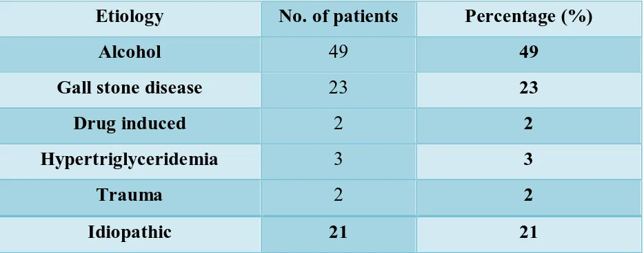
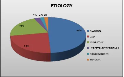
Etiology of acute pancreatitis:
common cause, in about 35 percent of cases. In a study done in New Delhi, India, gall stones and alcoholism were found to be the cause in 49% and 23.6% cases, respectively13.
The disease occurs at higher rate in young men and older women. Females are more prone to have gall stone pancreatitis and males are more prone to have alcohol induced pancreatitis14.
CAUSES OF ACUTE PANCREATITIS7:
Alcohol
Biliary tract disease Obstructive causes:
Choledocholithiasis
Ampullary carcinoma or pancreatic malignancy Papillary obstruction by worms/foreign bodies Pancreas divisum with minor duct obstruction Choledochocele
Duodenal diverticula at periampullary region Spasm sphincter of Oddi
Toxins or drugs:
5-Aminosalicylate (ASA) 6-Mercaptopurine (6-MP) Azathioprine
Cytosine arabinoside (cytarabine) Didanosine
Diuretic agents Estrogens, etc.
Probable Cause Acetaminophen α-Methyl-DOPA L-Asparaginase Isoniazid (INH) Phenformin, etc.
Trauma:
External / surgical traumatic injury to the abdomen.
Iatrogenic injury- postoperative trauma, post ERCP, post endoscopic sphincterotomy and manometry of sphincter of Oddi
Metabolic abnormalities: Hypercalcemia
Inherited conditions Infection:
Parasitic:- ascariasis, Clonorchis sinensis
Viral:- mumps, rubella, hepatitis A, B, non-A, non-B, coxsackie B, echo virus, adenovirus, CMV, varicella, EBV, HIV.
Bacterial: - mycoplasma pneumoniae, Campylobacter jejuni, Myco. tuberculosis, MAC, legionella pnemophila, leptospiral infection
Vascular causes:
hypo perfusion causing ischemia (e.g., after major cardiac vascular surgery) Athero-embolism
Vasculitis-SLE, PAN, malignant hypertension Miscellaneous causes:
Peptic ulcer penetration Cystic fibrosis
Crohn’s disease Reye’s syndrome Hypothermia
Idiopathic causes Gall stones
In 1901, Opie, at the Johns Hopkins Hospital in Baltimore, documented impaction of gallstone in the ampulla of Vater during the autopsy of a patient (operated on by Halsted) who had died due to gallstone pancreatitis and thereby first to describe the pathogenic mechanism of gallstone induced pancreatitis3.He suggested that the stone might have caused outflow obstruction from a common ‘biliopancreatic channel’. This led him to propose the "common-channel hypothesis3" in which a blockage below the junction of the biliary and pancreatic ducts would cause bile to flow into the pancreas, which could then be damaged by the detergent action of bile salts. Although this bile reflux theory was originally favored, most observers now believe that it is stone-induced pancreatic duct obstruction and ductal hypertension, rather than bile reflux that triggers acute pancreatitis.
Another proposed mechanism of causation postulates that passage of a gallstone through the sphincter of Oddi renders it momentarily incompetent, permitting the reflux of duodenal juice containing activated digestive enzymes into the pancreatic ductal system.
Microlithiasis (occult gall stones/biliary sludge) is a well-known cause of acute pancreatitis. The diagnosis of microlithiasis should be ruled out before labeling the disease as idiopathic pancreatitis. Biliary microscopy & endosonogaraphy are recommended nowadays to diagnose the microlithiasis.
Alcohol
other possible causes, even the first attack of pancreatitis is considered to be related to alcoholic pancreatitis. However, it is possible that a first attack of alcohol-related pancreatitis in the typical longstanding alcohol user is really the first manifestation of chronic pancreatitis. The disease can recur with continuous abuse of alcoholism. The nature of alcohol that was consumed (i.e., beer, wine, or hard liquor) is less significant than a daily intake of between 100 and 150 g of ethanol7.
Various theories have been put forward7, 8:
1. Alcohol consumption can alter lipid metabolism, and a transient hyperlipidemic state that causes hypertriglyceridemia and the generation of fatty acids as well as their ethyl ester metabolites, that can injure the pancreas.
2. Alcohol consumption causes intra pancreatic generation of oxygen free radicals, which can injure the pancreas.
3. It promotes secretion of pancreatic juice that is high in proteolytic enzyme content but low in enzyme inhibitor content. Enzyme activation can theoretically occur in these conditions and cause pancreatic injury.
5. Secretion of enzyme-rich fluid, deficient in enzyme inhibitors could also lead to precipitation of protein and calcium within this protein matrix, causing multiple ductal obstructions, while continued secretion can cause pressure to buildup and the formation of intra-ductal plugs, which cause ductal obstruction and ductal hypertension.
6. Ethanol causes focal ischemic injury to the gland, thereby transiently decreases pancreatic blood flow.
Hyperlipidemia
It is responsible in 1.5-4 % of cases. Triglyceride level > 1000 mg/dl increases the likelihood of developing pancreatitis. It is hyperlipidemia type I, IV or V that causes pancreatitis. It has been suggested that lipase can liberate large amounts of toxic fatty acids into the pancreatic microcirculation8. This could lead to endothelial injury, sludging of blood cells, and consequent ischemic states.
Hypercalcemia
Hypercalcemia secondary to hyperparathyroidism or any other cause can cause acute pancreatitis. The mechanism most likely involves hyper secretion and the formation of calcified stones intra ductally.
Tumours
About 1 to 2% of patients with acute pancreatitis may have pancreatic malignancy, in which an episode of acute pancreatitis could be the first clinical sign of a periampullary tumor. In both conditions, the pancreatitis occurs probably due to blockade of pancreatic secretion and its upcoming consequences.
Drugs
For practical reasons, it often is difficult to implicate a drug as the cause of pancreatitis. Many drugs can produce hyperamylasemia and/or abdominal pain, and a drug is considered to be a cause if the pancreatitis-like illness resolves with its discontinuation.
Infections
Miscellaneous Causes
The infestations by Ascaris lumbricoides and the liver fluke Clonorchis sinensis, which is endemic to China, Japan, and Southeast Asia, cause Oriental cholangitis, which is associated with cholangiocarcinoma obstructing the pancreatic duct.
A dominant gene mutation following Mendelian inheritance is known to result in hereditary pancreatitis. Whitcomb and associates described several families from various parts of the world were found to have mutations in the cationic trypsinogen gene PRSS1, which results in acute pancreatitis.
20 to 45% of patients with pancreas divisum (unfused ducts of Wirsung and Santorini) develop pancreatitis, but the failure of procedures to improve drainage of the lesser papilla in reducing attacks of pancreatitis, as well as the observed lack of ductal dilatation in such patients, contradicts pancreas divisum as an etiologic factor, rendering the role of this condition as yet unclear12.
Finally, no apparent cause can be ascribed to some episodes of acute pancreatitis, and these constitute the group referred to as idiopathic pancreatitis, which is the third most common cause of acute pancreatitis15.
Pathophysiology
Acute pancreatitis occurs in varying degrees of severity, the determinants of which are multifactorial. It is generally believed that acute pancreatitis is triggered by digestive enzymes which got activated inside acinar cells. This was thought to be counter acted by endogenously secreted pancreatic enzyme inhibitor. The ultimate severity depends upon the event that subsequently occurs following the acinar cell injury. The events are activation and recruitment of inflammatory cell, synthesis and release of cytokines and other chemical mediators of inflammation. Large amounts of liberated digestive enzymes however overwhelm the system as a whole.
There are three reasons for this theory7, 15:
(a) The pancreas is digestible by the activated enzymes of the duodenum.
(b) Activated digestive enzymes are found within the pancreas during pancreatitis.
(c) The histology of pancreatitis is suggestive of a coagulative necrosis.
According to “colocalization hypothesis” digestive enzymes are localized in cytoplasmic vacuoles which also contain the lysosomal hydrolase Cathepsin B, which is known to activate trypsinogen7. Recent studies suggest that cathepsin B activity inhibition by highly specific inhibitor, CA-074me, protects against intra-acinar cell activation of trypsinogen and hence pancreatitis. These findings suggest that the trypsinogen is activated because it erroneously colocalises in cytoplasmic vacuoles with cathepsin B.
Recent studies suggest that trypsin, once activated inside the colocalized vacuoles (appears similar to autophagic vacuoles), mediates the
during pancreatitis. Once inside the cytosol, it initiates apoptotic cell death by permeabilizing mitochondrial membranes, which allows cytochrome C to be released into the cytosol. This initiates the apoptotic cascade and ultimately the apoptotic death of the acinar cells.
FACTORS DETERMINING THE SEVERITY OF PANCREATITIS:
In addition to the cells of the immune system like neutrophils, the pancreatic acinar cells are also a source of inflammatory mediators during pancreatitis. The list of factors associated with pancreatitis and associated lung injury include: tumor necrosis factor alpha, monocyte chemotactic protein-1, Mob1, interleukin-1β (IL-1β), platelet activating factor, substance P, adhesion molecules [intercellular adhesion molecule-1 (ICAM-1) and selectins], IL-6, 8, 10, C5a, the CCR1 receptor and its ligands, granulocyte-macrophage colony-stimulating factor(GMCSF), macrophage migration inhibitory factor, COX-2, prostaglandin E1, nitric oxide (NO) and reactive oxygen species. The heat shock proteins are found to be protective in pancreatitis. The ultimate severity of pancreatitis and associated lung injury depends on the balance between the pro-inflammatory and anti-inflammatory factors7.
Several therapeutic regimens aimed at reducing the inflammatory response have been tested and include anti–tumor necrosis factor alpha antibody, IL-1 receptor antagonist, IL-10, anti-ICAM-1 and anti-CD3 Ab, rPAF acetyl hydrolase, and the calcineurin antagonist FK5068.
proinflammatory response. Mice in which TLR4 is genetically deleted have significantly reduced pancreatitis; this suggests that TLR4 is a significant promoter of proinflammation. However, this effect appears independently of lipopolysaccharides and is probably mediated by a hitherto unknown TLR4 agonist. It is likely that TLR4 antagonists would be a good therapy against pancreatitis15.
Initiation of injury
Block in pancreatic enzyme secretion
Co-localization of zymogen granules and lysosomal enzymes
Activation of trypsinogen & release of trypsin
Activation of other zymogens
Reactive oxygen Species Cytokines, adhesion molecules
Leucocyte infiltration
Release of further cytokines (TNF, IL-6, IL-8, PAF)
Inflammation SIRS
Ischemia Multi organ failure
Necrosis & Apoptosis
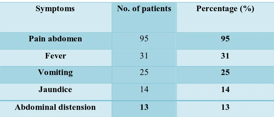
Clinical presentation:
The clinical presentation, diagnosis, and management of an acute attack of pancreatitis are similar regardless of whether that attack is acute or chronic pancreatitis. The acute pancreatitis can mimic like acute abdomen and should never be excluded in differential diagnosis8.
Abdominal pain, nausea, and vomiting are the predominant symptoms. Each episode begins with severe pain, following a substantial amount of meal. The cardinal symptom is usually epigastric pain, but can occur anywhere in the abdomen or lower chest. The pain was described as "knifing" or "boring through" to the back, and might be relieved by leaning forward(Mohmadian prayer position). Pain starts 12-48 hours after a bout of alcohol or after a large meal in
case of gall stone pancreatitis. Pain became generalized once peritonitis has been sets in8, 15.
Peritoneal dialysis, post-operative situations, legionnaire’s disease are well known for the occurrence of uncommon painless pancreatitis.
intense in necrotizing pancreatitis than in edematous pancreatitis. Although vomiting and retching may be relieved by passage of a nasogastric tube, the pain usually persists even after gastric decompression.
Fever is an important sign. Fever in the first week is due to acute inflammation mediated by cytokines. Fever in the second or third week is due to infected pancreatic necrosis. Fever in gall-stone induced pancreatitis, may be due to cholangitis and mandates prompt biliary decompression.
Physical Findings:
On examination, the patient may be tachypneic, hypotensive, and hyper thermic, and have tachycardia7, 8. The temperature was mildly elevated in uncomplicated pancreatitis. Voluntary and involuntary guarding may present over the epigastric region. The bowel sounds may be decreased or absent. There is usually no palpable swelling or masses. The abdomen may be distended with free intraperitoneal fluid, may associated with pleural effusion, particularly on the left side.
peritoneal cavity which may dissect via the soft tissues and appears as a bluish discoloration around the umbilicus (Cullen's sign) or in the flanks (Grey Turner's sign) and the inguinal region (Fox's sign) 17. Neither sign is pathognomonic of AP; actually the Cullen’s sign was first described with ruptured ectopic gestation.
The severe intravascular fluid loss may lead to acute renal shutdown with elevated BUN and creatinine levels. And also there may be hyperglycemia, hypoalbuminemia, and hypocalcemia that are sufficient enough to produce tetany in few cases.
Diagnosis:
The clinical diagnosis is one of exclusion and diagnosis may be difficult despite the plenty of investigation that are available.
Serum pancreatic enzymes:
Amylase, lipase, elastase and trypsin were released into the blood stream at the same time, but their clearance varied with different sensitivities, depends on the timing of blood sampling after the onset of disease.
damage, the pancreas may not release large amounts of enzymes into the circulation. It is important to recognize that, in patients with severe pancreatitis, frequent measurement of serum enzymes is not needed. Patients with alcoholic pancreatitis, in general, have a smaller increase in serum amylase levels. Because hyperamylasemia can be observed in many extra pancreatic diseases, measuring pancreatic-specific amylase (p-amylase) rather than total amylase, which also includes salivary amylase, makes the diagnosis more specific (88 to 93%).
The serum lipase estimation has been found to have high sensitivity and specificity in the diagnosis as there are no other sources of lipase15, 17. Total amylase is having a sensitivity of 84%, the serum P- amylase has 95% and lipase has 93%. Specificities for amylase, P-amylase and lipase respectively are- 88%, 93% and 96%, respectively. Thus P-amylase is the enzyme with the higher diagnostic value.
Immunologic assay like serum trypsinogen or immune lipase are generally less specific than the lipase assay. The increased urinary level of activation peptides released during either trypsinogen, procarboxypeptidase, or prophospholipase activation, may aid in predicting the severity of an attack.
Leucocyte migration and activation has considered as major determining factor of local & systemic complications8, 15.
Imaging:
In general, the plain chest and abdominal radiographs can be useful in the management by identifying other causes for the patient's symptoms (e.g., pneumonia, perforated hollow viscous, mechanical bowel obstruction). Plain abdominal X-ray findings are either generalized or local ileus (known as sentinel loop), colon “cut-off” sign or “renal halo” sign. A chest radiograph may show left pleural effusion, elevated left hemi diaphragm or basal atelectasis17.
Ultrasonography:
Abdominal ultrasound (US) examination is the gold standard for confirmation of gallstones pancreatitis. It also helpful to detect extra pancreatic ductal dilations & pancreatic edema, swelling, free peritoneal fluid and peripancreatic acute fluid collections (PFCs).It may not be sensitive in about 20% of cases, due to bowel gas interference with the imaging.
CT scan:
The contrast-enhanced computed tomography (CECT), has become gold standard for17
Diagnosis
Assessing the severity
The Balthazar scoring system and other similar grading systems have incorporated various CT findings such as inflammation and fluid collections in & around the pancreas to correlate radiographic appearance with morbidity and mortality19.
Early CT scans often fail to detect evolving necrosis, which become well demarcated by 2 to 3 days after the onset of symptoms. The CT scans are not useful in diagnosing necrosis or predicting the severity within the 24 hours of onset of illness. The sensitivity for identifying pancreatic necrosis using contrast-enhanced CT scan approaches 100%, 4 days from diagnosis. CT scans also been useful in the early diagnosis of infected pancreatic necrosis and image guided aspiration of necrosis, when patient not improving clinically or who experience clinical decline. In the patient with moderate renal impairment or allergy to intravenous contrast material, magnetic resonance imagining (MRI) may be useful. MRI has been found to have sensitivity and specificity similar to contrast-enhanced CT for detecting severe acute pancreatitis.
recurrent attacks of acute pancreatitis, without any obvious cause. It is useful in correcting potentially correctable lesions such as CBD stones with impaction, pancreas divisum, ampullary stenosis, pancreatic duct stenosis etc.
Mild acute pancreatitis (MAP) Vs. Severe acute pancreatitis (SAP):
Characteristic MAP SAP
Process Mild, self-limiting Fatal attack or failure to settle on supportive therapy
Course Resolves rapidly Emergence of local and systemic complications
Hallmarks Edematous, interstitial
inflammation of the pancreas
Extensive and prolonged, pancreatic and retroperitoneal inflammation with superimposed patchy or generalized areas of necrosis and hemorrhage in the pancreas and surrounding tissues
Fatality rate Does not exceed 3%
Fatality rate between 10-20%
CECT Normal in 15-30% of patients
May show pancreatic abscess, intra-abdominal fluid collection
Assessment of Severity:
Scoring systems in acute pancreatitis: The various prognostic scoring systems for assessing the severity will be discussed in detail later.
UK guidelines for the management of AP20:
The correct diagnosis has to be made within 48 hrs. of admission.
The etiology has to be determined in 80% of cases at least and idiopathic cause should not exceed 20%.
The serum lipase assay has been preferred over serum amylase assay for diagnosis the acute pancreatitis.
The contrast enhanced computed tomography has to be preferred over USG for detection of the presence/absence of pancreatitis.
Treatment:
There are two phases in evolution of an acute attack of pancreatitis. Both phases are overlapping on each other15, 17.
The second phase, that may last for weeks or months, is primarily characterized by the development of local complications that are, themselves, the results of necrosis, infection and pancreatic duct rupture.
The initial management of patients with pancreatitis focuses on early establishment the diagnosis, assessing the severity, treating the major symptoms, and haltering the disease progression. The treatment for acute pancreatitis is largely supportive. Since 15-30 % patients develop severe pancreatitis, so each and every patient should be treated aggressively. The main aim of the treatment is ‘allowing rest to the gland’ by oral feed and fluids restriction21
. The goal of initial management consists of adequate fluid replacement, correction of electrolyte imbalance, nutritional support and prevention of local & systemic complications.
Management of Pain
Good analgesics should be given to these patients as the pain can be very severe in intensity. Most patients require narcotic analgesics. Meperidine is preferred as morphine induces spasm of the sphincter of Oddi, which can, at least theoretically, worsen biliary pancreatitis.
Fluid and Electrolyte Management
utmost importance to prevent systemic complications, mainly acute renal insufficiency, that may occur with hypovolemia. Transudation of the fluid from intravascular space into the areas of inflammation (i.e., peripancreatic, retroperitoneum and into the pulmonary parenchyma and soft tissues elsewhere in the body) is the principle cause of hypovolemia. Furthermore, studies have shown that inadequate resuscitation may add upon as a significant risk that leads to further pancreatic injury.
Banks and colleagues have showed that while aggressive fluid resuscitation might not prevent the progression to develop pancreatic necrosis. The degree and intensity of monitoring depends upon the disease severity22.
Measurement of central filling pressures, using a Swan-Ganz or central venous pressure catheter, can be helpful in guiding fluid management, particularly when hypovolemia is combined with lung injury.
Nasogastric Decompression
The nausea and vomiting of pancreatitis can result in significant fluid as well as electrolyte losses and retching can lead to gastro-esophageal mucosal tears and result in upper gastrointestinal bleeding (i.e., the Mallory-Weiss syndrome). For symptomatic relief and to increase patient comfort, nasogastric decompression may be needed, although the institution of nasogastric drainage does not shown to alter the eventual outcome of an attack7, 8.
Prophylactic Antibiotics
infection has been the rationale behind the use of prophylactic antibiotics widely in patients with pancreatic necrosis. In severe pancreatitis, beneficial effects have been observed with regimens that included imipenem alone, imipenem with cilastatin, metronidazole and third-generation cephalosporin (cefuroxime). Because
Candida species are common inhabitants of the upper GI tract, Candida sepsis and secondary fungal infection of pancreatic necrosis is a risk in severe disease, and many surgeons advocate empirical therapy with fluconazole in severe acute pancreatitis.
The duration of treatment has not defined clearly. A treatment course of 1week to 4 weeks has been recommended commonly, but many of them limit the treatment to 2 weeks17.
According to the current UK guidelines (Johnson 2005), the duration of antibiotic prophylaxis is 1 to 2 weeks20.
Nutritional Support
The points favoring enteral nutrition are7, 15:
It might feasible, safe, and desirable in severe pancreatitis.
It has the advantage of avoiding the high cost of total parenteral nutrition (TPN) as well as its associated catheter-related complications.
The use of enteral nutrition may support intestinal mucosal integrity by avoiding the alteration in intestinal permeability & barrier function as seen with use of TPN.
Treatments of Limited or Unproven Value
In patients who develop severe disease, other treatment modalities may be tried. The antiproteases like gabexate/aprotinin, antisecretory agents like octreotide and anti-inflammatory drugs or PAF antagonists like lexipafant were found to be less useful15, 17.
Treatment of Early Systemic Complications of Pancreatitis
superimposed on other disease states that are characterized by peritonitis and hypovolemia8.
Cardiovascular collapse is largely caused by hypovolemia, and its management requires aggressive fluid and electrolyte repletion.
The pulmonary manifestations of pancreatitis include atelectasis and acute lung injury. The latter appears to be similar to the acute lung injury caused by other systemic processes, including septic shock, ischemia and reperfusion, and massive blood transfusion. Management includes good pulmonary toilet combined with close monitoring of pulmonary function. For many patients, intubation and respiratory support may be required.
Renal failure in pancreatitis is usually prerenal and is associated with a poor prognosis. In severe cases, dialysis, usually hemodialysis, may be required.
Stress-induced gastro duodenal erosions account for most of the gastrointestinal bleeding, prophylaxis with antacids, H2-receptor antagonists, or proton pump inhibitors may be appropriate.
can cause bleeding. Management of these complications of pancreatitis is similar to that involved when they occur in the absence of pancreatitis.
Some patients with severe pancreatitis develop disseminated intravascular coagulation, but it rarely causes bleeding, and prophylactic heparinization is usually not indicated.
Removal of precipitating factors, such as drugs or alcohol, is appropriate. Once the acute phase has been survived, usually by the end of the first week, and major organ failure is under control, then local complications become pre-eminent in the management of these patients.
An indication for operative intervention in acute pancreatitis is the drainage of an infected pancreatic necrosis. These patients require removal of as much as possible of the infected necrosis and drainage for the remaining viable exocrine tissue. Current opinion is against debridement in sterile necrosis unless it is accompanied by life threatening systemic complications17.
A pancreatic abscess occurs 2 to 6 weeks after an initial attack of acute pancreatitis, in contrast to infected necrosis which occurs in the first few hours or days. Treatment consists of external drainage, either by surgical or percutaneous catheter based measures17.
The presence of gallstones leading to choledocholithiasis is recognized as a major etiological factor worldwide. Endoscopic retrograde cholangio pancreatography (ERCP) has both diagnostic and most therapeutic utility in patients with biliary obstruction or cholangitis. By randomizing patients with AP to early ERCP versus no ERCP, both Neoptolemos and colleagues, and Fan and colleagues have showed a significant decrease in morbidity but there was no significant improvement in mortality with routine use of ERCP. A metacentric randomized control study in the ERCP group by Folsch and colleagues recently, have demonstrated increased complication rate and mortality rate, after excluding the patients with biliary sepsis or obstruction. It therefore, found that early ERCP may be harmful even in the absence of ongoing biliary obstruction. Magnetic resonance cholangio pancreatography (MRCP) is an additional alternative to ERCP as a diagnostic tool that avoids the risk of post procedure pancreatitis.
Surgical Management: Indications and Timing
There are very limited indications for surgical intervention; specifically, intervention may be needed to address the etiology of pancreatitis or its complications. Interventions, either surgical or endoscopic, to prevent recurrent gallstone pancreatitis are recommended in any patient with suspected choledocholithiasis. Delayed surgery is also, rarely needed for the treatment of local complications like pseudocysts17.
Complications17:
Complications may be classified as15, 17: I. LOCAL:
Fluid collections
Pancreatic ascites/pleural effusion
Pancreatic pseudocyst
Pancreatic necrosis
Infected pancreatic abscess
Hemorrhage/pseudo aneurysm
II. REGIONAL:
Venous thrombosis
Paralytic ileus
Intestinal obstruction
Cholestasis
III. SYSTEMIC:
A. Pulmonary
1. Pneumonitis, basal atelectasis
2. ARDS
3. Pleural effusion (L)
B. Cardiovascular
1. Hypotension
2. Hypovolemia
3. Sudden arrest &death
4. Nonspecific ECG(ST-T wave) changes
5. Pericardial effusion
C. Hematologic
1. Hemoconcentration
2. Disseminated intravascular coagulopathy
D. GI hemorrhage
1. Acid peptic disease
2. Gastric erosion
3. Portal/splenic vein thrombosis with variceal bleed
E. Renal
1. Oliguria
2. Azotemia
3. Renal vessel thrombosis
F. Metabolic
1. Hyperglycemic state
2. Hypocalcemic state
3. Hyperlipidemia (triglyceridemia)
4. Metabolic encephalopathy
5. Sudden loss of vision (Purtscher's retinopathy)
G. Central nervous system
2. Fat embolism occlusion
3. Alcohol withdrawal syndrome (AWS)
H. Fat necrosis
1. Intra-abdominal saponification
2. Subcutaneous tissue necrosis
SCORING SYSTEMS IN ACUTE PANCREATITIS
Pancreatitis is a serious disease with high morbidity and mortality rates. Some 80% were mild attack which recovers rapidly with conservative management. The rest of 20% were severe, with protracted course that needs intensive care and specialized management. Several predictors of severity are commonly used for this purpose24.
Scoring systems can be used to predict mortality, severity of disease and intensity of its complications. Prognostic factor analysis found to helpful in comparing the results, in-between the series of patients under study.
Several scoring scales exist that predict both mortality and morbidity in patients with acute pancreatitis.
These systems include25: Ranson’s criteria
Balthazar computed tomography (CT) grading Imrie Glasgow coma score (GCS)
Simplified acute physiology score(SAPS)
Marshall Multiple organ failure (MOF) score and
Acute physiology and chronic health evaluation (APACHE) I, II, III & O. The GCS and Ranson’s multiple scoring systems require 48 hours of data collection; however, APACHE can be calculated at any time and shows prognostic correlation with acute pancreatitis, as increasing scores are associated with poor prognosis.
Once the acute pancreatitis has been diagnosed, assessment of severity is extremely important for execution of appropriate measures, preferably in an ICU setup with close monitoring.
1) RANSON’S CRITERIA27
:
Criteria for Pancreatitis not due to gall stones: At admission or diagnosis:
Age more than55 years WBC count > 16,000/mm3 Blood sugar> 200 mg/dL Serum LDH> 350 IU/L
AST > 250 U/dL
During initial 48 hours:
Fall in hematocrit> 10 percentage points BUN elevation > 5 mg/dL
Serum calcium level < 8 mg/dL Arterial Po2 less than 60 mm Hg Base deficit more than 4 meq/L Estimated fluid sequestration > 6 L
Gall stone induced pancreatitis:
Recently, the cutoff values of these signs were modified in biliary pancreatitis. This limits the use of early prognostic signs; it now requires memorization of 18 separate parameters and etiology is not always known. Therefore the revisions for biliary pancreatitis have not had wide acceptance, and the original system is the one that is widely utilized31.
On admission or diagnosis: Age > 70 yrs
WBC count > 18,000/mm3 Blood sugar> 220 mg/dL Serum LDH> 400 IU/L
AST > 250 U/dL
Fall in hematocrit greater than 10 percentage points BUN elevation > 2 mg/dl
Serum ca2+ level < 8 mg/dl Base deficit more than 5 meq/L Estimated fluid sequestration > 4 L 2) IMRIE’S PROGNOSTIC CRITERIA: During initial 48 hours
WBC count > 15000/mm3 Blood sugar > 10 mmol/L
Serum urea > 16 mmol/L (no response to IV fluids) Po2 level < 60 mm Hg
Serum ca2+ level < 2 mmol/L Lactic dehydrogenase> 600 IU/L AST / ALT>200µm/l
Serum albumin level < 32 g/L
Ranson’s and Imrie’s scores indicate the severity at the time of admission and are not intended for monitoring the clinical course27.
3) BANK’S CLINICAL CRITERIA:
Cardiac Shock, tachycardia, arrhythmia, ECG changes
Pulmonary Dyspnoea, basal rales, PO2< 60 mm Hg, ARDS
Renal Urine output < 50 ml/h, rising BUN& creatinine
Metabolic Low Ca2+&pH; albumin
Haematological HCT, DIC
Neurological cerebral Irritation & confused state
GIT paralytic ileus, free fluid, hgic peritoneal tap
4) BALTHAZAR COMPUTED TOMOGRAPHY SEVERITY INDEX (CTSI):
Emil J. Balthazar et al, developed CTSI, a grading system used to determine the acute pancreatitis severity19, 33.
Prognostic Indicator Points Grade
Pancreatic inflammation
Normal pancreas
Focal or diffuse enlargement of the pancreas
Intrinsic pancreatic abnormalities with inflammatory changes in
peripancreatic fat
Single, ill-defined fluid collection or phlegmon
Two or more poorly defined collections or presence of gas in or
adjacent to the pancreas
Pancreatic necrosis None ≤ 30% > 30–50% > 50% 0 1 2 3 4 0 2 4 6 A B C D E
Modified CT Severity Index28
Prognostic Indicator Points
Pancreatic inflammation
Normal pancreas
Intrinsic pancreatic abnormalities with or without inflammatory changes in
peripancreatic fat
0
Pancreatic or peripancreatic fluid collection or peripancreatic fat necrosis Pancreatic necrosis None ≤ 30% > 30% Extrapancreatic complications
(one or more of pleural effusion, ascites, vascular
complications, parenchymal complications, or gastrointestinal tract involvement) 4 0 2 4 2
5) MODIFIED GLASGOW CRITERIA:
This one was useful in both alcoholic and biliary pancreatitis27. The score ≥ 3 means severe disease requires ICU care.
P - PaO2 <8kPa or < 60 mmhg A - Age more than 55 years old
N - Neutrophilia with WBC count>15x109/L C - Ca2+<2mmol/L or < 8 mg/dl
R - Renal function, Urea >16mmol/L or > 45 mg/dl E – Enzymes:- serum LDH >600 IU/L; AST>200 IU/L A - Albumin <3.2g/dL
S - Sugar: >10mmol/L or >180 mg/dl
According to this scoring system score of ≥ 2 indicates presence of organ failure. These scores were calculated within 72 hours of admission into the hospital. The organ failure was classified as27:
Transient (less than 48 hrs.) Persistent (more than 48 hrs.)
7) THE APACHE (ACUTE PHYSIOLOGICAL AGE AND CHRONIC HEALTH EVALUATION) SYSTEM
A number from zero to four was assigned to each measurement according to how far from normal the measurements vary.
When multiple values for the same measurement were available, the worst was chosen. The final score, which ranged from zero to 124, indicates how far from normal homeostasis a patient had strayed because of acute illness.
The true APACHE score was more difficult to calculate because of practical problems like collection of large number of variables. Also under the rules of APACHE system any unmeasured variable was assumed to be normal and weighted as zero. This gave rise to questions about the model’s general applicability. Another major criticism of original APACHE system was that the variables were chosen by a group of physicians and there was a potential of bias. These inaccuracies in the original APACHE system prevented its widespread use. However, it did serve as the prototype for the development of two subsequent systems.
APACHE II
most abnormal. APACHE II did not strictly depend on ICU setting only but it was found to be as reliable as APS outside the ICU settings. The age and chronic health problems were included in this score as they reflect the physiological reserve status.
APACHE II Score as published by Knaus et al is composed of three parts:
1) Acute Physiological Score (0 to 60 points) 2) Age points (0 to 6 points)
3) Chronic health points (0 to 5 points).
Range of potential score is 0 to 71 but scores above 40 are uncommon. Score above 30 are associated with mortality rate of at least 70%. Roumen et al, in their study on acute hemorrhagic necrotising pancreatitis, concluded that of Ranson, Imrie, Multiple organ failure (MOF) and Sepsis sensitivity score (SSS), APACHE II is the best for grading the severity of disease on admission32. It is well suited for stratification of patients and comparisons of treatment methods.
The advantages are:
1) Objective determination of AP within few hours of admission, which might helpful in detection of cases for ICU care or for clinical trial.
3) Ability to be recalculated daily. Sequential monitoring of APACHE II enables determination of improvement or deterioration in the physiologic status of the patient. Over the initial 48 h, the score increase significantly in those with severe disease (median increase three points) but decrease (median decrease one point) in patients with mild pancreatitis. Thus this might be useful for follow up of the disease course and helps to assess the therapeutic response.
4) This score was used universally for all serious illnesses, thereby avoiding the need for a separate grading for acute pancreatitis.
The major drawbacks of APACHE II are: 1) Complexity and poor feasibility
2) The ideal ‘cut-off’ score for APACHE II in acute pancreatitis remains to be determined
3) As shown by Wilson et al, the cut-off scores having the greatest prognostic values on admission are different from the peak scores during the hospital course. The use of a single cut-off score APACHE II ≥ 9, as suggested by Larvin and McMohan needs to be validated in more studies.
5) APACHE II generally underestimates mortality in many series of critically ill surgical patients as pre-ICU resuscitation not taken into account and young patients score few points despite severe pancreatitis.
6) APACHE II mortality predictions were based on treatment that lasts for 20 years ago.
APACHE III
In 1991, Knaus et al presented a revised and improved form of APACHE-II and termed it as the APACHE-III prognostic system34.
The following variables were included in this score; blood urea, urine output, Sr. albumin, Sr. bilirubin, blood glucose, pCO2 in comparison to APACHE-II.
APACHE III is regarded as a good prognostic scoring system by using it serially and sequentially in acute pancreatitis. APACHE III score shows significant differences in mild and severe AP and correlates well with severity35. APACHE evaluation proves very suitable for serial monitoring of patients and gives an objective indication of progress in the individual patient. Williams et al found that an APACHE III score >30 indicated a much higher morbidity and mortality rate36.
financially constrained ICUs. As compared to APACHE II, the data collection was very much complex, and it was not accurate for predicting the risk in post-op cases.
8) BISAP (The bedside index for severity in AP):
This new scoring system has been developed recently for early detection of patients with risk of in hospital mortality1.
The BISAP score has been developed and validated retrospectively on a large population based study, done by Cardinal Health Clinical Outcomes Research Database, Marlborough, USA38.
This score was published recently for clinical and research purpose, for its accuracy and reliability in patient stratification.
The BISAP includes38:
1) Blood urea nitrogen (BUN) >25 mg / dl. 2) Impaired mental status (GCS < 15). 3) SIRS.
4) Age >60 years. 5) Pleural effusion.
2) Respiratory rate > 20/min or PaCO 2 < 32 mm Hg. 3) Temperature >100.4 F or < 96.8 F / < 36 or > 38 ° C.
4) WBC count >12,000 or < 4,000 cells/mm3, or presence of more than 10% immature blasts.
(SIRS - Systemic Inflammatory Response Syndrome)
One point will be given for each variable present for a total of 5, score ranges from 0 to 5.
The presence of a pleural effusion was determined by a CT scan, chest radiograph or abdominal ultrasound obtained within 24 h of presentation. Imaging obtained within 24 h of presentation at the hospital of origin for transferred patients was also collected and reviewed.
A BISAP score of three or more has been found to have high mortality and have predicted the necrosis and organ failure very well39.
ADVANTAGES:
1. Simple and easy to calculate, usually done at the time of admission or within 24 hrs. Of hospitalization40.
2. The scores prediction ability was tested across 390 hospitals among large number (36,248) of populations, in contrast to other studies which were based on small number patients.
1. The Glasgow Coma Scale used for evaluating mental status was subject to interobserver variation.
2. It could not discriminate transient from persistent organ failure within 24 hrs. Of hospitalization.
3. This could not predict the preventable complications of acute pancreatitis like any other scoring system.
AIMS AND OBJECTIVES OF THE STUDY
To evaluate the role of BISAP score in place of traditional APACHE II scoring system in analyzing severity and early treatment intervention.
Stratification of the patients with acute pancreatitis according to their scores observed at the time of hospitalization.
MATERIALS AND METHODS
Study design: Comparative Analytical study.
Setting: Department of General Surgery, Govt. Stanley Medical College and Hospital, Chennai. The study was conducted after obtaining the Institutional Ethical Committee approval (annexure 2).
Inclusion criteria:
Age >20 years including both sexes.
Serum amylase/ Serum lipase equal to or more than 3 times the upper limit of normal.
Radiological evidence of presence of acute pancreatitis. Exclusion criteria:
Patients were excluded from the study if they were younger than 20 years. Proven cases of chronic pancreatitis.
Hereditary pancreatitis.
Patients with comorbidities like COPD, renal impairment, immunosuppressive state, etc.
Traumatic pancreatitis associated other visceral injuries.
First 100 patients attending the surgical emergency ward with clinical features of Acute Pancreatitis are evaluated clinically and subjected to laboratory and radiological investigations as per the designed proforma (annexure 1). Data pertinent to the scoring systems will be recorded within 24 h of admission to the hospital.
Once diagnosis is established the patient disease severity will be assessed by following two scoring systems
BISAP APACHE II
Statistical Analysis: Appropriate statistical tools.
For each of 100 patients included in the study, APACHE II and BISAP scores were calculated by using the APACHE II prognostic system in the manner described by Knaus et al and the Cardinal Health Database system for BISAP scoring.
Patients were classified to have mild or severe acute pancreatitis according to the definitions set by the Atlanta Classification guidelines (1992)42:
Severe attack--Criteria for severity included:
1) presence of one or more local complications: Pancreatic necrosis
Pancreatic abscess Pancreatic pseudo cyst.
Shock (systolic BP< 90 mm Hg).
Pulmonary insufficiency (PaO2< 60 mm Hg on room air). Renal failure (Sr. creatinine > 2mg/dl after fluid replacement).
Gastrointestinal bleeding (> 500 ml estimated loss of blood within 24 hrs.).
DIC (thrombocytopenia and hypofibrinogenemia and fibrin split products).
Severe hypocalcemia (<8 mg/dl).
Survivors were defined as patients discharged alive from the hospital and non-survivors were those who died from pancreatitis or its complications during hospitalization.
Biliary Pancreatitis was presence of gall stones/biliary sludge in the gall bladder or bile duct, which was documented by any radiological methods. Alcoholic Pancreatitis was considered, when the patient found to have regular high intake of alcohol daily, or if there was binge of alcohol consumption prior to the onset of illness and has no signs of other etiologies present. Idiopathic pancreatitis was the one with no identifiable etiological factor based on the history, or after initial investigations.
Patients were observed prospectively until discharge or death.
OBSERVATION &RESULTS
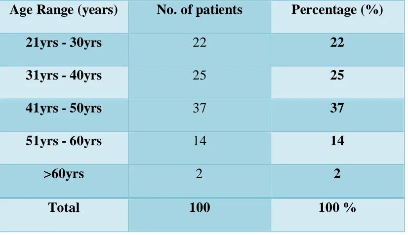
[image:86.612.68.481.304.542.2]This study was conducted in the department of general surgery, Govt. Stanley Medical College & Hospital, Chennai for a period of one year. The 100 persons with features of acute pancreatitis who fulfilled the inclusion cr iteria were enrolled in this study after obtaining an informed consent.
Table: 1 Age distribution
Age Range (years) No. of patients Percentage (%)
21yrs - 30yrs 22 22
31yrs - 40yrs 25 25
41yrs - 50yrs 37 37
51yrs - 60yrs 14 14
>60yrs 2 2
Total 100 100 %
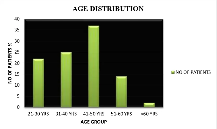
FIGURE -1
0 5 10 15 20 25 30 35 40
21-30 YRS 31-40 YRS 41-50 YRS 51-60 YRS >60 YRS
N
O
O
F
P
A
TI
EN
TS
%
AGE GROUP
AGE DISTRIBUTION