Copyright © 1999, American Society for Microbiology. All Rights Reserved.
Suppressor Mutations within the Core Binding Factor
(CBF/AML1) Binding Site of a T-Cell
Lymphomagenic Retrovirus
MARITA J. MARTINEY,1LAURA S. LEVY,2ANDJACK LENZ1*
Department of Molecular Genetics, Albert Einstein College of Medicine, Bronx, New York 10461,1and
Tulane University School of Medicine, New Orleans, Louisiana 701122
Received 20 August 1998/Accepted 2 December 1998
The transcriptional enhancer of the lymphomagenic mouse retrovirus SL3 contains a binding site for the transcription factor core binding factor (CBF; also called AML1, PEBP2, and SEF1). The SL3 CBF binding site is called the core. It differs from the core of the weakly lymphomagenic mouse retrovirus Akv by one nucleotide (the sequences are TGTGGTTAA and TGTGGTCAA, respectively). A mutant virus called SAA that was identical to SL3 except that its core was mutated to the Akv sequence was only moderately attenuated for lymphomagenicity. In most SAA-infected mice, tumor proviruses contained either reversions of the original mutation or one of two novel core sequences. In 20% of the SAA-infected mice, tumor proviruses retained the original SAA/Akv core mutation but acquired one of two additional mutations (underlined), TGCGGTCAA or TGTGGTCTA, that generated core elements called So and T*, respectively. We tested whether the novel base changes in the So and T* cores were suppressor mutations. SL3 mutants that contained So or T* cores in place of the wild-type sequence were generated. These viruses induced T-cell lymphomas in mice more quickly than SAA. Therefore, the mutations in the So and T* cores are indeed second-site suppressor mutations. The suppressor mutations increased CBF binding in vitro and transcriptional activity of the viral long terminal repeats (LTRs) in T lymphocytes to levels comparable to those of SL3. Thus, CBF binding was increased by any of three different nucleotide changes within the sequence of the SAA core. Increased CBF binding resulted in increased LTR transcriptional activity in T cells and in increased viral lymphomagenicity.
Transcriptional enhancers in murine leukemia viruses (MuLVs) are crucial genetic elements for determining viral pathogenicity. Enhancer sequences located in the unique 39 region (U3) in the long terminal repeat (LTR) determine the tissue specificity of virally induced disease, the fraction of the mice that develop tumors, and the length of latency prior to the appearance of tumors. MuLV enhancers usually contain tan-dem repeat units that are located starting approximately 170 bp upstream of the transcription initiation site in the 59LTR. Binding sites for various transcription factors are present within the repeats and the sequences flanking them (2, 3, 7, 12, 13, 21, 28, 33, 35–37, 41–45).
One element within the enhancers of MuLVs and related type C mammalian retroviruses including feline leukemia virus and gibbon ape leukemia virus is known as the enhancer core. The core was first identified in the simian virus 40 enhancer (19). Although the sequences of the 9-bp core elements are similar among simian virus 40, polyomavirus, and type C ret-roviruses, they are not identical. Mutagenesis studies showed that core elements are very important for the pathogenicity of the T-cell lymphomagenic MuLVs, Moloney MuLV (Mo-MuLV), and SL3 (14, 27, 34). In Mo-MuLV, point mutations in the core reduced viral potency and changed the disease specificity from T-cell lymphoma to erythroleukemia (34). The LTR enhancer of the lymphomagenic mouse retrovirus SL3 contains two 72-bp tandem repeats. Each repeat contains two slightly different core elements. Based on its position relative to other transcription factor binding sites, one of these (core I)
corresponds to the core element of Mo-MuLV (27). The SL3 and Mo-MuLV cores differ slightly in sequence (TGTGGT TAA and TGTGGTAAG, respectively). In addition, SL3 con-tains a second core element, termed core II, that has the sequence AGCGGTCTG (14, 27, 38). Mutation of the core I element of SL3 strongly decreased the pathogenicity of the virus (14, 27). Mutation of the core II element by itself had little effect on pathogenicity (14). However, when both core elements were mutated, the virus was only weakly pathogenic and most of the tumors were B-cell lymphomas (10).
Although all type C mammalian retroviruses have an iden-tifiable core element positioned equivalently to the SL3 core I element, the actual sequences of the elements vary somewhat among the different viruses (12). These differences can have large effects on viral pathogenicity. The core I element of SL3 (hereinafter termed the core element of this virus) and the core element of the weakly pathogenic MuLV Akv differ by 1 bp (the sequences are TGTGGTTAA and TGTGGTCAA, respectively). SAA is an engineered mutant of SL3 that is identical to SL3 except for the T to C change within the core elements in both enhancer repeat units in the viral LTR. Al-though SAA induced T-cell lymphomas in mice, the lym-phomagenicity of SAA was reduced compared to that of SL3 as the latency period to disease onset was increased (27). Thus, the 1-bp difference between the SL3 and Akv/SAA cores was important for viral pathogenicity. Nonetheless, it was surpris-ing that SAA was as potent as it was, because the T to C change substantially decreased the transcriptional activity of the SL3 LTR in T lymphocytes (27). Moreover, a 3-bp mutation in the SL3 core substantially inhibited viral lymphomagenicity (14). To account for these discrepancies, it was hypothesized that the 1-bp mutations in the cores of SAA had reverted, thus restoring the original SL3 sequence (27). These reversions * Corresponding author. Mailing address: Department of Molecular
Genetics, Albert Einstein College of Medicine, 1300 Morris Park Ave., Bronx, NY 10461. Phone: (718) 430-3715. Fax: (718) 430-8778. E-mail: [email protected].
2143
on November 9, 2019 by guest
http://jvi.asm.org/
This core element was termed So (27). Six of 38 mice had proviruses with a core sequence TGTGGTCTA (the novel nucleotide is underlined) that generated a core element that was termed T* (27). Since both So and T* retained the original C mutation and occurred independently in multiple mice, we hypothesized that they are core elements with second-site sup-pressor mutations.
In the study reported here, we tested this hypothesis. The LTR sequences of proviruses in tumors from mice containing So and T* core elements were recovered and used to replace the corresponding sequences of SL3 (27). The lymphomagenic potential of these viruses in mice was analyzed. To elucidate how the mutations generating So and T* might affect viral lymphomagenicity, we also assessed their effects on binding of core binding factor (CBF; also called AML1, Runt, PEBP2, and SEF1) (8, 17, 18, 24, 37, 40) and on the transcriptional activity of the viral LTR in T lymphocytes.
MATERIALS AND METHODS
Generation of viral genomes containing the So and T* cores.Infectious clones of the viral genome were generated in a manner similar to that previously described (27, 29, 30). The approach is summarized in Fig. 1. LTR sequences containing the So or T* cores were PCR amplified from proviruses present in DNA isolated from lymphomas induced by SAA (27). PCR products were gen-erated with the primers HM23 (59 TTCATAAGGCTTAGCCAGCTAACTG CAG 39) and HM22 (59GATGCCGGCACACACACACACACTCTCCC 39) at positions2470 to2443 and1272 to1244, respectively, relative to the tran-scriptional initiation site (Fig. 1) (27). PCR conditions involved a 30-cycle pro-gram of 1 min at 94°C, 1 min at 64°C, and 2 min at 72°C. The PCR products were digested withPstI andKpnI (Fig. 1A) and then subcloned into the corresponding sites of the pGEM 3Z(2) vector (Promega). LTR plasmid subclones were digested withBssHII andEcoRI, and a fragment containing the remainder of the SL3 genome including thegag,pro,pol, andenvgenes was inserted at those sites (Fig. 1B). This resulted in the formation of a plasmid subclone that contained the complete viral genome with a single LTR (Fig. 1B). These plasmids were cleaved withPstI to separate the viral and plasmid vector sequences. The viral fragments were self-ligated to form concatemers. This resulted in viral genomes that con-tained two identical LTRs, as previously described (27, 29, 30). Infectious virus was generated by transfection of viral genomes into NIH 3T3 mouse fibroblasts. 106cells per 60-mm2plate were seeded 24 h prior to transfection with Lipofectin
(GIBCO BRL). Cells were passaged 1:10 every third day. Supernatants collected from transfected cells at each passage were tested for reverse transcriptase activity (11). Approximately 3 weeks after transfection, viral stocks reached maximum reverse transcriptase levels. Aliquots of virus were frozen at this point, and the infectious virus titers were determined by XC plaque assays (31). PCR and sequencing of proviral DNA (described below) from the infected NIH 3T3 cells confirmed the presence of each mutation.
Tumorigenicity assays.Newborn NIH/Swiss mice (,1.5 days) were injected intraperitoneally with 0.1 ml of virus (104PFU, XC plaque assay). The diseased
animals were sacrificed and necropsied. Gross pathological examination always revealed enlargement of the thymus, spleen, peripheral lymph nodes, mesenteric lymph nodes, or liver. Enlarged organs were stored frozen at280°C until DNA was prepared from them. Southern blotting with a T-cell receptorbprobe was used to test whether the tumors were of T-cell origin (1, 15).
Analysis of viral enhancer sequences in infected cells.Proviral LTR DNA was amplified from tumors by using PCR primers HM22 and HM23 (Fig. 1A) as described above. PCR products were electrophoretically resolved on a nonde-naturing 5% polyacrylamide gel (27). The individual bands differed by multiples of 72 bp. Each band was excised and the DNA was isolated by using Qiaex II
scribed in the text. Transfection of cell lines was performed by the DEAE-dextran method as previously described (3, 32). 5.03106cells per plate were
pelleted and resuspended in 1 ml of TD (25 mM Tris-HCl [pH 7.4], 0.7 mM Na2HPO4, 5.1 mM KCl, 137 mM NaCl) containing 250mg of DEAE-dextran per
ml, 5mg of reporter plasmid DNA, and 1mg of a Rous sarcoma virus LTR-luciferase plasmid used as an internal control. The reagents were incubated at room temperature for 15 min. Five milliliters of medium supplemented with 10% fetal bovine serum was added, and incubation was continued for 20 min at 37°C. Cells were pelleted and resuspended in 5 ml of medium with serum. Cells were plated in 60-mm2dishes and harvested at 48 h. Cells were lysed by three cycles
of freeze-thawing, and protein concentrations were determined by Bradford assays (Biorad) (4). Aliquots of cell lysates were used for CAT or luciferase assays as previously described (45) except that the protocol included the use of 15ml of fluorescent BODIPY FL chloramphenicol substrate, FastCAT (Molec-ular Probes) instead of14C-labeled chloramphenicol. Silica gel thin-layer
chro-matography was performed in a sealed, equilibrated chrochro-matography chamber containing chloroform:methanol (9:1, vol/vol). CAT activity was quantified by calculating the percentage of chloramphenicol that was acetylated by using the STORM imager that scans for blue fluorescence. Each sample was normalized with its corresponding luciferase assay. All samplings were performed in dupli-cate and at multiple times. Means and standard deviations were calculated and plotted.
Cell lines.SL3H and Jurkat cells were grown in RPMI 1640 medium supple-mented with 10% fetal bovine serum, 100 U of penicillin per ml, 10 mg of streptomycin per ml, and 2 mM glutamine. L691-6 cells were propagated in Dulbecco’s modified Eagle media (DMEM) supplemented as described above. NIH 3T3 cells were grown in DMEM with 10% calf serum and other supple-ments as previously mentioned. All cells were maintained at 37°C in 100% humidity and 7.5% CO2.
EMSAs.Electrophoretic mobility shift assays (EMSAs) were performed with a double-stranded, 31-bp radiolabeled probe that contained the SL3 core I sequence as previously described (3). Nuclear protein extract from the mouse T-cell line WEHI 7.1 provided the source of CBF protein (3). Double-stranded, unlabeled oligonucleotides served as competitor DNAs. The sequences of the competitors and probe were as follows (only the sequences of the plus strands are shown): for SL3, 59ATCTGTGGTTAAGCACTAGGGCCCCGGCCCA 39; for Akv, 59ATCTGTGGTCAAGCACTAGGGCCCCGGCCCA 39; for So, 59ATC TGCGGTCAAGCACTAGGGCCCCGGCCCA 39; for T*, 59ATCTGTGGTC TAGCACTAGGGCCCCGGCCCA 39; and for mutated SL3 core (MUT), 59 ATCTGCCGTTAAGCACTAGGGCCCCGGCCCA 39. The binding reaction mixtures contained 10,000 dpm of SL3 probe labeled with [g-32P]ATP and T
4
polynucleotide kinase, 5% glycerol, 50 mM NaCl, 10 mM Tris (pH 8.0), 5 mM EDTA (pH 8.0), 1 mM dithiothreitol, 2mg of poly(dI-dC)z(dI-dC), and 1mg of sonicated salmon sperm DNA. Competitor DNAs were added as indicated. The binding reaction mixtures were incubated at room temperature for 15 min. The samples were run on a 5% polyacrylamide gel at 110 V for 193 Vh at room temperature. The recirculated electrophoresis buffer contained 6.7 mM Tris hydrochloride (pH 7.5), 3.3 mM sodium acetate, and 1 mM EDTA (pH 8.0) (3, 45). The gels were dried and exposed to a PhosphorImager screen for 24 h. The data were quantified and plotted.
RESULTS
Lymphomagenicity of second-site suppressor mutations
generating So and T* cores.To test the hypothesis that the So
and T* cores contain suppressor mutations, we engineered recombinant viruses containing these elements. The So and T* enhancer core sequences were detected initially in proviruses that were present in tumors of SAA-inoculated mice. Previous observations showed that the So and T* cores were present in proviruses where the LTR enhancers contained three 72-bp
on November 9, 2019 by guest
http://jvi.asm.org/
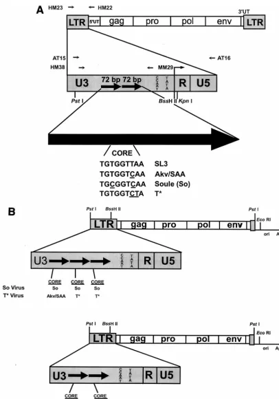
FIG. 1. Structures of the viruses and viral plasmids used in these studies. (A) Structures of the viral LTRs and sequences of the viral core elements. The top diagram shows the structure of the genome of proviruses of SL3 and the mutants derived from SL3. HM23 and HM22 are the PCR primers that were used to amplify the viral LTRs from tumor cell DNAs. The second diagram shows the structure of the viral LTRs. PCR primers used for sequencing studies are shown above the diagram. Restriction sites used for cloning experiments are shown below the diagram. The large arrow at the bottom represents one 72-bp enhancer repeat. The sequences of the cores present in the SL3, SAA, So, and T* enhancers are shown. Nucleotides that differ relative to the SL3 core are underlined. (B) Structures of the plasmids used to generate infectious virus particles. The top diagram represents the viruses with So and T* core mutations. The bottom diagram represents structures of the SL3 and SAA virus clones. In each diagram, the viral sequences are shown as boxes while the plasmid vector sequences are shown as lines. LTR sequences are enlarged to show the numbers of 72-bp enhancer repeats, which are indicated as black arrows. The sequence of the core in each repeat is shown below the LTR. Akv and SAA have the same core sequence; thus, this core is represented as Akv/SAA.
on November 9, 2019 by guest
http://jvi.asm.org/
enhancer repeat units were also reported to be present in tumors induced by a mutant of Mo-MuLV and in a tumor induced by feline leukemia virus (5, 25).
When proviruses in lymphomas induced by SAA contained three repeat units, they usually contained reversions or one of the putative suppressor mutations in at least two of the repeats (27). This was interpreted to mean that single base mutations in the core sequence occurred first and that this was followed by changes in the number of enhancer repeat units (27). We reasoned that viruses with the putative suppressor mutations present in more than one repeat unit would function as the most potently lymphomagenic viruses. Therefore, we isolated the LTR sequences that had So or T* cores present in multiple repeat units from proviruses present in SAA-induced lympho-mas (27). Consequently, the viruses that were tested were the viruses that we hypothesize actually caused the tumors in SAA-inoculated mice.
LTRs containing the So and T* cores were PCR amplified from DNA isolated from SAA-induced lymphomas (Fig. 1A). For the So core, a viral genome that had the mutation in three tandem repeat units was identified (27). For the T* core, a viral genome that had the mutation in the two promoter-proximal repeats of the three present in the provirus was identified (27). Restriction fragments containing the U3s of the amplified LTRs were used to replace the U3 sequences of an infectious clone of SL3 (Fig. 1B). Each of the plasmid clones contained a single LTR (Fig. 1B). Viral sequences were excised from the plasmids by digestion withPstI, self-ligated to generate con-catenates, and used to transfect NIH 3T3 fibroblasts as previ-ously described (27). This resulted in two identical LTRs in the progeny proviruses. Viral RNA transcribed from the trans-fected DNA generated infectious virus that spread and in-fected all the cells in the culture. After several passages of the cells over 3 weeks, the reverse transcriptase levels in the cul-ture supernatants reached a maximum level. XC cell assays indicated that both recombinant virus stocks had titers of about 104infectious virus units per ml. Stocks of SL3 and SAA were generated in parallel. These attained the same titers with the same kinetics as the mutants. Therefore, the differences among the core elements did not affect viral replication in fibroblasts.
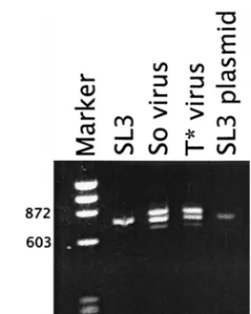
To verify that the viruses that contained the So and T* cores maintained the actual mutations, proviral DNA was PCR am-plified from the infected NIH 3T3 cells and sequenced. We previously showed that PCR amplification of viral DNA from infected cells resulted in multiple bands that came from pro-viruses with variable numbers of repeat units (27). Southern blotting confirmed that the bands were derived from proviruses and were actually present in the genomic DNA rather than resulting from polymerase jumping artifacts during PCR (27). DNA amplified from a control culture transfected with the infectious SL3 clone exhibited bands corresponding to LTRs
with one, two, and three repeats (Fig. 2). The two-repeat struc-ture predominated. Proviruses in the culstruc-tures infected with the recombinants containing So and T* core elements also showed bands corresponding to one, two, and three repeats (Fig. 2). Although the plasmids used to initiate the infection contained three repeat units, proviruses with two repeats were also abun-dant. We interpret this result to mean that reductions in the number of enhancer repeats occurred due to polymerase slip-page during viral replication. Most likely, viruses with the two LTR repeats have some replicative advantage over those with three repeats. Sequencing of the PCR-amplified bands indi-cated that the original mutations were indeed present in the viruses that replicated in NIH 3T3 cells. Therefore, the viral stocks used to infect mice consisted of mixtures of viral ge-nomes with varying numbers of tandem repeat units that re-tained the original core sequences.
Lymphomagenicity of the viruses containing the So and T* cores was examined by injecting the viruses into newborn NIH/ Swiss mice (Fig. 3). Parallel control studies were performed with SL3 and SAA viruses. SL3 virus caused tumors in 100% of infected mice, with a mean latency period of 69 days. SAA virus induced tumors in 88% of inoculated mice, with a mean latency of 112 days. It is unclear why only a fraction of the mice of this strain developed tumors after inoculation with the mu-tant. However, this effect was observed previously with other SL3 mutants in NIH/Swiss mice (29, 30). Of mice inoculated with the virus containing So core, 66% developed tumors, with a mean latency of 79 days. The decrease in the latency com-pared to that in mice inoculated with SAA was highly signifi-cant in a Studentttest (P,1026). Of the mice injected with the virus containing T* core, 62% developed tumors. The mean interval until onset of disease in the infected mice was 98 days. This result was also significant (P,0.001). The increased potency of the So- and T*-core-containing viruses strongly supported the argument that the single base pair substitutions relative to the Akv core in SAA were indeed second-site sup-pressor mutations.
Previous studies showed that mutations within the core ele-FIG. 2. LTR sequences amplified by PCR from proviral DNA isolated from infected NIH 3T3 cells. LTR sequences from cells infected with SL3, So-core-containing virus (So virus), or T*-core-So-core-containing virus (T* virus) are shown. LTR sequences from an SL3 plasmid are shown as a control for the size of a PCR product with two 72-bp repeats. Arrows on the right indicate the positions of fragments with one, two, or three 72-bp repeats. Marker isfX174 DNA digested withHaeIII. Numbers on the left indicate the sizes expressed in numbers of base pairs of two of the fragments in the marker lane.
on November 9, 2019 by guest
http://jvi.asm.org/
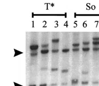
[image:4.612.362.504.70.248.2]ments sometimes altered the type of tumor that the virus caused (34). Therefore, it was important to test whether the tumors induced by the So- and T*-core-containing viruses were T-cell lymphomas. Pathologically, the tumors induced by both these viruses were identical to those induced by SL3 and SAA, and the affected mice showed grossly enlarged thymuses, spleens, lymph nodes, and livers. Southern blotting confirmed that the tumors contained rearrangements in T-cell receptorb chains (Fig. 4). Thus, the tumors induced by the So- and T*-core-containing viruses were T-cell lymphomas.
Enhancer sequences of proviruses in So and T*
virus-in-duced tumors.It was important to test whether the proviruses
in the lymphomas induced by the So- and T*-core-containing viruses retained the suppressor mutations within the proviral core sequences. Genomic DNA was prepared from four sep-arate tumors induced by each virus. Viral LTR sequences were PCR amplified and directly sequenced. The results are sum-marized in Table 1. All four lymphomas induced by the So virus contained proviruses with two and three LTR enhancer repeats. Sequencing analysis showed that all the cores in the amplified bands contained the So core (Table 1). LTRs with two repeats were amplified from all four lymphomas induced by the virus containing the T* core. In each case, the T* core sequence was present (Table 1). Three of the four tumors induced also contained proviruses with three LTRs. In one case, the structure was the same as the LTR of the virus in the original plasmid clone with T* cores in the promoter-proximal two repeats and an Akv/SAA core in the distal repeat (Table 1). In the other two cases, all three repeats had T* cores. In summary, the sequencing analysis showed that the viruses that were present in the tumors retained the original mutations. This observation provided additional strong evidence that the So and T* cores indeed contained suppressor mutations.
The enhancer core is one of many elements responsible for tumorigenicity in retrovirus-induced tumors. Mutations or de-letions of other factor binding sites can affect disease (9, 29, 34). In the 16 sequences that were obtained (Table 1), a total of two mutations were detected within the enhancer repeats. Both were outside the core element.
Effects of the So and T* enhancer cores on transcriptional
activity.The lymphomagenicity study showed that So and T*
enhancer cores rendered the viruses containing them more potent than the virus with the Akv/SAA core. The effects of MuLV LTR enhancers on transcription in the target cells for disease generally reflect the effects on tumorigenicity (32). We
[image:5.612.340.514.71.220.2] [image:5.612.53.294.72.228.2]therefore expected that the transcriptional activities of the So-and T*-core-containing enhancers would exceed that of SAA but be less than that of SL3 in T cells. To test this, LTR CAT plasmids (Fig. 5) were constructed with the LTRs of SL3, Akv, SAA, and the SAA-derived viruses with the suppressor muta-tions. U3 and R region sequences were inserted upstream of the CAT reporter gene as previously described (3, 32). We chose to test our set of CAT plasmids in T-cell lines because the SL3 virus caused disease specifically in T lymphocytes. In previous studies in T cells, SL3-CAT exhibited the greatest transcriptional activity, SAA-CAT displayed intermediate ac-tivity, and Akv-CAT had relatively low levels (27, 29, 30). In non-T-cell lines, the activities of SL3-CAT, SAA-CAT, and Akv-CAT were approximately equal (27). The same So- and T*-containing LTRs as were used to generate infectious virus (Fig. 1) were used to generate the CAT plasmids (Fig. 5). The abbreviations SoSoSo and CT*T* were used to symbolize these LTRs, where the symbols So, T*, and C represent the So, T*, and Akv/SAA cores, respectively. Each symbol represents the sequence of the core in an individual enhancer repeat. The number of symbols indicates the number of 72-bp enhancer repeats in the LTR, with the first symbol representing the FIG. 4. Southern blot analysis of T-cell receptorbrearrangements in tumors induced by the So- and T*-core-containing viruses. Four different lymphomas induced by the T*- (lanes 1 to 4) or the So-core-containing (lanes 5 to 8) virus were analyzed. Arrows to the left of the blot indicate the positions of the germline fragments detected by the probe.
TABLE 1. Sequences of core elements in proviral enhancers from tumor tissue
Individual mouse no.
Sequence of core elementa
2-Repeat 3-Repeat
So infected
2 So, So So, So, So
3 So, So So, So, So
5 So, So So, So, So
16 So, So So, So, So
T* infected
4 T*, T* Not present
7 T*, T* T*, T*, T*
8 T*, T* C, T*, T*
9 T*, T* T*, T*, T*
aThe sequence of the core element in each of the repeats is shown; the first abbreviation represents the core of the promoter-distal repeat, and the last represents the core in the promoter-proximal repeat. The sequence of So is TGCGGTCAA, that of T* is TGTGGTCTA, and that of C is the sequence found in the Akv/SAA core, TGTGGTCAA.
FIG. 3. Tumorigenicity of the So- and T*-core-containing viruses in NIH/ Swiss mice. Parallel control experiments were performed with SL3 and SAA.
on November 9, 2019 by guest
http://jvi.asm.org/
[image:5.612.311.550.551.686.2]distal repeat and the last representing the promoter-proximal repeat. In the So-CAT LTR, each of the three re-peats had a So core. In the T*-CAT LTR, two of the rere-peats had T* cores while the promoter-distal repeat retained the Akv core that was in the original mutant in the SAA-infected mouse. Because the So-CAT and T*-CAT constructs each had three 72-bp repeats, we generated for use as controls addi-tional LTR-CAT plasmids that contained SL3 core sequences and three enhancer repeats. TTT-CAT contained three peats, all with SL3 cores, and TTC-CAT contained three re-peats, the two distal ones containing SL3 cores and the pro-moter-proximal one containing an Akv/SAA core. Viral genomes containing these LTRs were initially identified in proviruses in SAA-induced tumors (27) and were obtained by PCR amplification of the viral genomes from the tumor sam-ples. These constructs allowed us to determine if a three-repeat structure had greater transcriptional activity than a two-repeat structure. Thus, we could distinguish between the effect of repeat number and the actual sequence of the core.
Wild-type SL3-CAT was used as a standard arbitrarily set at 100% activity. In the three T-cell lines tested, Akv-CAT had 10 to 20% as much activity as SL3-CAT (Fig. 6). SAA-CAT ex-hibited activity intermediate between those of SL3-CAT and Akv-CAT (Fig. 6). TTT-CAT and TTC-CAT had activities similar to that of SL3-CAT (Fig. 6). Therefore, the presence of a third SL3 core- or Akv core-containing repeat did not sig-nificantly alter the transcriptional activity of the LTR.
Both So-CAT and T*-CAT had higher transcriptional activ-ity than SAA-CAT. T*-CAT exhibited activactiv-ity comparable to those of SL3-CAT and the other three-repeat-containing LTRs (Fig. 6). So-CAT also had activity similar to those of SL3-CAT, TTT-CAT, and TTC-CAT, although it was consis-tently slightly higher than the other activities (Fig. 6). We conclude that changes in the core sequence increase the activ-ity of the viral LTR in T cells to a level similar to that of SL3. Presumably, the second-site suppressor mutations in the core element affect viral lymphomagenicity by increasing the tran-scriptional activity of the viral LTR in T cells.
Effects of second-site suppressor mutations on transcription
factor binding.The enhancer core of SL3 binds the
transcrip-tion factor CBF. Therefore, we tested whether the second-site suppressor mutations might increase transcriptional activity of the LTR in T cells by increasing CBF binding. CBF binding
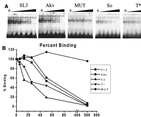
was assessed by using an EMSA. A nuclear extract from WEHI 7.1 T cells was used as the source of CBF protein. A radiola-beled probe containing the core element of SL3 was tested in the presence of increasing amounts of unlabeled competitor DNAs that were identical except for the mutations in the core element. The competitor DNAs contained the SL3, Akv, So, or T* cores. A mutated SL3 core (MUT) that does not bind CBF (43) was also tested as a negative control. Quantitation of the effects of the competitors showed that the SL3 core bound CBF slightly better than the Akv core (Fig. 7), consistent with previous observations (44). Quantitation of the EMSAs indi-cated that the So and T* cores bound CBF better than the Akv core and even slightly better than the SL3 core (Fig. 7). There-fore, increased binding of CBF to the So and T* cores corre-lated with the increased transcriptional activity in T cells and increased viral lymphomagenicity. When the oligonucleotides containing the So and T* cores were radiolabeled and used as probes in the EMSAs, no additional factors that bound to these sequences were detected (22). Thus, the So and T* enhancer cores appear to bind only CBF, implying that it is the critical transcription factor for the increased transcriptional and lymphomagenic effects of the suppressor mutations.
DISCUSSION
The lymphomagenicity data provided strong evidence that the So- and T*-core-containing viral variants contained sec-ond-site suppressor mutations within their core elements. The suppressor mutations were originally selected during the lym-phomagenic process in SAA-infected mice. They occurred within the same CBF binding sites as the original mutations and were detected in 20% of the lymphomas that developed in the SAA-infected mice. We previously showed that the SAA/ Akv core mutation was reverted in viruses present in 70% of the lymphomas in SAA-inoculated mice (27). Thus, both re-versions and suppressor mutations counteracted the detrimen-tal effects on pathogenicity of the original mutation.
Ethelberg et al. (9) identified second-site suppressor muta-tions that occurred in a mutant of SL3 in which both the core I and core II elements were altered by substitutions at three positions. However, in those studies, suppression was caused by a deletion of a binding site for the transcription factor nuclear factor 1 (NF-1) within the LTR enhancer repeats. In
FIG. 5. Structures of the plasmids used to measure transcriptional activities of the viral LTRs. Positions of the U3 and R sequences from the viral LTRs are shown. The CAT gene, ampicillin resistance gene (Apr), and plasmid origin of replication (ori) are also indicated. The arrow at the U3-R boundary indicates the direction of
transcription. Numbers represent distance from transcription initiation site. Black arrows in the boxes below the circles indicate the numbers of 72-bp repeats in the viral LTRs that were tested. The enhancer core sequences within each LTR of the plasmids are depicted below the arrows.
on November 9, 2019 by guest
http://jvi.asm.org/
our original analysis of tumor proviruses in 39 SAA-inoculated mice (27), in five different mice we detected proviruses that contained deletions of various sizes in the enhancer region. These deletions encompassed the NF-1 site in the promoter-distal, 72-bp repeat (26). Thus, it is likely that two distinct mechanisms, nucleotide substitutions within the core element and deletion of the NF-1 site, can lead to suppression of the single nucleotide mutation in SAA. However, Ethelberg et al. (9) did not detect any nucleotide substitutions within the core
element from mice inoculated with the core I-core II mutant. We hypothesize that this was because the mutant that was tested in those studies had multiple nucleotide substitutions within the core. Presumably, these had such a large effect on CBF binding that the mutations generating So and T* core elements failed to restore sufficient CBF binding and viral lymphomagenic activity for viruses containing the suppressor mutations to be selected.
The original So- and T*-core-containing virus clones that we generated in this study contained three 72-bp repeats within their LTRs (Fig. 1). Upon brief passage in NIH 3T3 fibroblasts, viruses with two repeats were abundant (Fig. 2). When the SL3 clone with two repeats was passaged in the same cells, genomes with three repeats were detectable but not nearly as abundant as genomes with two repeats (27). These observations suggest that the viruses with two repeats had some replicative advan-tage over those with three. Perhaps the genomes with two repeats are packaged more efficiently.
The presence of genomes with two repeats in the viral stocks used to inoculate mice means that the mice received a mixture of viruses with a variety of enhancer structures. In the case of the So-core-containing virus, the stock included viruses with enhancer repeats with SoSoSo and SoSo structures, where So indicates individual 72-bp units with a So core sequence. Pro-viruses with both types of enhancers were present in the lym-phomas induced by So virus (Table 1). In the case of the T*-core-containing virus, the mixture was more complex. The original clone had CT*T* enhancer structure. The two-repeat-containing viruses that formed by backward slippage during reverse transcription had the T*T* and CT* structures. These in turn could generate viruses with three repeats with the structures T*T*T*, CCT*, and the original CT*T* by forward slippage during reverse transcription in a subsequent replica-tive cycle. If multiple rounds of reverse and forward slippage occurred, then viruses with repeat structures CC and CCC should also have formed. Thus, mice inoculated with T*-core-containing virus probably received a mixture of isoforms dif-fering in their LTR enhancer structures. However, only T*T*T*, T*T*, and CT*T* were detected in the lymphomas that occurred in these mice (Table 1). This observation em-phasizes the strong selective pressure that the lymphomagenic process applies on viral enhancer structure.
The increased lymphomagenicity of the So-core-containing virus was correlated with increased CBF binding by the So core (TGCGGTCAA) compared to the SAA/Akv core (TGTGGT CAA) (Fig. 7). Thornell et al. (38) found that the mutation generating the So core increased CBF binding in the context of the SL3 enhancer (TGCGGTTAA versus TGTGGTTAA). Therefore, this mutation increased CBF binding in the context of either the SL3 or the SAA/Akv core. Using selected and amplified binding analysis, Melnikova et al. (23) also found CBF preferentially bound to DNA molecules containing a C at the position of the mutation generating So. However, a C occurs at this position in only 4 of 35 C-type mammalian retroviruses that were analyzed in an extensive comparison of viruses in this genus (12). The remaining 31 viruses have the T at this position (12), and several of these viruses were potent, T-cell lymphomagenic viruses. This suggests that the effect of a viral core element on pathogenicity depends on additional parameters than just the affinity of the binding site for CBF.
[image:7.612.55.290.68.569.2]The mutation generating T* (TGTGGTCTA) also increased CBF binding compared to the SAA/Akv core (Fig. 7). How-ever, Thornell et al. (38) found that this mutation in the con-text of the SL3 core (TGTGGTTTA versus TGTGGTTAA) had no effect on CBF binding. We interpret these observations to mean that the mutation generating T* affected CBF binding
FIG. 6. Transcription assays in (A) SL3H, (B) Lb91, and (C) Jurkat T-lymphocyte cell lines. Activities of the LTRs of the suppressor mutants are shown together with those of SL3, SAA, Akv, TTT, and TTC controls. The activity of the SL3 LTR in each cell line was set at 100%. Each error bar indicates one standard deviation.
on November 9, 2019 by guest
http://jvi.asm.org/
only when the preceding nucleotide in the core element was a C. Curiously, the selected and amplified binding analysis of Melnikova et al. (23) revealed that CBF bound to DNA mol-ecules with a T preferentially to molmol-ecules with an A at the position where the substitution led to the T* core element, even though almost all C-type retroviruses have the A at this position in their cores (12). One possibility to explain this is that neighboring nucleotides might affect which nucleotides at this position function best for CBF binding. Alternatively, the absence of a T at this position in all 35 C-type viruses analyzed might again reflect the possibility that the pathogenic activity of a viral core element depends on additional parameters than just the affinity of the binding site for CBF.
The increased binding of CBF due to the mutations gener-ating So and T* core elements was correlated with increased transcriptional activity of the viral LTRs in T cells relative to that of the SAA LTR (Fig. 6). The So- and T*-core-containing virus LTRs exhibited transcriptional activities in T cells com-parable to that of SL3. The So-containing LTR appeared even
slightly more active than the SL3 LTR. These results are con-sistent with the idea that relatively high levels of LTR tran-scriptional activity in T cells are necessary for T-cell lym-phomagenicity of the virus.
Although the So and T* cores were comparable to the SL3 core in CBF binding and transcriptional activity, the viruses containing them were slightly less lymphomagenic than SL3. Both viruses differed from SL3 in that they induced disease in fewer than 100% of inoculated mice (Fig. 3). T*-core-contain-ing virus induced lymphomas with a statistically significantly longer mean latency period than SL3 (P50.02). Although the mean latency periods to disease onset were not significantly different in the SL3 virus and So-core-containing virus (P5 0.16), the last mice infected with the latter virus to develop disease did so more slowly than any of the SL3-infected mice. One possible explanation for the lower lymphomagenicity of the viruses with So and T* cores is that the differences in CBF binding and transcriptional activity among the SL3, So, and T* cores were small and thus may have been affected by experi-FIG. 7. Binding of CBF to viral enhancer core sequences. (A) A radiolabeled oligonucleotide containing the core element from the SL3 LTR enhancer was used in EMSAs with crude nuclear extract from the WEHI 7.1 mouse T-cell line as a source of CBF. Five different, unlabeled, competitor DNAs were tested in increasing amounts of 0, 5, 12.5, 25, 50, and 500 ng per reaction as indicated. Competitor DNAs were isogenic with the probe except for the sequence of the core element. SL3, Akv, So, and T* indicate competitor DNAs from the corresponding viruses. MUT was a competitor DNA from SL3 with a 2-bp mutation that was previously shown to prevent CBF binding (43). (B) The amount of binding was quantified by PhosphorImager analysis and plotted. The amount of binding detected in each experiment with no competitor DNA was set at 100%.
on November 9, 2019 by guest
http://jvi.asm.org/
[image:8.612.65.532.68.454.2]mental variations. However, we were able to detect small dif-ferences between the CBF binding by the SL3 core and that by the SAA/Akv core that were consistent with those previously reported (44). Likewise, we also observed differences in tran-scriptional activity among LTRs containing the SL3, SAA, and Akv cores that were consistent with those previously observed (3, 27, 29, 30). Thus, it is conceivable that the So and T* cores indeed bind CBF and drive LTR transcriptional activity as effectively as the SL3 core. If so, then this raises the question why the viruses with So and T* core elements were less lym-phomagenic than SL3. Perhaps a certain level of CBF binding and transcription in T cells is necessary for viral nicity, but it is not sufficient for maximum viral lymphomage-nicity. If so, then this would suggest that the effect of the core on viral pathogenicity is determined by CBF binding affinity plus some additional process. Another possibility is that cul-tured lymphoma cell lines that were used to test the transcrip-tional activity of the viral LTRs did not precisely reflect the normal T lymphocytes in the mice.
In summary, suppressor mutations within the core element restored part or most of the lymphomagenic activity of SAA. Multiple selective pressures may have led to the presence of viruses with the mutations producing So and T* core elements in tumors in SAA-infected mice. The suppressor mutations may have allowed the viruses to replicate better in T cells, leading them to outgrow the original SAA mutant. It is also possible that proviruses that had suppressor mutations were more effective at activating cellular protooncogenes and thus causing proliferation of the tumor cells.
ACKNOWLEDGMENTS
We thank Angel Nieves and Joseph Pantginis for help with these studies.
This work was supported by NIH grants CA44822 and CA57337 to J.L. and by American Cancer Society grant RPG-94-012-VM to L.S.L. M.J.M. was supported by NIH training grant GM07491. Core facilities for oligonucleotide synthesis, PhosphorImager analysis, and DNA sequencing were supported by NIH Cancer Center Grant CA13330 to the Albert Einstein College of Medicine.
REFERENCES
1.Athas, G., B. Choi, S. Prabhu, P. Lobelle-Rich, and L. S. Levy.1995. Genetic determinants of feline leukemia virus-induced multicentric lymphomas. Vi-rology214:431–438.
2.Barat, C., and E. Rassart.1998. Members of the GATA family of transcrip-tion factors bind to the U3 region of Cas-Br-E and Graffi retroviruses and transactivate their expression. J. Virol.72:5579–5588.
3.Boral, A. L., S. A. Okenquist, and J. Lenz.1989. Identification of the SL3-3 virus enhancer core as a T-lymphoma cell-specific element. J. Virol.63:76– 84.
4.Bradford, M. M.1976. A rapid and sensitive method for the quantification of microgram quantities of protein utilizing the principle of protein-dye bind-ing. Anal. Biochem.72:248–254.
5.Brightman, B. K., C. Farmer, and H. Fan.1993. Escape from in vivo restric-tion of Moloney mink cell focus-inducing viruses driven by the Mo1PyF101 long terminal repeat (LTR) by LTR alterations. J. Virol.67:7140–7148. 6.Corcoran, L. M., J. M. Adams, A. R. Dunn, and S. Cory.1984. Murine T
lymphomas in which the cellularmyconcogene has been activated by retro-viral insertion. Cell37:113–122.
7.Corneliussen, B., A. Thornell, B. Hallberg, and T. Grundstro¨m.1991. Helix-loop-helix transcriptional activators bind to a sequence in glucocorticoid response elements of retrovirus enhancers. J. Virol.65:6084–6093. 8.Erickson, P., J. Gao, K.-S. Chang, T. Look, E. Whisenant, S. Raimondi, R.
Lasher, J. Trujillo, J. Rowley, and H. Drabkin.1992. Identification of break-points in t(8;21) acute myelogenous leukemia and isolation of a fusion transcript, AML1/ETO, with similarity to Drosophila segmentation gene, runt. Blood80:1825–1831.
9.Ethelberg, S., B. Hallberg, J. Lovmand, A. Luz, T. Grundstro¨m, and F. S. Pedersen.1997. Second-site proviral enhancer alterations in lymphomas induced by enhancer mutants of SL3-3 murine leukemia virus: negative effect of nuclear factor 1 binding site. J. Virol.71:1196–1206.
10. Ethelberg, S., J. Lovmand, J. Schmidt, A. Luz, and F. S. Pedersen.1997.
Increased lymphomagenicity and restored disease specificity of AML1 site (core) mutant SL3-3 murine leukemia virus by a second-site enhancer vari-ant evolved in vivo. J. Virol.71:7273–7280.
11. Goff, S., P. Traktman, and D. Baltimore.1981. Isolation and properties of Moloney murine leukemia virus mutants: use of a rapid assay for release of virion reverse transcriptase. J. Virol.38:239–248.
12. Golemis, E. A., N. A. Speck, and N. Hopkins.1990. Alignment of U3 se-quences of mammalian type C viruses: identification of highly conserved motifs and implications for enhancer design. J. Virol.64:534–542. 13. Gunther, C. V., and B. J. Graves.1994. Identification of ETS domain
pro-teins in murine T lymphocytes that interact with the Moloney murine leu-kemia virus enhancer. Mol. Cell. Biol.14:7569–7580.
14. Hallberg, B., J. Schmidt, A. Luz, F. S. Pedersen, and T. Grundstro¨m.1991. SL3-3 enhancer factor 1 transcriptional activators are required for tumor formation by SL3-3 murine leukemia virus. J. Virol.65:4177–4181. 15. Hedrick, S. M., D. I. Cohen, E. A. Nielsen, and M. M. Davis.1984. Isolation
of cDNA clones encoding T cell-specific membrane-associated proteins. Nature308:149–153.
16. Holland, C. A., C. Y. Thomas, S. K. Chattopadhyay, C. Koehne, and P. V. O’Donnell.1989. Influence of enhancer sequences on thymotropism and leukemogenicity of mink cell focus-forming viruses. J. Virol.63:1284–1292. 17. Kagoshima, H., K. Shigesada, M. Satake, Y. Ito, H. Miyoshi, M. Ohki, M. Pepling, and P. Gergen.1993. The Runt domain identifies a new family of heteromeric transcriptional regulators. Trends Genet.9:338–341. 18. Kamachi, Y., E. Ogawa, M. Asano, S. Ishida, Y. Murakami, M. Satake, Y. Ito,
and K. Shigesada.1990. Purification of a mouse nuclear factor that binds to both the A and B cores of the polyomavirus enhancer. J. Virol.64:4808–4819. 19. Khoury, G., and P. Gruss.1983. Enhancer elements. Cell33:313–314. 20. Lenz, J., D. Celander, R. L. Crowther, R. Patarca, D. W. Perkins, and W. A.
Haseltine.1984. Determination of the leukaemogenicity of a murine retro-virus by sequences within the long terminal repeat. Nature308:467–470. 21. Manley, N. R., M. O’Connell, W. Sun, N. A. Speck, and N. Hopkins.1993.
Two factors that bind to highly conserved sequences in mammalian type C retroviral enhancers. J. Virol.67:1967–1975.
22. Martiney, M. J., and J. Lenz.Unpublished results.
23. Melnikova, I. A., B. E. Crute, S. Wang, and N. A. Speck.1993. Sequence specificity of the core-binding factor. J. Virol.67:2408–2411.
24. Meyers, S., J. R. Downing, and S. W. Hiebert.1993. Identification of AML-1 and the (8;21) translocation protein (AML-1/ETO) as sequence-specific DNA-binding proteins: therunthomology domain is required for DNA binding and protein-protein interactions. Mol. Cell. Biol.13:6336–6345. 25. Miura, T., M. Shibuya, H. Tsujimoto, M. Fukusawa, and M. Hayami.1989.
Molecular cloning of a feline leukemia provirus integrated adjacent to the c-mycgene in a feline T-cell leukemia cell line and the unique structure of its terminal repeat. Virology169:458–461.
26. Morrison, H. L., and J. Lenz.1998. Unpublished results.
27. Morrison, H. L., B. Soni, and J. Lenz.1995. Long terminal repeat enhancer core sequences in proviruses adjacent toc-mycin T-cell lymphomas induced by a murine retrovirus. J. Virol.69:446–455.
28. Nielsen, A. L., P. L. Norby, F. S. Pedersen, and P. Jorgensen.1996. E-box sequence and context-dependent TAL1/SCL modulation of basic helix-loop-helix protein-mediated transcriptional activation. J. Biol. Chem.271:31463– 31469.
29. Nieves, A., L. S. Levy, and J. Lenz.1997. Importance of a c-Myb binding site for lymphomagenesis by the retrovirus SL3-3. J. Virol.71:1213–1219. 30. Pantginis, J., R. M. Beaty, L. S. Levy, and J. Lenz.1997. The feline leukemia
virus long terminal repeat contains a potent determinant of T-cell lym-phomagenicity. J. Virol.71:9786–9791.
31. Rowe, W. P., W. E. Pugh, and J. W. Hartley.1970. Plaque assay techniques for murine leukemia viruses. Virology12:1136–1139.
32. Short, M. K., S. A. Okenquist, and J. Lenz.1987. Correlation of leukemo-genic potential of murine retroviruses with transcriptional tissue preference of the viral long terminal repeats. J. Virol.61:1067–1072.
33. Speck, N. A., and D. Baltimore.1987. Six distinct nuclear factors interact with the 75-base-pair repeat of the Moloney murine leukemia virus en-hancer. Mol. Cell. Biol.7:1101–1110.
34. Speck, N. A., B. Renjifo, E. Golemis, T. Fredrickson, J. Hartley, and N. Hopkins.1990. Mutation of the core or adjacent LVb elements of the Moloney murine leukemia virus enhancer alters disease specificity. Genes Dev.4:233–242.
35. Sun, W., B. J. Graves, and N. A. Speck.1995. Transactivation of the Moloney murine leukemia virus and T-cell receptorb-chain enhancers bycbfandets requires intact binding sites for both proteins. J. Virol.69:4941–4949. 36. Sun, W., M. O’Connell, and N. A. Speck.1993. Characterization of a protein
that binds multiple sequences in mammalian type C retrovirus enhancers. J. Virol.67:1976–1986.
37. Thornell, A., B. Hallberg, and T. Grundstro¨m.1988. Differential protein binding in lymphocytes to a sequence in the enhancer of the mouse retro-virus SL3-3. Mol. Cell. Biol.8:1625–1637.
38. Thornell, A., B. Hallberg, and T. Grundstro¨m.1991. Binding of SL3-3 enhancer factor 1 transcriptional activators to viral and chromosomal en-hancer sequences. J. Virol.65:42–50.