1
A STUDY OF THE MICROBIOLOGICAL PROFILE IN
CHRONIC DACRYOCYSTITIS
DISSERTATION SUBMITTED FOR M.S.DEGREE
EXAMINATION
BRANCH III – OPHTHALMOLOGY
APRIL 2011
TIRUNELVELI MEDICAL COLLEGE
THE TAMILNADU DR. M.G.R. MEDICAL UNIVERSITY
2
CERTIFICATE
This is to certify that this dissertation entitled “A study of the Microbiological profile in Chronic Dacryocystitis” submitted by
Dr.A.Sukanya to the faculty of Ophthalmology The Tamil Nadu Dr. MGR Medical University, Chennai in partial fulfilment of the requirement for the award of M.S Degree in Branch III (Ophthalmology), is a bonafide research work carried out by her under our direct supervision and guidance.
Dr.A. Meenakshi Sundaram., Professor & HOD, Department of Ophthalmology,
Tirunelveli Medical College, Tirunelveli.
The Dean
3
Tirunelveli Medical College and Hospital, Tirunelveli-11.
Institutional Ethical Committee Certificate of Approval
4
DECLARATION
I solemnly declare that the dissertation titled " A study of the Microbiological profile in Chronic Dacryocystitis”is done by me at Tirunelveli Medical College hospital, Tirunelveli under the guidance and supervision of Prof. Dr.A.Meenakshi Sundaram M.S.
The dissertation is submitted to The Tamilnadu Dr. M.G.R.Medical University towards the partial fulfilment ofrequirements for the award of M.S. Degree (Branch III) in Ophthalmology.
Place: Tirunelveli
Date:
5
ACKNOWLEDGEMENT
I express my heartfelt gratitude to The Dean, Tirunelveli Medical College, Tirunelveli, for all the facilities provided for my study.
I take this opportunity to express my profound gratitude to
Dr.A.Meenakshi Sundaram M.S., Prof and HOD, Department of Ophthalmology for his support, guidance, advice and constant encouragement all through this study.
I am grateful to Dr.N. Palaniappan M.D., Prof and HOD Department of Microbiology, Associate professors, Assistant professors and staff of microbiology department for helping me in the timely completion of my study.
I am highly thankful to Dr.R.Mani M.S., Associate professor Department of Ophthalmology who helped me to sharpen my critical perceptions by offering valuable suggestions.
I am highly obliged to Dr. J.Kishore Kumar Jacob,
6
My special thanks to my postgraduate colleagues Dr.S.Hari Rama Subramanian and Dr.A. Subhalakshmi for their help and support.
I am very much thankful to the statistician, Mr. Arumugam for his invaluable support in helping me with the statistics and graphs.
I am immensely thankful to all my patients who volunteered whole heartedly for this study on informed consent, and who were responsible to make this work possible.
I express my sincere thanks to Broad Band Net café/DTP centre staffs for helping me to bring out the dissertation on time in a printed format.
7
TABLE OF CONTENTS
S.No Title P.No
1 INTRODUCTION 1
2 AIMS & OBJECTIVES 2
3 REVIEW OF LITERATURE 3
4 MATERIAL AND METHODS 29
5 OBSERVATION & RESULTS 34
6 DISCUSSION 53
8 CONCLUSION 58
BIBLIOGRAPHY
PR0FORMA
8
INTRODUCTION
Dacryocystitis is inflammation of the lacrimal sac usually secondary to obstruction of the nasolacrimal duct. The lacrimal drainage apparatus is an effective system for drainage of tears. When there is obstruction in the drainage apparatus, there is stasis of sac contents which forms a reservoir for development of infection. The close association of conjunctival and nasal mucosa with the sac makes it more prone to infection. The most common sources of infection are the nose, paranasal sinuses and pericystic tissues1.
9
AIM OF THE STUDY
1. To analyse the bacterial etiology in cases of chronic dacryocystitis reported at a tertiary level hospital.
2. To assess the association of chronic dacryocystitis with age, sex of patient, duration of symptoms and laterality.
10
REVIEW OF LITERATURE
Anatomy of lacrimal apparatus
Lacrimal gland
The lacrimal gland is a tubuloacinar gland with short branched tubules lying above and anterolateral to the eyeball and secretes tears through a series of ducts into the superior fornix. The lacrimal gland is divided by the superior transverse ligament of Whitnall into
A large orbital or superior part
A small palpebral or inferior part in continuity with the superior part.
Ducts of lacrimal gland
About 10-12 ducts pass downwards from the main gland and open into the lateral part of superior fornix3. Since all the ducts pass through the palpebral part of the gland, excision of the palpebral part alone amounts to excision of entire gland as far as secretory function of the gland is concerned.
Blood supply
11
Nerve supply
The sensory nerve supply to lacrimal gland is from the Lacrimal nerve, a branch of ophthalmic division of the fifth cranial nerve. The sympathetic nerve supply comes from the carotid plexus of the cervical sympathetics. The para sympathetic secretomotor fibres arise from the superior salivatory nucleus; pass through the Greater superficial petrosal nerve which joins with the Deep petrosal nerve to form the Nerve of pterygoid canal (vidian nerve). From here para sympathetic fibres after relaying in the Spheno palatine ganglion pass via the Zygomatic nerve on to the Lacrimal nerve to reach the lacrimal gland.
Accessory lacrimal glands
The accessory glands of Krause and Wolfring are located in the superior fornix and above the superior border of tarsus respectively
Tear film composition
Mucinous or inner layer secreted by goblet cells
Aqueous or intermediate layer secreted by main and accessory lacrimal glands
Oily or outer layer secreted by meibomian glands
Lacrimal puncta
12
punctum is slightly medial to the lower, their respective distances from the medial canthus being 6 and 6.5mm respectively4. The puncta are surrounded by a ring of dense fibrous tissue which keeps them patent. With each blink the puncta slide in the groove between the plica semilunaris and the eyeball.
Lacrimal canaliculi
The canaliculi are each 8-10 mm long. In 90% individuals they combine to form a common canaliculus that enters the lateral wall of the tear sac. A fold of mucosa, the valve of Rosenmuller, normally prevents tear reflux from the sac back into the canaliculi with operation of the tear pump.
Lacrimal sac
It lies in the lacrimal fossa located in the anterior part of medial orbital wall. The fossa is bounded by the anterior and posterior lacrimal crests. Medial to the sac is the middle meatus of the nose and anterior ethmoidal cells separated by the thin lacrimal bone and the thicker frontal process of maxilla. The angular artery and vein lies 7-8 mm medial to the medial canthal angle.
Nasolacrimal duct
13
fold (valve of Hasner). The duct is directed downwards laterally and slightly posterior.
Physiology of tear drainage
Tear drainage is brought about by an active lacrimal pump mechanism described by Rosengren-Doane5. It is constituted by fibres of the preseptal portion of the orbicularis which arise from the lacrimal fascia and the posterior lacrimal crest (Horner’s muscle). The contraction of the orbicularis muscle provides the motive force for drainage of tears.
Conjunctival flora
The conjunctiva and eyelids harbour many microorganisms which may be either resident flora or transient flora. Resident flora comprises of Staphylococcus epidermidis and Corynebacterium xerosis. The transient flora is composed of both pathogenic and non pathogenic organisms.
Normal conjunctival flora is held in check by the following factors
Flushing mechanism provided by tears
Bactericidal action of lysozymes present in tears
Phagocytosis of epithelial cells
Mechanical barrier of intact mucous membrane
14
The various pathogens found in the conjunctiva are as follows Gram positive
Diphtheroids
Staphylococcus aureus
Hemolytic and Non hemolytic Streptococci
Bacillus species
Gram negative
Hemophilus species
Moraxella species
Neisseria species
Enteric species
Escherichia coli
Klebsiella pneumonia
Enterobacterium species
Fungi
Aspergillus species
Mucor
Dematiaceous fungi
15
Evaluation of the nasolacrimal apparatus
Examination with diffuse illumination using magnification: is done to rule out causes of reflex hypersecretion located in lids, conjunctiva, cornea etc. This should exclude punctal causes of epiphora and any swelling in sac area.
Regurgitation test : a steady pressure is applied over lacrimal sac area. Reflux of mucopurulent discharge indicates chronic dacryocystitis with obstruction at lower end of nasolacrimal duct.
Fluorescein dye disappearance test6,7 : This is a physiologic test to analyse the lacrimal drainage which is useful in children and infants. The tears are stained with a moistened flourescein strip in each eye, patient is instructed not to wipe the eyes and blink at a normal rate and the tear film is observed after 5 minutes with cobalt blue filter of slit lamp. Persistence of dye and asymmetric clearance of dye from the conjunctival sac indicates a partial obstruction on the side retaining dye
Jones I test8 : Fluorescein in the tears is recovered in the inferior meatus by passing a cotton tipped applicator into the region of opening of NLD after 2 and 5 minutes. Staining of the applicator indicates patency. Non staining indicates anatomic or physiologic block.
16
lacrimal sac indicates physiologic block. Absence of dye indicates anatomical block.
Syringing : Topical anesthetic is instilled and the lower punctum is dilated with Nettleship’s punctum dilator. Saline is injected through a smooth tipped cannula passed into the lacrimal canaliculus 2 mm downwards and then turned medially to lie in the horizontal portion of the canaliculus. Irrigating solution is injected and results are observed. If saline passes freely into the nose or throat it indicates patent nasolacrimal system. On syringing the block may be either complete, partial or functional.
In complete NLD obstruction the fluid regurgitates through either the same punctum or the upper punctum depending on the level of obstruction.
In partial NLD obstruction there is a combination of saline reflux through upper punctum and the fluid passing into the patients throat.
In functional block the saline passes freely into the nose or throat, as a patent nasolacrimal system is present. However this irrigation is successful under increased hydrostatic pressure so there could still be a lacrimal pump failure.
17
punctum before withdrawal. The probe should not be forced through any area of resistance to avoid making a false passage.
In canalicular block the resistance is felt at or before 8mm. Both canaliculi are tested separately. In common canalicular block the obstruction is at 8-10 mm9 and the probe meets a soft resistance (Soft stop) and on moving the probe against the resistance , the tissues at the
medial canthus will be seen to move. In fibrosed small lacrimal sac or in nasolacrimal duct block, the probe meets a bony resistance beyond 10 mm (hard stop) and on moving the probe against it, there is no movement of tissues at the medial canthus .
Nasal endoscopy: is helpful in evaluation of nasal septal and turbinate diseases. It is now possible to directly visualise the nasal passages using endoscopic equipments. Presence of inferior turbinate hypertrophy or nasal polyps should be identified.
Mini endoscope known as dacryoscope allows direct visualisation of interior and lining of the lacrimal passages.
18
Dacryocystography : is used to delineate the anatomy of lacrimal system, define level of obstruction10 and identify the presence of any fistula, diverticula, stone or tumour in the sac. To perform it, a radio-opaque material such as lipiodol, dianosil or conray-280 is pushed into the sac using a lacrimal cannula. X rays are taken after 5 and 30 minutes to visualize the entire passage. For better anatomical visualization the modified technique known as ‘subtraction macrodacryocystography’ with canalicular catheterisation should be preferred.
Scintigraphy9 : It requires radioactive dye, sodium pertechnate, instilled into the tear film. The lacrimal sac area is scanned with gamma camera to follow the progress of the dye into canaliculi, sac, NLD, and nose.
19
Dacryocystitis
Dacryocystitis is the inflammation of the lacrimal sac. The disease has been known from earliest times owing to its grosser manifestations involving abscesses and fistulae on the face but was interpreted variously and the general term, ήγνλοψ (argilops, a fistula) was given to all swellings of inner canthus. In the middle of first century A.D., Vesalius and Fallopius described the lacrimal system with considerable accuracy. Further George E. Stahl of Halle in 1702 described that the pathological manifestations of ήγνλοψ were due to inflammation, not of the tissues generally but of the naso-lacrimal canal, these manifestations taking three forms - acute, chronic and hydropsia or ulceration ( i.e., with a fistula).
Duke elder remarked “Dacryocystitis - inflammation of lacrimal sac and duct – is a common and unpleasant disease, partly because of the troublesome and conspicuous symptoms it may cause, partly because it has little tendency to resolve and its adequate treatment presents considerable problems”.
It presents in three different forms :
Congenital dacryocystitis
Acute dacryocystitis
20
Congenital dacryocystitis
Congenital dacryocystitis is seen in 3-6% of otherwise healthy infants. It is due to a failure of canalization of the duct so that its lumen is blocked near the lower ostium by epithelial debris, by a membrane or by a stricture in the bony canal. Of these the commonest cause is an imperforate membrane leading onto epiphora. In 80 to 90% of cases, the residual membrane spontaneously dissolves within 2 to 4 months after birth. Majority of persistent cases will respond to conservative treatment with antibiotics and lacrimal sac massage.
Treatment includes
Proper counselling
Crigler’s massage – massage of the sac increases the hydrostatic
pressure and may rupture the membranous obstruction. In large majority of cases, the cause of failure of conservative management is the improper technique of sac massage. It is therefore imperative to explain the proper way of massaging the sac area. The mother should be instructed to compress the sac by applying pressure with the pulp of the index finger and with firm, gentle pressure slide the finger down for about one inch in the groove between medial inferior orbital rim and nose. Ten strokes should be applied four times a day.
21
Probing : should be delayed until the age of 12-18 months because spontaneous canalization occurs in 96% of cases53. Probing performed within the first 1-2 years of life has a very high success rate. It is carried out under general anaesthesia. The rationale is to manually overcome the obstructive membrane at the Hasner valve. After probing, the lacrimal system is irrigated with saline labelled with fluorescein. If fluorescein can be recovered by aspiration from the pharynx, successful probing is confirmed. Postoperative steroid antibiotic drops are used q.i.d for upto 3 weeks. If no improvement repeat probing is done after 6 weeks. Nasal endoscopic monitoring of probing is recommended to detect anatomical abnormalities. Results are excellent and 90% of children are cured by first probing and a further 6% by the second.
Lacrimal intubation : if second probing fails then temporary intubation with fine silastic tubes with or without balloon dilatation of the nasolacrimal duct may effect cure. Success rate of silicone intubation as been reported to be from 80 to 90% in congenital nasolacrimal duct obstruction 13,14.
22
In children all other causes of watering should be ruled out before the diagnosis of congenital dacryocystitis is made. The other causes of watering include outflow abnormality and reflex watering.
Congenital watering15
Outflow abnormality Reflex watering
Punctual agenesis Congenital glaucoma
Congenital dacryocystocele Epiblepharon Nasolacrimal duct obstruction Distichiasis Congenital lacrimal-cutaneousfistula
Acute dacryocystitis
Acute dacryocystitis is commonly caused by retention of tears leading on to secondary infection with bacteria. It is manifested by sudden onset pain, erythema and edema over sac area and epiphora.
Tenderness is localised in the medial canthal area.
Common organisms causing acute dacryocystitis are
Staphylococcus aureus
Staphylococcus epidermidis
Pseudomonas aeruginosa
Klebsiella and acenetobacter
23
Complications include
Lacrimal mucocele
Lacrimal fistula
Lacrimal abscess
Chronic conjunctivitis
Orbital cellulitis16
Orbital abscess16,17
Treatment
This includes institution of appropriate antibiotic therapy systemically and locally coupled with warm compresses over the inflamed area. Syringing or probing of an acutely inflamed sac is strictly contraindicated because of the risk of damage to the inflamed mucosa resulting in a fibrotic stricture. If untreated in the early stage, acute dacryocystitis may progress to form lacrimal abscess. In such cases, surgical drainage is indicated.
Dacryocystorhinostomy is usually necessary after the acute infection has been controlled and should not be delayed because of risk of recurrent infection.
Chronic dacryocystitis
24
swelling may be absent, although pressure over the sac commonly results in reflux mucopurulent material through the canalicular system onto the surface of the eye. Chronic dacryocystitis needs to be treated surgically prior to any intraocular surgeries.
A review of literature from Sattler (1885)30 to Reddy and Reddy (1955)31 on bacteriology of dacryocystitis reveals that a good number of organisms have been isolated from the sac-fluid. Of these, Streptococcus haemolyticus, Bacillus coli, Pneumococcus, Bacillus funduliformis,
Staphylococcus, Bacillus typhosus, Morax-axenfeld bacillus,
Pneumo-bacillus, Klebs-Loffler Pneumo-bacillus, bacillus Proteus vulgaris, Micrococcus
catarrhaliis, Bacillus fusiformis, Pfeiffer's bacillus, Friedlander's
bacillus, Koch's bacillus, Bacillus tetragenes, gram negative bacillus,
mycotic organisms have been found in pure cultures; the preponderating
organism being the pneumococcus. These organisms together with Staphylococcus have also appeared in mixed infections. While
Pneumococcus has been isolated in pure cultures in an overwhelming
majority, in mixed infections the Staphylococcus has been isolated in preponderating numbers.
varie-25
ties), Streptococcus (all varieties), Pneumococcus, Diphtheroids, were isolated in descending order of frequency.
Bale RN33 did a study of 100 consecutive cases of dacryocystitis, of which 43 were bilateral and 57 unilateral (total 143 eyes). It was found that the disease was more prevalent in females (57 per cent) than in males. The incidence of affection of left and right eyes was equal The disease has its commencement at 30 years age and maximum incidence in the 5th decade.The common organisms were D pneumonae, Coagulase negative Staphylococci, Niesseria catarrhalis, Coagulase positive
Staphylococci and Klebsiella. Mixed organism infection is not
uncommon.
Briscoe D et al,34 did a prospective study on changing isolates and antibiotic sensitivities of purulent dacryocystitis. The most common isolates were Pseudomonas (22%), Staphylococcus (13%), Enterobacter (10%), Citrobacter (10%), Streptococcus pneumonia, E.coli and Enterococcus (7%).
26
Huber Spitzy V et al,36 reported Staphylococcus as the most common organism. Also a significant number of gram negative bacilli were isolated of which E.coli was the most frequently grown.
Umesh Bareja et al,37 in their study on 114 eyes, in which 57.9% constituted gram positive cocci, found that the most effective antibiotic was cloxacillin with efficacy of 77%.
Sun X. et al,38 investigated a total of 100 samples obtained from lacrimal duct in 91 consecutive patients with chronic dacryocystitis, Staphylococcus species represented 34.5% of all strains, followed by Corynebacterium diphtheroids (15.5%). The sensitive test revealed
levofloxacin, ofloxacin and amikacin were the most effective antibiotics. Usha kim,39 studied 238 samples of dacryocystitis. Positive cultures were obtained from 197 samples and no growth in 41 samples. Of the positive cultures 124 were of gram positive organism and 93 were gram negative. The gram positive organisms were sensitive to chloramphenicol (98%),vancomycin (82%) and ofloxacin (75%). The gram negative organisms were sensitive to ofloxacin (83%), ciprofloxacin (81%), chloramphenicol (65%),gentamicin (60%), tobramycin (57%), amikacin(50%).
27
Staphylococcus aureus (10.8%) and Streptococcus pneumonia (8.7%)The
proportions of Staphylococcus aureus and Pseudomonas spp are higher in causing acute dacryocystitis, while the proportion of Coagulase negative Staphylococcus aureus is higher in chronic dacryocystitis. The
percentages of antibacterial resistant isolates were higher among bacterial species from chronic dacryocystitis.
Surgical management for NLD obstruction
The type of surgery depends on the level of obstruction
Obstruction at NLD or junction of NLD and sac - DCR
Obstruction at internal punctum or common canaliculus – DCR with silicone tubes
Canalicular obstruction at or beyond 8 mm from the punctum – canaliculo DCR
Obstruction less than 8mm from punctum in both canaliculi – conjunctivo DCR
Dacryocystorhinostomy is of three types
Conventional DCR
Endonasal DCR
Laser assisted DCR ( transcanalicular and endonasal )
Conventional DCR
28
Indications
NLD block in adults, chronic dacryocystitis and mucocele
NLD block in children not responding to probing and lacrimal intubation
Partial block in significantly symptomatic patients
As part of conjunctivo and canaliculo DCR
Contraindications
Malignancy of lacrimal sac
Dry eye syndrome
Blood dyscrasias
Acute dacryocystitis
Children below 3 years
Atrophic rhinitis
Rhinosporidiosis
Preoperative work up
• Hb
• Urine analysis
• Bleeding time, clotting time
• Blood pressure
29
• ENT examination to be done to rule out atrophic rhinitis, turbinate hypertrophy, deviated nasal septum, nasal polyps , malignancy
• Adequate treatment of acute dacryocystitis
• NSAIDS and anticoagulates should be stopped 3 days prior to surgery
• Should start instillation of antibiotic eye drops and nasal decongestants in ipsilateral nostril twice a day for 3 days prior to surgery. Sedatives may be given to relieve anxiety of the patient
Nasal packing
It helps in mucosal decongestion by pressure and vasoconstriction. Nasal pack consists of half inch wide and 16 inches long ribbon gauze soaked in xylocaine 2% with adrenaline. Naphazoline drops are instilled into the nasal cavity. The ipsilateral nasal cavity is packed under direct visualization using nasal speculum and packing forceps.
Surgical technique
30
with 4.0 silk in each flap. Identify and expose the anterior limb of medial palpebral ligament. It maybe cut or just dissected out.
Expose the periosteum over the anterior lacrimal crest and above it. Incise it 3 to 4 mm anterior and parallel to the anterior lacrimal crest. Reflect the periosteum from underlying bone and reach upto the anterior lacrimal crest. With the blunt end of the lacrimal sac dissector force a hole in the thin bone at the junction of lacrimal bone with the frontal process of maxilla in the centre or in posterior third of the lacrimal fossa. Fracture out a small piece to allow the finest bone punch into the hole and gradually enlarge the opening until the whole of lacrimal fossa is removed thus exposing the nasal mucosa. Anterior and posterior flaps are made in the lacrimal sac and in the nasal mucosa by an H-shaped incision. The posterior flaps are sutured together with 1 to 3 interrupted 5-0 chromic catgut sutures using half circle needle. Similarly the anterior flaps are sutured with sufficient tension on the flap to prevent it from collapsing. If MPL is incised it should be sutured with 4-0 or 5-0 vicryl. Skin is sutured with 5-0 prolene using continuous or interrupted sutures.
Complications of DCR
Hemorrhage – intranasal bleeding from nasal mucosa requires nasal packing for 24 hours. Injection ethamsylate or vitamin K may be given
31
2. Blockage of anastomosis due to improper suturing, redundant flaps, bony fragments, post operative hematoma
3. Post operative soft tissue infection
Sump syndrome
Management of failed DCR
Patients with mucopurulent discharge – repeat DCR with silicone tube intubation
In case of common canalicular block– canaliculo-DCR with silicone
tube intubation
In case of canalicular block – conjunctivo- DCR
Endonasal DCR18,19 Indications
Chronic dacryocystitis with NLD block
Mucocele
Contraindications
Lacrimal sac tumours
Dacryoliths
Procedure
32
the upper or lower canaliculus into the lacrimal sac. The light is located endoscopically on the lateral wall of the nose, and its position is noted. A 1 cm diameter circle of mucosa is removed at the site of transillumination to expose the underlying bone. Osteotomy is made using curette, punch, chisel, electric burr and lasers. In laser assisted procedures20 the laser used to make the osteotomy are Holmium YAG and Argon laser (blue green). The lacrimal sac is opened with a 45○ cutting forceps, and the opening is enlarged to approximately 1 cm. Metal stents attached to the silastic tubing at either end are passed through the upper and lower canaliculi . This helps in maintaining patency as the flaps are not anastomosed in this procedure.
Advantages
No cutaneous scar
Bloodless surgery
Speedy recovery
Day care procedure
Medial canthal anatomy is not disturbed
Bilateral DCR can be done in the same sitting
Disadvantages
High cost
33
Intranasal manipulations needed at times like inferior turbinate fracture or septoplasty
Canaliculo DCR
This procedure is done in cases with obstruction of common canaliculus. Following skin incision, dissection is carried out till the MPL. The common canaliculus is freed from the medial canthal tendon and all pericanalicular fibrous tissue is excised thoroughly. The most medial patent part of common canaliculus is intubated from the punctum by two ends of a silicone tube. The common canaliculus is sutured to the flaps of the lacrimal sac and DCR completed as usual.
Very often the sac is also scarred and the patent canalicular remnant may be sutured directly to the nasal mucosa. However a minimum of 8 mm of the canalicular system must be present to allow this anastomosis without undue tension. If less than 8 mm of the canalicular system is patent then conjunctivo DCR is considered
Conjunctiva DCR
34
reach the lacrimal sac. A Lester Jones tube is then advanced through this track to reach the lacrimal sac. The anterior flaps and nasal mucosa are then sutured together and DCR completed as usual.
An important aspect in postoperative care is educating the patients regarding care of tube and prevention of its extrusion. Extrusions are managed by immediate replacement.
Balloon catheter dilatation
Becker et al22 described this method which is effective in both congenital and acquired nasolacrimal duct obstruction. A guide wire is passed into the lacrimal sac over which a balloon catheter is guided and inflated. This is done twice for 10 minutes. Success rate is around 95% in congenital cases and 70% in acquired cases.
Dacryocystectomy
35
36
MATERIALS AND METHODS
The study of microbiological profile in chronic dacryocystitis and their susceptibility and resistance to antibiotics in South Tamil Nadu was carried out in the Department of Ophthalmology, Tirunelveli Medical College, Tirunelveli.
Settings : Ophthalmology ward, microbiological lab Study design : Single centre observational prospective hospital
based study
Period of study : June 2009 to June 2010 Ethical approval : Obtained
Tirunelveli Medical College Hospital is a tertiary care centre in South Tamil Nadu. The patient population is a fairly representative sample of the disease pattern in this region.
Inclusion Criteria:
Patients between 16-80 years of age.
All patients with complaints of mucopurulent discharge, epiphora, and sac abscess were included in this study.
Exclusion Criteria:
Patients less than 16 years of age.
37
Those who have been treated with systemic or topical antibiotics within 1 week of presentation were excluded from the study.
All cases of pseudoepiphora and epiphora caused by diagnoses other than nasolacrimal duct obstruction were also excluded from this study.
Procedure
Specimens for microbiological analysis were obtained by wiping a broth-moistened swab across the lower conjunctival cul-de-sac and also from everted punta by applying pressure over the lacrimal sac area. Surgically excised lacrimal sacs were collected and were also subjected to microbiological analysis. Those cases with mucoid or mucopurulent discharge on syringing of the lacrimal sac were advised surgery. In patients undergoing dacryocystectomy, a prior ENT clearance was obtained and the sacs were collected intraoperatively and subjected to microbiological examination.
38
inoculated media were incubated aerobically. The inoculated Sabouraud’s dextrose agar was incubated at 27◦C, examined daily, and discarded at 3 weeks if no growth was seen. The inoculated blood agar, chocolate agar, thioglycollate broth, brain–heart infusion broth were incubated at 37◦ C, examined daily, and discarded at 7 days if growth was not seen.
Microbial cultures were considered significant if
Growth of the same organism was demonstrated on more than one solid-phase medium.
or
There was confluent growth at the site of inoculation on one solid medium.
or
Growth of one medium was consistent with direct microscopy findings (ie, appropriate staining and morphology with Gram stain)
or
The same organism was grown from more than one specimen.
39
The broth culture was then allowed to incubate at 37◦C until a slightly visible turbidity appeared (usually 2–5 h), and the turbidity of the inoculum was compared with 0.5 Macsarland standard. Standardized bacterial inoculum was inoculated on the Mueller–Hinton agar using a sterile, non-toxic swab evenly over the entire surface of the agar plate to obtain a uniform inoculum. Blood agar was used for Streptococci and other fastidious bacteria. The inoculated plates were then allowed to dry for 3–5 min. The antibacterial impregnated discs were applied with a gap of 24mm between them and the plates were incubated at 37◦C within 15 min after applying these discs.
40
41
OBSERVATION AND RESULTS
Percentage distribution of the patients according to their sex
Total number of patients
100
The percentage distribution of the patients in table 1 reveals that the incidence of chronic dacryocystitis is more (64%) among females than males (36%).
64%
42
Table : 1
Percentage distribution of the patients according to their sex
Total number of male patients
Total number of female patients
36 64
The percentage distribution of the patients in table 1 reveals that incidence of chronic dacryocystitis is more (64%) among females
36%
male females
43
Table : 2
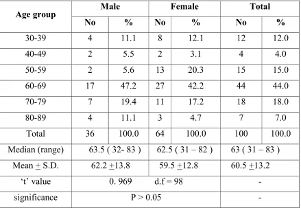
Age and sex distribution of the patients
Age group Male Female Total
No % No % No %
30-39 4 11.1 8 12.1 12 12.0
40-49 2 5.5 2 3.1 4 4.0
50-59 2 5.6 13 20.3 15 15.0
60-69 17 47.2 27 42.2 44 44.0
70-79 7 19.4 11 17.2 18 18.0
80-89 4 11.1 3 4.7 7 7.0
Total 36 100.0 64 100.0 100 100.0
Median (range) 63.5 ( 32- 83 ) 62.5 ( 31 – 82 ) 63 ( 31 – 83 ) Mean + S.D. 62.2 +13.8 59.5 +12.8 60.5 +13.2
‘t’ value 0. 969 d.f = 98 -
significance P > 0.05 -
The above table describes the median age of males to be 63.5 years and that of females 62.5 years. The mean ages of males and females were 62.2 +13.8 and 59.5 +12.8 years respectively. The difference between the mean age of the sexes was not statistically significant ( P > 0.05 )
Comparison of age and sexwise distribution of chronic dacryocystitis
0 5 10 15 20 25 30 35 40 45
30-39 40-49
44
Comparison of age and sexwise distribution of chronic dacryocystitis
49 50-59 60-69 70-79
male female total
Comparison of age and sexwise distribution of chronic dacryocystitis
45
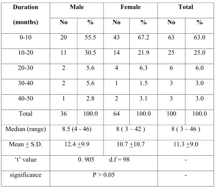
Table 3
Comparison of the sexwise distribution of chronic dacryocystitis Duration
(months)
Male Female Total
No % No % No %
0-10 20 55.5 43 67.2 63 63.0
10-20 11 30.5 14 21.9 25 25.0
20-30 2 5.6 4 6.3 6 6.0
30-40 2 5.6 1 1.5 3 3.0
40-50 1 2.8 2 3.1 3 3.0
Total 36 100.0 64 100.0 100 100.0
Median (range) 8.5 (4 - 46) 8 ( 3 – 42 ) 8 ( 3 – 46 ) Mean + S.D. 12.4 +9.9 10.7 +10.7 11.3 +9.0
‘t’ value 0. 905 d.f = 98 -
significance P > 0.05 -
Comparison of sex
0 10 20 30 40 50 60 70
0-10 10-20
46
Comparison of sex wise distribution of chronic dacryocystitis
20 20-30 30-40
40-wise distribution of chronic dacryocystitis
-50
47
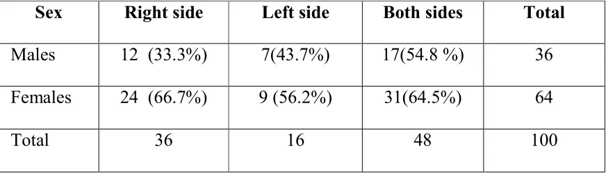
Table : 4
Laterality of dacryocystitis in males and females
Sex Right side Left side Both sides Total
Males 12 (33.3%) 7(43.7%) 17(54.8 %) 36 Females 24 (66.7%) 9 (56.2%) 31(64.5%) 64
Total 36 16 48 100
Sexwise comparison of laterality of chronic dacryocystitis
0 5
males females
48
Sexwise comparison of laterality of chronic dacryocystitis
10 15 20 25 30
both eyes left eye right eye
Sexwise comparison of laterality of chronic dacryocystitis
49
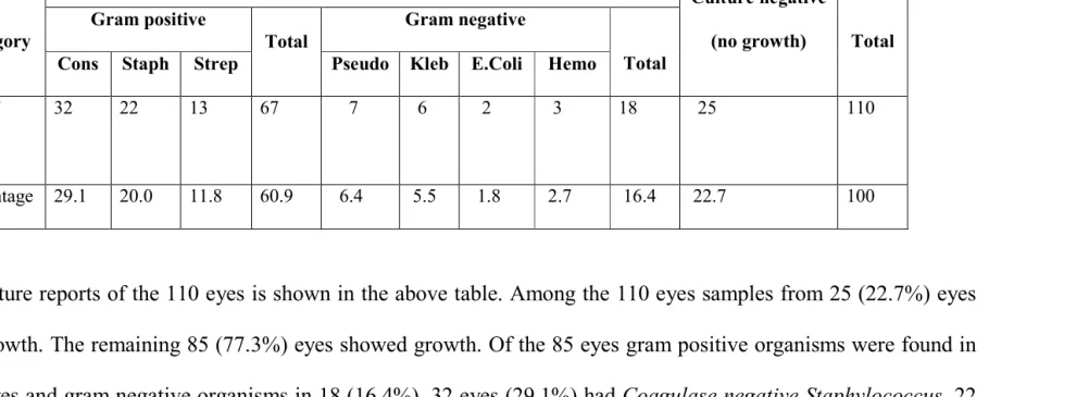
Table 5
Percentage distribution of culture positive and culture negative results
Category
Culture positive
Culture negative
(no growth) Total
Gram positive
Total
Gram negative
Total
Cons Staph Strep Pseudo Kleb E.Coli Hemo
No. of
eyes
32 22 13 67 7 6 2 3 18 25 110
Percentage 29.1 20.0 11.8 60.9 6.4 5.5 1.8 2.7 16.4 22.7 100
The culture reports of the 110 eyes is shown in the above table. Among the 110 eyes samples from 25 (22.7%) eyes
showed no growth. The remaining 85 (77.3%) eyes showed growth. Of the 85 eyes gram positive organisms were found in
67 (60.9%) eyes and gram negative organisms in 18 (16.4%). 32 eyes (29.1%) had Coagulase negative Staphylococcus, 22
eyes (20.0%) had Staphylococcus aureus, 13 eyes (11.8%) had Streptococcus species,7 eyes (6.4%) had Pseudomonas
Distribution of culture positive and culture negative results
Strep 15%
Pseudo 8%
50
Distribution of culture positive and culture negative results
CoNS 38%
Staph 26% Kleb
7%
E.coli 2%
Hemo 4%
Distribution of culture positive and culture negative results
Surgical treatment of chronic dacryocystitis Total number of
patients
Total number of
110
This table shows the surgical treatment done for those cases which had regurgitation of pus on syringing. DCR was done in 18 eyes (16.4%) and DCT was done in 92 eyes (83.6%).
Surgical treatment of chronic dacryocystitis
0 10 20 30 40 50 60 70 80 90 100 DCT 51 Table 6
Surgical treatment of chronic dacryocystitis Total number of patients
undergoing DCT
Total number of patients undergoing DCR
92
This table shows the surgical treatment done for those cases which had regurgitation of pus on syringing. DCR was done in 18 eyes (16.4%) and DCT was done in 92 eyes (83.6%).
rgical treatment of chronic dacryocystitis
DCR
surgery
Total number of patients undergoing DCR
18
Association between age and surgical procedure done
Treatment
Age of patients < Median Median + No. % No.
DCR 16 88.9 2 DCT 33 35.9 59 Total 49 44.5 61
The above table shows that the age was associated with the surgical procedure done. DCR was done for younger patients and DCT was done for older patients. The association was statistically very highly significant (P<0.001).
Association between age and the surg
0 10 20 30 40 50 60 DCR 52 Table 7
Association between age and surgical procedure done Age of patients
Χ2 (chi-square)
d.f. Median + Total
No. % No. %
11.1 18 100.0
15.05 1 64.1 92 100.0
55.5 110 100.0
above table shows that the age was associated with the surgical procedure done. DCR was done for younger patients and DCT was done for older patients. The association was statistically very highly significant
Association between age and the surgical procedure done
DCT
<MEDIAN
>MEDIAN
significance
P<0.001
above table shows that the age was associated with the surgical procedure done. DCR was done for younger patients and DCT was done for older patients. The association was statistically very highly significant
ical procedure done
<MEDIAN
53
Table 8
Percentage distribution of sensitivity of different antibiotics to organisms
The sensitivity of the drugs to the organisms is shown in the above table. Bacterial isolates showed higher sensitivity
to Gatifloxacin (84.7%) followed by Cefazolin (80%), Moxifloxacin (76.5%) and Ciprofloxacin (72.9%). Least sensitive
among the ten drugs was Gentamicin (44.7%).
S.no Drug
CoNS n=32 Staph n=22 Strep n=13 Pseudo n=7 Kleb n=6 E.coli n=2 Hemo n=3 Total n=85
No % no % no % no % no % no % no % no %
1 GA 31 96.9 21 95.6 12 92.3 1 14.3 5 83.3 2 100.0 0 0 72 84.7
2 M 28 87.5 19 86.4 12 92.3 0 0 5 83.3 1 50.0 0 0 65 76.5
3 C 27 84.4 18 81.8 10 76.9 1 14.3 5 83.3 1 50.0 0 0 62 72.9
4 V 23 71.9 15 68.2 10 76.9 1 14.3 3 50.0 0 0 0 0 52 61.2
5 O 16 50.0 10 45.5 9 69.2 6 85.7 5 83.3 2 100.0 3 100.0 51 60.0
6 CZ 26 81.3 17 77.3 8 61.5 7 100.0 6 100.0 1 50.0 3 100.0 68 80.0
7 A 26 81.3 16 72.7 4 30.8 4 57.1 4 66.7 2 100.0 0 0 56 65.9
8 T 21 65.6 14 63.6 5 38.5 7 100.0 4 66.7 1 50.0 3 100.0 55 64.7
9 GM 16 50.0 10 45.5 3 23.1 1 14.3 4 66.7 1 50.0 3 100.0 38 44.7
0 5 10 15 20 25 30 35
GA M C
Percentage distribution of sensitivity of different antibiotics to organisms
54
V O CZ A T GM
Percentage distribution of sensitivity of different antibiotics to organisms
GM CF
CoNS Staph Strep Pseudo Kleb E.coli Hemo
55
Table 9
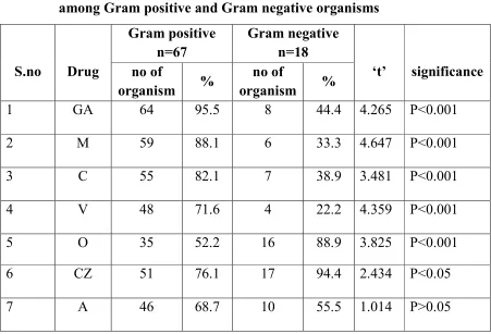
Comparison of distribution of percentages of antibiotic sensitivity among Gram positive and Gram negative organisms
S.no Drug
Gram positive n=67
Gram negative n=18
‘t’ significance no of
organism %
no of
organism %
1 GA 64 95.5 8 44.4 4.265 P<0.001
2 M 59 88.1 6 33.3 4.647 P<0.001
3 C 55 82.1 7 38.9 3.481 P<0.001
4 V 48 71.6 4 22.2 4.359 P<0.001
5 O 35 52.2 16 88.9 3.825 P<0.001
6 CZ 51 76.1 17 94.4 2.434 P<0.05
7 A 46 68.7 10 55.5 1.014 P>0.05
The comparison of sensitivity of antibiotics between gram positive and gram negative organisms is shown in the above table. Gram positive bacteria were more sensitive to gatifloxacin, moxifloxacin, ciprofloxacin and vancomycin. The above sensitivities were statistically very highly significant ( P<0.001). The antibiotics such as ofloxacin, cefazolin, tobramycin and cefotaxime had more sensitivity with gram negative than gram positive organisms and the sensitivities were statistically significant (P<0.05). The antibiotics amikacin and gentamicin had no significant
Comparison of distribution of percentages of antibiotic sensitivity among Gram positive and
0 10 20 30 40 50 60 70 80
GA M C
56
Comparison of distribution of percentages of antibiotic sensitivity among Gram positive and
Gram negative organisms
V O CZ A T GM
Comparison of distribution of percentages of antibiotic sensitivity among Gram positive and
CF
gram +ve gram -ve
57
Table 10
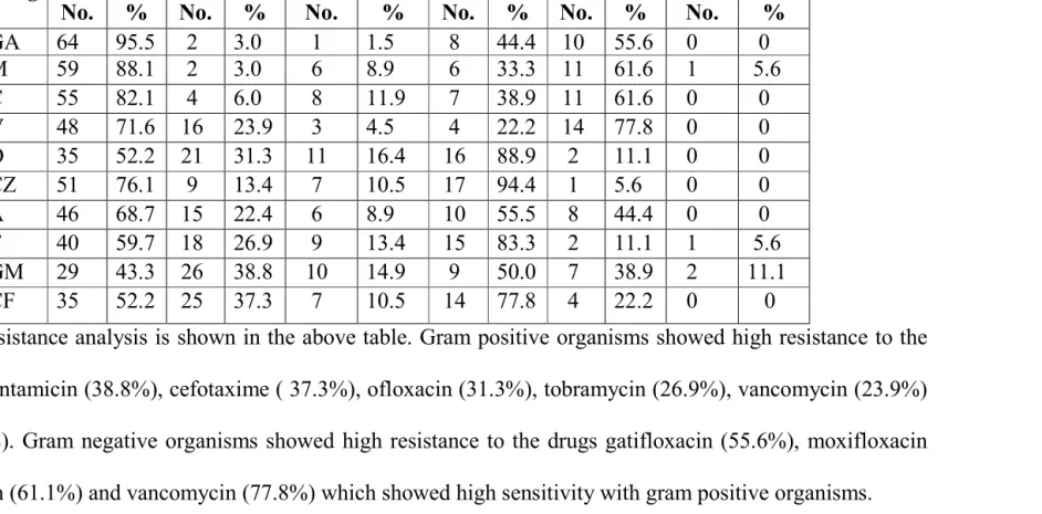
Analysis of invitro resistance pattern
s.no Drug
Gram positive n = 67 Gram negative n = 18
Sensitivity Resistance Intermediate Sensitivity Resistance Intermediate No. % No. % No. % No. % No. % No. %
1 GA 64 95.5 2 3.0 1 1.5 8 44.4 10 55.6 0 0
2 M 59 88.1 2 3.0 6 8.9 6 33.3 11 61.6 1 5.6
3 C 55 82.1 4 6.0 8 11.9 7 38.9 11 61.6 0 0
4 V 48 71.6 16 23.9 3 4.5 4 22.2 14 77.8 0 0
5 O 35 52.2 21 31.3 11 16.4 16 88.9 2 11.1 0 0
6 CZ 51 76.1 9 13.4 7 10.5 17 94.4 1 5.6 0 0
7 A 46 68.7 15 22.4 6 8.9 10 55.5 8 44.4 0 0
8 T 40 59.7 18 26.9 9 13.4 15 83.3 2 11.1 1 5.6
9 GM 29 43.3 26 38.8 10 14.9 9 50.0 7 38.9 2 11.1
10 CF 35 52.2 25 37.3 7 10.5 14 77.8 4 22.2 0 0
The in vitro resistance analysis is shown in the above table. Gram positive organisms showed high resistance to the
antibiotics namely gentamicin (38.8%), cefotaxime ( 37.3%), ofloxacin (31.3%), tobramycin (26.9%), vancomycin (23.9%)
and amikacin (22.4%). Gram negative organisms showed high resistance to the drugs gatifloxacin (55.6%), moxifloxacin
Analysis of invitro resistance in gram positive organisms
0 10 20 30 40 50 60 70
GA M C
58
Analysis of invitro resistance in gram positive organisms
V O CZ A T GM CFCF
Analysis of invitro resistance in gram negative organisms
0 2 4 6 8 10 12 14 16 18
GA M C
59
Analysis of invitro resistance in gram negative organisms
V O CZ A T GM CFCF
60
DISCUSSION Sex distribution
The number of male patients in our study was 36 and females 64. Thus there was a female preponderance.
Duke – Elder1,41 states that while the disease in newborn, affects both sexes equally, its occurrence among adults is in the ratio of 75-80% - females to 25-30% - males. Meller (1929) Ruiz Barranco and Martinez Roman (1966) stated that this difference was due to a narrower bony nasolacrimal canal in females.
Heinoven (1920) blamed that the high incidence amongst females is due to the fact that females had a higher nasal index.
Bharathi MJ et al,40 in his study found overall female to male ratio was 3.9:1 and females (80.9) were more in number than males (19.1) The incidence of dacryocystitis in females had been recorded by Traquair41 as 83 percent by Summer Skill, W. Ft 43 as 73 per cent , Sood et al 44 as 63.3 percent and by Bale RN 33 as 57 per cent. This is in concurrence with our study.
Age
61
Bale RN 33 reports in his study that nearly 78% of cases were over the age of 30 years Amongst this the peak was at 51-60 years of age (26%).
Matthew W. et al45 reported the mean age of presentation as 60.7 yrs.
Bharathi MJ et al40 in his study found that patients with age greater than 30 years were significantly more in number in chronic dacryocystitis (90%) than those aged less than 31 years (10%).
Laterality
In our study in males, right eye was involved in 33.3%, left eye in 43.7% and both eyes in 54.8%. In females the incidence in right eye was 66.7%, left eye was 56.2% and both eyes was 64.5% The total incidence in right eye was 36%, incidence in left eye was 16% and involvement of both eyes was 48%.
The affection of side was found by Sood et al44 as 50 each right and left. Veris46 observed the occurrence to be on the leftas 66%.
Bale RN33 found that the incidence was 51.04% in left eye H.Basil Jacobs (1959)47 in his study found that right side was involved in 53 cases left side in 37 cases and 14 cases were bilateral.
62
Thus there is no predilection to any side and it may affect both sides equally.
Duration of symptoms
It was observed that 63% of patients presented to us within the first 10 months and 25% within the next 10 months of the onset of symptoms.
Duct patency
On syringing of the total 200 eyes, nasolacrimal duct was found to be patent in 53 eyes, regurgitation of clear fluid was seen in 37 eyes and regurgitation of pus was seen in 110 eyes. Thus the number of eyes with infection was 110.
Bacteriological profile
The most common gram positive organism cultured in our study was Coagulase negative Staphylococcus (29.1%) followed by Staphylococcus aureus (20%) and then Streptococcus (11.8%).
Similar incidence was reported by Bharathi MJ et al40, in cases of chronic dacryocystitis, CoNS (563 of 1275; 44.2%) followed by S.aureus (138 of 1275; 10.8%) and S. pneumoniae (111 of 1275;8.7%) were found to be the predominant bacterial pathogens.
63
Streptococcus species represented 20% in our study which is
higher than Huber Spitzy et al36 (2%), Coden et al51 (2.3%) and Hartikainen et al52 (5%).
In our study Gram negative organisms contributed to 16.4% of all isolates. The most frequently isolated species being Pseudomonas aeruginosa (7/18; 6.4%) followed by Klebsiella (6/18; 5.5%).
Similarly Das JK et al48 found gram negative organisms to be 25% with a predominance of Pseudomonas aeruginosa.
Coden DJ et al50 observed gram negative organisms in 27% of all isolates, including Pseudomonas in 9%.
Huber Spitzy et al36 reported gram negative organisms accounting for 26% isolates, the most frequent being E.coli (12%)
Surgery
In our study out of 110 eyes, DCR was done in 18 eyes (16.4%) and DCT was done in 92 eyes (83.6%). DCR was done for younger patients (less than 40 years of age) and DCT was done for older patients. The association was statistically very highly significant (P<0.001).
Antibiotic Sensitivity
64
organisms were highly sensitive to Cefazolin (94.4%) .The above sensitivities were statistically very highly significant (p<0.001). Neither gram positive nor gram negative organisms (p>0.05) had significant sensitivity to Amikacin and Gentamicin.
The analysis of invitro resistance showed that Gram positive organisms had high degree of resistance to Gentamicin (38.8%), Cefotaxime (37.3%) Ofloxacin (31.3%) and Tobramycin (26.9%). For gram negative organisms Vancomycin (77.8%), Moxifloxacin (61.1%) and Ciprofloxacin (61.1%) had highest resistance. Least resistance was seen with Cefazolin (5.6%).
65
CONCLUSION
In our study, incidence of chronic dacryocystitis was found more in females than males, the mean age of presentation was 60.5+13.2 years, most of the patients presented to us within 10 months of the onset of symptoms. DCT was done in 92 eyes and DCR was done 18 eyes. The most common micro organism isolated was Coagulase negative Staphylococcus followed Staphylococcus aureus & Streptococcus
species. Gram positive organisms showed highest sensitivity to Gatifloxacin , Moxifloxacin and Gram negative organisms to Cefazolin . The high rate of micro-organism positive cultures suggests that adult patients should be treated for their lacrimal sac infection before any intraocular surgery because of the potential risk of post operative infection. Bacterial flora is abundant at the eyelid margin and the setting is conducive to a possible spontaneous mutation that can cause antibiotic resistance. Hence a prudent use of antibiotics is essential. Unnecessary usage of antibiotic leads to emergence of resistance. Thus in cases of regurgitation it is better to use Gatifloxacin or Moxifloxacin as they are more effective.
66
BIBLIOGRAPHY
1. Duke- Elder S. System of Ophthalmology Vol XII. The Ocular Adnexa. Part II St. Louis, CU Mosby Co., 1974, PP.699-714.
2. Kushner BJ. Congenital Nasolacrimal system obstruction. Arch Ophthalmol 1982; 100:697.
3. Khurana AK, Indu khurana, Anatomy and Physiology of eye. (CBC publication ) 1998: 337-364
4. Wolff’s Anatomy of Eye and orbit 8th edition.
5. Bilyk JR, Jokobie FA. Embryology and Anatomy of the Orbit & Lacrimal system In Duane T (Ed) Foundation Vol 1 Philadelphia; JB Lippincott, 2000.
6. Katowito JA, Welsh MG. Timing of intial probing and irrigation in congenital Nasolacrimal duct obstruction. Ophthalmology 1987; 94:698
7. Zappia RJ. Milder B : Lacrimal drainage function. Part 11: The fluorescein dye disappearance test. Opthalmology 74: 160-162, 1972. 8. Zappia RJ. Milder B : Lacrimal drainage function. Part 1. The Jones
fluorescein test. Am J Opthalmol 1972; 74: 154.
67
10. Mallik SRK, Chatterjee DI. Dacryocystography of normal and disturbed lacrimal passage, Orient Arch Ophthal, 1970; 8:5
11. Russell EJ, Czervionke L, Huckman M, et al : CT of inferomedial orbit and lacrimal drainage apparatus: Normal and pathologic anatomy. AJR 145: 1147-1154, 1985.
12. Robb RM. Success rate of nasolocrimal duct probing at time intervals after 1 year of age. Ophthalmol 1998; 105 : 1301
13. Leone CR Jr, Van Gemert JV. The success rate of silicone inubation in congenital lacrimal obstruction. Ophthalmic Surgery 1990; 21:91 14. Ratliff CD, Meyer DR. Silicone intubation without intranasal fixation
for treatment of congenital nasolacrimal duct obstruction Am J of Ophthal 1994; 118:781.
15. Evaluation and Management of the Tearing patient. American Academy of Ophthalmology, Basic and Clinical Science Course 2004-5; Vol 7.
16. Myren Yanoff, Jay S. Duker. The lacrimal drainage system, chapter – 98, In: Ophthalmology, 2nd edition. Mosby publication, pp.761-769. 17. Kikkawa DO, et al : orbital cellulitis and abscess secondary to
dacryocystitis. Arch ophthalmol 2002, 120:1096-99.
68
19. Woog JJ, Metson R, Puliafito CA, Holmium: YAG endonasal laser DCR. Am J Ophthalmol 1993; 116: 1-10.
20. Gonnering RS, Lyon DB, Feschner JC et al Endoscopic laser assisted lacrimal surgery. Am J Ophthalmol 1991; 11: 152.
21. Jones LT. Conjunctivodacryocystorhinostomy. Am J Ophthalmol 1965; 59:773.
22. Becker BB, Berry FD, Koller H, Balloon catheter dilatation for treatment of congenital nasolacrimal duct obstruction. Am J Ophthalmol 1996; 121:304.
23. Duke-Elder S, MacFaul PA. The ocular adnexa. In: Duke-Elder S (ed). System of Ophthalmology, Vol 13. Part II. Lacrimal, Orbital and Para-orbital Diseases. Henry Kimpton: London, 1974, pp 715–718. 24.Holds JB, Anderson RL, Wolin MJ. Dacryocystectomy for the
treatment of dacryocystitis patients with Wegener's granulomatosis. Ophthalmic Surg 1989; 20(6): 443–444.
25.Boynton, JR, Anawis MA. Role of dacryocystectomy in the management of failed dacryocystorhinostomy associated with chronic dacryocystitis. Ophthalmic Surg Lasers 1996; 27(2): 133–136.
69
27.Bartley GH, Nichols WL. Hemorrhage associated with dacryocystorhinostomy and the adjunctive use of Desmopressin in selected patients. Ophthalmology 1991; 98: 1864.
28.Meyers EF. Cocaine toxicity during dacryocystorhinostomy. Arch Ophthalmol 1980; 98: 842–843.
29.Meyer DR. Comparison of oxymetazoline and lidocaine versus cocaine for outpatient dacryocystorhinostomy. Ophthalmic Plastic Reconstruct Surg 2000; 16(3): 201–205
30.Sattler (1885), Text Book of Ophthalmology, Duke Elder, V., p. 5318.
31.Reddy, P. S., and Reddy, D. B. (1955) Journal of I. M. A., 24, 413
32. Prasad B, Ram D, Prasad G. Bacterial flora in chronic dacryocystitis. Indian J Ophthalmol 1958;6:68-70.
33.Bale RN. Dacryocystitis : Bacteriological study and its relation with nasal pathology.Indian J Ophthalmol 1987;35:178-82.
34.Briscoe D, Rubowitz A, Assia EI. Changing bacterial isolates and antibiotic sensitivities of purulent dacryocystitis. Orbit 2005; 24: 95-98
70
36.Huber-spitzy V Steinkogler FJ, Huber E, Arocker – Mettinger E, Schiffbanker M. Acquired dacryocystitis microbiology and conservative therapy. Acta Ophthalmol (Copenh) 1992, 70 : 745-749. 37.Umesh Bareja et al Clinico Bacteriological Correlates of Congenital
dacryocystitis. Indian J Ophthalmology 1990; 38: 66-69.
38.Sun X, Liang Q, Luo S, Wang Z, Li R, Jin X. Microbiological analysis of chronic dacryocystitis ophthalmic physiol opt 2005; 25: 261-263. 39.Kim Usha, MD, Sankaranarayan smitha, Shah N, Lalitha P, Kelkar R,
et al in Spectrum and the susceptibilities of Microbial isolates in cases of congenital Nasolacrimal Duct obstruction. ( J AAPOS 2006 Oct; 10:469-472 )
40. MJ Bharathi et al, Retrospective analysis on comparative bacteriology of acute and chronic daryocystitis, Eye 2007 June 29.
41. Duke Elder S. Disease of lacrimal passage; System of Ophthalmology. Vol XIII- Part- II Mosby publication, 1974, 675-724pp.
42.Traquair (1940) Trans Ophthalm Society, UK, 60, 127.
43.Summer Skill, WA (1949) Trans Ophthalm Soc, UK, 69,494. 44. Sood NN, Ratnaraj A, Balaraman G, Madhavan HN: chronic
71
45.Matthew. W.Lee – wing, MD, FRCSC, Micheal E, et al Ophthalmology 2001; 108: 2038 – 2040 by AAO.
46.Veris (1955) The Lacrimal system, Clinical applications, page 73, Grune & Stratton, New York
47.Jacob Basil H, Symptomatic Epiphora Br. J of Ophthal, 1959; 43:415-418.
48.Dalgleish R. et al, Idiopathic Accquired lacrimal drainage obstruction. Br. J of Ophthalmol 1967, 51: 463-468.
49.Das JK,Deka AC, Kuri GC, Bhattacharjee K, Das D, Gogoi K, Bacteriology of chronic dacryocystitis in adult population in North East India, Orbit 2008- 27 pages 243-247.
50.M.Chaudry, A.Bhattari, SK.Adhikar,DR. Bhatta- Nepalese Journal of Ophthalmology. Vol 2, No2 (2010).
51.Coden DJ, Hornblass A, Hass BD Clinical Bacteriology of dacryocystitis in adults. Ophthal plast reconstr surg 1993; 9:125-31. 52.Hartikainen J, Lehtonen OP, Saari KM. Baceriology of Lacrimal duct
obstruction in adults BJO 1997, 81: 37-40.
53.Grover AK. In Modern Ophthalmology ed 3, Vol II. LC Dutta (ED) New Delhi : Jaypee 2005; 708
72
PROFORMA
S.No M.R.No. Date Name
Age/Sex Occupation
H/o Presenting illness : Watering Discharge Pain
Swelling in sac area Duration
H/o Past illness : Previous similar episodes Side involved
Duration
Previous surgeries ( DCT/DCR ) H/o ENT problems
H/o Systemic illness Ocular examination
Lids Upper puncta Lower puncta Conjunctiva
Cornea
Anterior chamber Iris
Pupil Lens
73
Investigation
Right eye left eye ROPLAS
Syringing of duct ENT Opinion
Clinical Diagnosis :
Culture report
Organism grown in culture: Antibiotic more sensitive :
Treatment planned
Antibiotic given : Surgical procedure done :
74
S. no
Op/Ip
cases Age Duration Sex
Patency Treatment Organism
Grown Culture Sensitivity
RE LE RE LE RE LE G M C V O CZ A T G CF
1 28752 33 5 M - RP - DCR - Pseudo R R R S R S S S R S
2 166628 51 6 F RP RC DCT - NO NO - - - -
3 191830 71 4 F RP RC DCT - CoNS NO S S S S S S S S S S
4 36380 53 3 F RC RC - - - NO - - - -
5 36827 62 4 M - RP - DCT - Hemo R R R R S S R S S S
6 36624 83 16 M RP - DCT - NO - - - -
7 206076 54 8 F RC - - - NO - - - -
8 38935 61 6 F RP RC DCT _ CoNS - S S S R R I I S S S
9 38928 63 7 F RC - - - NO - - - -
10 41903 34 8 F RC - - - NO - - - -
11 223080 65 13 M RP - DCT - Staph - S S S S R S S I S S
12 43765 55 6 F RC RP - DCT NO Staph S S S S S S S S I S
13 44723 61 5 F - RP - DCT - Hemo R R R R S S R S S S
14 44192 34 6 M RP RC DCR - Staph NO S S S R I S S R R I
15 45735 81 10 M RP RC DCT - Kleb NO S S R R S S S S R S
16 240481 66 9 M RP RC DCT - CoNS NO S S S S S S S S R R
17 247806 32 15 F - RP - DCT - ST S S S S S R R R S S
75
19 260212 56 9 F RP - DCT - CoNS - I R S R R S S S R R
20 272344 52 6 F RP - DCT - ST - S S S S S I R I I S
21 272307 62 5 M RP - DCT - Staph - S S S R R S I S S S
22 204914 61 14 F RP - DCT - NO - - - -
23 188910 31 9 F - RP - DCR - Pseudo R I R R S S R S S S
24 121510 61 6 M RP - DCT - CoNS - S S I I R S S S S S
25 21196 63 5 F - RP - DCT - ST S S S S S S R R R S
26 94768 63 8 F RP - DCT - Kleb - S S S R S S S S R S
27 14255 32 4 M RP RC DCR - Staph NO R I S S S S S R R R
28 36020 65 3 F RC - - - NO - - - -
29 30102 64 6 F RP RC DCT - NO NO - - - -
30 16980 83 8 M RP - DCT - ST - S S S S S R R S R S
31 12435 53 9 F RP - DCT - Staph - S S I S S R I S S S
32 15121 72 6 F RP - DCT - CoNS - S S R S S S S S S S
33 82661 35 14 F RP - DCR - NO - - - -
34 12863 73 20 M - RP - DCT - Pseudo R R R R S S S S I S
35 61161 72 6 M RP RP DCT DCT NO NO - - - -
36 14021 64 8 F RP RC DCT - CoNS NO S S S S S S S S S S
37 24327 65 22 M - RP - DCT - Staph S S S R I R R S R R
38 21062 36 3 F RP RC DCR - CoNS NO S S S S S S S I I I
76
40 18045 73 8 F RP - DCT - ST - S S S S S I R I S S
41 10420 68 6 F RP - DCT - CoNS - S S S R I S S R R R
42 11130 39 3 F RP RC DCR - CoNS NO S S S S R S S S S S
43 13028 61 16 M RC RC - - - NO - - - -
44 11047 67 8 F RP RC DCT - Kleb NO S S S S S S R R S S
45 12252 73 15 M RP RP DCT DCT NO Staph S I I S R I R S R R
46 14238 63 6 F RP - DCT - CoNS - S I I S I S S S S R
47 12220 64 8 F RP - DCT - NO - - - -
48 13422 72 3 F RP - DCT - CoNS - S S S R R S S S S S
49 13542 61 13 M RP - DCT - kleb - S R S S S S S R S S
50 13719 32 14 F RP RC DCR - NO NO - - - -
51 20229 82 6 F RP - DCT - CoNS - S I S S S S S S S S
52 23409 63 5 M RP RP DCT DCT CoNS CoNS S S S S S S S I I R
53 23167 42 4 M RP - DCR - Staph - S S S S R S S S R R
54 28481 52 37 M RP RC DCT - CoNS NO S S S S I S S S S S
55 83812 53 22 F - RP - DCT - Staph S S S S S S S S I I
56 11388 62 8 M RP RC DCT - NO NO - - - -
57 11421 33 6 F RP RP DCR DCR CoNS CoNS S S S S S S S S I I
58 10142 66 18 M RP RC DCT - Staph NO S S S S R S S S S S
59 13424 71 3 F RP RC DCT - Staph NO S S S S I S S S R R
77
61 66336 54 15 F RP RP DCT DCT ST ST S S S S R S S S R R
62 24271 71 9 M RC RP - DCT NO CoNS S S S S S S S S S S
63 28831 51 13 F - RP - DCT - CoNS S S S R I I I I I R
64 23312 77 38 F RP RP DCT DCT Staph Staph S S I R S R R R S S
65 22441 82 8 F RP RC DCT - Pseudo NO R R S R S S R S I R
66 20112 62 10 M - RP - DCT - ST S S S S S S I R R S
67 28382 43 9 F RP - DCR - Staph - S S S S R S S S R R
68 14402 81 8 F RP - DCT - CoNS - S S I R R R R S S S
69 13312 61 4 F RP - DCT - CoNS - S S S S S S S R R R
70 12880 73 16 M RP - DCT - NO - - - -
71 42551 64 15 F RP RC DCT - NO - - - -
72 42861 65 14 F RP RP DCT DCT NO Staph S S S S R S S S S S
73 42632 66 6 F RP RC DCT - Staph NO S S S S S S S S S S
74 21118 74 42 F - RP - DCR CoNS E.Coli S S S R R R S S S R
75 28743 64 8 M RP RP DCT DCT CoNS NO S I I S I S S S S S
76 28431 67 13 F RP RP DCT DCT ST ST S S R R I S S S R R
77 24021 35 15 M RP RC DCR - Staph NO S S S R R S S S S S
78 14422 61 8 F RC RP - DCT - CoNS S S S S S S S R I I
79 14851 63 46 M RP RP DCT DCT NO NO - - - -
80 15631 74 8 F - RP - DCT - Kleb S S S R S S S S S S