CLINICAL STUDY AND PATCH TESTING
IN CONTACT DERMATITIS DUE TO
METALS
Dissertation Submitted in
fulfillment of the university regulations for
MD DEGREE IN
DERMATOLOGY, VENEREOLOGY AND
LEPROSY
(BRANCH XX)
THE TAMILNADU DR.M.G.R.MEDICAL UNIVERSITY
CHENNAI
CERTIFICATE
Certified that this dissertation entitled ‘CLINICAL STUDY AND PATCH TESTING IN CONTACT DERMATITIS DUE TO METALS’ is a bonafide work done by Dr.N.ROKINI, Post Graduate Student of the Department of Dermatology, Venereology and Leprosy, Madras Medical College, Chennai – 600003, during the academic year 2009 – 2012. This work has not previously formed the basis for the award of any degree.
Prof Dr. S. JAYAKUMAR, M.D.,D.D.,
Professor and Head of the Department, Department of Dermatology and Leprosy, Madras Medical College,
Chennai –600003.
Prof. Dr. V.KANAGASABAI, M.D.,
Dean,
DECLARATION
I, Dr.N.ROKINI, solemnly declare that dissertation titled, “CLINICAL STUDY AND PATCH TESTING IN CONTACT DERMATITIS DUE TO METALS” is a bonafide work done by me at the Department of Dermatology and Leprosy, Madras Medical College, Chennai-3 during the period of October 2009 to September 2011 under the supervision of Prof. Dr. S. JAYAKUMAR, M.D., D.D, Professor and HOD, The Department of Dermatology and Leprosy, Madras
Medical College, Chennai. The dissertation is submitted to Tamilnadu Dr. M.G.R. Medical University, towards partial fulfillment of
requirement for the award of M.D. Degree (Branch-XII A) in
DERMATOLOGY, VENEREOLOGY AND LEPROSY.
Signature of the candidate
Place: Chennai
SPECIAL ACKNOWLEDGEMENT
My sincere thanks to Prof. Dr. V.KANAGASABAI, M.D.,
Dean, Madras Medical College for allowing me to do this dissertation
ACKNOWLEDGEMENT
I am gratefully indebted to Prof. Dr.S.Jayakumar M.D.,D.D.,
Professor and Head, Department of Dermatology and Leprosy for his
invaluable guidance, motivation and help throughout the study. I would
like to express my sincere and heartfelt gratitude to Prof.Dr.V.Sudha M.D.,D.V.,D.D Director, Institute of Venereology for her guidance.
I am grateful to Prof Dr.S.Nirmala M.D., Head of department, Department of Occupational dermatology and contact dermatitis for her
invaluable guidance and help. I sincerely thank Prof Dr.R. Arunadevi M.D.,D.D., Department of leprology, for her guidance throughout the
study. I thank Prof Dr.C.Janaki M.D.,D.D., Department of
Dermatology (Mycology) for her priceless support.
I express my sincere gratitude to Prof Dr.V.Sampath M.D.,
Department of Dermatology and Leprosy, Prof Dr.R.Priyavathani Annie Malathy M.D., Department of Occupational Dermatology and contact dermatitis. I also thank Prof Dr.P.Elangovan, M.D.,D.V., Institute of Venerelogy for his valuable support.
I wish to thank Dr.D.Prabhavathy M.D., D.D., Former
Professor, Department of Dermatology and Leprosy,
Occupational Dermatology and contact dermatitis, for their constant
support and motivation.
My heartfelt thanks to Dr.Afthab Jameela Wahab M.D., D.D, and Dr.N.Saravanan M.D (DVL)., Assistant Professors, Department of Occupational Dermatology and contact dermatitis for their valuable
support and guidance. My sincere thanks to Dr.J.Manjula M.D.,DNB., Dr.G.K.Tharini M.D., Dr.R.Madhu M.D.,D.C.H., Dr.S.J.Daniel M.D.,DVL., Dr.C.Vijayabhaskar M.D.,D.C.H, and Dr.S.Madhavi M.D.,DVL., Assistant Professors, Department of Dermatology for their kind support and encouragement.
I am inclined to thank Dr.V.Thirunavukarasu M.D.,D.V., Dr.P.Mohan M.D.,D.V., Dr.V.N.S.Ahamed Shariff M.D (DVL)., Dr.P.Prabhakar M.D (DVL)., Assistant Professors, Department of Venereology for their help and suggestions.
I thank my former Assistant Professors Dr.A.Hamedullah M.D.,D.D., Dr.S.Kumaravel M.D.,D.D., Dr.N.Hema M.D., Dr.K.Venkateswaran M.D.,D.V., Dr.S.Kalaivani M.D.,D.V., Dr.S.Arunkumar M.D.,D.V, for their valuable support.
I duly acknowledge the paramedical staff and my colleagues for
their help and favour. Last but not the least I am profoundly grateful to
CONTENTS
Sl.No Title Page No.
1. INTRODUCTION 1
2. REVIEW OF LITERATURE 3
3. AIMS AND OBJECTIVES 38
4. MATERIALS AND METHODS 39
5. OBSERVATIONS AND RESULTS 43
6. DISCUSSION 59
7. CONCLUSION 70
8. ANNEXURES
REFERENCES
PROFORMA
MASTER CHART
ABBREVIATIONS
INTRODUCTION
Metals are the most frequent contact allergens and sensitivity
found both in occupational and non occupational group. Contact
dermatitis to metals are increasing due to the rapid growth of
industrialization and due to the vast increase in occupations in the
construction industry.
Most of the metals cause some form of skin reactions as allergic
or irritant contact dermatitis.1 Allergic contact dermatitis to metals
occurs only if the metal salts are in solution, as occurs with perspiration
or exposure with body fluids. In addition to direct skin contact with
metals, dermatitis to metals can occur due to ingestion or implantation
of metals. Allergic sensitivity to a metal is highly specific, but cross-
sensitivity with other metals can occur. Many of the cross reactions
between metals are actually co- reactions, which occur due to
simultaneous exposure to two or more metals.
Co- reactions are more common among cobalt, nickel and
chromium. Polysensitization, which is considered to represent
susceptibility to delayed-hypersensitivity in general, is also associated
with the concurrent reactions to the metals. Hence, not only coupled
exposure, but also individual susceptibility may be responsible for
Lymphocyte testing for metal allergy produces non specific
reactions and correlation of positive tests with clinical finding is lacking.
Currently patch testing remains the “gold standard” for diagnosis.
OCCUPATIONAL DERMATOSIS
Occupational exposure to metals especially chromium, nickel,
cobalt are more common. A medical definition adopted by the
committee on the occupational dermatosis of the American medical
association (1939) was, ‘An occupational dermatosis is a pathological
condition of the skin for which occupational exposure can be shown to
be a major causal or contributory factor’. An occupational dermatosis is
defined as 'a skin disease that would not have occurred if the patient had
not been doing the work of that occupation.3
Evidences in favor of an occupational origin are:
• Working in contact with an agents known to have caused similar
skin changes.
• Occurrence of similar dermatosis in fellow workers or those
within the same occupation.
• Correct time relationship between exposure and dermatitis.
• Type and site of lesions consistent with information of exposure.
• Attack of dermatitis appearing after exposure, followed by
improvement or clearing after exposure ceases.
• History and examination corroborated by patch test results
Of all the occupational dermatosis, contact dermatitis is the most
REVIEW OF LITERATURE
CONTACT DERMATITIS
Contact Dermatitis is an inflammatory response of skin to an
exogenous substance. It is classified as
‐ Irritant contact dermatitis
‐ Allergic contact dermatitis
‐ Photoallergic, Phototoxic dermatitis
‐ Non eczematous reaction
IRRITANT CONTACT DERMATITIS
It is a non immunologic local inflammatory reaction characterized
by erythema and edema following single or repeated application of a
substance.5 Concentrated salt solution of nickel, chromium, cobalt can
produce irritant contact dermatitis. Irritant contact dermatitis may be
‐ Acute irritant contact dermatitis
‐ Delayed irritancy
‐ Cumulative irritant contact dermatitis.
ALLERGIC CONTACT DERMATITIS
Allergic contact dermatitis is due to delayed type of
HISTORY
The term “ALLERGIE” was first coined by Von Pirquet in 1906.
It was derived from Greek “Allos” & “ergon” meaning other or different
work.6Allergic sensitization of skin was first proved experimentally by
Bloch & Steiner woerlich using Primula extract on humans .7Jadassohn
who described contact allergy to mercury in 1895 is considered the
“Father of contact dermatitis”.8
In 1927, Landsteiner published studies regarding antigen
containing “Simple chemical compounds”. Landsteiner &Chase
published their findings that both contact allergy to small molecular
allergens & delayed type hypersensitivity to microbial antigens could be
passively transferred with lymphocytes in Guinea pigs.
PATHOGENESIS:
In allergic contact dermatitis the induction of sensitivity is the
primary event to occur before clinical expression.
Immunology of allergic contact dermatitis involves 2 main processes
(1) Sensitization ( induction or afferent limb)
SENSITISATION
The main events are
(1) Binding of allergens to skin components.
An allergen, usually a hapten comes in contact with
langerhan’s cell & associates with the MHC class II
molecules.10 This occurs in 6 hours.11 This requires
costimulatory factors such as IL 1β, TNFα, GM-CSF by
Matzinger’s danger hypothesis.12
(2) Recognition of complete or conjugated antigen.
The antigen presenting cell (APC) carries the antigen via
lymphatics to paracortical areas were it is apposed to ‘T’
lymphocytes. Apposition is assisted by physical factors &
cellular adhesion molecules like LFA-1 on CD4 cells with
ICAM-1 on langerhans cells. This relases cytokines IL-1,
IL-2.13
(3) Proliferation & dissemination of sensitized ‘T’ lymphocytes.
These cytokines leads to “Blast” formation in lymph
nodes& proliferation of antigen specific CD8, CD4 cells.14
These ‘T’ cells disseminate. Contact hypersensitivity is
through a subset of T cells expressing CLA (cutaneous
CCR 10, the receptor for chemokine CCL 27 of basal
keratinocytes leading to localization to the site of
sensitization.15The cytotoxic T cells induce keratinocyte
death by Fas ligand & Perforin.16
ELICITATION
The allergen specific T lymphocytes persist at the site of original
contact for some months following sensitization, leading to ‘retest’ or
‘flare up’ reactions following re exposure at a distant site. So, on re
exposure to a specific allergen in sufficient concentration, the clinical
reaction develops much more quickly, usually within 24 to 48 hours, but
may vary from few hours to many days. APC bind with specific T
lymphocytes in epidermis leading to rapid elicitation.17 IL-1 secreting
lymphocytes may acquire HLA DR status and also present antigens.
PREDISPOSING FACTORS
INDIVIDUAL FACTORS
(1) Constitution
Capacity for sensitization varies from person to person.
Fillagrin null mutation carrier status is associated with
nickel allergy and self reported intolerance to ornamental
(2) Sex
Women have stronger CMI responses than men. Females
are more prone to develop sensitivity to particular
substances like nickel.19
(3) Race
Racial difference exist, but it is a reflection of exposure
rather than predisposition.20
(4) Age
Age has a little influence. Number of positive patch test
reaction tends to increase with age due to accumulation of
allergies acquired over life time.21 Nickel is the most
common sensitizer in almost all studies pertaining to
paediatric studies.22 According to An Goossens et al, the
most important allergens observed in children are metals,
topical medications and cosmetics. Ear piercing along with
atopy have been regarded as major risk factors for the
development of nickel sensitization especially in girls.23
(5) Medication
Prednisolone more than 15 mg /day & potent topical
steroids suppress allergic reactions.24
(6) Local factors
Preexisting irritant dermatitis affects barrier function of
increased with hand eczema.25 Longer the duration of
eczema greater is the sensitivity.
(7) Atopic diathesis
Atopics with dry skin are more prone to contact dermatitis.
Rystedt in his study concluded that persistent eczema is
often found in atopic dermatitis patients. In 1985, Neilson
et al studied the association of atopy, wet & dry occupation
and domestic works as a risk factor for hand eczema.
Atopic dermatitis increased the chances of developing hand
eczema by 3 fold. Atopy doubles the effect of irritant
exposure. Also atopic workers developing hand eczema
have a poorer prognosis than non atopics, as they have
persistent dermatitis even if they change their jobs.26
II- ENVIRONMENTAL FACTORS
(1) Climate
By virtue of varying UV exposure, heat, relative humidity
liability to contact dermatitis varies. Friction, pressure,
perspiration predisposes to nickel dermatitis.27 Photo
contact allergies are common in summer.
(2) Socioeconomic factors
Socioeconomic factors also play a role in allergic contact
dermatitis. Pattern of jewellery and cosmetic usage varies
(3) Cultural factors.
Safety pins made up of stainless steel contains nickel in its
composition. Safety pins have ubiquitous usage across India and
all the safety pins used by the participants in the study of Sharma
AD et al showed positive result with dimethylglyoxime test.28
This should be a major cause of worry for its potential impact on
most women across India. In the era of modernization the practice
of tattooing & body piercing has increased the risk of contact
with potent allergens like nickel.29
III-CHEMICAL
Most allergens and haptens are electrophilic atoms (positively
charged & electron deficient) which interact with nucleophilic atoms by
covalent bondings to form a hapten protein complex (complete antigen).
Most metals Ni2+, Cr3+, Co2+, Hg2+ are haptens.30
The risk of sensitization depends not only on the amount of
allergen applied but also to the duration of exposure, frequency of
exposure, and to the condition of skin such as preexisting dermatitis.
Those with contact allergies are more susceptible to become sensitized
CLINICAL EXAMINATION Eczematous responses
Eczemas may be acute, sub acute, chronic. The distribution of
dermatitis may suggest a cause, for example that due to nickel.
Depending on the distribution the patterns are
(1)Primary patterns
Anatomical patterns give a clue to the specific cause. Some
allergens will be limited to the site of contact while others may spread to
other sites by fingers.
(2)Secondary patterns
Contact dermatitis may not be localized to the primary site.
Seconday patterns may be due contamination of the allergen or due to an
‘id like’ spread. In the later, local aggrevation may precede secondary
spread by several days.
Systemically reactivated contact dermatitis
Ingestion or other systemic exposure to a contact allergen in an
already sensitized individual may result in a number of different patterns
of skin eruptions like focal flare of previous dermatitis or patch test site,
the body folds, eyelids, buttocks induced by oral challenge with nickel
in allergic patients leads to the ‘baboon syndrome’.31
PHOTOALLERGIC CONTACT DERMATITIS
Certain substances are transformed into photosensitizers after
irradiation with UV or short wave visible radiation. Upon absorption of
photons by the antigen, photoactivation to an excited state is produced.
The photoactivated molecules are transformed to new substances which
acts as irritants or haptens.32
INVESTIGATIONS
HISTOPATHOLOGY
Biopsies play a little role in contact dermatitis. Spongiosis
is most marked in irritant contact dermatitis along with epidermal
necrosis, acantholysis and pustulation. In Allergic contact dermatitis
early lesions of acute stage shows spongiosis. Intra epidermal vesicles
are formed due to rupture of intercellular attachments. There is a
superficial dermal infiltrate of lymphocytes, macrophages, and
Langerhans cells around blood vessels. In the subacute stage,which is a
transient phase, the vesicles tends to disappear and spongiosis decreases.
appearing. The chronic stage is characterized by hyperkeratosis,
acanthosis, broadening of rete ridges with elongation.33
PATCH TESTING
The diagnosis of allergic contact dermatitis is made by patch
testing and of photoallergic contact dermatitis by photopatch test. Patch
testing relies on the observation that primed antigen-specific T
lymphocytes will be present throughout the body. Patch test reactions
properly obtained and interpreted are acceptable as ‘scientific proof’ of a
state of allergic sensitization.
INDICATIONS 34
1 . Eczematous disorders where contact allergy is suspected or is to
be excluded.
2 . Eczematous disorders failing to respond to treatment as expected.
3 . Chronic hand and foot eczema.
4 . Persistent or intermittent eczema of the face, eyelids, ears and
perineum.
5 . Varicose eczema.
METHODS
The basis of testing is to elicit an immune response by
allergen and assessing the degree of response. The amount of allergen is
defined by its concentration in the vehicle and the amount applied.
Chambers or discs are used to ensure occluded contact with the skin.
The fixing tape should be non-occlusive, non-allergenic and non-irritant.
Patch testing should not be carried out in patients with active eczema
because it may reduce the threshold of activity and cause non-specific
reactions. The patches should not be exposed to the sun or other sources
of UV light.35 Corticosteroids and other immunosuppressive drugs
should be stopped before patch testing as they may reduce or extinguish
the positive patch tests in sensitized subjects.
The commonest system used to apply allergens is the Finn
chamber on a scanpor tape. The chambers consist of small occlusive
aluminium discs, mounted on a non occlusive tape.
PATCH TEST DOSE
If petrolatum is used as the vehicle with disposable syringes as the
containers, a length of 5 mm of test substance in vehicle is applied. For a
Finn chamber, 20 mg of allergen in petrolatum dispersion has been
shown to be the optimum dose.36 If the vehicle is a fluid, digital pipette
should be used to deliver 15 µl to a filter paper in the chamber. With
sensitization increases with the concentration and amount of test
substance applied.
MARKING
Test sites must be marked with indelible ink or stratum corneum
stains. The patient should be instructed not to bathe or shower for the
duration of the tests, and to avoid exercise or other activity likely to
dislodge the patches.
EXPOSURE TIME
Well-established allergens are conventionally tested in such
concentrations that a 48-h exposure under an occlusive patch will
generally allow penetration of an amount sufficient to provoke a
reaction. An ideal regimen is a 48-h application time, with readings
taken 1 h after removal and again 48 h later, that is Day 2 and Day
4,with the same observer performing the reading.37 A single day 2
reading is not advisable as it may allow some marginal irritants to be
labeled as allergens and positive reactions to more poorly absorbed
allergens may be missed.38 Immediately after removal of the patch tests,
there may be erythema from the stripping action of the tape, especially
in dermographic subjects, and this must be allowed to settle.
Furthermore, some reactions may take up to 1 h to develop once the
pressure of the strips has been released and the infiltration allowed to
READINGS AND INTERPRETATION:
Recording of patch-test reactions is done according to the
International Contact Dermatitis Research Group ( ICDRG).39
- Negative reaction.
?+ Doubtful reaction, faint erythema only.
+ Weakly (non vesicular) positive reaction. Erythema,
infiltration, possibly papules
++ Strong (vesicular) positive.
Erythema, infiltration, papules and vesicles.
+++ Extreme positive reaction.
Intense erythema, and infiltration and coalescing vesicles. Bullous reaction.
IR Irritant reaction.
NT Not tested.
Patch-test results should be recorded objectively, and the
interpretation of the results should be recorded separately. Once they
have developed, positive allergic reactions often persist for several days.
The strength of the reaction depends on barrier function, the presence or
absence of sweating, the atmospheric humidity, test material, technique
and the reactivity of the individual.
RELAVENCE OF PATCH TEST:
Once a decision has been reached that a patient has an allergic
re-
examining the patient’s history, distribution of rash and materials with
which there has been contact. In many cases relevance can be clearly
established and avoidance advice given.
FALSE POSITIVE REACTIONS40
The common causes for false positive reactions are,
1. Excessive concentration
2. Impure substance (contaminants)
3. Irritant vehicle
4. Uneven dispersion
5. Current or recent dermatitis at patch-test site
6. Current dermatitis at distant sites
7. Pressure effect of hard materials
8. Adhesive tape reactions
10. ‘Angry back’ reaction causing intensification of weak irritants41,42
11. Artefact
FALSE NEGATIVE REACTIONS43
The common causes for false negative reactions are
1. Insufficient concentration.
2. Insufficient amount applied.
3. Failure to perform delayed readings.
5. Failure to perform photopatch testing in a photosensitizing
substance.
6. Inappropriate vehicle.
7. Substance degraded.
8. Pretreatment of patch-test site with topical corticosteroids.
9. UV irradiation of patch-test site.44
10.Systemic treatment with immunosuppressants.
PHOTOPATCH TESTING45
Photo patch testing is done to investigate patients with
eczematous eruptions predominantly affecting light- exposed sites and
who have worsening of lesions following sun exposure.
An UV-A source is required. Dose to be applied is 5- 10 J/cm2.
Application of the allergens is performed in an identical fashion to
conventional patch tests, except that they must be applied in duplicate.
One set is irradiated and the other (the control) is not. The control site
and the rest of the skin must be covered with an opaque material during
irradiation of the photopatch-test site. The common method followed is
to apply the allergens on day 0.The patches are removed; results are read
on day 2. On the same day allergens on one side are irradiated. Results
are again read on day 4. If the same allergen provokes an equally strong
strongly positive on both the irradiated site and the nonirradiated site, it
indicates combined contact & photocontact allergy. If the reaction is
positive is only on the irrradiated site, it indicates purely photocontact
allergy.
COMPLICATIONS OF PATCH TESTING
1. Active sensitisation.
2. Irritant reactions.
3. “Ectopic” flare of dermatitis.41,42
4. Generalized flare of dermatitis.
5. Anaphylactoid reactions.
6. Pruritus.
7. Folliculitis.
8. Pigmentary changes.
9. Scarring.
10.Edge effects.
11.Infections.
MULTIPLE PATCH TEST REACTIONS
Causes of multiple patch test reactions are
1. Non specific Hyperreactivity.
2. Multiple primary hypersensitivities.
Non specific Hyperreactivity
The threshold at which a false-positive irritant reaction develops
differs from individual to individual and may even be variable in the
same subject. During active dermatitis, uninvolved skin, even at distant
body sites, exhibits increased susceptibility to irritant reactions. This
‘status eczematicus’ may lead to false-positive patch-test results. It has
become an established tenet that ‘eczema creates eczema’. When this
affects adjacent patch-test sites it is often referred to as ‘spillover’,
‘excited skin’ or ‘angry back’. Rietschel has proposed that ‘stochastic
resonance’ may be involved, that is signal amplification by immune
mediated events.46
Multiple primary Hypersensitivities47
Multiple primary specific (or concomitant) sensitivities to
substances that are unrelated chemically are frequent among patients
with contact dermatitis. Patients with a long history of dermatitis are
those most likely to accumulate several primary sensitivities, because of
the opportunities to encounter new allergens under conditions
favourable for sensitization. Sensitization is facilitated if an allergen is
Cross reactions48
Cross-sensitization is defined as the phenomenon where
sensitization engendered by one compound (the primary allergen),
extends to one or more other compounds, the secondary allergens, as a
result of structural similarity. Enantiospecificity or stereospecificity may
lead to cross-reactivity with some isomers and not others.
Other tests for contact allergic dermatitis
• Open test: Allergen applied , left to dry and read as for a standard
patch test.
• Repeat open application test (ROAT)49: Allergen applied
repeatedly over antecubital fossa for a fixed duration or until elicitation of positive reaction.
• Usage test: Suspected product is used in its usual manner for
several days and the site of application observed.
• Prophetic patch test/ repeat insult patch test: Test agent is
applied repeatedly (10-14) applications under occlusion. After a 1-week of test free period, individual is challenged with the test agent again.
• In vitro tests: Leukocyte migration inhibition test, Lymphocyte
CONTACT DERMATITIS TO METALS
CHROMIUM
Chromium is a chemical element with a symbol ‘Cr’ & atomic
number 24. It derives name from the Greek word “CHROMA” meaning
colour. It is ubiquitous and the fourth most common material in earths
crust. Chromium is distributed widely in both earth and sea. It is more
abundant than cobalt, copper, zinc, molybdenum, lead, nickel, cadmium.
It is a steel grey, lustrous hard metal that takes a high polish with a high
melting point.
Chromium is a transition metal that shows several different
oxidation states ranging from –II to +VI. However only the trivalent
Cr(III) and hexavalent Cr(VI) oxidation states are sufficiently stable to
act as haptens50. Hexavalent chromate in chromic acid, chromates and
dichromates of potassium, sodium, and ammonium is the commonest
sensitizer. The prevalence of sensitivity is commoner in men. Chromium
has been of special concern to the dermatologists since 1827. William
cummin in Scotland first described chrome ulcers, but there has been a
special interest since Hermanni in 1901 showed that contact dermatitis
could be produced by chromium. It took a furthur 24 years until
Parkhurst (1925) showed by patch testing that the dermatitis was due to
nasl ulceration, bronchitis, and carcinogenesis particularly of the lung.
The main source of hexavalent chromium is cement. There by the 3
major sources of chromate exposure are
(1) Construction materials including cement, concrete, bricks,
drywall etc.51
(2) Leather.52
(3) Metal works using chromium.
ROLE OF CHROMATES IN INDUSTRIAL DERMATITIS
Construction works: Allergic cement dermatitis is usually due to
dichromates found in cement and is highest amongst workers
handling wet cement. Plaster-like mixtures used in building
repairs contain chromates.
Metal workers and welders of chromium steel alloys.
Primer paints containing zinc chromate.
In the automobile industry: chromate dip to prevent corrosion of
nuts and bolts is a cause of chromate sensitization. In the diesel
locomotive radiator fluids, chromates are used to prevent rust of
radiators and pipelines.
Workers exposed to green pigments: Dyemakers, colour makers,
paint makersand painters.
Workers using Engraving solutions.
Pulp and paper industry workers.
Artificial flower makers.
Pottery workers.
Woodworkers.
Workers in explosive manufacturing.
ROLE OF CHROMATES IN CONSUMER ARTICLES
• Chrome-plated materials. Nickel acts as the offending agent after
leaching of chrome layer.
• Chrome-tanned leather goods including shoes, gloves and other
wearable items and accessories.
• Cosmetics containing chromate-containing pigment (green
colours).
• Disinfecting and bleaching agents where chromates are used for
colour and stabilizing properties.
• Safety matches, chromates commonly found in unlit and charred
match heads.
• Green felt fabric used to cover snooker and card tables, chromates
used in fabric dye.
• Internal exposure from dental or orthopaedic implants that contain
chromates.
CLINICAL MANIFESTATION Chrome ulcers
Hexavalent chromium is ulcerogenic. Chromates have a
corrosive, necrotizing effect on living tissue forming ulcers or chrome
holes.53 Chrome ulcers on skin & perforation of nasal septum can occur
in workers exposed to chrome dust & solution in tanning, electroplating
industries.54 Typical chrome ulcer is a crusted, painless, punched out
ulcer with undermined, indurated border. Samitz et al recommended
10% ascorbic acid in an ointment can be applied to nasal septum for
prevention of chrome ulcer and prompt washing with antichrome
solution of sodium pyrosulfite after contact.55
Irritant contact dermatitis:
Chrome in cement can produce irritant contact dermatitis from its
alkaline, hygroscopic & abrasive properties.
Allergic contact dermatitis:
Allergic eruptions are insidious, persistent & prone to relapse.
Although the eruption may at times be acute with oozing, it has a greater
tendency to be dry, to fissure and to lichenify.56 In occupational
population allergic contact dermatitis is exclusively seen. Chromate
eruptions may mimic nummular eczema, atopic dermatitis,
neurodermatitis, dry forms of dermatophytosis and primary irritant
reactions. Chromate allergy in cement exposed individual develops
following years of chronic low grade exposure.57 Chromates in cement
may cause an Air borne contact dermatitis.58
Hand eczema
In sensitized individuals aside from cement, dermatitis of hands
can occur from the contact of leather gloves, matches, antirust
compounds, yellow green paints, and certain glues. Chromate ingestion
can produce pompholyx like eruptions & flares of chromate dermatitis
in sensitive individuals.59 Once acquired chromate dermatitis of hands
tends to be chronic.
Chronicity of chromate dermatitis
Once chromate sensitivity is established they become more
severe, more extensive & take longer to clear with each exposure, even
after prompt removal from the chemical.60 Burry & Kirk labeled
chromate sensitized industrial workers “CHROME CRIPPLES”.61
Systemic exposure to chromium by ingestion is a cause for chronicity in
chrome sensitive individuals.62 Chromium occurs in highest
cloves(1.50mg/g). Minute quantities are found in dairy products, meat
and fish.63
Photosensitive eczemas
There is an increased photosensitivity in chromium dermatitis.
There are reports of seasonal variation in with a peak in late summer.64
Photosensitivity reactions to chromate & cobalt have been reported by
Tronnier et al.65
Cement dermatitis and chromates
The main sensitizers in cement are dichromates, but other metals
like Nickel and Cobalt may be present. Soluble Chromates have been
found in most of 25 samples of British cement.
The patterns of cement contact dermatitis 66are
1. Dryness irritation.
2. Acute irritant contact dermatitis (cement burns).
3. Chronic (cumulative irritant) contact dermatitis.
4. Allergic contact dermatitis.
Not only construction workers, but also Artists and do it yourself
home builders are at risk of hazards of cement dermatitis.51 Changing
work to avoid contact with cement does not seem to improve the
hardening. Ferrous sulfate has been added to cement manufactured in
Denmark, since September 1981.
Addition of ferrous sulphate to fresh cement reduces the water
soluble hexavalent chromate to trivalent chromate making it less
allergenic.69 Denmark passed legislation requiring the use of cement
with lower levels of hexavalent chromium in 1983; Finland followed at
the beginning of 1987 and Sweden in 1989. A statistically significant
decrease in the number of workers with allergic cement eczema was
found in the cohort exposed to cement with the lower water soluble
chromate concentration.70
Patch testing for chromium
Sensitivity is demonstrated by a closed patch test with potassium
dichromate 0.5% in petroleum.
Chromium spot test (Diphenyl carbazide test)71
It is a qualitative test to detect hexavalent chromium. The object
is placed in water to extract chromium and a few drops of concentrated
sulfuric acid and 0.5% diphenyl carbazide in ethanol are added to the
object. A red –violet colour develops if the object contains Chromium.
NICKEL
Nickel is a chemical element with the symbol Ni, and atomic
number 28. It is a silvery white lustrous metal with a slight golden tinge.
It has a slow range of oxidation at room temperature and is corrosion
resistant. It is widely used as alloys with copper, aluminium, lead, silver
and gold. Through out the world, nickel is reported to be one of the most
common causes of allergic contact dermatitis particularly in women.72,73
“Nickel is with you and does things for you from the time you get up in
the morning until you go to sleep at night.” This phrase from the
brochure “The Romance of Nickel” clearly shows that this metal is
present in a large variety of products, and therefore is almost impossible
to avoid.74,75
Ear piercing at an early age seems to increase the risk of incurring
Ni sensitivity.29 In men Ni dermatitis has been deemed to be
predominantly of occupational origin. Nickel solutions can pass through
rubber gloves.
Industrial exposure to nickel76 occurs in
• Metal refining industries from the ore,
• Nickel plating industries,
• Hairdressers[ permanent wave solutions of ammonium
thioglycolate leach nickel from scissors]
• Retail clerks,
• Printing industries,
• Food service workers, cleaners [involving wet works].
In women nickel dermatitis occurs both occupationally and non
occupationally. Women are commonly exposed to nickel in ornamental
jewellery."Hypoallergenic" solid gold (12 carat or more) and silver
jewellery are safe. Nine carat gold and white gold both contain nickel.
The composition of some alloys77 are
*Chromel alloy : Ni 90% & Cr 10%
*Nichrome alloy: Ni 80% &Cr 20%.
From 2001, the European Union Nickel Directive has limited
nickel in items intended for direct and prolonged contact with skin, such
as jewellery, watches, buttons, spectacle frames, etc. The limit value for
nickel release is 0.5 mg/cm 2/week. The nickel content in piercing posts
has to be below 0.05%.78 Denmark has banned the sale of any jewellery
or clothing accessory that releases more than 0.5 mg/cm 2/week of nickel
since 1990.79 Nickel was voted “ The Allergen of the year 2008” by
nickel triggers an inflammatory response by directly activating
Human TLR4.80
The common nickel containing foods are green beans, broccoli,
canned vegetables, canned fruit, dried fruit, nuts, cocoa, and chocolate.81
Clinical pattern of Nickel dermatitis
The dermatitis are classified into 2 groups
(1)Primary or Localized
In affected individuals, dermatitis develops in places
where nickel- containing metals are in direct contact with the
skin. The most common sites are the earlobes (from ear rings), the
wrists (from a watch strap) and the lower abdomen (from a jeans
stud). Bracelets, bangles, metallic spectacle frames, safety pins,
etc can also cause contact dermatitis.82 This contact may present
as acute dermatitis or become dry, thickened and pigmented
(chronic dermatitis). The lesions may occasionally be papular.
Nummular eczemas are also frequently associated.
(2) Secondary
These eruptions behave like an autosensitive
haematogenous spread similar to id phenomenon. Involvement of
dermatitis). It can also present as pompholyx type of eruption,
Flexural eczema, Baboon syndrome, Erythema muliforme type of
eruption, urticaria, or Prurigo.83
The patterns of hand eczema’s in nickel sensitive patients are
(1) Localized dermatitis due to direct contact.84
According to Suman et al, one of the commonest allergens
in hand eczemas of Indian patients were nickel (20%) and
chromium (20%).85
(2)Pompholyx or dishydrosiform dermatitis
Pompholyx can occur due to ingestion of nickel containing
foods.83 Unfortunately, it is not possible to avoid ingesting nickel
as it is present in most of the foodstuffs. The concept of ingested
nickel as a cause of hand eczema is supported by the clinical
improvement brought about by the chelating agent disulfiram.86
Patch testing for Nickel
According to Cronin et al Ni sulfate 5% in a vehicle of aquaeus
solution has an even distribution of nickel salt. Other antigenic
Nickel Spot test (Dimethyl glyoxime test)
This test has been modified & popularized by Fisher.88 The kit
contains
‐ 1% Dimethyl glyoxime in ethanol solution
‐ 10% Ammonium hydroxide solution.
A few drops of each are successively applied to a cotton tipped
applicator, which is rubbed against a metal object. Dimethylglyoxime
reacts with nickel ions in the presence of ammonia, giving a strawberry
red colour in the cotton. A worn area of the surface should be checked,
especially in chrome-plated objects, to avoid a false negative test.
If the spot test is negative, other tests, such as atomic absorption
spectrometry or the calorimetric method, may be performed to detect
nickel.
COBALT
Cobalt is a chemical element with symbol Co & atomic number
27. It is a shiny grey brittle, magnetic metal widely used in additives to
produce a blue colour in porcelain, glass potteries, & enamel. In earth it
Occupational cobalt dermatitis occurs in
‐ Industries using hard metal.
‐ Paint industries.
‐ Cement industries.90
‐ Carbide industries.
‐ Polyester resin manufacturing.
‐ Pottery workers.
‐ Manufacturing of alloys containing cobalt.
Clinical manifestation
As cobalt is invariable contaminant of nickel, the pattern of cobalt
allergy can be identical to those of nickel allergy. Presence of cobalt in
cement may induce a clinical pattern identical to allergy from chromate.
Certain plastics may release cobalt salts and induce contact sensitivity.
Isolated cobalt allergy in hard metal workers, pottery and glass
industries are associated with hand dermatitis. Dental plates and fillings
may release sufficient cobalt to produce stomatitis or vesicular hand
eczema in sensitive patients. Rarely a widespread, disseminated or
nummular eruption may occur.91
Photosensitization dermatitis due to Cobalt
Romageura et al found few patients with chronic photocontact
dermatitis were sensitive to Cobalt Salts. Photooxidation tests proved
Patch testing
Testing is done with Cobalt chloride – hexahydrate 1% aquous by
Indian Standard series.
PREVENTION OF CONTACT DERMATITIS
Principles of prevention can be related to two categories,
individual and collective, and further divided into primary, secondary
and tertiary. Primary prevention focuses on the induction of contact
sensitization and control of exposure. Secondary prevention relates to
elicitation, and tertiary to measures for established and continuing
dermatitis. Protective measures, work practice, and physician education
should be improved to prevent or manage such problems.93 The various
steps that can be taken for prevention of contact dermatitis are
1. Allergen containment and replacement.
2. Legal and regulatory measures.
3. Corporate responsibility.
4. Domestic precautions and hygiene.
5. Barrier method for preventing contact.
6. Wear protective clothing and employ no-touch techniques.
Protective measures for occupational chromate exposure
8. Addition of other chemicals (e.g. iron sulphate94 or
combination of barium hydrate, nitrate and chloride with lead
acetate) to cement or mortar to neutralize the chromate
content.
9. Application of a barrier cream made from ascorbic acid
(vitamin C) and EDTA (a chemical which binds metals).
10. Impregnation of ascorbic acid into filters of respirators
enhances protection against inhalation of chromate dust,
particularly useful in chromate-sensitive printing and
lithography workers.
11. Using disposable hand towels by workers in the chromate
industry to avoid cross contamination.
12. To thoroughly wash all clothing contaminated with
chromates.
13. Wearing thick absorbent socks and using other nonchromate
chemicals for tanning and curing leather can minimize this
form of exposure.95,96
Prevention of nickel contact dermatitis
14. Sterile stainless needles should be used for piercing. After
piercing wear only nickel-free earrings for at least the first
15. Substitute products made of materials that do not cause
reactions.97
TREATMENT
I Avoidence advice
The first principle of management is to give advice on avoidance
tailored to an individual. Examples of specific avoidance measures
include clothing free of nickel-containing studs, zips, etc. It is advisable
to stress that allergy does not disappear when the dermatitis clears but
that the risk of relapse after further contact with the allergen persists
throughout life.
II Active treatment:
The mainstay of treatment of allergic contact dermatitis is
avoidance of the causative factors, although topical corticosteroids will
be required in most instances.
1. For acute weeping dermatitis, wet dressings with saline soaks,
aluminium acetate, or Silver nitrate can be given.
2. Emollients and soap substitutes to be used.
3. Secondary infection is treated with Antibiotics.
4. Antihistamines for pruritus.
6. In severe widespread eruptions , systemic steroids may be
necessary.98
7. Recalcitrant disabling cases may require immunosuppressive
therapy such as Azathioprine and Cyclosporine.
PROGNOSIS
Prognosis of allergic contact dermatitis depends on its cause and
feasibility of avoiding repeated and continued exposure of the causative
allergen. The prognosis is poor for those allergic to Nickel and
Chromium as a result of their ubiquity in the environment. According to
the results of a follow-up questionnaire of the study by
Dooms-Goossens A et al, more than 70% of the patients allergic to chromates or
nickel continued to suffer from contact eczema after the diagnosis had
been made and therapeutic measures taken.99Once acquired this contact
sensitivity tends to persist.100
Chronicity of contact dermatitis is attributed to the following factors,
1. Impaired barrier function of the skin.
2. In appropriate treatment.
3. Ingestion of allergens.
4. Secondary infection.
5. Autosensitisation.
6. Stress.
7. Constitutional factors.
8. Inherent tendency of eczemas to become chronic.
AIMS OF THE STUDY
1. To study the incidence of sensitization to metals like chromium,
nickel, and cobalt among patients with a presumptive diagnosis of
contact dermatitis to metal antigens.
2. To study the age and sex incidence among patients of contact
dermatitis to metals antigens.
3. To study the association of contact dermatitis to metal antigens
with atopy.
4. To find the incidence of occupational and nonoccupational causes
of metal contact dermatitis.
5. To study the association between the duration of exposure to
metal antigens required for clinical manifestation in the
occupational group.
6. To study the various combinations of presentations among
chromium, nickel and cobalt and to compare the results with the
literature.
STUDY DESIGN
SAMPLE
277 cases of contact dermatitis with a history of exposure to
metals attending the OPD of occupational contact dermatitis section,
department of dermatology, Rajiv Gandhi Government General Hospital
(RGGGH), Chennai from October 2009 to October 2010 were included
in the study. The study was approved by the institutional ethical
committee, RGGGH & Madras medical college, chennai. A written
consent form was signed by all.
METHODS
A detailed history of the patients including the age, sex, chief
complaints, the type of occupation and the duration of exposure to
metals in their occupation were noted. Based on the morphology and
distribution of the lesions patients were diagnosed as allergic contact
dermatitis, irritant contact dermatitis and photocontact dermatitis.
Clinical pattern’s of the distribution of the contact dermatitis was noted.
History, symptoms and signs suggestive of atopy, past history of similar
illness and any drug intake both prior and after the onset of lesions were
noted. Patients with history and clinical features compatible with contact
Procedure: PATCH TESTING was done for all the patients with the
three metal antigens available in the Indian standard series.
- 0.1% Potassium dichromate.
- 5 % Nickel sulphate.
- 5 % Cobalt sulphate.
Patch testing was deferred in patients with
*Active disease (Acute Eczema)
*on Systemic Steroids
*on Antimetabolites.
Inclusion Criteria: All the patients of allergic contact dermatitis
and photoallergic contact dermatitis with positive patch test results and
all patients of irritant contact dermatitis with irritant reaction were
included for analysis.
Exclusion Criteria: Patients with contact dermatitis and who
patch tested negative for the metals antigens.
STATISTICAL ANALYSIS:
Analysis was done using the SPSS software. Whereever required
Pearsons chi square was used to calculate the P value. Two by two
PROCEDURE OF PATCH TESTING:
The patch test allergens used were from the Indian Standard Series approved by the Contact and Occupational Dermatoses Forum of India (CODFI).
Patch testing was done as follows,
1. The protective foil of the finn chambers was removed and the
patch test unit was placed on the table with the aluminium
chambers facing up.
2. 5mm length of the allergens from the syringe was put in the
centre of the aluminium chambers.
3. Aqueous allergens (nickel and cobalt) were applied using a filter
paper disc.
4. The upper back of the patient was gently cleaned with sterile
gauze before application of antigens.
5. Allergens were applied on the patch test unit with the first
allergens in the top right hand corner and then downwards in the
region of upper back.
6. The control was applied on the left side of upper back in parallel
to the allergens on the right side.
8. Reading was taken after 1 hour.
9. A second reading was taken on day 4.
INSTRUCTIONS GIVEN TO THE PATIENTS
Following instructions were given to the patients
1. Patch test to be left in place for two days and two nights.
2. Not to take bath or wash the back during the period.
3. To avoid tight garments.
4. To avoid exercise or any other activity causing sweating.
5. To avoid friction or rubbing and lying on back.
6. To avoid scratching the patch test site.
7. To avoid exposure to sunlight/ UV light.
8. To report immediately if there is any severe itching or irritation.
9. To come for patch test reading after 48 and 96 hours.
The readings on both sides were compared. The readings were
then interpreted according to the guidelines devised by the International
OBSERVATION
A total of 277 patients with a history of an exposure to metal
antigens were recruited for the study. Based on the history and clinical
morphology, they were diagnosed as allergic contact dermatitis, irritant
contact dermatitis and photo allergic contact dermatitis. All cases were
patch tested with three metal antigens: chromium, nickel and cobalt.
Patients with history of exposure to other allergens were patch tested
with appropriate antigens from the Indian standard series.
237 patients with positive patch test reaction to the metal antigens
were included for analysis. 40 patients who tested negative to all three
[image:54.595.123.486.465.622.2]metals antigens were excluded from the analysis.
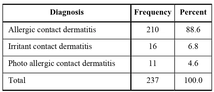
TABLE 1 – CLINICAL DIAGNOSIS
Diagnosis Frequency Percent
Allergic contact dermatitis 210 88.6
Irritant contact dermatitis 16 6.8
Photo allergic contact dermatitis 11 4.6
Total 237 100.0
Out of the 237 patients analysed, 210(88.6%) had allergic contact
dermatitis, 16 patients had irritant contact dermatitis and 11 had
photoallergic dermatitis. In our study, allergic contact dermatitis
Male 55% Female
45%
Gender distribution
n=107
n=130
ACD ICD
Photo Dermatitis
ACD- 88.6%
ICD- 6.75%
0 5 10 15 20 25 30
<= 20 yrs 21 - 30 yrs 31 - 40 yrs 41 - 50 yrs > 50 yrs 2.3
24.6
26.2
25.4
21.5
6.5
25.2
27.1
22.4
18.7
Per
centa
ge
Age Group
0 10 20 30 40 50 60 70 80 90 Cement Ornaments & metallic objects
Leather Paint Metal polishing
Others 83.8
3.1 3.8 5.4 2.3
In our study, cement topped the list as the source of metal
exposure with 129 patients (54.4%), followed by ornaments & metallic
objects exposure with 86 patients (36.3%). One mechanic and one
worker in glue packaging who had metal exposure were the least in the
study and they were grouped in the category ‘Others’. Among the 129
patients in the cement exposure group, males were the majority (109
patients) constituting 84.4%.Out of the 86 patients in the ornaments &
metallic objects exposure group, females constituted 95.35%. This
association was statistically significant (P value- <0.001). This may be
due to fact that males were more commonly employed in the
construction industry and females have the passion of wearing
[image:60.595.198.414.504.597.2]ornaments more.
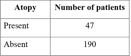
TABLE 5- INCIDENCE OF ATOPY
Atopy Number of patients
Present 47
Absent 190
Of the 237 cases 47 cases (19.8%) were atopic individuals by
47
190
18.90%
30.80%
18.10% 13.90%
9.30% 4.20%
4.60%
Extremities
Hands Foot
Extremities & trunk
Head, neck & shoulder ABCD
TABLE 8- GRADING OF PATCH TESTS GRADING NO: of positive reactions
1+ 238
2+ 78
3+ 11
Irritant reaction 16
343 positive reactions were found in 237 patch tested patients. Of these 238 reactions was 1+ and grading 3+ was the least in the study.
TABLE 9- INCIDENCE OF SENSITIZATION TO METALS
(In isolation or in association)
Metals Isolated Associated Total P value
No: % No: %
Chromium 111 60.3% 73 39.6% 184
<0.001
Nickel 29 32.2% 61 67.7% 90
Cobalt 10 14.5% 59 85.5% 69
X2 (Chi square test of independence)= 49.4
Chromium was an isolated sensitizer in 111patients (60.3%) while
in 73 patients (39.6%) chromium sensitivity was associated with either
nickel or cobalt sensitivity. Nickel and cobalt most commonly presented
as co sensitizers. In 67.7% patients nickel and in 85.5% cobalt were
associated with other metal sensitivity. This association of chromium
sensitization presenting commonly in isolation, and nickel & cobalt
sensitization presenting in association with other metals is statistically
[image:65.595.102.514.365.502.2]238 78
11 16
1+
2+
3+
0.00% 10.00% 20.00% 30.00% 40.00% 50.00% 60.00% 70.00% 80.00% 90.00%
Chromium Nickel Cobalt 60.30%
32.20%
14.50% 39.60%
67.70%
85.50%
Isolated sensitization Associated sensitization (in isolation & association)
Due to the simultaneous exposure to other antigens, 77 patients
were also patch tested with other allergens from the Indian standard
[image:68.595.95.515.260.584.2]series.
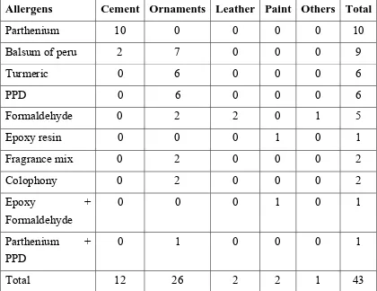
TABLE 10– ALLERGENS ASSOCIATED WITH METAL ALLERGY
Allergens Cement Ornaments Leather Paint Others Total
Parthenium 10 0 0 0 0 10
Balsum of peru 2 7 0 0 0 9
Turmeric 0 6 0 0 0 6
PPD 0 6 0 0 0 6
Formaldehyde 0 2 2 0 1 5
Epoxy resin 0 0 0 1 0 1
Fragrance mix 0 2 0 0 0 2
Colophony 0 2 0 0 0 2
Epoxy + Formaldehyde
0 0 0 1 0 1
Parthenium + PPD
0 1 0 0 0 1
Total 12 26 2 2 1 43
Parthenium was the most common allergen to be associated with
metal allergy found positive in 11 patients. Balsum of Peru and
10
9
6 6
5 2
2 1 1 1
Parthenium
Balsum of peru
Turmeric
PPD
Formaldehyde
Fragrance mix
Colophony
Epoxy resin
Epoxy resin & Formaldehyde
Parthenium
Balsum of peru
Turmeric Formaldehyde
TABLE 11- PATCH TEST RESULTS to INDIVIDUAL METALS.
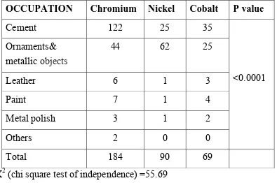
OCCUPATION Chromium Nickel Cobalt P value
Cement 122 25 35
<0.0001 Ornaments&
metallic objects
44 62 25
Leather 6 1 3
Paint 7 1 4
Metal polish 3 1 2
Others 2 0 0
Total 184 90 69
X2 (chi square test of independence) =55.69
Chromium sensitivity was the most common in occupations
involving cement, leather, paint, and metal polish exposure. Chromium
was also the most common metal to be sensitive in a mechanic and a
glue packaging worker. Nickel was the most common antigen in those
exposed to ornaments & metallic objects.This association of chromium
in cement, leather, paint, metal polish exposure and the association of
nickel in ornaments & metallic objects exposure group was statistically
significant (P value- <0.001).
As cement and ornaments & metallic objects constituted the
0 20 40 60 80 100 120 140 Cement Ornaments & metallic objects
Leather Paint Metal
polish Others 122 44 6 7 3 2 25 62
1 1 0 0
35
25
3 4 2 0
CHROMIUM
NICKEL
CEMENT EXPOSURE
TABLE 12- OCCUPATION involving CEMENT EXPOSURE
OCCUPATION MALE FEMALE TOTAL
Mason 67 10 77
Tile layer 10 - 10
Plumber 9 - 9
Electrician 8 - 8
Building contractors 5 - 5
Watchman 5 - 5
Mason& Farmer 4 4 8
Vendor 1 2 3
Housewife (cement exposure) - 4 4
TOTAL 109 20 129
Males predominated in the cement exposure group. Overall in
both sexes, masons topped the list with a total of 77 patients (59.7%).
Tile layers (7.75%) and plumbers (6.97%) followed masons in
frequency. Housewives formed the least number (4 patients) of this
group. They were sensitized to cement due to the construction and repair
[image:74.595.107.515.184.433.2]
and 8.5% after 30 years of exposure. The mean duration of exposure was 5.3 years.
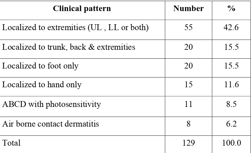
TABLE 15-CLINICAL PATTERNS IN CEMENT EXPOSURE
Clinical pattern Number %
Localized to extremities (UL , LL or both) 55 42.6
Localized to trunk, back & extremities 20 15.5
Localized to foot only 20 15.5
Localized to hand only 15 11.6
ABCD with photosensitivity 11 8.5
Air borne contact dermatitis 8 6.2
Total 129 100.0
Majority of the patients (55) developed contact dermatitis
localized to the extremities. This pattern of dermatitis localized to either
the upperlimb or the lowerlimb or both contributed to 42.6% of the
group. Foot eczema and dermatitis localized to the trunk and extremities
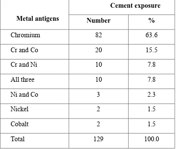
TABLE 16-PATCH TEST POSITIVITY IN CEMENT EXPOSURE
Metal antigens
Cement exposure
Number %
Chromium 82 63.6
Cr and Co 20 15.5
Cr and Ni 10 7.8
All three 10 7.8
Ni and Co 3 2.3
Nickel 2 1.5
Cobalt 2 1.5
Total 129 100.0
Chromium was the most common metal to be positive in 122
patients (94.57%) of the cement exposure group. This sensitization was
isolated in 82 patients (63.6%) and associated with other metals in 40
patients (31%). Cobalt allergy was most commonly associated with
chromium presenting in 30 patients (23.25%). Among this group, 14
patients developed irritant reaction to chromium. Nickel and cobalt
predominantly presented in association with other metals in the cement
3 27 36 37 26 0 5 10 15 20 25 30 35 40
<20 21-30 31-40 41-50 >50
Age distribution in cement exposure
Age in years No: of cases 0 10 20 30 40 50 60 70 80 MALE (84.5%) FEMALE (15.5%)
42.60%
11.60% 15.50%
15.50%
6.20% 8.50%
Clinical patterns in cement exposure
Extremities
Hand Foot
Trunk & extremities
ABCD with photosensitivity
9.30%
34.90%
21.70% 17.80%
7.80% 8.50%
1 - 5 yrs
5 - 10 yrs 10 -20 yrs
< 1 yr > 30 yrs
82 10
20
10
2 2 3
Cr Cr + Ni Cr + Co ALL 3 Ni Co Ni + Co
Cr + Ni Cr + Co
ALL 3
Ni Co
Ni + Co
[image:79.595.117.499.176.512.2]
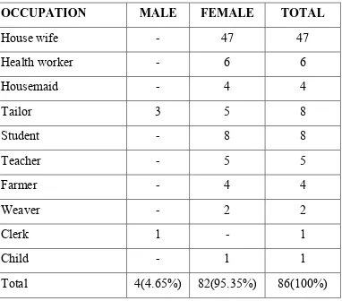
ORNAMENTS & METALLIC OBJECTS EXPOSURE TABLE 17– OCCUPATIONS involving ORNAMENTS &
METALLIC OBJECTS
OCCUPATION MALE FEMALE TOTAL
House wife - 47 47
Health worker - 6 6
Housemaid - 4 4
Tailor 3 5 8
Student - 8 8
Teacher - 5 5
Farmer - 4 4
Weaver - 2 2
Clerk 1 - 1
Child - 1 1
Total 4(4.65%) 82(95.35%) 86(100%)
Among the 86 patients in the ornaments & metallic objects
exposure group females formed the majority. Housewives were more
commonly exposed to ornaments in our study, constituting 54.65% (47
patients) of the total. The youngest in the age group was a female child,
18 months old sensitized to nickel from a pendant and the bangles.
Occupational exposure to scissors & needles was found in 8 tailors and
This pattern was due to exposure to ornaments in the form of earrings,
chains and safetypins. The next common pattern was hand eczema in 17
patients. Two farmers, who had ABCD, were sensitive to both nickel
and parthenium. Since they presented with ABCD, nickel sensitivity in
them was probably of past relevance. Ten patients had dermatitis
[image:81.595.118.491.322.556.2]localized to the feet, of which 6 were also sensitive to detergents.
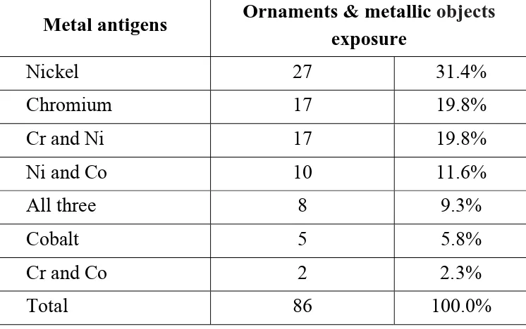
TABLE 20- PATCH TEST POSITIVITY IN ORNAMENTS & METALLIC OBJECTS EXPOSURE GROUP
Metal antigens Ornaments & metallic objects exposure
Nickel 27 31.4%
Chromium 17 19.8%
Cr and Ni 17 19.8%
Ni and Co 10 11.6%
All three 8 9.3%
Cobalt 5 5.8%
Cr and Co 2 2.3%
Total 86 100.0%
6 24 22 16 18 0 5 10 15 20 25 30
<20 21-30 31-40 41-50 >50
17 27 5 17 2 10 8
Patch test positivity in ornaments & metallic objects exposure
CHROMIUM NICKEL COBALT Cr + Ni Cr + Co Ni + Co ALL 3
Cr
Ni
Co Cr +Ni
Ni + Co ALL 3 51.20% 15.10% 19.80% 11.60% 2.40% exposure
Head, neck & shoulders Extremities
Hand
ALLER
RGIC CON
EX
NTACT D
XTREMI
DERMAT ITIES
LOWER LIMBS
IRRITANT CONTACT DERMATITIS TO CEMENT
CONTACT DERMATITIS TO NECKCHAIN
CONTACT DERMATITIS TO SAFETY PIN
CONTACT DERMATITIS TO BANGLES
POMPHOLYX IN NICKEL SENSITIVE PATIENT
CONTACT DERMATITIS- SPECTACLE FRAME & SAFETY PIN
CONTACT DERMATITIS TO FOOTWEAR (LEATHER)
HISTOPATHOLOGY OF ALLERGIC CONTACT DERMATITIS ( FEATURES OF CHRONIC ECZEMA )
PATCH TEST READING - 1+ (CHROMIUM)
PATCH TEST READING – 1+ (CHROMIUM)
PATCH TEST READING – 2+ (CHROMIUM)