A PREDICTOR OF RUGAE PATTERN AND RELATIVE
CORRELATION BETWEEN RUGAE PATTERN, LIP
PRINT AND FINGER PRINT
DISSERTATION
Submitted to The Tamil Nadu Dr. M.G.R Medical University
in partial fulfillment of the requirement for the degree of
MASTER OF DENTAL SURGERY
BRANCH IX
ORAL MEDICINE AND RADIOLOGY
This is to certify that the dissertation titled “
A Study of Intercanine
Width and Arch Form as a Predictor of Rugae Pattern and Relative
Correlation Between Rugae Pattern, Lip Print and Finger Print
” is a
bonafide record of the work done by
Dr. T. Kartheesan,
under our guidance
during his postgraduate study period of 2015-2018. The dissertation is
submitted to
The Tamil Nadu Dr. M.G.R Medical University, Chennai,
in
partial fulfillment of the requirement for the Degree of
Master of Dental
Surgery in Oral Medicine and Radiology
,
Branch IX.
It has not been
submitted (partial or full) for the award of any other degree or diploma.
.
Guide
Dr. TATU JOY E, MDS
Professor and HOD
Department of Oral Medicine and Radiology
Sree Mookambika Institute of Dental Sciences
Kulasekharam.
Co-Guide
Dr. SHASHI KIRAN M, MDS
Reader
Department of Oral Medicine and Radiology
Sree Mookambika Institute of Dental Sciences
This is to certify that this dissertation work titled “A Study of
Intercanine Width and Arch Form as a Predictor of Rugae Pattern and
Relative Correlation Between Rugae Pattern, Lip Print and Finger
Print” of the candidate Dr. T. Kartheesan, with registration Number
241527252 for the award of MASTER OF DENTAL SURGERY in the
branch of Oral Medicine and Radiology, [Branch- IX]. I personally verified
the urkund.com website for the purpose of plagiarism Check. I found that the
uploaded thesis file contains from introduction to conclusion pages and result
shows 12 percentage of plagiarism in the dissertation.
Guide & Supervisor sign with Seal.
Date:
SCIENCES, KULASEKHARAM
ENDORSEMENT BY THE PRINCIPAL / HEAD OF THE
INSTITUTION
This is to certify that the dissertation entitled
“A Study of Intercanine Width
and Arch Form as a Predictor of Rugae Pattern and Relative Correlation Between
Rugae Pattern, Lip Print and Finger Print”
is a bonafide research work done byDr. T. Kartheesan,
under the guidance of Dr. Tatu Joy E, MDS, Head of theDepartment, of
Oral Medicine and Radiology
, Sree Mookambika Institute ofDental Sciences, Kulasekharam.
Dr. Elizabeth Koshi, MDS,
PRINCIPAL
Sree Mookambika Institute of Dental Sciences, V.P.M Hospital Complex,
Padanilam, Kulasekharam,
I hereby declare that this dissertation
“A Study of Intercanine Width
and Arch Form as a Predictor of Rugae Pattern and Relative Correlation
Between Rugae Pattern, Lip Print and Finger Print”
is a bonafide record
of work undertaken by me and that this thesis or a part of it has not been
presented earlier for the award of degree, diploma, fellowship or similar title
of recognition.
Dr. T. Kartheesan,
MDS Student
Department of Oral Medicine and Radiology
Sree Mookambika Institute of Dental Sciences
I extend my profound sense of gratitude to my guideDr. Tatu Joy E, MDS,
Professor & Head, Department of Oral Medicine and Radiology, Sree Mookambika
Institute of Dental Sciences whose constant strive for excellence was the beacon for
my study. I thank him for the invaluable guidance, constant encouragement and
immense patience with me at every step of this endeavor. I am most grateful for my
co-guide Dr. Shashi Kiran M, MDS, Reader Dept of Oral Medicine and Radiology
who has been very helpful and kind to me.
I would like to acknowledge the help and support given by
Dr. Velayudhan Nair MBBS, MS Chairman and Dr. Rema V Nair MBBS, MD, DGO
Director Sree Mookambika Institute of Dental Sciences without which
my study would not have been possible. I also extend my gratitude to
Dr. Elizabeth Kozhi, MDS, Principal, Sree Mookambika Institute of Dental
Sciences, for the motivation and support.
I take this opportunity to express my sincere gratitude to my teachers
Dr. Eugenia Cherubin J, MDS, Associate Professor, Dr. Redwin Manchil Dhas P, MDS,
and Dr. Farakath Khan M, MDS, Senior Lecturers, whose constant encouragement
and support helped me through the course of the work.
I am thankful to Dr. Sarath Babu for providing me with his timely
statistical analysis involved in this study.
I wish to express my heartfelt thanks to my wife Dr. Soundari Bala for her
Mr. Thangasamy and Mrs. Mehala and my in laws Mr. Sakthivel Murugan and
Mrs. Muthulekshmi for their encouragement, love, great sacrifices and innate
confidence without which I wouldn’t have been where I am today.
I gratefully acknowledge my batchmates Dr. Lekshmi P.S, Dr. Divya N.L,
my beloved fellow post graduates Dr. Sajitha Jasmin, Dr. Tanuja, Dr. Dhanya,
Dr. Monisha, Dr. Godwi, and Dr. Janitha for their motivation and encouraging
words.
Sl. No Index Page No
1. List of abbreviations i
2. List of tables ii
3. List of graphs iii
4. List of color plates iv
5. List of annexure v
6. Abstract vi-vii
7. Introduction 1
8. Aim and objectives 4
9. Review of literature 5-39
10. Materials and Methods 40-42
11. Results and observations 43-53
12. Discussion 54-58
13. Summary and Conclusion 59
14. Bibliography viii-xxii
ŝ
AFIS - Automated finger print identification system
BC - Before Christ
CAD CAM - Computer aided design-computer aided manufacturing
CRL - Crown Rump Length
DNA - Deoxy Ribo Nucleic Acid
3D - 3 Dimensional
FBI - Federal Bureau of Investigation
ICW - Intercanine width
IAFIS - The integrated fingerprint identification system
MCI - Mean canine index
ŝŝ
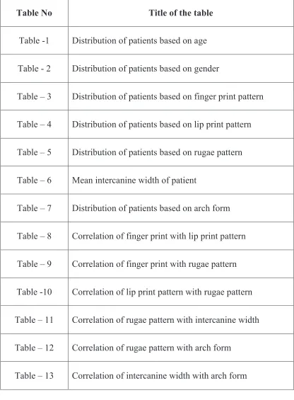
Table No
Title of the table
Table -1
Distribution of patients based on age
Table - 2
Distribution of patients based on gender
Table – 3
Distribution of patients based on finger print pattern
Table – 4
Distribution of patients based on lip print pattern
Table – 5
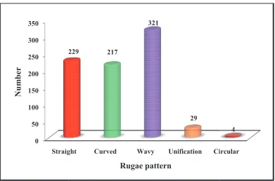
Distribution of patients based on rugae pattern
Table – 6
Mean intercanine width of patient
Table – 7
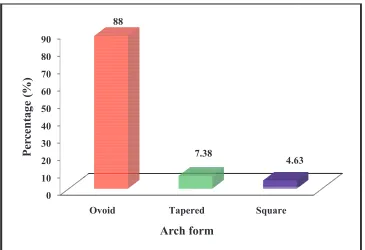
Distribution of patients based on arch form
Table – 8
Correlation of finger print with lip print pattern
Table – 9
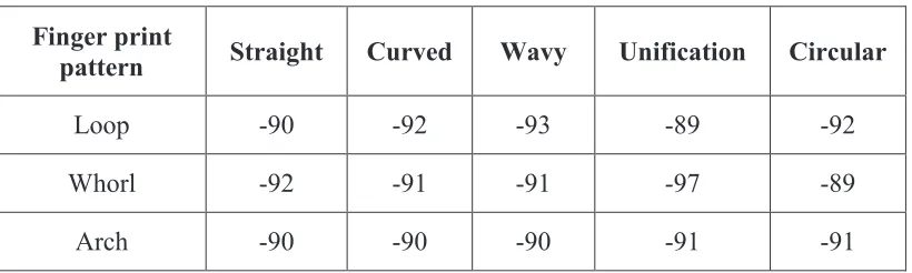
Correlation of finger print with rugae pattern
Table -10
Correlation of lip print pattern with rugae pattern
Table – 11
Correlation of rugae pattern with intercanine width
Table – 12
Correlation of rugae pattern with arch form
ŝŝŝ
Graph No
Title of the Graph
Graph - 1
Distribution of patients based on age
Graph - 2
Distribution of patients based on gender
Graph - 3
Distribution of patients based on finger pattern
Graph - 4
Distribution of patients based on lip print pattern
Graph - 5
Distribution of patients based on rugae pattern
ŝǀ
Color Plate No
Title of color plate
CP - 1
Sample Collection Method
CP - 2
Sample collectors
CP - 3
Rugae pattern
CP - 4
Lip print pattern
CP- 5
Finger print pattern
CP-6
Arch form
ǀ
No
Title
Annexure -1
Certificate from Institutional Research Committee
Certificate from Institutional Human Ethics Committee
Annexure - 2
Patient information sheath
English
Malayalam
Tamil
Annexure - 3
Patient consent form
English
Malayalam
Tamil
BACKGROUND:
Human identification is of paramount importance and it is indeed
challenging considering the fact that every individual has distinct traits. Although
DNA comparisons, dental records and finger print analysis are common techniques
employed to ensure fast and secure identification, there are certain crime and mass
disaster scenarios where other supplemental aids become essential. Some times
odontometric methods are preferred. The finger print and palatal rugae are broadly
used because of its feasibility and accuracy, which are matters of relevance for
practical usefulness.
AIMS AND OBJECTIVES:
1. To determine the correlation of the pattern of lip print ,rugae and finger print
in males and females
2. To analyze the rugae pattern and its correlation with arch shape and inter
canine width
3. To analyze the predominant relative pattern of lip prints, finger prints, and
rugae in males and females.
MATERIALS AND METHODS:
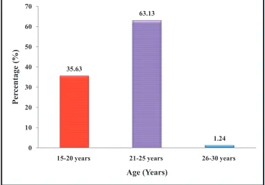
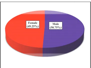
In this study a sample of 800 individuals (404 males and 396 females) of age
group 18-30 years were selected from Sree Mookambika Institute of Dental
sciences, Kulasekharam. Lip prints, finger prints and rugae pattern were collected
from each individual. Analysis of lip print was done based on classification of
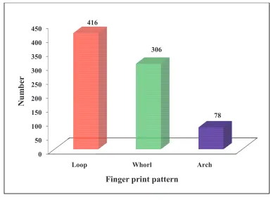
Suzuki and Tsuchihashi. The analysis of finger print was done based on
Kotze. Intercanine width was measured by using digital vernier caliper. Arch form
were analyzed by Chuck classification.
RESULTS:
The correlation of rugae pattern, lip prints and finger prints by were showed
no statistical significance in both males and females. Also intercanine width and
arch form in the determination of rugae pattern did not show any significance in
both males and females. The results are correlated well with other studies.
CONCLUSION:
In our study, negative correlation was observed between lip print, finger
print and rugae pattern. Therefore it was concluded that no pattern among these
three parameters will predict other two patterns in human identification. Likewise,
change in arch form and intercanine width do not show any change in rugae pattern.
Hence, palatal rugae pattern can be used as stable marker for personal identification.
KEY WORDS:
Arch form, finger print, intercanine width, lip print, rugae pattern.
ͳ
Human identification play an important role in forensic investigation .It is
indeed challenging considering the fact that every individual has their own unique
character. This requires a combination of different procedures to individualize a
person or object. Identity is a set of physical characteristics, functional or psychic,
normal or pathological that define an individual.1There are many techniques
available for personal identification. They are DNA comparisons, finger print
analysis, lip print analysis, palatal rugae pattern and bite mark analysis, dental
records, anteroposterior metric data, retinal scan, age, race, sex, stature etc. Among
which DNA comparisons and finger print analysis are commonly used technique.
Meanwhile other supportive evidenceslike tattoos piercings, associated clothing,
eyewitness, documents and belongings also become inevitable because personal
identification involves a combination of different techniques for identifying a person
or object.2
Forensic dentistry is a specialized branch of forensic medicine and may be
described as that part of odontology which deals with handling and examination of
dental evidence from which a proper evaluation &presentation of dental findings can
be made. In the world, No two person look alike and each are unique and this
concept of uniqueness is utilized in human identification procedures.3Gender
discrimination is the important aspect of personal identification procedures that aid
in the establishment of biological profile from the skeletal &dental remains.4
Labial mucosa shows wrinkles and grooves and is called sulci labiorium. it
forms a specific pattern called lip print, the study of lip print is known as
cheiloscopy.5 Finger shows epidermal ridges on distal portion of finger and
ʹ
known as dactyloscopy.6 Likewise palate shows surface elevations &depression and
is called rugae pattern, study of rugae pattern is called as rugoscopy or palatoscopy.
Rugae are not damaged from trauma due to their internal position in the oral cavity
and are insulated from heat by tongue and buccal fat pads.7 In one study, it was
reported that no two palates are alike in their configuration and that the palatal print
did not change.8 In identical twins also, the studies indicated that the patterns may be
similar but not identical. Events contributing to changes in Rugae pattern.9,10
• Finger sucking in childhood
• Persistent pressure due to orthodontic treatment
• Local effect on lateral rugae after tooth extraction(mainly effects the lateral
part of rugae)
• Changes in the lateral edge of the rugae with orthodontic tooth movement.
Odentometric parameters show ethnic, racial and to some extent
geographical variations and they are least vulnerable to sudden accidental changes
and help in collecting appropriate dental evidence. Teeth unlike soft tissue remains
throughout the life and even after death.11,12
The study of permanent Maxillary Canine teeth offers certain advantages.
These teeth are less affected by periodontal diseases and are also less exposed to
plaque. Moreover, they are less prone to calculus and abrasion from brushing. Over
and above, maxillary canines are the last to be extracted with respect to age.13 After
numerous investigations, there is small agreement as to the best size and shape for
ideal arch form. For more than 100 years, researchers have been trying to define
͵
represented by an geometric formula.14Chuck was first to classify arch forms as
tapered, square and ovoid.
Lip prints are unique to an individual and are applicable mostly in
identifying the living. Since lip prints are usually left at crime scenes they can
provide direct link to the suspect. Lip prints can be recorded and used as evidence in
nabbing suspects in crime scene.3 Finger print is a known method for gender
determination and personal identification. Finger print can be taken even from a
decomposed body either from the peeled off epidermis of the finger or from the
dermis when epidermis is lost.15 Even when most of anatomical structures are
destroyed, burnt, or dehydrated, the palate remains intact and thus helps in personal
identification based on the rugae pattern. The palatal pattern is characteristic of
every individual and does not change as a result of growth. The relationship between
intercanine distance and the lateral dimension of palatal rugae remained consistent
pre and post orthodontic treatment. This could be because the intercanine width is
maintained in most of the treatment modality and rugae is stable.16
In this study, we analyzed the predominant pattern and correlation of the
pattern of lip print, rugae and finger print in males and females. Also we analyzed
the rugae pattern and its correlation with arch shape and inter canine width. Thereby,
a correlation if present among these various parameters of personnel identification
will help in the human identification, especially in case of mass disasters and crime
scenes where often complete human remains are not found and identification is
Ͷ
1. To determine the correlation of the pattern of lip print, rugae and finger
print in males and females.
2. To analyze the rugae pattern and its correlation with arch shape and inter
canine width.
ϯ͘ To analyze the predominant relative pattern of lip prints, finger prints and
ͷ
PALATAL RUGAE
Palatal rugae are also called as ‘plicapalatinae transversae’ and ‘rugae
palatine’ which refer to the epithelial ridges on the anterior part of palatal mucosa,
present on each side of the median palatal raphae and behind the incisive papilla.
They also known as transverse palatine folds, refers to the irregular elevations of the
mucosa seen on the anterior third of the palate. This projection runs in a transverse
direction from the palatine raphae located on the mid-sagital plane. These rugae
have significant characteristic features and they are unique patterns in each
individual and remain stable from the time of the development until death.18,19,20
Anatomically, the rugae consist of around 37 ridge and oblique ridges that
radiate out tangentially from the incisive papillae. Winslow was the first person to
describe rugae. The study of palatal rugae is called as rugoscopy and it is applied in
the fields of dentistry like forensic odontology, prosthodontics and
orthodontics.18,20,21
DEVELOPMENT:
The palatal rugae appear towards the third month of intrauterine life, from
the covering connective tissue in the palatine process of maxillary bone. Its
development and growth are mutually controlled by epithelial-mesenchymal
interactions, where specific extracellular matrix molecules are expressed during
development.9,22
The first rugae can be distinguished in human embryos of 32mm next to the
incisive papilla. Then, in the prenatal stage they are relatively prominent. The palatal
adolescence they acquire the final shape in each individual. Once they are formed
they may experience changes in their size due to growth of the palate, but its shape
is maintained even after the growth was completed.9,23
At the 550 mm stage of embryonic development, there are five to seven
rather symmetrically disposed ridges, with the anterior ones beginning at the raphe,
the others more laterally, towards the end of intra-uterine life the pattern of the rugae
becomes less regular, posterior one disappearing and those anterior become
considerably more pronounced and compressed.24 According to Carrea(1937) the
rugae pattern is formed by the 12th to 14th week of prenatal life, and it remains stable
throughout the person’s life.25,26
PHYSIOLOGY
Physiologically the palatal rugae are involved in the oral phase of
swallowing and help to improve the relationship between food and taste receptors in
the dorsal surface of the tongue. It also have a significant role in speech and in the
suckling in children.9,23,26
The number of rugae on each side of the palate varies between 3-5 and the
rugaedonot extend to the posterior palatal half of the hard palate. They never cross
the midline. The anterior rugae usually are more prominent than the posterior rugae.
Most of the rugae are curved and rest are angular and the last rugae has the higher
probality of division where medial and lateral parts are not connected and donot
continue in their axial orientation. Fragmentary rugae frequently are present,
particularly in the posterior half of the rugae territory. The shape, length, width,
people. Variation exists in a lesser extent in the left and right sides of same person.
The inclination of the rugae to the sagittal plane also shows marked differences on
both sides. Bilateral symmetry also exists in the rugae pattern.22
VARIOUS APLICATIONS OF PALATAL RUGAE
In orthodontics:
Palatal rugae is being used in orthodontics as a suitable soft tissue reference
points. It does not change in the adult hood after the complete eruption of teeth and
no significant alteration in length occurs. They serve as a suitable reference points
from which the clinician can derive the reference planes necessary for longitudinal
cast analysis. Positional changes of posterior teeth in the antero-posterior direction
are relavant to the diagnosis and correction of sagittal occlusal abnormalities and
arch length discrepancies. The maximum mean change in distance between the
rugae in the antero-posterior plane was0.41mm.27,28 The palatine rugae can be used
as reference points for measuring tooth movement in a manner comparable with
cephalometric superimpositions.27
Palatine rugae in speech and palatal prostheses:
In prosthesis, when there is a changed relationship of the tongue to a palate,
the speech is altered. Speech may require surface texture to orient the tongue. The
palatine rugae often can serve as a cue for pronunciation of certain letters like S, Sh
which requires contact of tongue to the palatal rugae. Palatography served as a basis
for determining the shape of the anterior palatal vault to determine the contact
position of the tongue to the palate for specific sounds. The re-creation of the palatal
ͺ
Palatal rugae in cleft palate:
The early detection of the sub mucosal cleft-palate is necessary in children as
the diagnosis depends on the patient’s clinical history and intra oral examination.
Rugae are used to measure the distances on the cleft palate from the period of birth
to the time of early mixed dentition period. These distance of the cleft are measured
by using fixed reference points of the palatal rugae to estimate the changes that
occurred in the anterior palate during various changes of orthodontic therapy and
growth.27
APPLICATION OF RUGAE PATTERN IN FORENSIC ODONTOLOGY
Rugae patterns contribute reliable details to forensic odontology in
identification of the deceased. Even in extreme cases of trauma or incineration the
rugae remains protected due to its anatomical position. Rugae are well protected
from heat as the lips, tongue and buccal fat pads act as insulators and remain
undisturbed. Palatoscopy or rugoscopy is the name given to the study of palatal
rugae in order to establish a person’s identity.28
Rugae changes throughout early childhood and adolescence. Changes that
occur in rugae are related only to their length which helps to compare the
ante-mortem data to the postante-mortem data.29
The rugae have been successfully identified in severely burnt edentulous
body and by comparing the pattern on the victim’s old denture, this indicates that
rugae are stable in adult life. Thus, palatal rugae appear to possess the features of an
ideal forensic identification parameter due to its uniqueness, postmortem resistance
ͻ
Palatoscopy used in necro-identification holds good enough due to its
resistance to decomposition up to 7 days. Therefore anatomical structures such as
palatal rugae may assume more importance in future.21
History:
Rugae were first described by Winslow in 1732. The earliest illustration of
palatal rugae was probably by Santorini in 1775, wherein he put drawing depicting 3
wavy lines crossing the midline of palate.27,29 The application of palatal rugae
patterns for personal identification was first suggested by Allen in 1889.31 The first
palatal system classification system was put forth by Goria in 1911 in two days: by
specifying the number of rugae and specifying the extent of the rugal zone relative
to the teeth. Palatal rugoscopy was first proposed in 1932, by a Spanish investigator
named Trobo Hermosa. In 1937, Carrea conducted a detailed study and established a
method to classify palatal rugae.24,27
In 1955, Lysell suggested that the palatine rugae might possess unique
characteristics that could be used in pattern identification. In 1983, Brinon,
following the studies of carrea, divided palatal rugae in to two groups (fundamental
and specific) in a similar way to that done with fingerprints.22,25
Sassouni (1957) stated that it is possible to devise a classification based on
the symmentry, number and shape of papillae. When Sassouni(1957) tested the
classification, he was able to identify a person without difficulty. The palatal rugae
can be used in same way as fingerprints; however, as the rugae are composed only
of soft tissue, they are not present in skeletons and stated that no palatal rugae are
ͳͲ
CLASSIFICATION:
1. Lysell classification (1955):26
Palatal rugae were classified depending on its length
• Primary : 5mm or more
• Secondary: 3-5mm
• Fragmentary: 2-3mm
• Rugae smaller than 2mm are disregarded
2. By Kapali et al (1997):33,34
Based on shape of palatal rugae
• Curved
• Wavy
• Straight
• Circular
Modification of Kapali’s classification
• Converging
• Curved
• Wavy
• Straight
• Circular
• Furcated
3. Thomas and Kotze Classification (1983):27(most accepted classification)
According to the shape of palatal rugae, palatal rugae classified in to-
ͳͳ
• Curved: simple crescent shape which curved gently. Evidence of even
the slightest bend at the termination or origin of a rugae lead to a
classification as curved.
• Wavy: the basic shape of the rugae was serpentine; however if there
was a slight curve at the origin or termination of curved rugae it was
classified as wavy.
• Circular type to be classified as circular, a rugae needed to display a
definite continuous ring formation.
• Unification type occurs when two rugae are joined at their origin or
termination. Unification in which two rugae began from the same origin
but immediately diverged were classified as diverging and which
converged were classified as converging.
SOME STUDIES ON PALATAL RUGAE:
Kapali S, Townsend G, Richards T. (1997) did a longitudinal study on serial
dental casts of ten Aborigines, from 6-20 years. They examined rugae patterns and
compared it between different ethnic groups of 100 dental casts of Australian
Aborigines and 200 casts of Caucasians, ranging in age from 13 to 17 years and
observed features like number, length, shape, direction and unification of rugae.
According to their study the most common shapes in both ethnic groups were wavy
and curved forms, whereas straight and circular types were least common. There
was a statistically significant association between rugae forms and ethnicity, straight
forms being more common in Caucasians whereas wavy forms were common in
ͳʹ
Fahmi F, AL-shamrani SM, Talic YF. (2001) did a study to identify and
compare the rugae pattern in Saudi males and females from 120 samples using
Thomas Kotze et al classification. They identified that female had a significant
difference in the converge type while the male had a significant difference in the
circular type and suggested rugae can be used as an adjuvant tool in determination of
sex in Saudi population.35
Hermosilla VV, San Pedro VJ, Cantin IM, Sauzo GIC. (2009) analysed 120
subjects of the both sexes, between 15 to 20 years old and fully dentated, using
rugoscopy to determine the shape, size, number and position of the palatal rugae.
They stated that most prevalent palatal rugae shape was sinuous (43%) followed by
curve (27%), line (15%), point (11%) and polymorphic varieties (4%). The average
number of rugae was 12.27, being higher in male than female. They concluded that
the analysis of the palatal rugae and their features can be used as a reliable guide to
the forensic identification.9
For identifying the predominant pattern of rugae in Indian population from
Madhya Pradesh and kerala groups, a study was conducted by AparnaPaliwal et al
(2010) using 60 maxillary cast with an age group 17-23 years in both sexes. The
results showed that straight rugae pattern on the right side of the palate were more in
the male subjects and found to be significantly predominant in the Madhya Pradesh
population, whereas wavy shape was predominant in keralapeople; however, rugae
pattern on the right side of the palate in female subjects exhibited no significant
ͳ͵
Kamala R, Gupta N, Bansal A, Sinha A. in 2011 did a study where they
randomly selected sample group of 1000 subjects comprising 500 males and 500
females of age from 3 to 51 years. They clearly demonstrated that palate do not
undergo any changes except in length due to normal growth, remaining in the same
position throughout a person’s entire life. Thus, palatal rugae appear to possess the
features of an ideal forensic identification parameter.37
Bharat ST, Kumar GR, Dhanapal R, Saraswathi TR. in 2011 studied the
differences of palatal rugae patterns in males and females by a cross sectional study
in a hospital based coastal Andhra population. 100 preorthodontic model cast were
studied and Thomas classification was adopted for analysis. Discriminant analysis
showed 78% accuracy with actual data.18
Gandikota C, Venkata TP, Challa P, Juvadi SR, Mathur A. in 2012 did a study
to use palatal rugae as an aid to classify malocclusion in untreated class II div I
malocclusion in comparison to normal class I occlusions. They observed that there was
a significant constriction of the palatal rugae in class II div I than the class I individuals
and that there was a distinct pattern of palatal rugae between the two groups.38
Manjunath S, BakannavarSM, Pradeep Kumar G, Vrinda J, Prabhu N,
Kamath A. et al (2012) did a study to analyses the differences in the rugae pattern
among the Indian males and females in a small portion of a population with 63
subjects of which 32 were males and 31 were females of age group from 17-25
years. They found that the females had more number of rugae in the right side and
showed increased in number of curved and straight shapes rugae pattern than
ͳͶ
Shanmugam S, Anuthama K, Shaikh H, Murali K, Suresan V, Nisharudeen
K. et al (2013) suggested that palatal rugaen can be used to differentiate the
population among the north Indians and south Indians. They did a study using 940
individuals of 18-23 years of age. They observed that the wavy and curved are
common in both groups. On applying chi square test evaluation found that
classification had accuracy of 87.8%.40
Rajan VP, John JB, Stalin A, Priya G, Abuthagir A (2013) did a study to
assess the morphology of the rugae pattern in 5-15 years old children with gender
dimorphism. He observed that there was an increased female prediction in the total
count and primary rugae pattern. On comparing the shapes of rugae in male and
female, the study models showed a predominance of wavy shape followed by
curved. Circular pattern was not seen in the study population. There was significant
statistical difference in the direction and unification of rugae among males and
females were reported.41
Bajracharya D, Vaidya A, Thapa S, Shrestha S (2013) conducted a study on
200 Nepalese (100 male and 100 female) to determine the number and pattern of
palatal rugae. They observed that the number of primary rugae did not show any
statistical significant difference between the gender groups while a wavy pattern was
predominant followed by curved, straight, branched and circular in their
population.20
Madhusudan K, Rajesh N, Umesh K, Sangeetha R, Patel H, Patel R (2014)
did a study on palatal rugae pattern its association with lineage by using 30 families.
ͳͷ
offspring matched with either parents. Thus, they concluded that there was
statistically significance resemblance of offspring palatal rugae patterns with
parent’s palatal rugae patterns.42
For comparing palatal rugae pattern among male, female, and transgender
population of the Bhopal city, a study was conducted by Saxena E, Chandrasekar
BR, Hongal S, Torwane N, Goel P, Mishra P (2015) using 148 subjects in the age
group of 17-35 years. The results showed that statistically significant difference with
regard to some parameters like number of rugae, fragmentary rugae, wavy rugae,
curve rugae, forwardly directed and backwardly directed rugae between transgender
and other gender groups were present.43
Oberoi IS, Chalkoo AS, Dhingra A (2016) evaluated of rugae pattern in
individuals of known population by using 120 subjects of which 60 males and 60
females in the age group of 17to25 years. They found that straight and wavy variety
arethe most commonly seen rugae pattern in both males and females.44
LIP PRINTS
The study lip print is known as Cheiloscopy. (The name derived from Greek
words cheilos means lips and skopein means to see). The importance of cheiloscopy
is linked to the fact that the lip prints are unique to one person, except in
monozygotic twins. Lip grooves are permanent and unchangeable like finger print,
palatal rugae. It is possible to identify the lip print patterns as early as the sixth week
in uterine life. From that moment onwards, the lip groove pattaerns are
unchangeable or rarely changeable and resisting many afflictions such as herpetic
ͳ
History:
The biological feature of lip groove patterns was first described by Fisher in
1902, it was only in 1930, that Diou de Lille developed some studies which led to lip
print use in criminology.46 In 1932, Edmond Locard, one of France’s greatest
criminologists, acknowledged the importance of cheiloscopy.47 In 1950, Le Moyer
Synder, in his book “Homicide Investigation”, mentioned the possibilities of using
lip prints in the matter of human identification.48 Sometime later, Santos in 1960,
suggested that the fissures and the criss- cross lines in the lips could be divided in to
different groups (simple and compound), and each group could be further divided in
to eight subtypes.49
Renaud, in 1972, studied 4000 lip prints and confirmed the singularity of
each one, supporting the idea of lip print singularity. Two years later, Suzuki and
Tsushihashi developed another study which resulted in a new classification for lip
prints. This study, made over long period of time, enabled the authors to confirm not
only lip print singularity, but also lip response to trauma; in fact, these authors
observed that after healing, the lip pattern was equal to that before the injury
occurred.5,50
Development:
Lip development occurs in 4th -8th week of intra uterine life. Upper lip
formation appears for the first time at stage 16 of intra uterine life and their
formation gets completed by stage 20 of intra uterine life. Each maxillary processes,
grows medially and fuses, first with lateral nasal process and then with the medial
ͳ
Lower lip development appears for the first time at stage 11 and their
formation gets completed by stage 15 of intra uterine life. The mandibular processes
of the two sides grow each other and fuse in midline. They now form the lower
margin of the stomatodaeum. Fused mandibular processes give rise to lower lip and
lower jaw.47,51
Anatomy:
Lips are two, highly sensitive mobile folds, composed of skin, muscle,
glands and mucous membrane. They surround the oral orifice and form the anterior
boundary of the oral cavity. Anatomically, whether covered with skin or mucosa, the
surface that forms the oral sphincter is the lip area. There is an upper lip and lower
lip; the two lips are joined at the corners of the mouth- the commissures- and
separated by the buccal fend.47,51
There are two different kinds of lip covering-skin or mucosa. When the two
meet, a white wavy line is formed- the labial cord. Where identification is concerned,
the mucosal area holds the most interest. This area, also called Klein’s zone, is covered
with wrinkles and grooves that form a characteristic pattern-lip print.46
However, this is not only the area that deserves careful study. In fact, in
cheiloscopy, one should also analyse lip anatomy, considering their thickness and
the position. The lips can be horizontal, elevated or depressed and according to their
thickness, it is possible to identify the the following four groups: (1) thin lips
(common in European Caucasian); (2) medium lips (from 8 to 10mm, are the most
common type; (3) thick or very thick lips (usually having the inversion of the lip
ͳͺ
Lip Print classification:
1. Suzuki and Tsuchihashi classification (1970)5
Type I – complete vertical
Type’II – incomplete vertical
Type II – Branched
TYPE III - Intersected
TYPE IV – Reticular pattern
Type V – Irregular
2. Renaud classification:53
A – Complete vertical
B – Incomplete vertical
C – Complete bifurcated
D – Incomplete bifurcated
E – Complete branched
F – Incomplete branched
G – Reticular pattern
H – X or coma
I – Horizontal
J – Other forms (ellipse, triangle)
3. Afchar-Bayat classification:54
A1 – Vertical and straight grooves, covering the whole lip
A2 – Like the former, but not covering the whole lip
B1 – Straight branched grooves
ͳͻ
C – Converging grooves
D – Reticular pattern grooves
E – Other grooves
.APPLICATION OF LIP PRINTS IN FORENSIC ODONTOLOGY:
Lip prints in a crime scene investigation is very important in establishing the
true nature of the facts and link a subject to a specific location if found on clothes or
other objects, such as glasses, cups or even cigarette’s buts.55,56 When searching for
lip prints, one must always consider that all lipstick smears coloured; in fact, in
recent years the cosmetic industry has been developing new lipsticks which do not
leave a variable smear or mark when they come in contact with different items, these
are called persistent lipsticks.57
Cheiloscopy is a process which provides both qualitative and quantitative
results by examination of system of furrows on the red part of human lips. The
investigation of lip prints is very useful elementary in resolving a criminal act.
Traditional lipstick produces a print that is easily identifiable but lip prints made
without lipstick are invisible, requiring reagents that are more sensitive than
conventional materials to locate and develop lip prints.58
SOME STUDIES ON LIP PRINTS:
Shailesh MG, Indurkar A, Shirish D, Rahul B. in 2009 conducted a study on
cheiloscopy for sex determination by using 140 subjects of which 70 males and 70
females in the age group of 0-70 years. They found that 67 out 0f 70 lip prints of
females were correctly identified and 65 0ut of 70 males were correctly diagnose as
males. Type C (47.14%) was the most commonly occurring trend in females,
ʹͲ
Preeti S, Susmita S, Vanita R. in 2009 compared the 50 reliability of
cheiloscopy and palatoscopy in human identification by using 100 subjects of which
50 males and 50 females. The study not only showed that palatal rugae and lip prints
are unique to an individual, but also that lip prints is more reliable for recognition
the sex of an individual.60
Nagasupriya A, Dhanapal R, Reena K, Saraswathi TR, Ramachandran CR.
Did a study on patterns in 2011 by using 200 students of which 100 males and 100
females in the age group of 18-27 years. The study showed that lip and finger
patterns did not reveal statistically significant results with the gender, the correlation
between lip and finger patterns for gender identification, was statistically significant.
In males, branched type of lip patterns associated with arch, loop and whorl type of
finger pattern was significant. In females, vertical lip pattern associated with arch
finger pattern and reticular lip pattern associated with whole finger patterns were
most significant.15
Amith HV, Anil VA, Nagesh L in 2011 conducted a study on lip prints by
using 200 dental students. They found that type II pattern was dominant in males in
the third and fourth quadrants. Type III pattern did not occur in third and fourth
quadrants at all. It has occurred first and second quadrant in males. The results were
found to be statistically significant.61
Koneru A, surekha R, Ganesh SN, Vanishree M, Ramesh DNSV, Patel RS in
2013 compared the lip prints in two different populations of India by using 60
subjects which included 30 each from Kerala and Manipuri. The study showed that
ʹͳ
was common in females. Type 1 pattern was common in both populations, with an
incidence of 28.3%. Furthermore, Type 1 pattern was found to be more common in
Kerala females and Manipuri males when compared to their counterparts. Type 1’
and Type 4 were predominant in Kerala and Type 5 in Manipuri.62
Ghimire N, Nepal P, Upadhay S, BudhathotkiSS,Subba A, Kharel B in 2013
did a study on lip print pattern by using 200 Nepalese undergraduate student. They
concluded that lip print pattern can be used as an additional tool for personal
identification and sex determination.63
Murugan M, Karikalan T in 2014 conducted a study on correlation between
the pattern of finger print and lip print by using 300 students. They concluded that
individual parameters, i.e., lip print and finger print patterns play an important role
in forensic identification. Correlation of these two parameters did not show any
significant association, hence, these combinations as such cannot be used in
individual identification.64
Peeran SW, Naveenkumar PG, Abdalla KA, Abdelkader F, Azaruk A,
Manipady S in 2015 did a study on lip print patterns among adults of Sebha city,
Libya by using 104 subjects in the age group of 18-35 years. Result showed that
type I lip print pattern was seen in 53.37% and 60.07% of lip quadrants in males and
females, respectively. 27 (25.96%) subjects had same lip print pattern in all the four
quadrants.65
Rachna K, Shreenivasa MP, Padar SS, Nishat S, Suraksha B in 2015
conducted a cross sectional study on cheiloscopic patterns in Indian population and
ʹʹ
380 females in the age group of 1-80 years. They found that type I was the most
prevalent cheiloscopic pattern. Based on the method of sex determination using lip
prints, the percentage of individuals correctly identified was less.66
FINGER PRINT:
Study of fingerprints as a method of identification is known as
Dactylography or Dactyloscopy or Dermatoglyphics and at present, also as
Henry-Galton system of identification.67 The word Dactylography is taken from two Greek
words, dactylos meaning ‘finger’ and graphein meaning ‘to write’. It is the study of
impression patterns formed by the papillary ridges on the bulbs of fingers and
thumbs, when taken upon unglazed paper with the help of printer’s ink.
Dactylography is a progressing science and new methods for the recording, lifting
developing of prints under different field conditions, including those from the
decomposed body, are being introduced regularly.68
History:
Finger prints as a form of identification have been used atleast since 7000 to
6000 BC by the ancient Assyrians and Chinese. Bricks used in houses in the ancient
city of Jericho were sometimes imprinted by pairs of thumb prints. In the mid
1800’s, scientific studies were begun and established two critical characteristics of
finger prints that are true till to thisday: no two finger prints from different fingers
have been found to have the same ridge pattern and finger print ridge patterns are
unchanging throughout the life. These studies led to the use of finger prints for
criminal identification, first in Argentina in 1896, then at Scotland Yard in 1901, and
to other countries in the early 1900’s. Computer processing of finger prints began in
ʹ͵
process theseImages. Since then, automated finger print identification systems
(AFIS) have been employed widely among law enforcement agencies throughout the
world. In the late 1990s, the introduction of inexpensive finger print capture devices
and the development of fast, reliable matchingalgorithms have set the stage for the
expansion of finger print matching to personal use. There are various theories
proposed for finger pattern.69
Development:
There are various theories proposed for finger pattern formation. Kollmann
in 1883 speculated that ridge pattern is formed as a result of a folding process
induced by differential growth. This idea was promoted by Bonnevie in 1920 who
stated that there will be intense cell proliferation in the basal layer of the epidermis
which results in cylindrical cells. Finally, epidermis evades the dermis, resulting in
the formation of primary ridges. There is evidence that primary ridge system
changes till 16th week of pregnancy and after 16th week it becomes permanent and
remains unchanged throughout the life. Even in superficial skin injuries, finger
patterns will be reformed without any change because they are encoded at the
interface between dermis and epidermis. Another view regarding the formation of
primary ridges is linked to the nervous system. Dell and Munger in 1986 stated that
before ridge formation, finger tips are innervated by a hexagonal pattern of axons
determining the growth of primary ridges formation. But, this theory was refuted by
Morohunfolaet al. 1992 who proved the formation of primary ridges even in the
absence of innervations.6 Till now, there is no commonly accepted mechanism for
ʹͶ
Fingerprint biometrics
Fingerprints have been used in criminal investigations as a means of
identification for centuries. It is one of the most important tools of crime detection
because of their robustness and uniqueness. A fingerprint is the pattern of friction
ridges and valleys on the surface of a fingertip. In order to match a print, a
fingerprint technician digitalizes or scans the print obtained at a crime scene and
computer algorithms of a biometric system locate all the unique minutia and ridge
points of a questioned print. These unique feature sets are then matched against a
stored fingerprint database.
The Integrated Automated Fingerprint Identification System (IAFIS) is a
national automated fingerprint identification and criminal history system maintained
by the Federal Bureau of Investigation (FBI). IAFIS provides automated fingerprint
search capabilities, latent searching capability, electronic image storage, and
electronic exchange of fingerprints and responses. IAFIS houses the fingerprints and
criminal histories of 70 million subjects in the criminal master file, 31 million civil
prints and fingerprints from 73,000 known and suspected terrorists processed by the
U.S. or by international law enforcement agencies.
The average response time for an electronic criminal fingerprint submission
is about 27 min, while electronic civil submissions are processed within an hour and
12 min. IAFIS processed more than 61 million ten-print submissions during Fiscal
Year 2010. In September 2014, the FBI announced that its Next Generation
Identification system was at full operational capability and effectively replaced
ʹͷ
Fingerprint classification:
1. Henry’s classification71
• Arch pattern – plain, radial, ulnar, tented
• Loop pattern – radial, ulnar
• Whorl pattern – plain, central pocket, double loop, accidental
2. Michael and Kucken’s classification6
• Arch
• Loop
• Whorl
• Composite
3. Cummins classification72
• Arch
• Loop
• Whorl
SOME STUDIES ON FINGERPRINTS:
Nagasupriya A, Dhanapal R, Reena K, Saraswathi TR, Ramachandran CR.
Did a study on patterns in 2011 by using 200 students of which 100 males and 100
females in the age group of 18-27 years. The study showed that lip and finger
patterns did not reveal statistically significant results with the gender, the correlation
between lip and finger patterns for gender identification, was statistically significant.
In males, branched type of lip patterns associated with arch, loop and whorl type of
finger pattern was significant. In females, vertical lip pattern associated with arch
finger pattern and reticular lip pattern associated with whole finger patterns were
ʹ
Sayed YK, Goudar ES, Sayeda YK (2013) did a study on fingerprint pattern
and gender distribution of fingerprint in and around Bijapur by using 500 males and
500 females in the age group of 18-60 years. They concluded that predominant
pattern among male and female is ulnar loop. The ridge count in male was slightly
more than in female. 73
Mutalik VS, Menon A, Jayalakshmi N, Kamath A, Raghu AR. Correlated the
lip print, fingerprint and palatal rugae in 2013 by using 100 females. They
concluded that reticular pattern of lip print, wavy pattern of palatal rugae and loop
pattern of finger prints were predominant patterns. Correlation of three parameters
did not reveal significant differences.74
Murugan M, Karikalan T in 2014 conducted a study on correlation between
the pattern of finger print and lip print by using 300 students. They concluded that
individual parameters, i.e., lip print and finger print patterns play an important role
in forensic identification. Correlation of these two parameters did not show any
significant association, hence, these combinations as such cannot be used in
individual identification.64
Hansi DB, Ashish DB, Neeti SK (2014) conducted a study on distribution of
fingerprint patterns in an Indian population by using 256 males and 280 females.
The result showed that ulnar loop (51.3%) was found to be the most common pattern
observed. No statistically significant difference were observed between distribution
of fingerprint patterns in males and females.75
Harsha L, Gifrina J in 2015 conducted a study on correlation of lip print,
fingerprint and blood group in a Tamil Nadu based population by using 150
ʹ
pattern of clustering the results were not statistically significant. Apart from forensic
identification, fingerprint patterns can also predict susceptibility to diseases.76
Mohammed A, Naveen KM, Kavita DB, Shyam PR (2015) conducted a
study on cheiloscopy and dactyloscopy by using 200 subjects of which 100 males
and 100 females in the age group of 18-30 years. The result showed that type I with
whorl finger pattern was predominant in males, whereas in females predominant lip
pattern recorded was type II with loops fingerprint pattern.77
Madhsudan K, Patel HP, Umesh K, Chavan S in 2015 correlated
dactyloscopy, chiloscopy and dental caries among dental students if VisNagar Town
Gujarat by using 233 dental students. The result showed that prevalence of dental
caries was higher among subjects with loop pattern compared to other thumb print
patterns. Prevalence of dental caries was higher among subjects with branched
groove pattern compared to other lip print patterns.78
Abdullah SF, Rahman AFNA, Abas ZA in 2015 conducted a study on
classification of gender by using fingerprint ridge density in Northern part of
Malaysia by using 50 subjects. They concluded that woman tends to have a greater
density compared to man. Ridges density is highly trusted to be one of the best
criteria for feature extraction in gender classification.79
Sandeep VB, Anand BK in 2017 conducted a study on fingerprint pattern
and distribution in and around Nanded district of Maharashtra state by using 3504
individuals in the age group of 18-60 years. The result showed that principal pattern
among both male and female was loop, also they showed that the frequency of fork
ʹͺ
CANINE IN FORENSIC ODONTOLOGY:
Teeth are considered as the vital components of the masticatory apparatus
and being very hard structures, resistant to any kind of destructive forces therefore
considered as a good source of material for civil & medico-legal identification.81
Determination of sex is one of the significant aspects of forensic odontology.
Morphological difference in the form, either in shape or size between individuals of
different sexes can be termed as sexual dimorphism. Sex determination by specific
features of teeth is based mainly on the comparison of dimensions of the teeth in
males and females.
The mean age of eruption of mandibular canines is about 10.87 years. They
are least affected by the periodontal diseases and possess better resistance to severe
trauma such as air disasters, storms and tornadoes or fire disasters. These attributes
of mandibular canines can make them reliable to be used as the key teeth for
personal identification.82
HISTORY:
Black, (1902) was the first to establish the mesio distal diameter of the teeth
in North America white population, later Nelson (1938), Ballard (1944) and wheeler
(1961) conducted studies to find out the mesio distal crown width in a particular
population.83
Garn et al (1966) stated that males tend to have more square dimensions of
teeth that females show greater size reduction bucco-lingually than mesiodistally.84
In 1972 Ditch and Rose were the first to prove that teeth diameters can be
successfully used in determining sex in poorly preserved and fragmentary skeletal
ʹͻ
Scott and Turner (1997) showed that many non-metric dental traits are
highly positive correlated with tooth size because they both are genetically
determined.
Dentition should be treated as a unit, considering the correlation among all
teeth, to determine the difference between sexes. Teeth must have a context of other
measurements from the same individual with which to be evaluated. This implied
that sex dimorphism is more correctly illustrated when whole male dentition was
compared to the whole female dentition.86
Methods of sexual dimorphism assessment:
Dental features in sex identification can be broadly classified in to
non-metrical and non-metrical methods.
Non metrical features are based on the presence or absence of a particular
morphological feature. The non-metric features are crown and root, features such as
upper incisor shoveling, cusp of Carabelli, hypocone and protostylid are heritable
and therefore, help in establishing population group or ethnicity.87
Metric features are based on tooth measurements. The use of metrical
approach in sex estimation is more structured, less subjective and furthermore, it can
be repeated to validate the obtained results. The bucco-lingual and mesiodistal tooth
dimensions, termed linear measurements may be used for determining sex based on
the differences in tooth size and tooth proportions. In addition to linear
measurements, diagonal measurements are useful in measuring rotated, crowded and
͵Ͳ
Various odontometric studies:
Mohammed Q, AL-Rifaiy AM, Aleem AM, IgbalA,Nazeer K (1997)
conducted a study to investigate whether dimorphism of permanent mandibular
canine and intercanine distance play a role in establishing sex identity. They
observed that the mean values for left and right mandibular and maxillary canine
width were less for females than for males and the difference were not statistically
significant. The mean value for both the arches intercanine distances for females
were less than males, a statistically significant increase in intercanine distance was
observed in both arches in males. The analysis using the canine width and
intercanine distance of the maxilla and mandible showed 55.07% and 65.48% was
accuracy for the classification of sex.89
Nair P, Rao BB, Annigeri RG (1999) checked an ability to determine the
gender in south Indian population of an age group 15-21 years by comparing the
observed mean canine index MCI with standard MCI value. They stated that
accuracy for sexual identification was 84.3% in males and 87.5% in females.90
Kaushal S, Patnaik VVG, Agnihotri G (2003) did a study using mandibular
canine and found a statistically significant dimorphism in the mandibular canines. A
sample of 60 subjects from North Indian population was selected for the study. The
mandibular left canine exhibit greater sexual dimorphism and they concluded that if
the width of the canine is greater than 7mm, the probability of the sex of the person
under consideration being male was 100%.91
Singh SP, Goyal A (2006) did a study on 110 subjects from the age group of
͵ͳ
be more than that of the females and the ratio of the mesiodistal crown dimension of
the maxillary lateral incisors to the maxillary central incisors was 80% in females
and 78% in males. The total arch length in males was 117.77mm in maxilla and
111.60mm in mandible, while in females the values were 113.98mm in maxilla and
107.10mm in mandible.92
Acharya A, Mainali P (2007) found reverse dimorphism in the mesiodistal
dimension of mandibular second premolars in a Nepalese population. The finding
could be attributable to evolution resulting in a reduction in sexual dimorphism,
causing an overlap of tooth dimensions in males and females.93
Suazo GI, Cantin IM, Lopez FB, Sandoval MC, Torres MS, Gajardo RP et al (2008)
investigated sexual dimorphism in the size of permanent teeth among Chilean
population using mesiodistal and buccolingual diameters. The values were larger in
males. A significant difference in mean BL diameter was observed from the right
central, lateral, canine, molar P value < 0.05%. Mandibular left central, lateral
incisors, canine exhibited more significant sexual dimorphism with P value 0.01.
The result showed that sexual dimorphism can be found in all group of teeth.94
Karen B, Chahavi G. (2009) reported that the mean values of the
buccolingual and mesiodistal dimensions of the mandibular left canine were greater
in females. These findings could be attributable to evolution resulting in a reduction
in sexual dimorphism, causing an overlap of tooth dimensions in male and
females.95
Khangura RK, SircarK,Singh S, Rastogi V (2011) showed the mesiodistal
͵ʹ
mesiodistal values of all maxillary incisors and canines exhibited larger mean values
comparison in males but only canine values were found to be statistically significant
for sexual dimorphism.96
Dhara HP, Patel SV, Zalawadia AZ, Patel SM (2012) showed that the sexual
dimorphism in mesio-distal diameter is more in right maxillary canine (8.87%) than
the left maxillary canine (7.26%). Sexual dimorphism in canine index is more in
right maxillary canine (1.93%) than the left maxillary canine (0.61%).97
Gupta S, Chandra A, Gupta OP, Verma Y, Srivastava S (2014) showed that
MD of maxillary canine was significantly greater in males than in females. They
conclude that sexual dimorphism was more on right permanent maxillary canine
than in left. Increased intercanine width in males favours the teeth in sex
identification.85
Shivhare P, Shankarnarayan L, Basavaraju SM, Gupta A. (2015) conducted
a study on intercanine width as a tool in two dimensional reconstruction of face by
using 90 subjects of which 45 males and 45 females. They concluded that
intercanine width showed a significant relationship with different points. The width
varied with age and gender.98
Kulkarni SP, Badole SM, Wasekar RR, Ullakhan FAT. (2016) conducted a
study on mandibular canine index, palatine rugae and cheiloscopy as a predictor in
sex determination by using 300 subjects of which 150 males and 150 females. The
study showed that all three methods are reliable for sex determination. However,
cheiloscopy was the better predictor of sex than the mandibular canine indices and
͵͵
ARCH FORM:
The achievement of a stable, functional, and esthetic arch form has long been
one of the primeobjectives of orthodontics. Consideration of the arch form is of
paramount importance, because it is imperative that the arch form should be
examined before embarking upon the treatment as this gives valuable information
about the position into which teeth can be moved if they are to be stable following
treatment. Several Orthodontics prefer a single form for all malocclusions.
Now, the arch form is adopted based on maximum function and close to
perfect aesthetic as per the orthodontist’s opinion. With the exponential use of
computers and advancing technology, the approach of custom designing arch forms
have gained importance and may provide the optimum solution for accurately
obtaining the ideal orthodontic arch form, thus catering to each individual’s aesthetic
and function.
History:
Since the beginning of orthodontics, many dentists had tried to identify
“Ideal arch form” which will be suitable for a large part of the population. Shapes
investigated were based on the mathematical formulae which included cubic spline,
conic section and polynomial function. Begole devised a method of arch form
developmentusing a mathematical principal, in which arch is shaped by forming a
cubic spline curve. These shapes proposed by different people have been questioned
for their validity thus receiving a mixture of acceptance and criticism.100
However, with time, the need for a custom arch form that fits each
͵Ͷ
Basic arch forms:
Chuck GC in 1932 classified arch forms as tapered, square and ovoid, which
constitute the basic arch forms.101
Ovoid Arch Forms
Being the most preferred arch form, it results in minimal post-treatment
relapse. It shows a greater intercuspal distance than tapered form.
Tapered Arch Forms
It has the narrowest intercanine width and is useful early in treatment for
patients with narrow, tapered arch forms, especially in cases with gingival recession
in the canine and premolar regions.
Square Arch Forms
For patients with broad arches, this is used. The square form is useful in
maintaining expansion in upper-arch after rapid expansion. When superimposed, the
three shapes vary mainly in intercanine and inter-first-premolar width, giving a
range of approximately 6 mm in this area.
Other Arch Forms:
Bonwill –Hawley Arch Form
With the need for custom arch forms, came the earliest method of measuring
arch length and arch width was given by Bonwill in 1887.102 He used three
anatomical landmarks in mandible and constructed a triangle. By using
mathematical geometry, shape, size and position of each tooth was approximated.
This popula