on the Establishment and Maintenance of Latency in the ROSA26R
Reporter Mouse Model
M. P. Nicoll, J. T. Proença, V. Connor, and S. Efstathiou
Division of Virology, Department of Pathology, University of Cambridge, Cambridge, United Kingdom
Herpes simplex virus 1 (HSV-1) can establish life-long latent infection in sensory neurons, from which periodic reactivation can occur. During latency, viral gene expression is largely restricted to the latency-associated transcripts (LATs). While not essential for any phase of latency, to date the LATs have been shown to increase the efficiency of both establishment and reactivation of latency in small-animal models. We sought to investigate the role of LAT expression in the frequency of latency establishment within the ROSA26R reporter mouse model utilizing Cre recombinase-encoding recombinant viruses harboring deletions of the core LAT promoter (LAP) region. HSV-1 LAT expression was observed to influence the number of latently infected neurons in trigeminal but not dorsal root ganglia. Furthermore, the relative frequencies of latency establishment of positive and LAT-negative viruses are influenced by the inoculum dose following infection of the mouse whisker pads. Finally, analysis of the in-fected cell population at two latent time points revealed a relative loss of latently inin-fected cells in the absence of LAT expression. We conclude that the HSV-1 LATs facilitate the long-term stability of the latent cell population within the infected host and that interpretation of LAT establishment phenotypes is influenced by infection methodology.
H
erpes simplex virus 1 (HSV-1) is a ubiquitous human patho-gen responsible for a number of important diseases, ranging from recurrent oral and genital ulceration to viral keratitis, en-cephalitis, and disseminated infection of neonates. During lytic replication,⬎80 genes are expressed from the virus genome in an extensive transcription cascade, but in stark contrast, the latent transcription program is largely restricted to a single 8.3-kb tran-script termed the latency-associated trantran-script (LAT) located within the repeats flanking the unique-long (UL) region of thevirus genome (25,31,41,46). The majority of data with regard to LAT function have been elucidated from studies using small-ani-mal models of HSV-1 infection. These models have shown that the LATs are not essential for the establishment or maintenance of infection or for reactivation from latency (15,36,40). Despite this, LAT-null mutants have been reported to reactivate with reduced efficiency in rabbit models of spontaneous reactivation (28) and induced reactivation (12) as well as in mouse ganglia explanted or induced to reactivatein vivo(21,35,44). On the basis of the uti-lization of contextual analysis of DNA (CXA-D) to enumerate viral DNA-positive cells, it has been proposed that this deficiency is the result of a reduced capacity for latency establishment, sug-gesting that the LATs function during entry into latency, rather than directly during reactivation (44). Importantly, this study was able to quantify the frequency of HSV genome-positive cells dur-ing latency, and by increasdur-ing the latent burden of LAT-null virus to wild-type virus levels, the reactivation deficit of the LAT-defi-cient mutant was reversed (43). Nonetheless, other studies have concluded that LAT-negative HSV-1 and -2 mutants reactivate with reduced efficiency even when total virus DNA loads during latency approach wild-type levels (12,13,21). In addition, a num-ber of studies have demonstrated the stability of wild-type latent viral DNA loads over time (9,11), and an additional study dem-onstrated no role for LATs in latency maintenance (36). However, with the appreciation that the number of HSV genomes per latent neuron can range between⬍10 and⬎1,000 copies (32),
measure-ments of total HSV DNA loads during latency may not provide an accurate reflection of the number of infected neurons. Given these uncertainties, it remains unclear whether the LATs function dur-ing establishment and/or reactivation and whether they have ad-ditional functions involved in the maintenance of latent infection. We have previously described the ROSA26R reporter mouse model of infection allowing historical marking of neuron infec-tion via the use of HSV-1 strain SC16 recombinants expressing Cre recombinase (29). In this study, we used this system to inves-tigate both latency establishment and long-term stability by gen-erating mutants lacking the core LAT promoter (LAP). These vi-ruses were shown to be capable of establishing latency at a greater frequency than LAT promoter-positive HSV-1, but this pheno-type was sensitive to the input virus dose. Furthermore, results of comparisons of marked-cell numbers 30 days postinfection (d.p.i.) and⬎110 d.p.i. suggest that the latent cell reservoir is less stable in the absence of LAT expression.
MATERIALS AND METHODS
Cells and viruses.All viruses were derived from HSV-1 strain SC16 (12a). Baby hamster kidney (BHK) cells were utilized for virus stock production and plaque assays, unless otherwise stated, and were maintained in com-plete Dulbecco’s modified Eagle’s medium (DMEM) containing 10% fetal calf serum (FCS), 10% tryptose phosphate broth, 2 mML-glutamine, penicillin (100 U/ml), and streptomycin (100g/ml).
MRC-5 cells utilized for explant reactivation assays were main-tained with complete DMEM, additionally supplemented with
am-Received14 March 2012Accepted6 June 2012 Published ahead of print13 June 2012
Address correspondence to S. Efstathiou, [email protected]. Copyright © 2012, American Society for Microbiology. All Rights Reserved.
doi:10.1128/JVI.00652-12
The authors have paid a fee to allow immediate free access to this article.
on November 7, 2019 by guest
http://jvi.asm.org/
photericin B (Fungizone; 2.5g/ml) and 1⫻nonessential amino acids (PAA) for long-term culture.
Plasmids.All HSV-1 genetic coordinates used throughout this study were determined on the basis of a sequence available at GenBank (acces-sion number NC_001806) (24).
pPSTD1 (2) comprises a 3.3-kb HpaI fragment of HSV-1 strain SC16 (HSV-1 nucleotides [nt] 117010 to 120301), containing the HSV-1 LAT promoter.
pPSTD⌬LAT was constructed by the removal of the 203-bp PstI ment (HSV-1 nt 118664 to 118867) from pPSTD1. The 203-bp PstI frag-ment corresponds to the HSV-1 core LAP elefrag-ment at⫺142 to⫹61 bp relative to the minor LAT transcription start site (nt 118803) (38).
pPSTD⌬LAT-CMVGFP was derived from pPSTD1 and contains a 1,369-bp NsiI-PstI fragment derived from pcDNA3 (Invitrogen) encod-ing green fluorescent protein (GFP) under the control of the human cy-tomegalovirus (HCMV) major immediate early promoter (MIEP) in place of the 203-bp PstI core LAP. GFP is transcribed by the HCMV MIEP in the opposite orientation to the LAT locus.
Construction and characterization of recombinant viruses.HSV CMVCre⌬LAT-GFP was constructed by cotransfecting pPSTD⌬ LAT-CMVGFP linearized with BamHI and HSV CMVCre (29)-infected cell DNA to produce a virus in which the 203-bp core LAP sequence was replaced by the HCMV MIEP GFP cassette in both Repeat-Long (RL)
genomic regions. GFP is transcribed by the HCMV MIEP in the opposite orientation to the LAT locus.
HSV CMVCreREV was constructed by cotransfecting pPSTD1 linear-ized with BamHI and HSV CMVCre⌬LAT-GFP-infected cell DNA to produce a GFP-negative virus restored for the core LAP in both RL genomic regions.
HSV CMVCre⌬LAT was constructed by cotransfecting pPSTD⌬LAT linearized with BamHI and HSV CMVCre⌬LAT-GFP-infected cell DNA to produce a GFP-negative virus retaining the 203-bp LAP sequence de-letion in both RLgenomic regions.
All virus recombinants were isolated and plaque purified by limiting dilution. Viral genomic structures were confirmed by restriction endonu-clease digestion and Southern blot hybridization (Fig. 1b).
In vitrogrowth curve analyses were performed by infecting mono-layers of BHK cells with 0.01 PFU/cell. Cells were incubated for 1 h, and extracellular virus was inactivated with citric acid solution (135 mM NaCl, 10 mM KCl, 40 mM citric acid). Infected cell monolayers were sampled at set time points over a 72-h period and stored at⫺70°C prior to assay.
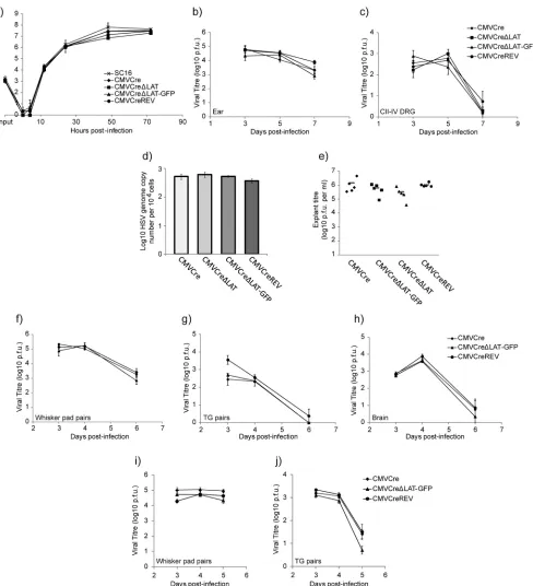
In vivogrowth curves utilized female BALB/c mice (Harlan, United Kingdom) at 8 weeks of age. All infections were conducted using iso-fluorane anesthesia. Ear pinna infections were conducted by pipetting 20l of virus inoculum containing 2⫻106PFU onto the left ear FIG 1Generation of Cre reporter viruses bearing deletions of the core LAT promoter. (a) Genomic structures of HSV CMVCre, HSV CMVCre⌬LAT, HSV CMVCre⌬LAT-GFP, and HSV CMVCreREV. All four viruses harbor an HCMV MIEP-Cre recombinase expression cassette within the nonessential HSV-1 US5
locus. (b) Genomic structures as analyzed by Southern blot hybridization. Restriction digest with HpaI demonstrates all predicted restriction fragments. Deletion of the core LAT promoter within HSV CMVCre⌬LAT and HSV CMVCre⌬LAT-GFP as well as its rescue within HSV CMVCreREV was confirmed by PstI restriction digest. The 3.3-kb HpaI fragment encoded within pPSTD1 was utilized as a radiolabeled probe. (c) LAT expression was quantified by qRT-PCR from total RNA extracted from TG latently infected with all four viruses, utilizing primers for major LAT and cyclophilin RNA. Histograms represent the mean (⫾ SEM) numbers of major LAT RNA copies per 103copies of cyclophilin RNA from triplicate PCRs.
on November 7, 2019 by guest
http://jvi.asm.org/
[image:2.585.90.487.63.391.2]pinna. Light scarification (20 shallow scratches) with a 27-gauge nee-dle was then applied to the ear pinna through the inoculum. Mice were killed by CO2asphyxiation, and the inoculated ear and ipsilateral CII, CIII, and CIV dorsal root ganglia (DRG) were dissected into complete DMEM for viral titration following storage at⫺70°C. Samples were freeze-thawed, homogenized, and freeze-thawed once more prior to assay. Whisker pad inoculation was conducted via the same method-ology, except that animals were infected by scarification of both whis-ker pads by the use of 40l of viral inoculum harboring 106PFU. Mice were killed by CO2asphyxiation, and whisker pads and trigeminal ganglia (TG) and brain tissue were dissected into complete DMEM for viral titration following storage at ⫺70°C. Samples were freeze-thawed, homogenized, and freeze-thawed once more prior to assay.
ROSA26R reporter mice (39) were used for thein vivocharacterization of HSV recombinants encoding Cre recombinase as previously described (29,30). Groups of adult mice (⬎8 weeks of age) that differed in age by less than 2 weeks were infected upon the ear pinna or whisker pads. Mice were killed by CO2asphyxiation at various times postinfection, and the ipsilat-eral CII, CIII, and CIV DRG (following ear infection) or both TG (follow-ing whisker pad infection) were dissected, pooled, and fixed on ice for 1 h in 4% paraformaldehyde–phosphate-buffered saline (PBS) and incubated in X-Gal (5-bromo-4-chloro-3-indolyl--D-galactopyranoside) as de-scribed previously (20). Marked-cell counts to determine potential differ-ences between virus groups were performed in a blinded manner to avoid experimental bias. This study predominantly utilized male mice. Where that was not possible, groups of equal numbers of males and females were used but were divided evenly between LAT-positive and LAT-negative mutants. The sex of the ROSA26R mice did not affect the phenotypes described in this study (data not shown).
All animal experiments were performed under United Kingdom Home Office Project License 80/2205.
DNA extractions for real-time quantitative PCR (qPCR) were per-formed on individual TG or pooled CII, CIII, and CIV DRG from five mice. The ganglia were homogenized and incubated in 0.5% sodium do-decyl sulfate (SDS) and 50 g of proteinase K/ml in TE buffer (10 mM Tris HCl, 1 mM EDTA [pH 8]) overnight at 37°C. Samples were sonicated, and phenol chloroform was extracted before purification with Qiagen PCR purification columns. qPCRs were conducted as previously described (7). Virus genome was quantified with ICP0 promoter-specific primers, and adenine phosphoribosyltransferase (APRT) was utilized as a cellular DNA control. PCRs were run as duplex reactions. The ICP0 promoter forward primer was GGAAAGGCGTGGGGTATAA, the reverse primer was AAC GTAGGCGGGGCTTC, and the TaqMan probe was TCGCATTTGCAC CTCGGCAC. The APRT forward primer was GGGGCAAAACCAAAAA AGGA, the reverse primer was TGTGTGTGGGGCCTGAGTC, and the TaqMan probe was TGCCTAAACACAAGCATCCCTACCTCAA (8). PCR products were quantified using a Corbett Research Rotor-Gene and associated software as the copy numbers per PCR, calculated from tripli-cate results from each PCR. A standard curve for each gene region was generated using dilutions of appropriate plasmids. Reaction conditions utilized were 15 min at 95°C, with 45 cycles of 15 s at 95°C and 60 s at 60°C. LAT RNA expression was assessed from pooled TG from five latently infected mice per virus. Ganglia were removed into 1 ml of RNAlater (Qiagen), prior to RNA extraction. Total RNA was extracted and DNase I treated from pooled TG by the use of TRIzol reagent and a PureLink RNA minikit (Invitrogen). RNA was reverse transcribed (RT) with a TaqMan MicroRNA reverse transcription kit (Invitrogen), according to the man-ufacturer’s protocol. RT reactions were primed with random primers (In-vitrogen) at a concentration of 0.2gl⫺1. qPCRs of samples and RT-negative controls were conducted with the aforementioned reaction conditions utilized for DNA load quantification, utilizing primers for mouse cyclophilin and major LAT RNA. The LAT forward primer was CCAGGCAGTAAGACCCAAGC, the reverse primer was GGCCGGTGT CGCTGTAAC, and the TaqMan probe was TCCCACCCCGCCTGTGTT TTT. The cyclophilin forward primer was GTCTCCTTCGAGCTGTT
TGC, the reverse primer was GAGGAACCCTTATAGCCAAATCC, and the TaqMan probe was ACAAAGTTCCAAAGACAGCAGAAAACTTTC. Whole-ganglion preparation and immunohistochemistry for HSV-1 antigen detection during latency was performed as described by Sawtell (33). However, antigen detection was conducted with anti-HSV-1 rabbit polyclonal (Dako) and horseradish peroxidase (HRP)-conjugated donkey anti-rabbit (Amersham Biosciences) antibodies. Staining was developed with a 3,3=-diaminobenzidine (DAB) peroxi-dase substrate kit (Vector Laboratories). Detection of antigen-positive cells was determined alongside naive and acutely infected tissue as negative and positive controls, respectively. All counts to determine potential differences between virus groups were performed on blinded material to avoid experimental bias.
Statistical analysis of ROSA26R mouse ganglion marked-cell popula-tions was conducted using the Mann-Whitney U and Kruskal-Wallis tests for paired- and multiple-group analyses, respectively.
RESULTS
In vitroand in vivocharacterization of HSV-1 recombinants deleted for the core LAT promoter.Cre recombinase-expressing viruses carrying deletions of the LAP with or without an HCMV MIEP GFP expression cassette as well as a LAP revertant virus were constructed on the HSV-1 SC16 genomic background as described in Materials and Methods. The predicted structures of these viruses are represented inFig. 1aand were confirmed by Southern blot hybridization (Fig. 1b) using the purified 3.3-kb HpaI DNA fragment from pPSTD1 as a probe. Wild-type 3.3-kb and 3.5-kb HpaI restriction fragments (from the IRLand TRL,
respectively) and the core LAP 203-bp PstI restriction fragment were detected from both HSV CMVCre and HSV CMVCreREV digests. Both HSV CMVCre⌬LAT and HSV CMVCre⌬LAT-GFP were negative for the core LAP 203-bp PstI fragment. Detection of 4.5-kb and 4.7-kb HpaI restriction fragments confirmed the inser-tion of the HCMV MIEP GFP expression cassette in both the IRL
and the TRLgenome regions of HSV CMVCre⌬LAT-GFP (Fig.
1b). Quantitative reverse transcriptase PCR (qRT-PCR) analysis of RNA recovered from trigeminal ganglia (TG) of mice latently infected with both⌬LAT mutants (HSV CMVCre⌬LAT and HSV CMVCre⌬LAT-GFP) demonstrated a 4,000-to-8,000-fold ablation of major LAT expression in comparison to either HSV CMVCre or the HSV CMVCre⌬LAT-GFP-revertant virus designated HSV CMVCreREV (Fig. 1c). Low-multiplicity-of-infection (low-MOI) growth curves demonstrated that thein vitroreplication kinetics of all recombinants were highly similar (Fig. 2a). Acute replication kinetics were assessedin vivoutilizing the mouse ear pinna infec-tion model. Assessment of infectious virus titers at 3, 5, and 7 d.p.i. showed that replication results for all recombinants in both ears (Fig. 2b) and in pooled CII, CIII, and CIV dorsal root ganglia (DRG) were similar (Fig. 2c). All viruses displayed equivalent lev-els of latency establishment within BALB/c DRG in comparison to the previously characterized Cre-expressing recombinant SC16 strain HSV CMVCre (29) as observed by real-time PCR quantifi-cation of latent viral DNA loads from pooled CII, CIII, and CIV DRG dissected 70 d.p.i. (Fig. 2d). In addition, explant reactivation analysis demonstrated that all viruses were reactivation compe-tent and generated similar amounts of virus following 5 days of culture of DRGs dissected 40 d.p.i. (Fig. 2e). Acute-phase replica-tion kinetics were also determined following whisker pad infec-tion with HSV CMVCre, HSV CMVCre⌬LAT-GFP, and HSV CMVCreREV. Titers achieved within the periphery, TG, and brain tissue were similar for all viruses in BALB/c mice (Fig. 2fto
on November 7, 2019 by guest
http://jvi.asm.org/
FIG 2Cre reporter virus growth characterization. (a) Low (0.01) MOI.In vitrogrowth curves of recombinant viruses and wild-type strain SC16 from duplicate experiments performed in BHK cells are shown. (b and c)In vivoreplication of recombinant viruses in the ears (b) and pooled CII, CIII, and CIV DRG (c) 3, 5, and 7 d.p.i. following inoculation of BALB/c mice on the left ear with 2⫻106PFU. Data points represent mean (⫾SEM) viral titers from five mice at each time
point. (d) Virus latent DNA loads. qPCR was performed on DNA extracted from pooled CII, CIII, and CIV DRG from five latently infected mice, utilizing primers for the ICP0 promoter and cellular APRT. Values represent mean (⫾SEM) numbers of HSV genome copies per 104copies of APRT from triplicate PCRs. (e)
Explant reactivation competence of HSV recombinants. CII, CIII, and CIV ganglia from latently infected mice were cultured for 5 days and assayed for reactivating virus. Each point represents viral titers detected per mouse, and the bars represent the averages of these data. (f to h)In vivoreplication of recombinant viruses in both whisker pads (f), TG pairs (g), and brain (h) 3, 4, and 6 d.p.i. following inoculation of BALB/c mice on both whisker pads. Data points represent mean (⫾SEM) viral titers from five mice at each time point. (i and j)In vivoreplication of recombinant viruses in both whisker pads (i) and TG pairs (j) 3, 4, and 5 d.p.i. following inoculation of ROSA26R mice on both whisker pads with 106PFU. Data points represent mean (⫾SEM) viral titers from five mice
at each time point. No mortality was observed in any of the above-described experiments.
on November 7, 2019 by guest
http://jvi.asm.org/
[image:4.585.49.537.62.598.2]h), as well as in the periphery and TG of ROSA26R reporter mice (Fig. 2iandj). Using these assays, we conclude that LAT-positive and LAT-negative HSV-1 recombinants replicate in similar man-ners bothin vitroandin vivo. Furthermore, these assays revealed that LAT-positive and LAT-negative recombinant viruses both establish and reactivate from latency within the DRGs in compa-rable manners.
Latency establishment is enhanced in the absence of LAT ex-pression within the trigeminal ganglion.It has been reported that a major phenotype observed with LAT-deficient mutants is that of a reduced efficiency for reactivation (12,21,28,35), and there is evidence to indicate that the reactivation deficit observed with LAT mutants can be explained by a decrease in the efficiency of latency establishment (42,44). Whether LATs have a dedicated function in promoting reactivation is less clear, although recently it has been proposed that LATs function to maintain thein vivo
reactivation competence of HSV-1 following repeated hyperther-mic stress (43).
We sought to investigate whether disparities existed between the numbers of infected cells in which HSV-1 enters latency in the presence and absence of LAT expression by utilizing the ROSA26R reporter mouse model of HSV-1 latency (29,30). In this model, expression of Cre recombinase from the virus leads to excision of aloxP-flanked neomycin resistance gene cassette situated between the constitutive ROSA26 promoter and downstreamlacZreporter gene within the infected cell. As a result,lacZis constitutively expressed from the cell genome. Thus, infection of mice with HSV-1 encoding Cre recombinase under the control of the HCMV MIEP results in the permanent genetic marking of latently infected neurons that can be visualized and accurately quantified following incubation with X-Gal (29,30,47).
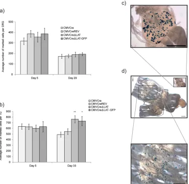
Forty ROSA26R mice were inoculated on the left ear with 106PFU of HSV CMVCre, HSV CMVCre⌬LAT, HSV CMVCre⌬LAT-GFP, or HSV CMVCreREV in groups of 10 and sampled at 5 and 29 d.p.i. (Fig. 3a). CII, III, and IV DRG were dissected and fixed, and following incubation with X-Gal, the numbers of reporter
gene-FIG 3LAT promoter-negative HSV-1 establishes latency at a higher frequency than the wild type in the TG. (a and b) Detection of Cre-marked cells following infection of ROSA26R mice with HSV CMVCre, HSV CMVCre⌬LAT, HSV CMVCre⌬LAT-GFP, and HSV CMVCreREV in the DRG (a) and TG (b). Histograms represent the mean (⫾SEM) numbers of positive cells per ganglion detected at the specified time points postinfection.Pvalues of 0.005 and 0.058 are represented by ** and *, respectively. (c and d) Light micrographs of latently infected DRG (c) and TG (d, panels i and ii). Bars, 1 mm. A single HSV CMVCre-infected mouse was euthanized following whisker pad infection when pathology reached a predetermined threshold of severity. No other mortality was recorded.
on November 7, 2019 by guest
http://jvi.asm.org/
[image:5.585.101.487.67.443.2]positive cells were enumerated (Fig. 3a andc). During acute infection, at 5 d.p.i., the average numbers of marked cells per ganglion were similar across all experimental groups: 317.2⫾ 32.4, 355.4⫾47.1, 386.6⫾54.4, and 387.1⫾31.3 positive cells per ganglion (⫾standard errors of the means [SEM]) for HSV
CMVCre, HSV CMVCre⌬LAT, HSV CMVCre⌬LAT-GFP,
and HSV CMVCreREV, respectively (P for comparisons to HSV CMVCre results⫽0.77, 0.34, and 0.15 for HSV CMVCre⌬LAT, HSV CMVCre⌬LAT-GFP, and HSV CMVCreREV, respectively). Consistent with previous reports (29,30), a decrease in the numbers of marked cells was observed in the transition from acute to latent infection (day 29 p.i.) and, as observed for the day 5 acute-infection time point, the average numbers of marked cells per ganglion were similar across all experimental groups: 158.7⫾19.8, 189.8⫾22.5, 193.1⫾15.0, and 175.3⫾16.4 positive cells per ganglion (⫾SEM) for HSV CMVCre, HSV CMVCre⌬LAT, HSV CMVCre⌬LAT-GFP, and HSV CMVCreREV, respectively (Fig. 3a) (Pfor comparisons to HSV CMVCre results⫽0.44, 0.32, and 0.65 for HSV CMVCre⌬LAT, HSV CMVCre⌬LAT-GFP, and HSV CMVCreREV, respectively). These results are consistent with the equivalent virus DNA loads de-tected during latency (Fig. 2d) and together suggest that LAT expres-sion does not affect latency establishment in the DRG.
It has been previously shown that establishment and reactiva-tion phenotypes of LAT-deficient mutants show a strong anatom-ical dependence on the TG but not DRG (35). To investigate whether establishment deficits could be detected in TG of reporter animals, 55 ROSA26R mice were infected via scarification of the whisker pads with HSV CMVCre, HSV CMVCre⌬LAT, HSV CMVCre⌬LAT-GFP, or HSV CMVCreREV in groups of 15 ani-mals (or groups of 10 aniani-mals, for HSV CMVCre⌬LAT-GFP). TG were removed from five mice per group at 5, 33, and 110 d.p.i. and individually analyzed for marked cells (Fig. 3d, panels i and ii). During acute infection (5 d.p.i.), the average numbers of marked cells per ganglion were once again highly similar across all exper-imental groups, though at 2-fold-higher numbers than were ob-served in DRG: 633.5 ⫾ 49.2, 601.1 ⫾ 58.8, 631.6 ⫾ 103.0, and 629.5⫾ 48.7 marked cells per ganglion (⫾SEM) for HSV CMVCre, HSV CMVCre⌬LAT, HSV CMVCre⌬LAT-GFP, and HSV CMVCreREV, respectively (Fig. 3b) (Pfor comparisons to HSV CMVCre results⫽0.73, 0.96, and 0.79 for HSV CMVCre⌬LAT, HSV CMVCre⌬LAT-GFP, and HSV CMVCreREV, respectively). During latency (33 d.p.i.), both HSV CMVCre and HSV CMVCreREV marked-cell populations had decreased by an average of 19%, to 489.7⫾45.0 and 542.5⫾52.0, respectively. In contrast, we observed that both⌬LAT virus infections led to an average increase in the numbers of marked cells during the transition from acute infection (day 5 p.i.) to latent infection (day 33 p.i.) (Fig. 3b), with 762.4⫾63.5 and 729.7⫾81.1 marked cells per ganglion (⫾SEM) detected for HSV CMVCre⌬LAT and HSV CMVCre⌬LAT-GFP, respectively, at the latter time point. This divergence of marked-cell populations at 33 d.p.i. was significant:P⫽0.0058 and 0.0587 (compared to HSV CMVCre) and 0.0172 and 0.0376 (compared to HSV CMVCreREV) for HSV CMVCre⌬LAT and HSV CMVCre⌬LAT-GFP, respectively. These data show that, following whisker pad infection of ROSA26R reporter mice, two HSV-1 recombinants deficient for LAT expression establish latency in the TG at a higher frequency than both parental and revertant viruses that are wild type for the LAT promoter locus. LAT expression does not influence the average DNA copy number per infected cell. It has been shown previously that HSV-1 genome copy numbers per infected cell during latency are
highly variable, ranging from one to⬎1,000 copies (32). Within this study, the majority of infected neurons possessed 1 to 10 or 10 to 100 HSV-1 genome copies at a range of inoculum doses. LATs have been shown to suppress lytic gene expression and replication in mouse neurons and neuronal cell lines (10,22) and to encode microRNAs (miRNAs) that can suppress translation of the key IE gene products ICP0 and ICP4 in anin vitromodel system (45). It has been hypothesized that LAT expression may resist the entry of HSV-1 into lytic replication following infection with high num-bers of virus particles (42). If that hypothesis is correct, in the absence of LAT expression a greater proportion of neurons in-fected with high HSV-1 genome loads would die, eventually re-sulting in a population of latent neurons that would, on average, possess fewer genome copies per cell than observed with wild-type HSV-1. Of importance, the HSV-1 genome copy number per neu-ron appears to positively correlate with the reactivation compe-tence of different HSV-1 strains (34) and may thus represent an important determinant of recrudescence. In order to determine whether expression of LATs influences the average latent genome copy number per cell, 26 mice were infected via the whisker pads with 106PFU of either HSV CMVCre⌬LAT-GFP or HSV
CMVCreREV in groups of 13. At 30 d.p.i., DNA was extracted from individual TG for HSV-1 genome quantification (n⫽14) and the other 12 TG from each virus-infected group were fixed for X-Gal incubation and quantification of marked-cell numbers. In the latter procedure, HSV CMVCre⌬LAT-GFP and HSV CMVCreREV possessed an average of 595.25⫾38.68 and 444.5⫾ 17.61 marked cells per TG (⫾SEM), respectively (P⫽0.0032). For each virus-infected group, total HSV-1 genome copies for each TG were divided by these data to obtain an estimate for the average genome copy number per marked cell. We calculated av-erage genome copy numbers per marked cell⫾SEM of 90.0⫾ 19.6 and 94.5 ⫾ 17.4 for HSV CMVCre⌬LAT-GFP and HSV CMVCreREV viruses, respectively. These data suggest that, fol-lowing inoculation with 106PFU (a dose at which LAT-negative
mutants establish latency at a greater frequency than wild-type virus), LAT expression does not influence the average genome copy number during the establishment of latency in the mouse.
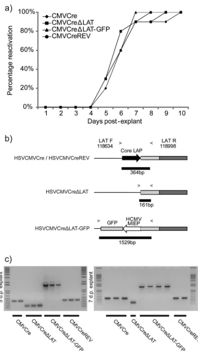
Absence of LAT expression is not associated with a reactiva-tion deficit in the ROSA26R mouse.Assessment of reactivation competence during initial characterization of latent DRG infec-tion demonstrated that all of the viruses were competent for reac-tivation during explant culture (Fig. 2e). To differentiate the ki-netics of virus recovery, ROSA26R mice were infected via the whisker pad and, 49 d.p.i., individual latently infected TG were cut into five equally sized pieces and cultured on MRC-5 monolayers. When cytopathic effect (CPE) was observed in these monolayers, the ganglion was scored positive for reactivation. Utilizing this system, we observed no gross deficit in reactivation kinetics in comparisons of LAT-positive and -negative viruses (Fig. 4a). All recovered viruses were identified as representing the correct input via PCR utilizing primers flanking the core LAT promoter (Fig. 4b andc).
The capacity of LAT-negative virus to establish latency with increased efficiency in the TG is lost with increasing virus inoc-ulum. Due to the observed differences between DRG and TG infection models in latency establishment, we next sought to de-termine whether our observations were the result of ganglion-specific differences or methodological differences in our infection procedures. ROSA26R mice were inoculated with 105, 107, and
on November 7, 2019 by guest
http://jvi.asm.org/
6⫻107PFU of either HSV CMVCre⌬LAT-GFP or the revertant HSV CMVCreREV on both whisker pads. At time points during latency (⬃30 d.p.i.), mice were killed and both TG dissected for marked-cell enumeration. The number of TG analyzed, the d.p.i. of dissection, and the average marked-cell numbers for all infec-tion doses are presented inTable 1. Following infection with 105
PFU, we noted that latent HSV CMVCreREV-infected cell num-bers were once again lower than those seen with the⌬LAT mutant but that this time they were lower by⬃50%, a markedly increased difference from previous 106PFU infections. In contrast, when
inoculum doses were increased to 107PFU and beyond, this trend was reversed, with HSV CMVCre⌬LAT-GFP infections now re-sulting in fewer latently infected cells than HSV CMVCreREV infections (Fig. 5a).
In the light of this dose dependence of latency establishment within the TG, we reevaluated the possibility that these differences were tissue specific by infecting ROSA26R mice with HSV
CMVCre⌬LAT-GFP or HSV CMVCreREV via the ear route of infection at doses of 105and 107PFU. At 30 d.p.i., mice were killed and CII, III, and IV DRG dissected for marked-cell enumeration. We observed no significant difference between the frequencies of latency establishment of the two viruses at either dose, with aver-age numbers of marked cells per DRG⫾SEM of 160.8⫾13.7 and 150.4⫾18.7 at the 105PFU dose and 189.1⫾16.0 and 174.0⫾
16.9 at the 107PFU dose for HSV CMVCre⌬LAT-GFP and HSV CMVCreREV, respectively (Fig. 5b).
Together, these dosage data suggest that the effect of LAT ex-pression on HSV-1 latency establishment is anatomically localized to the TG.
The maintenance of HSV-1 latency is impaired in the absence of LAT expression.Experimental infection of ROSA26R reporter mice with wild-type and LAT-negative HSV-1 recombinants demonstrates that the frequency of latency establishment in TG can be enhanced, with no effect upon the average DNA copy num-ber. Additionally, we have shown that in the absence of LAT ex-pression, the frequency of establishment of HSV-1 latency is sen-sitive to increasing concentrations of viral inoculum. Next, we assessed the long-term stability of cell marking during experimen-tal infection of the ROSA26R mouse TG. In three independent experiments, mice were infected with 106PFU on both whisker
[image:7.585.63.262.64.416.2]pads and ganglia were dissected and incubated in X-Gal at latent (29 to 33 d.p.i.) and “late-latent” (110 to 140 d.p.i) time points. The viral recombinants tested, numbers of TG analyzed, d.p.i. of dissection, and average marked-cell numbers at each time point are presented inTable 2. The average numbers of marked cells at either time point were utilized to estimate the rate of change over the duration of infection. An assumption was made that the rate of change in cell numbers was constant over time. In agreement with published observations, viruses that were wild type for the LAT promoter demonstrated a relatively stable population of marked cells between the early and late time points during latency (29), with HSV CMVCre and HSV CMVCreREV losing averages of 0.24 and 0.07⫾0.56 (SEM) marked cells per TG per day, respectively (Fig. 6a). In contrast, both HSV CMVCre⌬LAT-GFP and HSV CMVCre⌬LAT exhibited a loss of marked cells between 29 and 33 d.p.i. and 110 and 140 d.p.i., with the two mutants losing an aver-age of 1.72 and 1.63⫾0.6 (SEM) marked cells, respectively, per
TABLE 1Summary of individual experiment data from inoculum dosage study
Dose (PFU)a
Avg (range) no. of marked cells per TG
% difference (LAT⫹vs
LAT⫺) Pb nc
Day p.i.d
HSV
CMVCre⌬LAT-GFP HSV CMVCreREV
105
282.1 (0–825) 92.9 (0–245) 67.1 0.0340 10 42
402.2 (213–475) 248.7 (47–446) 38.2 0.0062 10 36
106
729.7 (270–1,105) 542.5 (334–840) 25.7 0.0380 10 33
715.0 (219–1,022) 564.0 (321–935) 21.1 0.2700 10 30
595.3 (385–850) 444.5 (318–541) 25.3 0.0032 12 30
595.0 (271–1,009) 423.3 (213–617) 28.9 0.0017 18 29
107 387.7 (246–521) 488.0 (371–646) ⫺25.9 0.1400 10 35
6⫻107 533.1 (321–791) 645.5 (439–833) ⫺21.1 0.0120 20 30
a
At the 106
PFU dose, a single HSV CMVCre⌬LAT-GFP-infected mouse was killed when pathology reached a predetermined threshold of severity. No mortality was recorded at any other dose.
bPvalues obtained using Mann-Whitney U test. c
Numbers of ganglia enumerated per virus-infected group.
dNumbers of days postinfection that TG were dissected for analysis. FIG 4Explant reactivation kinetics are unimpaired in the absence of LAT
expression following latency in the TG. (a) ROSA26R mice were inoculated with 106PFU of each recombinant virus per whisker pad. At 40 d.p.i., TG were
dissected (n⫽10 per recombinant), cut into five pieces, and plated onto MRC-5 cell monolayers, with pieces from one whole TG per dish. Plates were scored positive for reactivation upon observation of CPE within monolayers. (b) PCR with primers flanking the LAT region was designed to identify each recombinant. (c) Reactivating virus identity was successfully confirmed by PCR. The identities of viruses reactivating 5 and 7 days postexplant are shown. No mortality was recorded within this experiment.
on November 7, 2019 by guest
http://jvi.asm.org/
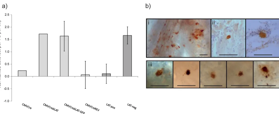
[image:7.585.298.544.553.669.2]TG per day (Fig. 6a). Of note, while decreases in the sizes of the latent cell populations were consistently observed during LAT-negative infection, statistical significance was not achieved within individual experiments. However, statistical analysis of aggregate data from infections 29 to 30 d.p.i. and 135 to 140 d.p.i. (Table 2) determined the loss of cells during HSV CMVCre⌬LAT-GFP in-fection to be significant (P⫽0.02). HSV CMVCreREV marked-cell populations did not change significantly (P⫽0.28). These data suggest that LAT-positive virus latency is largely stable, with an average loss of 0.11⫾0.40 marked neurons per TG per day in comparison to the 1.66⫾0.35 marked neurons lost per TG per day during LAT-negative virus latency (Fig. 6a). While LAT ex-pression can be observed to maintain the stability of the latently infected cell reservoir, it is not clear how this is accomplished. To ascertain whether an increased level of spontaneous reactivation occurs in the ROSA26R mouse in the absence of LAT expression, latently infected TG were dissected, fixed, and processed for whole-mount detection of lytic antigen-positive neurons. Utiliz-ing an anti-HSV polyclonal antibody, we were able to detect very low levels of lytic gene-positive cells at 44, 60, 66, and 69 d.p.i. From all time points, a total of six, three, and four positive cells
were detected in TG infected with HSV CMVCre (n⫽40 TG), HSV CMVCre⌬LAT-GFP (n⫽44 TG), and HSV CMVCreREV (n⫽46 TG), respectively (Fig. 6b). TG positive for HSV-1 antigen typically contained just one HRP-labeled cell and could be de-tected from 20-m-thick sections (Fig. 6b, panel ii) and from whole mounted ganglia (Fig. 6b, panel iii). These data are consis-tent with previous estimates of spontaneous reactivation with the mouse TG (23). These data indicate that spontaneous reactivation within the mouse TG is not dependent on LAT expression. With regard to maintenance, we observed that the mechanism of LAT-negative HSV-1-infected cell loss throughout latency does not op-erate via an increase in spontaneous reactivation, as measured using a polyclonal antibody. These data suggest that infected cells may be lost via apoptosis throughout latency, and such a mecha-nism clearly warrants further investigation.
DISCUSSION
In this paper, we have utilized the ROSA26R reporter mouse model in conjunction with Cre recombinase-expressing HSV-1 recombinants to investigate the role of LATs in the establishment and maintenance of neuronal infection latency. Previous studies have established that expression of Cre recombinase under the control of the HCMV MIEP results in the stable and efficient marking of latently infected neurons (29,30, 47). This system therefore provides an amenable approach allowing direct mea-surement of the frequency of latently infected cellsin vivo.
Previous studies have determined the frequency of latently in-fected neurons following infection of scarified corneas with 2⫻ 105PFU of HSV-1, a procedure that results in 10% to 20%
mor-tality of infected mice (42). Using this infection model, it has been shown that the HSV-1 strain KOS LATs are required for the effi-cient establishment of latency (44). Of importance, in this model system analysis at the single-neuron level has revealed that LAT-negative mutants establish only one-third as many latent infec-tions as wild-type virus (44). Utilizing ROSA26R reporter mice, we report that, following low-dose (ⱕ106PFU) whisker pad fection with HSV-1 strain SC16 LAT-deficient viruses, an in-creased frequency of latency was observed relative to wild-type or revertant viruses. We define doses ofⱕ106PFU as “low” doses due
to the observation that, following whisker pad inoculation with 104PFU, we were unable to efficiently infect the ROSA26R TG
[image:8.585.138.451.63.188.2](data not shown). Further, we were able to manipulate the relative
TABLE 2Summary of individual experimental data from maintenance study
Expt and strain
Avg (range) no. of marked cells per TG at the indicated time point
na Pb
Early Late
1 33 d.p.i. 110 d.p.i.
HSV CMVCre 490 (244–639) 471 (223–714) 8–10 0.76 HSV CMVCre⌬LAT 762 (506–1,028) 630 (303–885) 10 0.13 HSV CMVCreREV 543 (321–935) 455 (173–719) 10 0.41
2 30 d.p.i. 135 d.p.i.
HSV CMVCre⌬LAT-GFP 715 (270–1,105) 480 (88–843) 10 0.14 HSV CMVCreREV 564 (334–840) 587 (245–857) 10 0.65
3 29 d.p.i. 140 d.p.i.
HSV CMVCre⌬LAT-GFP 595 (271–1,009) 481 (214–783) 18–20 0.06 HSV CMVCreREV 423 (213–617) 503 (260–756) 18 0.16 a
Numbers of ganglia enumerated per virus-infected group.
bPvalues obtained using Mann-Whitney U test.
FIG 5In the absence of LAT expression, the frequency of latency establishment is enhanced at “low” doses but diminishes at increasing doses. (a) R26R mice were infected on both whisker pads at a range of doses (105to 6⫻107PFU) and TG dissected⬃30 d.p.i. for marked-cell quantification. Histograms represent
the mean (⫾SEM) number of positive cells per ganglion at each dose.Pvalues of⬍0.00005,⬍0.002, and 0.01 are represented by ***, **, and *, respectively. (b) R26R mice were infected on the left ear at 105or 107PFU doses. CII, III, and IV DRG were dissected 30 d.p.i. for marked-cell quantification. Histograms represent
the mean (⫾SEM) number of positive cells per ganglion at each dose.
on November 7, 2019 by guest
http://jvi.asm.org/
[image:8.585.41.287.535.706.2]efficiencies of establishment of infections by LAT-positive and LAT-negative viruses by altering the inoculum dose. Starting at an inoculum dose of 106PFU, we observed that LAT-positive virus
established latency at a reduced frequency, with just 75% of the number of neurons in which latency was established by LAT-neg-ative virus. Dropping the inoculum dose to 105PFU further exac-erbated this difference, to between 33% to 62% of the total LAT-negative latent population. Conversely, increasing the inoculum dose beyond 106PFU led to a loss of this phenotype, with
LAT-positive virus establishing latency in 1.21 to 1.26 times as many neurons as LAT-negative virus. Though not similar in magnitude, the latter establishment phenotype is more consistent with previ-ously reported data (44).
Taken together, these data suggest that the latency establish-ment phenotypes of HSV-1 mutants in the presence and absence of LAT expression are highly sensitive to the infection methodol-ogy, a fact that may confound absolute interpretation of LAT functionin vivo. Despite the loss of the low-dose phenotype at increasing amounts of inoculum virus, we were unable to recapit-ulate previously reported data demonstrating severely reduced es-tablishment efficiency in the absence of LAT expression (44). HSV-1 strains of higher virulence, different LAT mutations, greater infection doses (or at least more efficient infection meth-odologies), and differing inoculation sites may account for such observations. Despite such variables, a dose-dependent effect upon establishment may be a general feature of mouse infection and it is likely that these variables would simply affect the range of doses at which this is evident.
Previous studies have investigated the role of the inoculum dose in latency establishment following infection with wild-type HSV-1 and LAT-deficient mutants. A study utilizing the rabbit corneal model of infection found no difference in latent viral DNA loads in the TG at doses of 500 and 50,000 PFU (26). We have found that estimates of total latent viral DNA loads are a relatively
insensitive measure by which to evaluate establishment efficiency. Thus, we have been unable to detect a statistically significant dif-ference between wild-type and LAT-deficient mutants in total la-tent DNA loads (P⬎0.37) despite a 30% increase in latent cell numbers following infection of reporter mice with a LAT-defi-cient mutant. Our inability to detect a statistically significant dif-ference between LAT-positive and LAT-negative virus in total la-tent virus DNA loads is consisla-tent with a number of previous studies with HSV-1 (3,12,28) and HSV-2 (18,48,49). In contrast, Hoshino et al. (13) demonstrated in 2009 that an HSV-2 (strain 333) LAT promoter mutant displayed a clear dose dependence during latency establishment within mouse TG but that, at the lowest doses employed, the latent DNA load of a LAT-null virus was lower than that of the corresponding revertant virus. Whether the reduced establishment of latency observed following low-dose HSV-2 infection relates to differences in the function of the HSV-1 and HSV-2 LATs or to virus strain and/or animal model differences is currently unclear. In our study, dose-dependent ef-fects on establishment were observed within the TG but not the DRG, suggesting that, in addition to virus strain and/or animal model differences, the anatomical site of latency can impact virus mutant phenotypes.
We have been unable to observe increased titers during acute infection of the mouse with LAT-negative viruses and yet have observed increased numbers of marked cells at latency. Ongoing studies in our laboratory utilizing a fluorescent reporter mouse strain, allowing specific analysis of live marked cells, have deter-mined that HSV-1 DNA is detectable only within the marked-cell population (unpublished observations). As such, increased neu-ronal cell marking by LAT-deficient mutants represents Cre re-combinase-mediated reporter gene activation as a consequence of infection rather than a bystander effect. However, at this time, we cannot determine the LAT mechanism that results in this anatom-ically distinct and dose-dependent establishment phenotype.
FIG 6Maintenance of the latent cell reservoir is unstable in the absence of LAT expression. (a) R26R mice were infected on both whisker pads with 106PFU and
TG dissected at latency (29 to 33 d.p.i.) and “late” latency (110 to 140 d.p.i.) for marked-cell quantification. Histograms represent the mean number of marked cells lost per TG per day⫾SEM. “LAT-pos” denotes both HSV CMVCre and HSV CMVCreREV data. “LAT-neg” denotes both HSV CMVCre⌬LAT and HSV CMVCre⌬LAT-GFP data. (b) Light micrographs of HSV-1 antigen-positive cells as detected via whole-ganglion immunohistochemistry. An example of acute infection within the TG at 4 d.p.i. is displayed in panel i. Panel ii displays antigen-positive cells detected in 20-m-thick sections of latently infected TG at 44 d.p.i. Panel iii displays examples of antigen-positive cells detected at 44 d.p.i. to 69 d.p.i. All bars represent 100m.
on November 7, 2019 by guest
http://jvi.asm.org/
[image:9.585.48.538.62.262.2]HSV-1 LATs have multiple functions that include repression of viral IE gene expression (5,10,22,37,45), inhibition of apop-tosis/promotion of cell survival (1,4,14,27,42), and regulation of histone-mediated repression of the HSV-1 genome during latency (6,19,50). Given these functions, it would perhaps be predicted that at low doses of infection, removal of LAT-mediated IE gene repression would favor lytic cycle entry in primarily infected neu-rons and increased spread within ganglionic tissue. In contrast, at high input doses, if LAT-mediated effects mitigating apoptosis were not present, a high proportion of primarily infected neurons might be killed without supporting virus replication, thus limiting virus spread through the tissue. While we were not able to detect increased replication efficiency of HSV CMVCre⌬LAT-GFP within the TG following whisker pad infection (Fig. 2), the exper-iments may have been of insufficient statistical power to observe the presumably minor increase in replication titers needed to es-tablish latency in ⬃33% more cells. However, this predicted “changing dominance” of LAT functions at low and high doses of viruses presents an appealing interpretation of our data and re-quires further investigation.
In this study, we also provide evidence for a role of LAT in maintaining latent infection of the mouse TG. By enumerating the number of marked cells in the ROSA26R TG at “early” (29 to 33 d.p.i.) and “long-term” (110 to 140 d.p.i.) times during latency, we consistently observed reductions in marked-cell populations dur-ing LAT-negative virus but not LAT-positive infection. These data suggest that LATs actively play a role in the maintenance of the latent cell reservoir. An assumption made in the interpretation of our current data is that the distribution of latency among distinct neuronal subpopulations is equivalent to that following wild-type virus infection. However, further work is necessary to define the neuronal populations latently infected by both LAT-deficient and wild-type viruses to investigate the possibility that mutant viruses establish latency in a neuronal population in which latency is in-trinsically less stable.
By utilizing a previously described methodology for the detec-tion of antigen-positive cells in whole-mount TG samples (33), we investigated whether the loss of marked cells was due to an in-crease in spontaneous reactivation during LAT-negative infec-tion. However, upon interrogation of latently infected tissue, we found that antigen-positive cells could be detected at similarly low levels per ganglion for all viruses. These findings are consistent with earlier reports (23) and together suggest that LAT is not es-sential for spontaneous reactivation within the mouse TG. It is clear that the observed loss of marked cells during ROSA26R la-tency is not a result of increased spontaneous reactivation, as as-sessed by virus detection with an anti-HSV-1 polyclonal antibody. Whether this cell loss is due to an elevated frequency of apoptosis, possibly in response to the earliest stages of reactivation, is under investigation.
Previous investigations of HSV-1 latency maintenance have relied upon the assessment of total viral DNA within infected tis-sues (9,11,36). However, those studies do not provide a direct insight into the number of latently infected cells. With regard to the nonuniformity of viral DNA copy numbers per cell (32), it is thus currently unclear whether a loss of infected cells would man-ifest in a proportional loss of total viral DNA within the ganglion. A previous study (17) revealed that disruption of splicing of the primary LAT resulted in an approximately 10-fold reduction in the latent genome copy number in mouse TG. However, due to
the assessment of a single latent time point, it was not possible to discern whether the observed phenotype was due to a deficit in the establishment or maintenance of latency.
Of great interest, a recent report determined that, following repeated induction ofin vivoreactivation by hyperthermic stress, a LAT-negative virus suffered a reduced capacity to reactivate be-tween 30 days and 42 weeks postinfection (43). These data are consistent with our findings, as a loss of infected cells over a pro-longed period of time could be hypothesized to reduce reactiva-tion frequency within the TG. While we did not observe a differ-ence in spontaneous reactivation frequency (as measured by HSV-1 lytic antigen-positive cell numbers), our analyses were conducted both earlier and without reactivation stimuli. Thefore, a larger reduction in the latent cell population may be re-quired to observe a reactivation deficit.
It is important that all viruses utilized within this study were constructed by replacing the viral US5 gene with a Cre
recombi-nase expression cassette. This mutation removes expression of glycoprotein J (gJ), a protein of known antiapoptosis function (16), and may thus sensitize the recombinants in this study to the effects of apoptosis. Nonetheless, we observed no effect upon the
in vivoreplication kinetics in comparisons of gJ-deficient
LAT-positive and LAT-negative viruses. In addition, stable mainte-nance of latency was observed during infection with gJ-deficient LAT-positive viruses, indicating that gJ antiapoptotic functions play little or no role in latency. Studies of LAT-negative HSV-1 gene expression have shown elevated lytic transcript accumula-tion during latency (5), consistent with increased lytic gene ex-pression during acute infection (10). In the context of LAT-defi-cient mutants, such “leaky” expression of genes in the lytic cycle could increase the probability of virus reactivation and/or the en-gagement of an apoptotic program that in the absence of LATs cannot be effectively curtailed.
In summary, our studies suggest that the HSV-1 latency-associ-ated transcripts function to (i) enhance the efficiency of latency es-tablishment following high-dose infection, (ii) reduce the efficiency of latency establishment following low-dose infection, and (iii) facil-itate the long-term maintenance of the latent cell reservoir.
ACKNOWLEDGMENTS
This research was supported by the Wellcome Trust (project grant 086403/Z/08/7) and the Medical Research Council (MRC) UK (MR/ J002232/1). M.N. is supported by an MRC (UK) Ph.D. studentship.
REFERENCES
1.Ahmed M, Lock M, Miller CG, Fraser NW.2002. Regions of the herpes simplex virus type 1 latency-associated transcript that protect cells from apoptosis in vitro and protect neuronal cells in vivo. J. Virol.76:717–729. 2.Arthur JL, Everett R, Brierley I, Efstathiou S.1998. Disruption of the 5=
and 3=splice sites flanking the major latency-associated transcripts of her-pes simplex virus type 1: evidence for alternate splicing in lytic and latent infections. J. Gen. Virol.79(Pt 1):107–116.
3.Bloom DC, Devi-Rao GB, Hill JM, Stevens JG, Wagner EK.1994. Molec-ular analysis of herpes simplex virus type 1 during epinephrine-induced reac-tivation of latently infected rabbits in vivo. J. Virol.68:1283–1292. 4. Branco FJ, Fraser NW. 2005. Herpes simplex virus type 1
latency-associated transcript expression protects trigeminal ganglion neurons from apoptosis. J. Virol.79:9019 –9025.
5.Chen SH, Kramer MF, Schaffer PA, Coen DM.1997. A viral function represses accumulation of transcripts from productive-cycle genes in mouse ganglia latently infected with herpes simplex virus. J. Virol.71: 5878 –5884.
6.Cliffe AR, Garber DA, Knipe DM. 2009. Transcription of the herpes
on November 7, 2019 by guest
http://jvi.asm.org/
simplex virus latency-associated transcript promotes the formation of fac-ultative heterochromatin on lytic promoters. J. Virol.83:8182– 8190. 7.Coleman HM, et al.2008. Histone modifications associated with herpes
simplex virus type 1 genomes during quiescence and following ICP0-mediated de-repression. J. Gen. Virol.89:68 –77.
8.Dush MK, Sikela JM, Khan SA, Tischfield JA, Stambrook PJ. 1985. Nucleotide sequence and organization of the mouse adenine phosphori-bosyltransferase gene: presence of a coding region common to animal and bacterial phosphoribosyltransferases that has a variable intron/exon ar-rangement. Proc. Natl. Acad. Sci. U. S. A.82:2731–2735.
9.Efstathiou S, Minson AC, Field HJ, Anderson JR, Wildy P. 1986. Detection of herpes simplex virus-specific DNA sequences in latently in-fected mice and in humans. J. Virol.57:446 – 455.
10. Garber DA, Schaffer PA, Knipe DM.1997. A LAT-associated function reduces productive-cycle gene expression during acute infection of mu-rine sensory neurons with herpes simplex virus type 1. J. Virol.71:5885– 5893.
11. Hill JM, et al.1996. Quantitation of herpes simplex virus type 1 DNA and latency-associated transcripts in rabbit trigeminal ganglia demonstrates a stable reservoir of viral nucleic acids during latency. J. Virol.70:3137– 3141.
12. Hill JM, Sedarati F, Javier RT, Wagner EK, Stevens JG.1990. Herpes simplex virus latent phase transcription facilitates in vivo reactivation. Virology174:117–125.
12a.Hill TJ, Field HJ, Blyth WA.1975. Acute and recurrent infection with herpes simplex virus in the mouse: a model for studying latency and re-current disease. J. Gen. Virol.28:341–353.
13. Hoshino Y, Pesnicak L, Straus SE, Cohen JI. 2009. Impairment in reactivation of a latency associated transcript (LAT)-deficient HSV-2 is not solely dependent on the latent viral load or the number of CD8(⫹) T cells infiltrating the ganglia. Virology387:193–199.
14. Inman M, et al.2001. Region of herpes simplex virus type 1 latency-associated transcript sufficient for wild-type spontaneous reactivation promotes cell survival in tissue culture. J. Virol.75:3636 –3646. 15. Javier RT, Stevens JG, Dissette VB, Wagner EK.1988. A herpes simplex
virus transcript abundant in latently infected neurons is dispensable for establishment of the latent state. Virology166:254 –257.
16. Jerome KR, et al.1999. Herpes simplex virus inhibits apoptosis through the action of two genes, Us5 and Us3. J. Virol.73:8950 – 8957.
17. Kang W, Mukerjee R, Fraser NW.2003. Establishment and maintenance of HSV latent infection is mediated through correct splicing of the LAT primary transcript. Virology312:233–244.
18. Krause PR, et al.1995. Expression of the herpes simplex virus type 2 latency-associated transcript enhances spontaneous reactivation of genital herpes in latently infected guinea pigs. J. Exp. Med.181:297–306. 19. Kwiatkowski DL, Thompson HW, Bloom DC.2009. The polycomb group
protein Bmi1 binds to the herpes simplex virus 1 latent genome and maintains repressive histone marks during latency. J. Virol.83:8173– 8181.
20. Lachmann RH, Efstathiou S.1997. Utilization of the herpes simplex virus type 1 latency-associated regulatory region to drive stable reporter gene expression in the nervous system. J. Virol.71:3197–3207.
21. Leib DA, et al.1989. A deletion mutant of the latency-associated tran-script of herpes simplex virus type 1 reactivates from the latent state with reduced frequency. J. Virol.63:2893–2900.
22. Mador N, Goldenberg D, Cohen O, Panet A, Steiner I.1998. Herpes simplex virus type 1 latency-associated transcripts suppress viral replica-tion and reduce immediate-early gene mRNA levels in a neuronal cell line. J. Virol.72:5067–5075.
23. Margolis TP, et al.2007. Spontaneous reactivation of herpes simplex virus type 1 in latently infected murine sensory ganglia. J. Virol.81:11069 – 11074.
24. McGeoch DJ, et al.1988. The complete DNA sequence of the long unique region in the genome of herpes simplex virus type 1. J. Gen. Virol.69(Pt 7):1531–1574.
25. Nicoll MP, Proença JT, Efstathiou S.2012. The molecular basis of herpes simplex virus latency. FEMS Microbiol. Rev.36:684 –705.
26. O’Neil JE, et al.2004. Wide variations in herpes simplex virus type 1 inoculum dose and latency-associated transcript expression phenotype do not alter the establishment of latency in the rabbit eye model. J. Virol. 78:5038 –5044.
27. Perng G-C, et al.2000. Virus-induced neuronal apoptosis blocked by the herpes simplex virus latency-associated transcript. Science287:1500 –1503.
28. Perng GC, et al.1994. The latency-associated transcript gene of herpes simplex virus type 1 (HSV-1) is required for efficient in vivo spontaneous reactivation of HSV-1 from latency. J. Virol.68:8045– 8055.
29. Proença JT, Coleman HM, Connor V, Winton DJ, Efstathiou S.2008. A historical analysis of herpes simplex virus promoter activation in vivo reveals distinct populations of latently infected neurones. J. Gen. Virol. 89:2965–2974.
30. Proença JT, et al.2011. An investigation of HSV promoter activity com-patible with latency establishment reveals VP16 independent activation of HSV immediate early promoters in sensory neurones. J. Gen. Virol.92(Pt 11):2575–2585.
31. Rock DL, et al.1987. Detection of latency-related viral RNAs in trigem-inal ganglia of rabbits latently infected with herpes simplex virus type 1. J. Virol.61:3820 –3826.
32. Sawtell NM.1997. Comprehensive quantification of herpes simplex virus latency at the single-cell level. J. Virol.71:5423–5431.
33. Sawtell NM.2003. Quantitative analysis of herpes simplex virus reactiva-tion in vivo demonstrates that reactivareactiva-tion in the nervous system is not inhibited at early times postinoculation. J. Virol.77:4127– 4138. 34. Sawtell NM, Poon DK, Tansky CS, Thompson RL.1998. The latent herpes
simplex virus type 1 genome copy number in individual neurons is virus strain specific and correlates with reactivation. J. Virol.72:5343–5350. 35. Sawtell NM, Thompson RL.1992. Herpes simplex virus type 1
latency-associated transcription unit promotes anatomical site-dependent estab-lishment and reactivation from latency. J. Virol.66:2157–2169. 36. Sedarati F, Izumi KM, Wagner EK, Stevens JG.1989. Herpes simplex
virus type 1 latency-associated transcription plays no role in establishment or maintenance of a latent infection in murine sensory neurons. J. Virol. 63:4455– 4458.
37. Shen W, et al.2009. Two small RNAs encoded within the first 1.5 kilo-bases of the herpes simplex virus type 1 latency-associated transcript can inhibit productive infection and cooperate to inhibit apoptosis. J. Virol. 83:9131–9139.
38. Soares K, et al. 1996. cis-acting elements involved in transcriptional regulation of the herpes simplex virus type 1 latency-associated promoter 1 (LAP1) in vitro and in vivo. J. Virol.70:5384 –5394.
39. Soriano P.1999. Generalized lacZ expression with the ROSA26 Cre re-porter strain. Nat. Genet.21:70 –71.
40. Steiner I, et al.1989. Herpes simplex virus type 1 latency-associated tran-scripts are evidently not essential for latent infection. EMBO J.8:505–511. 41. Stevens JG, Wagner EK, Devi-Rao GB, Cook ML, Feldman LT.1987.
RNA complementary to a herpesvirus alpha gene mRNA is prominent in latently infected neurons. Science235:1056 –1059.
42. Thompson RL, Sawtell NM.2001. Herpes simplex virus type 1 latency-associated transcript gene promotes neuronal survival. J. Virol.75:6660 – 6675.
43. Thompson RL, Sawtell NM.2011. The herpes simplex virus type 1 latency associated transcript locus is required for the maintenance of reactivation competent latent infections. J. Neurovirol.17:552–558.
44. Thompson RL, Sawtell NM.1997. The herpes simplex virus type 1 laten-cy-associated transcript gene regulates the establishment of latency. J. Vi-rol.71:5432–5440.
45. Umbach JL, et al.2008. MicroRNAs expressed by herpes simplex virus 1 during latent infection regulate viral mRNAs. Nature454:780 –783. 46. Wagner EK, Bloom DC.1997. Experimental investigation of herpes
sim-plex virus latency. Clin. Microbiol. Rev.10:419 – 443.
47. Wakim LM, Jones CM, Gebhardt T, Preston CM, Carbone FR.2008. CD8(⫹) T-cell attenuation of cutaneous herpes simplex virus infection reduces the average viral copy number of the ensuing latent infection. Immunol. Cell Biol.86:666 – 675.
48. Wang K, Pesnicak L, Guancial E, Krause PR, Straus SE. 2001. The 2.2-kilobase latency-associated transcript of herpes simplex virus type 2 does not modulate viral replication, reactivation, or establishment of la-tency in transgenic mice. J. Virol.75:8166 – 8172.
49. Wang K, Pesnicak L, Straus SE.1997. Mutations in the 5=end of the herpes simplex virus type 2 latency-associated transcript (LAT) promoter affect LAT expression in vivo but not the rate of spontaneous reactivation of genital herpes. J. Virol.71:7903–7910.
50. Wang Q-Y, et al.2005. Herpesviral latency-associated transcript gene promotes assembly of heterochromatin on viral lytic-gene promoters in latent infection. Proc. Natl. Acad. Sci. U. S. A.102:16055–16059.