Open Access
Research
Cellular responses to modified
Plasmodium falciparum
MSP1
19
antigens in individuals previously exposed to natural malaria
infection
Christian MF Okafor*
1,5, Chiaka I Anumudu
1, Yusuf O Omosun
1,6,
Chairat Uthaipibull
3,7, Idowu Ayede
2, Henrietta O Awobode
1,
Alex B Odaibo
1, Jean Langhorne
3, Anthony A Holder
3, Roseangela I Nwuba
1and Marita Troye-Blomberg
4Address: 1Cellular Parasitology Programme, Department of Zoology University of Ibadan, Ibadan, Nigeria, 2Oni Memorial Children's Hospital,
Ring Road, Ibadan, Nigeria, 3Division of Parasitology, MRC National Institute for Medical Research, Mill Hill, London NW7 1AA, UK, 4Department of Immunology, Wenner-Gren Institute, Arrhenius Laboratories, Stockholm University, Stockholm, Sweden, 5College of Art and
Sciences, Northwest University, 5520, 108th Ave. NE, Kirkland WA 98033, USA, 6Department of Biotechnology, Bells University of Technology,
Sango-Otta, Nigeria and 7Protein-Ligand Engineering and Molecular Biology Laboratory, National Center for Genetic Engineering and
Biotechnology (BIOTEC), Thailand Science Park, Pathumthani, Thailand
Email: Christian MF Okafor* - [email protected]; Chiaka I Anumudu - [email protected];
Yusuf O Omosun - [email protected]; Chairat Uthaipibull - [email protected]; Idowu Ayede - [email protected]; Henrietta O Awobode - [email protected]; Alex B Odaibo - [email protected]; Jean Langhorne - [email protected]; Anthony A Holder - [email protected]; Roseangela I Nwuba - [email protected]; Marita Troye-Blomberg - [email protected] * Corresponding author
Abstract
Background: MSP1 processing-inhibitory antibodies bind to epitopes on the 19 kDa C-terminal region of the Plasmodium falciparum merozoite surface protein 1 (MSP119), inhibiting erythrocyte
invasion. Blocking antibodies also bind to this antigen but prevent inhibitory antibodies binding, allowing invasion to proceed. Recombinant MSP119 had been modified previously to allow inhibitory but not blocking antibodies to continue to bind. Immunization with these modified proteins, therefore, has the potential to induce more effective protective antibodies. However, it was unclear whether the modification of MSP119 would affect critical T-cell responses to epitopes
in this antigen.
Methods: The cellular responses to wild-type MSP119 and a panel of modified MSP119 antigens
were measured using an in-vitro assay for two groups of individuals: the first were malaria-naïve and the second had been naturally exposed to Plasmodium falciparum infection. The cellular responses to the modified proteins were examined using cells from malaria-exposed infants and adults.
Results: Interestingly, stimulation indices (SI) for responses induced by some of the modified proteins were at least two-fold higher than those elicited by the wild-type MSP119. A protein with four amino acid substitutions (Glu27→Tyr, Leu31→Arg, Tyr34→Ser and Glu43→Leu) had the highest stimulation index (SI up to 360) and induced large responses in 64% of the samples that had significant cellular responses to the modified proteins.
Published: 23 November 2009
Malaria Journal 2009, 8:263 doi:10.1186/1475-2875-8-263
Received: 31 July 2009 Accepted: 23 November 2009 This article is available from: http://www.malariajournal.com/content/8/1/263
© 2009 Okafor et al; licensee BioMed Central Ltd.
Conclusion: This study suggests that specific MSP119 variants that have been engineered to improve their antigenicity for inhibitory antibodies, retain T-cell epitopes and the ability to induce cellular responses. These proteins are candidates for the development of MSP1-based malaria vaccines.
Background
The development of an effective malaria vaccine remains a major public health challenge. Merozoite surface pro-tein (MSP)-1 of Plasmodium falciparum is being developed as a vaccine candidate to protect against the erythrocytic stages of the malaria parasite [1,2]. Much of the work has been focused on the 19 kDa C-terminal region of MSP1 (called MSP119[3]). Protection against challenge infection following immunization in rodent and monkey models of malaria has been reported [4-11]. However, sero-epide-miological studies [12-16] and vaccine trials [17] in human populations have given conflicting results con-cerning the protective role of anti-MSP1 antibodies, which may be explained by differences in the fine specificities of the MSP119-specific antibodies [18,19]. It can be con-cluded from these studies that in the humoral control of malaria infection, the fine specificity of the antibody response may be crucial to inhibit erythrocyte invasion by merozoites.
The MSP1 precursor is cleaved into four fragments on the merozoite surface and at invasion the C-terminal 42 kDa fragment (MSP142) is processed further into two smaller fragments: a 33 kDa polypeptide (MSP133) and the C-ter-minal MSP119, which remains on the parasite surface dur-ing invasion of red blood cells (RBC). MSP1 has been reported to elicit three types of antibody that can bind MSP142[2,20,21]. These are a) inhibitory antibodies, which inhibit the cleavage of MSP142 and thus invasion of RBC; b) blocking antibodies, which have overlapping spe-cificities and compete with inhibitory antibodies for bind-ing to the antigen, thereby allowbind-ing processbind-ing and invasion to occur even in the presence of inhibitory anti-bodies; and c) neutral antibodies that are neither inhibi-tory nor blocking. Significantly, it has been shown that all these types of MSP119-specific antibodies are part of the natural immune response to MSP1 in malaria-exposed individuals [22,23]. Thus, the rational design of an MSP1-based malaria vaccine for the preferential induction of processing-inhibitory antibodies with the appropriate specificities is an important goal. The relative abundance of these protective antibodies in relation to the detrimen-tal (blocking) antibodies in any infection is one of the important factors that may determine the outcome of that infection [2,3,20,22].
The MSP119 epitopes recognized by inhibitory and block-ing monoclonal antibodies (mAbs) have been mapped using site-directed mutagenesis, PEPSCAN, and nuclear magnetic resonance (NMR) [21,24,25]. A number of sin-gle and multiple amino acid substitutions in MSP119 has been made, which had either no effect, or reduced, or completely abolished the binding of individual mAbs [21]. Recent data have shown that polyclonal antibodies in sera obtained from individuals living in a malaria endemic area recognize and bind to the modified antigens [22,23]. A vaccine based on one of these modified pro-teins could be designed to induce inhibitory but not blocking Abs and thus provide a focused polyclonal anti-body response to inhibit RBC invasion and cleavage of MSP1 [21,22].
vaccine that would induce both protective antibodies and suitable cellular responses.
Methods
Subjects and samples
Volunteers were recruited in Ibadan, Nigeria where P. fal-ciparum malaria is perennial. High malaria transmission occurs from April to November, corresponding to the rainy season (March to October), all samples were col-lected in March. The study protocol was reviewed and approved by the Joint University of Ibadan/University College Hospital Ethical committee. Informed consent was obtained from all participating individuals or legal guardians of children enrolled in the study. The donors were divided into two groups; the first was between three months and seven years of age (six individuals) and had clinical malaria symptoms, while the second aged between 13 yrs and 43 yrs (30 individuals) was without clinical malaria at recruitment. Venous blood (up to 4 ml from children and 7 ml from adults) was collected in heparinized tubes and transported immediately in insu-lated boxes to the laboratory for processing at the Depart-ment of Immunology, Wenner-Gren Institute, Stockholm University, Sweden. Giemsa-stained thick-blood films were examined for the presence of malaria parasites. A group of seven malaria-unexposed European donors pro-vided control samples for the study.
Peripheral blood mononuclear cells (PBMC)
Human PBMC were harvested from whole blood approx-imately 24 hrs after phlebotomy. PBMC were prepared by Ficoll-Hypaque (Pharmacia, Uppsala, Sweden) density gradient centrifugation according to the manufacturers' instructions. The harvested cells were suspended in freez-ing medium [10% dimethyl sulfoxide (DMSO), 20% fetal calf serum (FCS), RPMI 1640, HEPES and L-glutamine], frozen at 1°C min-1 to -70°C in plastic containers (Nal-gene Cryo, Nalge Company, USA), and then transferred into liquid nitrogen until used in in vitro assays. The cells had a demonstrated >95% viability when compared with frozen cells collected in Sweden.
Antigens
Cells in the proliferation assays were stimulated by anti-gens at a predetermined final concentration of 1 μg/ml.
Wild-type (WT) and modified MSP1 proteins
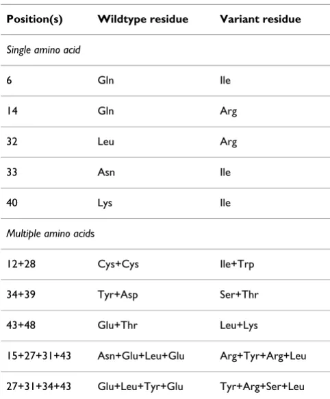
The recombinant wild-type and modified MSP119 anti-gens fused to Schistosoma japonicum glutathione S-trans-ferase (GST) have been described previously [21]. Table 1 shows the specific modifications in the primary sequence of the MSP119 antigens used; residues are numbered from the N-terminus of MSP119.
P. falciparum antigen
Crude P. falciparum antigen (F32) was used as a positive control for confirming previous infection or lack of expo-sure to P. falciparum in exposed and naïve individuals [31].
Control antigens
Phytohemagglutinin (PHA; Sigma, Poole, UK) was used as positive control for testing lymphocyte viability and responsiveness. Recombinant GST was used as a control antigen.
Cell proliferation assays
[image:3.612.313.554.118.409.2]Frozen PBMCs were thawed, washed three times, and resuspended at a final concentration of 106 viable cells/ml in complete medium [RPMI 1640 containing 2 mM L-glutamine, 50 U/ml penicillin, 0.1 mg/ml streptomycin, 10 mM HEPES, 0.22% (vol/vol) sodium bicarbonate and 10% heat-inactivated non-immune FCS]. The cells were then plated into sterile, round-bottomed 96-well micro-titer plates at 105 viable cells/well in 100 μl culture medium. Antigens were diluted to 2 μg/ml in complete medium and 100 μl of the antigen solution added to the wells containing the cells (in triplicates), giving a final
Table 1: The panel of modified MSP119 antigens
Amino acid substitutions in the primary sequence
Position(s) Wildtype residue Variant residue
Single amino acid
6 Gln Ile 14 Gln Arg 32 Leu Arg 33 Asn Ile 40 Lys Ile
Multiple amino acids
12+28 Cys+Cys Ile+Trp 34+39 Tyr+Asp Ser+Thr 43+48 Glu+Thr Leu+Lys
antigen concentration of 1 μg/ml. Unstimulated cultures served as negative controls, and those stimulated with PHA as positive controls. The plates were incubated at 37°C in a humidified atmosphere containing 5% CO2 for 6 days. On the 5th day of incubation, 100 μl of superna-tant were removed from each well and replaced with 100
μl of fresh medium containing [3H] thymidine (1 μCi/ well; Amersham Life Sciences, Little Chalfont, UK) for a further 16-18 hours. Cells were harvested onto filter mats using a cell harvester (Tomtec) and incorporation of radi-oactivity was measured using a Microbeta counter. Prolif-erative responses expressed as the geometric mean of radioactivity incorporation in counts per minute were determined for each antigen, and the stimulation index (SI) was calculated as the ratio of incorporation by anti-gen-stimulated cells to that of the unstimulated control cells. Subjects whose cells responded to one or more
vari-ants with a SI two-fold or more above that of the GST con-trol were grouped as responders whereas others with a SI similar to or lower than that of GST were not considered to be responders. This cut-off was set to ensure that we were measuring a response to MSP119 and not GST.
Statistics
Correlation coefficients and stimulation indices were plotted using SPSS and MS Excel software (p = 0.05 was considered significant).
Results
Parasitemia of donors
Microscopic examination of blood smears from the 36 donors in this study showed that 20 (56%) were positive for P. falciparum, while no parasites were detected in the blood of the remaining donors. Parasite burden in
[image:4.612.61.538.306.660.2]indi-Mean T cell responses in malaria-exposed and un-exposed individuals
Figure 1
Mean T cell responses in malaria-exposed and un-exposed individuals. Responses to the wildtype-MSP119, modified
MSP119 antigens, PHA, GST and F32 (a crude malarial antigen preparation) in seven malaria-exposed individuals (shaded boxes)
viduals decreased significantly with increasing age among the subjects (p = 0.021). Among the parasitemic subjects, 6 of 20 were symptomatic at the time of recruitment with parasite densities ranging between 280 and 72,727 para-sites/μl blood, while the remaining 14 donors who were asymptomatic had a parasitemia that ranged between 40 and 600 parasites/μl blood (data not shown).
Cellular proliferation in response to the wild-type and modified MSP119 antigens
The stimulatory capacity of each antigen in the panel was assessed. In vitro cellular responses to the WT MSP119 and a panel of ten variants with one or more amino acid sub-stitution [21] (Table 1) were studied, using PBMC from the 36 naturally exposed and 7 malaria-naïve donors. Pro-liferative responses to both the WT and the modified anti-gens were observed and a stimulation index was calculated for each. Though not all the malaria-exposed
donors responded to both WT and modified antigens, these individuals had significantly higher proliferative responses than the malaria naïve group (Figure 1). In addition, for the malaria-exposed individuals, it was observed that most of the responses to the modified MSP119 antigens were significantly greater than the response to the WT molecule.
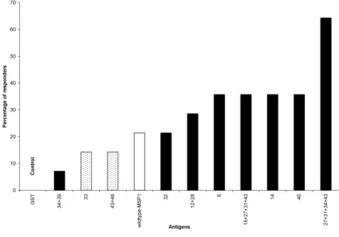
Cells from 14 (39%) of the exposed individuals had a sig-nificant proliferative response to the panel of MSP119 anti-gens (SI range >2 - 360). These individual were grouped as responders, while the remaining 61% of malaria-exposed donors that responded poorly to the antigens were grouped as non-responders. A modified protein, 27+31+34+43 with four amino acid substitutions (Table 1), had the most intense and consistently highest T-cell stimulatory effect, with a mean SI as high as 360 and elic-ited a response in the greatest number of donors: nine of
[image:5.612.64.540.324.657.2]Prevalence of response to MSP119 variant and wildtype antigens
Figure 2
Prevalence of response to MSP119 variant and wildtype antigens. Prevalence of T cell responses to the antigens for 14
14 (64%) responders had a significant response to this variant (Figure 2). In these same nine individuals, the responses to the 27+31+34+43 variant were significantly higher than the responses to the wild-type (WT-MSP119) antigen. Using cells collected from the same individual the SI was 240-fold greater than the SI seen in response to WT-MSP1. More than half of the nine (5/9) individuals that responded to the 27+31+34+43 variant had a SI that was at least five-fold higher than that seen in response to the wild-type antigen.
Three single amino acid substitution variants 6, 14 and 40 (Table 1) and the 15+27+31+43 variant (a four-residue substitution variant) all elicited high responses in 5/14 (36%) of the responders (Figure 2). Four of 14 (27%) responders had a significant response to the 12+28 variant
which lacks a disulfide bond (Cys12→Ile and
Cys28→Trp). The 34+39 variant (Tyr34→Ser,
Asp39→Thr) elicited a response in only one of the
responders. Two of the 14 (14%) responders had responses to the 33 (Asn33→Ile) and 43+48 (Glu43→Leu
and Thr48→Lys) variants; the intensity of these responses were higher than the responses to the GST control protein but lower than the responses to WT-MSP119.
Intense responses were also elicited by the 15+27+31+43 variant in five of the responders with a mean SI of 314.6. Three out of six individuals that responded to 15+27+31+43 had a SI that was at least five-fold greater than that to the wild-type protein. Variants 14 and 12+28 induced a mean SI of 315.7 and 269, respectively. The data revealed that all the high stimulation indices (SI >100), occurred with the MSP119 variants containing two or more amino acid substitutions except for the variant 14 which had a single amino acid change of Gln to Arg at position 14. All the variants in the panel that induced an intense response consistently induced a ≥3-fold stronger response than the WT protein.
The difference between the two 4-amino acid substitution variants, 27+31+34+43 and 15+27+31+43 is a
substitu-tion Asn15→Arg in 15+27+31+43 and Tyr34→Ser in
Cellular responses to wildtype-MSP119 antigen and the two variants with four amino acid substitutions
Figure 3
Cellular responses to wildtype-MSP119 antigen and the two variants with four amino acid substitutions.
27+31+34+43 and 15+27+31+43 MSP119 variants in 5 individuals of different ages and malaria exposure. OM21 and OM08 had
27+31+34+43. The responses to these two variants were compared in five individuals that responded to both vari-ants (Figure 3). For this small group, the data suggested that the 27+31+34+43 variant preferentially induced a response in cells obtained from children whereas 15+27+31+43 had a greater effect with cells from adults. The response to the WT antigen did not change signifi-cantly with increasing age (Figure 3).
Discussion
This study investigated the proliferative cellular responses to wild-type MSP119 and several variant proteins in indi-viduals naturally exposed to the P. falciparum parasite. The results revealed that the modified MSP119 antigens induced stronger and more frequent responses as com-pared to the wild type MSP1. In particular, the responses seen to most of the modified antigens with two or more amino acid substitutions were stronger than the response to the native antigen. These findings indicate that the anti-genic epitopes in some of the MSP119 variants were not compromised by the specific amino acid modifications; some of these modifications actually enhanced the response to the proteins.
The MSP-119 sequence is highly conserved, largely with diversity at only four positions in its 96 amino acid resi-dues [32]. This study has shown that selective replacement of up to four amino acid residues in the MSP119 sequence did not compromise the linear epitopes that are recog-nized by T-cells.
In the population tested, a response to one or more vari-ants was noted in 38% of individuals, whereas only 22% had a proliferative response to WT-MSP119. In previous work to evaluate human T-cell responses to WT-MSP119 antigen a prevalence of 26% responders was observed in Gambian and Kenyan populations, with marked individ-ual variations [27,28].
The higher prevalence of cellular proliferative responses to
the modified MSP119 noted in this study might be
explained by structural changes induced by the modifica-tions. It has been suggested that the relatively poor cellu-lar response to MSP119 is due to its structural complexity and resistance to proteases. Thus with 6 disulfide bonds it is impossible to load MSP119 onto MHC class II molecules and it can only be available for loading when the bonds have been reduced [29,30]. A form of MSP119 that is more accessible to proteases would allow improved processing by antigen-presenting cells (APC), which could enhance the cellular responses and thus vaccine efficacy [29].
Human PBMC recognized and responded significantly to eight out of ten variants, which had been designed to either reduce or abolish the binding of known blocking
mAbs [21] (Figure 1). Among those individuals whose cells responded, the prevalence of the responses to each variant ranged from 7% to 64% of the population. More people responded to the 27+31+34+43 variant than any of the other variants tested (Figure 2). The ability of the cells to respond to more than one variant was age depend-ent, thus cells from adults responded to more variants than the cells from children. This suggests a possible role of previous and repeated exposure to infection in the breadth of recognition and response to these variants (Fig-ure 3).
The two mutant proteins with four amino acid substitu-tions designated 27+31+34+43 and 15+27+31+43, char-acterized as having epitopes for MSP1-processing inhibitory antibodies, but no binding affinity for known blocking mAbs [21] were the most frequently recognized and they induced some of strongest cell responses observed. The 27+31+34+43 mutant induced the strong-est proliferative response (SI up to 360) and was the anti-gen most frequently recognized by cells from malaria-exposed adults and children. Each individual that responded to the 15+27+31+43 variant also responded either with equal or greater intensity to the 27+31+34+43 variant, suggesting a better recognition of the 27+31+34+43 variant by the responding cells. This is
attributable to the Tyr34→Ser modification in the
27+31+34+43 MSP119 variant compared with the
Asn15→Arg substitution in the 15+27+31+43 variant. This result is consistent with the suggested role of Tyr34 at the interface between the two epidermal-growth factor (EGF) domains in providing structural stability; the replacement of tyrosine by serine may result in an antigen that can be processed more readily. From these results, and the earlier antigenicity studies, the 27+31+34+43 MSP119 variant would be a favored variant for develop-ment of a MSP1-based vaccine. However, additional stud-ies are needed to determine whether the difference in the cellular responses to these two closely related proteins is a result of recognition by individual cell types (host factors) or a result of the structural perturbations due to the amino acid substitutions in the MSP1 sequence.
Conclusion
be useful to properly characterize the phenotypes of the proliferating cells. In addition the role of individual host genetics, and other factors in the modulation of cellular responses to these variants cannot be overemphasized. The potential in vitro correlates of protective immunity defined in this study represent an important step forward in the search for and evaluation of an efficient MSP1-based malaria vaccine.
Abbreviations
GST: glutathione S-transferase; MSP: merozoite surface protein; PBMC: peripheral blood mononuclear cells; RBC: red blood cell; SI: stimulation index; WT: wildtype.
Competing interests
The authors declare that they have no competing interests.
Authors' contributions
CMFO, CIA, RIN, MTB AAH, CU, IA and JL made substan-tial contributions to the conception and the design of the study, or acquisition, analysis and interpretation of data; YOO, HOA and ABO were involved in drafting the manu-script or revising it critically for content; and RIN, JL, AAH and MTB, gave final approval of the version to be pub-lished.
Acknowledgements
The authors do not have a commercial or other association that might pose a conflict of interest. Financial support was provided by the Swedish Agency for Research Development with Developing Countries (SIDA/SAREC), the European Commission through contract LSHP-CT-2004-503578 and the EUROMALVAC Consortium Contract QLK2-CT-2002-01197; the Medical Research Council, UK; and the Multilateral Initiative on Malaria (MIM) project grant (A10581) to Dr. RI Nwuba through the UNICEF/UNDP/ World Bank/WHO Programme for Research and Training in Tropical Dis-eases (TDR). CMF Okafor was a recipient of MIMPAC Network Exchange Programme scholarship and C. Uthaipibull was a recipient of a Thai Gov-ernment Scholarship. We acknowledge with appreciation, the cooperation of the individuals and parents who gave consent for their children to par-ticipate in the study. We also acknowledge the contributions of Femi Ibra-him, K. Akpala and Salah Eldin to this work.
References
1. Angov E, Aufiero BM, Turgeon AM, Van Handenhove M, Ockenhouse CF, Kester KE, Walsh DS, McBride JS, Dubois MC, Cohen J, et al.:
Development and pre-clinical analysis of a Plasmodium falci-parum Merozoite Surface Protein-1(42) malaria vaccine. Mol Biochem Parasitol 2003, 128:195-204.
2. Holder AA, Guevara Patino JA, Uthaipibull C, Syed SE, Ling IT, Scott-Finnigan T, Blackman MJ: Merozoite surface protein 1, immune evasion, and vaccines against asexual blood stage malaria. Parassitologia 1999, 41:409-414.
3. Holder AA: The carboxy-terminus of merozoite surface pro-tein 1: structure, specific antibodies and immunity to malaria. Parasitology 2009, 136:1445-1456.
4. Chang SP, Case SE, Gosnell WL, Hashimoto A, Kramer KJ, Tam LQ, Hashiro CQ, Nikaido CM, Gibson HL, Lee-Ng CT, et al.: A recom-binant baculovirus 42-kilodalton C-terminal fragment of Plasmodium falciparum merozoite surface protein 1 protects Aotus monkeys against malaria. Infect Immun 1996, 64:253-261. 5. Daly TM, Long CA: A recombinant 15-kilodalton carboxyl-ter-minal fragment of Plasmodium yoelii yoelii 17XL merozoite
surface protein 1 induces a protective immune response in mice. Infect Immun 1993, 61:2462-2467.
6. Hirunpetcharat C, Tian JH, Kaslow DC, van Rooijen N, Kumar S, Ber-zofsky JA, Miller LH, Good MF: Complete protective immunity induced in mice by immunization with the 19-kilodalton car-boxyl-terminal fragment of the merozoite surface protein-1 (MSP1[19]) of Plasmodium yoelii expressed in Saccharomyces cerevisiae: correlation of protection with antigen-specific antibody titer, but not with effector CD4+ T cells. J Immunol
1997, 159:3400-3411.
7. Kumar S, Collins W, Egan A, Yadava A, Garraud O, Blackman MJ, Guevara Patino JA, Diggs C, Kaslow DC: Immunogenicity and effi-cacy in aotus monkeys of four recombinant Plasmodium falci-parum vaccines in multiple adjuvant formulations based on the 19-kilodalton C terminus of merozoite surface protein 1. Infect Immun 2000, 68:2215-2223.
8. Ling IT, Ogun SA, Holder AA: Immunization against malaria with a recombinant protein. Parasite Immunol 1994, 16:63-67. 9. Singh S, Kennedy MC, Long CA, Saul AJ, Miller LH, Stowers AW:
Bio-chemical and immunological characterization of bacterially expressed and refolded Plasmodium falciparum 42-kilodalton C-terminal merozoite surface protein 1. Infect Immun 2003,
71:6766-6774.
10. Stowers AW, Chen Lh LH, Zhang Y, Kennedy MC, Zou L, Lambert L, Rice TJ, Kaslow DC, Saul A, Long CA, et al.: A recombinant vac-cine expressed in the milk of transgenic mice protects Aotus monkeys from a lethal challenge with Plasmodium falciparum. Proc Natl Acad Sci USA 2002, 99:339-344.
11. Stowers AW, Cioce V, Shimp RL, Lawson M, Hui G, Muratova O, Kaslow DC, Robinson R, Long CA, Miller LH: Efficacy of two alter-nate vaccines based on Plasmodium falciparum merozoite surface protein 1 in an Aotus challenge trial. Infect Immun
2001, 69:1536-1546.
12. Dodoo D, Aikins A, Kusi KA, Lamptey H, Remarque E, Milligan P, Bos-omprah S, Chilengi R, Osei YD, Akanmori BD, et al.: Cohort study of the association of antibody levels to AMA1, MSP119, MSP3 and GLURP with protection from clinical malaria in Ghana-ian children. Malar J 2008, 7:142.
13. Braga EM, Barros RM, Reis TA, Fontes CJ, Morais CG, Martins MS, Krettli AU: Association of the IgG response to Plasmodium fal-ciparum merozoite protein (C-terminal 19 kD) with clinical immunity to malaria in the Brazilian Amazon region. Am J Trop Med Hyg 2002, 66:461-466.
14. Dodoo D, Theander TG, Kurtzhals JA, Koram K, Riley E, Akanmori BD, Nkrumah FK, Hviid L: Levels of antibody to conserved parts of Plasmodium falciparum merozoite surface protein 1 in Ghanaian children are not associated with protection from clinical malaria. Infect Immun 1999, 67:2131-2137.
15. Egan AF, Morris J, Barnish G, Allen S, Greenwood BM, Kaslow DC, Holder AA, Riley EM: Clinical immunity to Plasmodium falci-parum malaria is associated with serum antibodies to the 19-kDa C-terminal fragment of the merozoite surface antigen, PfMSP-1. J Infect Dis 1996, 173:765-769.
16. Tolle R, Fruh K, Doumbo O, Koita O, N'Diaye M, Fischer A, Dietz K, Bujard H: A prospective study of the association between the human humoral immune response to Plasmodium falciparum blood stage antigen gp190 and control of malarial infections. Infect Immun 1993, 61:40-47.
17. Ogutu BR, Apollo OJ, McKinney D, Okoth W, Siangla J, Dubovsky F, Tucker K, Waitumbi JN, Diggs C, Wittes J, et al.: Blood stage malaria vaccine eliciting high antigen-specific antibody con-centrations confers no protection to young children in West-ern Kenya. PLoS One 2009, 4:e4708.
18. Corran PH, O'Donnell RA, Todd J, Uthaipibull C, Holder AA, Crabb BS, Riley EM: The fine specificity, but not the invasion inhibi-tory activity, of 19-kilodalton merozoite surface protein 1-specific antibodies is associated with resistance to malarial parasitemia in a cross-sectional survey in The Gambia. Infect Immun 2004, 72:6185-6189.
19. Okech BA, Corran PH, Todd J, Joynson-Hicks A, Uthaipibull C, Egwang TG, Holder AA, Riley EM: Fine specificity of serum anti-bodies to Plasmodium falciparum merozoite surface protein, PfMSP-1(19), predicts protection from malaria infection and high-density parasitemia. Infect Immun 2004, 72:1557-1567. 20. Guevara Patino JA, Holder AA, McBride JS, Blackman MJ: Antibodies
Publish with BioMed Central and every scientist can read your work free of charge "BioMed Central will be the most significant development for disseminating the results of biomedical researc h in our lifetime."
Sir Paul Nurse, Cancer Research UK
Your research papers will be:
available free of charge to the entire biomedical community
peer reviewed and published immediately upon acceptance
cited in PubMed and archived on PubMed Central
yours — you keep the copyright
Submit your manuscript here: BioMedcentral and erythrocyte invasion are blocked by naturally acquired
human antibodies. J Exp Med 1997, 186:1689-1699.
21. Uthaipibull C, Aufiero B, Syed SE, Hansen B, Guevara Patino JA, Angov E, Ling IT, Fegeding K, Morgan WD, Ockenhouse C, et al.:
Inhibitory and blocking monoclonal antibody epitopes on merozoite surface protein 1 of the malaria parasite Plasmo-dium falciparum. J Mol Biol 2001, 307:1381-1394.
22. Nwuba RI, Sodeinde O, Anumudu CI, Omosun YO, Odaibo AB, Holder AA, Nwagwu M: The human immune response to Plas-modium falciparum includes both antibodies that inhibit merozoite surface protein 1 secondary processing and block-ing antibodies. Infect Immun 2002, 70:5328-5331.
23. Omosun YO, Adoro S, Anumudu CI, Odaibo AB, Uthiapibull C, Holder AA, Nwagwu M, Nwuba RI: Antibody specificities of chil-dren living in a malaria endemic area to inhibitory and block-ing epitopes on MSP-119 of Plasmodium falciparum. Acta Trop
2009, 109:208-212.
24. Dekker C, Uthaipibull C, Calder LJ, Lock M, Grainger M, Morgan WD, Dodson GG, Holder AA: Inhibitory and neutral antibodies to Plasmodium falciparum MSP119 form ring structures with their antigen. Mol Biochem Parasitol 2004, 137:143-149.
25. Morgan WD, Frenkiel TA, Lock MJ, Grainger M, Holder AA: Precise epitope mapping of malaria parasite inhibitory antibodies by TROSY NMR cross-saturation. Biochemistry 2005, 44:518-523. 26. Hirunpetcharat C, Vukovic P, Liu XQ, Kaslow DC, Miller LH, Good
MF: Absolute requirement for an active immune response involving B cells and Th cells in immunity to Plasmodium yoe-lii passively acquired with antibodies to the 19-kDa carboxyl-terminal fragment of merozoite surface protein-1. J Immunol
1999, 162:7309-7314.
27. Egan A, Waterfall M, Pinder M, Holder A, Riley E: Characterization of human T- and B-cell epitopes in the C terminus of Plasmo-dium falciparum merozoite surface protein 1: evidence for poor T-cell recognition of polypeptides with numerous disulfide bonds. Infect Immun 1997, 65:3024-3031.
28. Udhayakumar V, Anyona D, Kariuki S, Shi YP, Bloland PB, Branch OH, Weiss W, Nahlen BL, Kaslow DC, Lal AA: Identification of T and B cell epitopes recognized by humans in the C-terminal 42-kDa domain of the Plasmodium falciparum merozoite surface protein (MSP)-1. J Immunol 1995, 154:6022-6030.
29. Hensmann M, Li C, Moss C, Lindo V, Greer F, Watts C, Ogun SA, Holder AA, Langhorne J: Disulfide bonds in merozoite surface protein 1 of the malaria parasite impede efficient antigen processing and affect the in vivo antibody response. Eur J Immunol 2004, 34:639-648.
30. Quin SJ, Langhorne J: Different regions of the malaria mero-zoite surface protein 1 of Plasmodium chabaudi elicit distinct T-cell and antibody isotype responses. Infect Immun 2001,
69:2245-2251.
31. Troye-Blomberg M, Perlmann H, Patarroyo ME, Perlmann P: Regula-tion of the immune response in Plasmodium falciparum malaria. II. Antigen specific proliferative responses in vitro. Clin Exp Immunol 1983, 53:345-353.