Azari Mehrdad Mohammad Ali. Periodontoprotective action of oral gel "Biotrit-Denta" in rats with sugar diabetes, received orthodontic treatment. Journal of Education, Health and Sport. 2018;8(8):1186-1192. eISNN 2391-8306. DOI
http://dx.doi.org/10.5281/zenodo.1451740
http://ojs.ukw.edu.pl/index.php/johs/article/view/6164
The journal has had 7 points in Ministry of Science and Higher Education parametric evaluation. Part b item 1223 (26/01/2017). 1223 Journal of Education, Health and Sport eISSN 2391-8306 7
© The Author(s) 2018;
This article is published with open access at Licensee Open Journal Systems of Kazimierz Wielki University in Bydgoszcz, Poland
Open Access. This article is distributed under the terms of the Creative Commons Attribution Noncommercial License which permits any noncommercial use, distribution, and reproduction in any medium, provided the original author (s) and source are credited. This is an open access article licensed under the terms of the Creative Commons Attribution Non commercial license
(http://creativecommons.org/licenses/by-nc/4.0/) which permits unrestricted, non commercial use, distribution and reproduction in any medium, provided the work is properly cited. This is an open access article licensed under the terms of the Creative Commons Attribution Non commercial License (http://creativecommons.org/licenses/by-nc/4.0/) which permits unrestricted, non commercial
use, distribution and reproduction in any medium, provided the work is properly cited. The authors declare that there is no conflict of interests regarding the publication of this paper.
Received: 02.08.2018. Revised: 14.08.2018. Accepted: 31.08.2018.
UDK 616.31:616.37:616.179:379:008
PERIODONTOPROTECTIVE ACTION OF ORAL GEL "BIOTRIT-DENTA" IN RATS
WITH SUGAR DIABETES, RECEIVED ORTHODONTIC TREATMENT
Azari Mehrdad Mohammad Ali
Odessa National Medical University
Abstract
Background. Significant changes in diabetes mellitus are observed in the oral cavity,
which can affect by treatment measures. The purpose of this study was to determine the
therapeutic effect of oral phytogel "Biotrit-Denta" on periodontal tissues of rats with
experimental diabetes mellitus and received orthodontic treatment.
Methods. Type 1 diabetes mellitus (DM1) was reproduced by alloxan. From 12-th day of
the experiment rats were fixed in the month with orthodontic springs. One group of rats from the
first day of the experiment received daily oral application gel "Biotrit-Denta". The duration of the
experiments was 35 days. In the gum the level of inflammation markers (elastase, MDA), activity
of urease, lysozyme, catalase were determined. The bone tissue of the alveolar process, the
activity of alcoline (AlF) and acidic (AcF) phosphatases, the content of calcium and protein were
Results. Diabetes mellitus causes in periodontis of rats the development of inflammation,
dysbiosis, decrease in immunity, antioxidant defense and the mineralizing activity of bone tissue.
The orthodontic operation was no effect on the periodontal parameters that were changed in
diabetes. Oral applications of gel "Biotrit-Denta" eliminated the phenomens of inflammation,
dysbiosis and normalized the mineralising activity of bone tissue.
Conclusion. Oral applications of gel "Biotrit-Denta" have periodontoprotective effect.
Keywords: diabetes mellitus, paradontium, dysbiosis, inflammation, oral gel
"Biotrit-Denta".
INTRODUCTION
As is known, diabetes mellitus causes significant disturbances in metabolism, immune
system, endoecology and in systems of organism adaptation [1-6]. Significant changes in diabetes
mellitus are observed in the tissues of the oral cavity [7-10], which can affect the effectiveness of
treatment measures produced in patients with diabetes mellitus.
The purpose of this study was to determine the therapeutic and prophylactic effects of the
mucosa-adhesive oral phytogel "Biotrit-Dent", containing biologically active substances from
wheat seedlings, lecithin, calcium citrate, NaF and ascorbic acid, on periodontal tissues of rats
with experimental diabetes mellitus and received standard orthodontic treatment (installation of
springs).
MATERIAL AND RESEARCH METHODS
The experiments were performed on 24 white Wistar rats (males, 4 months old, initial
living weight 181 ± 9 g) distributed into 4 equal groups: 1st control (intact), 2nd, 3rd and 4th -
Type 1 diabetes mellitus (DM1), which was reproduced once / in peritoneal administration of
alloxan at a dose of 100 mg / kg [11]. Rats of the 3rd and 4th groups, starting from the 12th day
of the experiment, were fixed in the mouth with orthodontic springs [12]. Rats of the 4th group
from the first day of the experiment received daily oral applications of Biotrit-Denta gel at a dose
of 0.5 ml / rat. The duration of the experiment was 35 days, after which the rats were sacrificed
under thiopental anesthesia (20 mg / kg) by total bloodletting from the heart. The gum was
excised and the alveolar process of the lower jaw was allocated.
In the gum homogenate, the level of biochemical markers of inflammation was
[14], lysozyme activity (index of non-specific immunity level) [14] and antioxidant catalase
enzyme activity [13]. The antioxidant-prooxidant index of API was calculated from the ratio of
catalase activity and MDA content [13], and the degree of disbiosis according to Levitsky [14]
was calculated from the ratio of relative activities of urease and lysozyme.
In the homogenate of the bone tissue of the alveolar process, the activity of alkaline (AlF)
and acidic (AcF) phosphatases [15], calcium content [15] and protein [15] was determined. The
mineralizing index (MI) was calculated from the ratio of AlF / AcF, and the degree of
mineralisation (DM) was calculated from the ratio of calcium and protein content [16].
The results of the studies were subjected to a standard statistical treatment.
Phytogel "Biotrit-Denta" was prepared in accordance with the recipe RC U
20.4-13903778-032 / 15: 2018, using a dietary supplement with the same name (TU U
013903778-45-97) in an amount of 10% of the mass of the gel CMC-Na.
RESULTS AND DISCUSSION
Table 1 presents the results of the determination in the gums of the rats of two markers of
inflammation, elastase and MDA. From these data it is clear that in diabetes mellitus the level of
elastase is significantly increased by 36.2% and MDA by 90.5%. The orthodontic operation did
not significantly affect the elevated level of elastase and MDA, whereas the Biotrit-Dent gel
applications completely normalized both indices, which indicates a high anti-inflammatory
activity of the gel.
Table 1. Effect of Biotrit-Denta gel on the level of markers of inflammation in the gum of
the rats with diabetes mellitus, who received orthodontic treatment (M ± m)
No Groups Elastase, μkat/kg MDA, mmol/kg
1 Control 27,1±1,2 9,5±0,8
2 Type 1 diabetes mellitus (DM1) 36,9±2,0
р<0,01
18,1±1,4 р<0,01 3 DM1 + orthodontic treatment
(OT)
35,3±1,8 р<0,01; р1>0,3
17,4±1,2 р<0,01; р1>0,3
4 DM1 + ot+ gel «Biotrit_denta» 28,8±11,5
р>0,3; р1<0,05 р2<0,05
11,2±0,9
р>0,05; р1<0,01 р2<0,01
Notes: p - in comparison with gr. No. 1; p1 - in comparison with gr. № 2; p2 - in comparison with
Table 2 presents the results of determining the activity of urease, lysozyme and the degree
of dysbiosis in the gingiva. It can be seen that rats with diabetes mellitus almost 2.8 times
increased urease activity, which indicates a significant increase in microbial contamination of the
gum. The activity of lysozyme in the gum decreases by 37.4%. Orthodontic surgery is not
significantly affected on these indicators, however oral applications of the Biotrit-Dent gel
[image:4.612.72.534.250.396.2]significantly reduce the activity of urease and practically normalize the level of lysozyme.
Table 2. Effect of Biotrit-Denta gel on the activity of urease and lysozyme in the gum of the rats
with diabetes mellitus, who received orthodontic treatment.
No Group μkat/kg Urease, Lysozyme,
units/kg
Degree of dysbiosis, units
1 Control 0,51±0,09 219±27 1,0±0,2
2 Type 1 diabetes mellitus (DM1)
1,42±0,20 р<0,01
137±24 р<0,05
4,5±0,4 р<0,01 3 DM1 + orthodontic
treatment (OT)
1,39±0,21 р<0,01; р1>0,5
144±21 р<0,05; р1>0,5
4,1±0,3 р<0,01; р1>0,3
4 DM1 + ot+ gel «Biotrit_denta»
0,67±0,12 р>0,1; р1<0,05
р2<0,05
207±22 р>0,3; р1<0,05
р2>0,05
1,4±0,2 р>0,2; р1<0,01
р2<0,01
In rats with diabetes, the degree of dysbiosis increases 4.5 times, but the gel applications
are normalized.
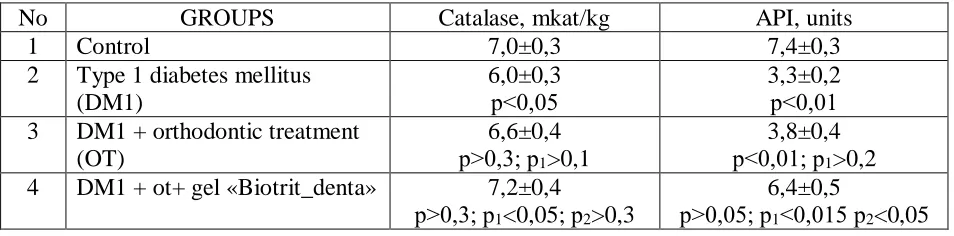
Table 3 presents the results of determining the activity of catalase and the API index in
the gingiva. It can be seen that in rats with diabetes the catalase activity is reliably reduced and
the API index is 2.2 times higher. These indicators are also reduced to the same extent after
orthodontic surgery. The application of the gel completely restores the activity of lysozyme and
[image:4.612.65.542.604.722.2]significantly increases the level of API.
Table 3. Effect of Biotrit-Denta gel on catalase activity and antioxidant prooxidant index (API) in
the gum of the rats with diabetes mellitus, who received orthodontic treatment
No GROUPS Catalase, mkat/kg API, units
1 Control 7,0±0,3 7,4±0,3
2 Type 1 diabetes mellitus (DM1)
6,0±0,3
р<0,05 3,3±0,2 р<0,01 3 DM1 + orthodontic treatment
(OT)
6,6±0,4 р>0,3; р1>0,1
3,8±0,4 р<0,01; р1>0,2
Table 4 shows the results of the determination of phosphatase activity in the bone tissue.
From these data it can be seen that in diabetes mellitus, the activity of alkaline phosphatase is
almost halved and the activity of AcF is increased by 1.5 times, which leads to a triple reduction
in the MI index. A similar change in the level of phosphatases is observed after orthodontic
[image:5.612.64.541.227.387.2]surgery. Gel applications completely restore the normal level of phosphatases and MI index.
Table 4. Effect of Biotrit-Dent gel on the activity of phosphatases in bone tissue the alveolar
process of the lower jaw of rats with diabetes mellitus, who received orthodontic treatment
No Group
Alkaline Phosphatase, μkat/kg Acid Phosphatase, μkat/kg Mineralising Index (MI), units
1 Control 102,5±6,9 8,8±0,7 11,6±0,3
2 Type 1 diabetes mellitus (DM1)
51,3±8,7
р<0,01 13,6±0,9 р<0,01 3,8±0,2 р<0,01 3 DM1 + orthodontic
treatment (OT)
67,2±9,0 р<0,05; р1>0,05
11,9±1,1 р<0,05; р1>0,1
5,6±0,3 р<0,01; р1<0,05
4 DM1 + ot+ gel «Biotrit_denta»
101,8±10,2 р>0,5; р1<0,01
р2<0,05
9,1±0,8 р>0,3; р1<0,01
р2<0,05
11,2±0,4 р>0,3; р1<0,01
р2<0,01
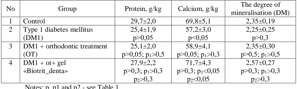
Table 5 shows the results of the determination of protein and calcium in bone tissue. As
can be seen from these data, the protein content in the bone changes little, while the calcium
content significantly decreases in rats with diabetes and completely normalizes after gel
[image:5.612.56.556.543.694.2]applications.
Table 5. Effect of Biotrit-Dent gel on protein and calcium content in bone tissue periodontal rats
with diabetes mellitus receiving orthodontic treatment (M ± m)
No Group Protein, g/kg Calcium, g/kg The degree of
mineralisation (DM)
1 Control 29,7±2,0 69,8±5,1 2,35±0,19
2 Type 1 diabetes mellitus (DM1) 25,4±1,9 р>0,05 57,2±3,0 р<0,05 2,25±0,25 р>0,3 3 DM1 + orthodontic treatment
(OT)
25,1±2,0 р>0,05; р1>0,5
58,9±4,1 р>0,05; р1>0,3
2,35±0,30 р>0,5; р1>0,5
4 DM1 + ot+ gel «Biotrit_denta»
27,9±2,2 р>0,3; р1>0,3
р2>0,3
71,7±4,3 р>0,3; р1<0,05
р2<0,05
2,57±0,27 р>0,3; р1>0,3
р2>0,3
The degree of bone mineralization due to large variability of the data does not change
significantly.
CONCLUSIONS
1. Diabetes mellitus causes in rats development in the periodontis of inflammation,
dysbiosis and a decrease in the mineralising activity of bone tissue.
2. Orthodontic operation has practically no effect on the periodontal parameters that are
lower in diabetes.
3. Mucozo-adhesive gel "Biotrit-Denta" has a periodontoprotective effect, eliminating the
phenomena of inflammation, dysbiosis and normalizing the mineralising activity of bone tissue.
REFERENCES
1. Orlovsky MA. Experimental studies of type I diabetes mellitus: the causes of inter- and
intraspecies differences in resistance to diabetogenic factors. Journal of the AMS of Ukraine.
2006; 12(2): 255-268. (in Russian)
2. Mazurina NK. Violations of the hypothalamic-pituitary-adrenal system in diabetes
mellitus. Problems of endocrinology. 2007; 53(2): 29-34. (in Russian)
3. Dedov II, Khaitov RM, Alekseev LP, Boldyreva MN, Shestakova MV, Trofimov D. Yu.,
Kuraeva TL, Peterkova VA. Use achievements in the field of molecular immunogenetics in a
clinic of type 1 diabetes mellitus. Bulletin of the Russian Academy of Medical Sciences. 2008;
20: 45-51. (in Russian)
4. Belyaeva NA, Mikhailova DG, Egorova EN, Gogina ED, Torshkova MA. Nonspecific
adaptive reactions and immune status in patients with type 2 diabetes mellitus. Clinical laboratory
diagnostics. 2010; 3: 14-18. (in Russian)
5. Mikaelyan NP, Terentyev AA, Gurina AE, Smirnov VV. Violations of the functions of
the membrane-receptor apparatus of blood cells in children with type 1 diabetes mellitus.
Biomedical chemistry. 2011; 57(6): 642-649. (in Russian)
6. Kuzmenko DI, Klimentyeva TK. Ceramide: the role in the apoptosis and pathogenesis of
insulin resistance. Overview. Biochemistry. 2016; 81(9): 1155-1171. (in Russian)
7. Sykes LM, Sukha A. Potential risk of serious oral infections in the diabetic patient: a
8. Fesenko UA, Malygina DA. Influence of diabetes mellitus on clinical and laboratory
indices of inflammatory diseases of maxillofacial area in children. Dentist. 2010; 1(139): 32-33.
(in Russian)
9. Skiba AV, Tereshina TP, Dmitrieva NB. Diabetes and periodontal disease. Visceral
dentistry. 2012: 6 (Special Edition): 82-86. (in Russian)
10. Mutoh T., Honda E., Matsumoto K. et al. Study of oral microflora in diabetes mellitus
patients. J. Dent. res. 2000; 79(Spec. is.): 2013.
11. Kikhtyak OP, Skripnik NV. Possibilities of recreation of diabetes mellitus are in
anexperiment. Experimental and clinical physisology and biochemistry. 2004; 2: 118-120. (in
Ukrainian)
12. Gorokhovskiy VN, Denga OV, Denga AE, Mirchuk BN. Modeling of the orthodontic
movement of teeth in rats. In book. Schneider SA, Levitsky AP "Experimental stomatology". Part
1. Experimental models of dental diseases. Odessa, 2017: 128-132. (in Russian)
13. Levitsky AP, Denga OV, Makarenko OA [and others]. Biochemical markers of
inflammation of the tissues of the oral cavity: methodical recommendations. Odessa, KP of the
OSG, 2010: 16. (in Russian)
14. Levitsky AP, Makarenko OA, Selivanskaya IA [and others]. Enzymatic method for
determining oral dysbiosis for screening pro and prebiotics: guidelines. Kiev, State
Pharmacological Center, 2007: 22. (in Russian)
15. Levitsky AP, Makarenko OA, Denga OV [and others]. Experimental methods of
research of stimulants of osteogenesis: methodical recommendations. Kiev, State
Pharmacological Center, 2005: 50. (in Russian)
16. Levitsky AP, Makarenko OA, Khodakov І. V. [ta іn.]. Enzymatic method