This is a repository copy of
Development of a biochemical marker to detect current breast
milk intake
.
White Rose Research Online URL for this paper:
http://eprints.whiterose.ac.uk/149334/
Version: Published Version
Article:
Addison, R., Hill, L., Bode, L. et al. (7 more authors) (2019) Development of a biochemical
marker to detect current breast milk intake. Maternal & Child Nutrition. ISSN 1740-8695
https://doi.org/10.1111/mcn.12859
[email protected]
https://eprints.whiterose.ac.uk/
Reuse
This article is distributed under the terms of the Creative Commons Attribution (CC BY) licence. This licence
allows you to distribute, remix, tweak, and build upon the work, even commercially, as long as you credit the
authors for the original work. More information and the full terms of the licence here:
https://creativecommons.org/licenses/
Takedown
If you consider content in White Rose Research Online to be in breach of UK law, please notify us by
bs_bs_banner
O R I G I N A L A R T I C L E
Development of a biochemical marker to detect current breast
milk intake
Ruth Addison
1|
Lauren Hill
2|
Lars Bode
3|
Bianca Robertson
3|
Biswa Choudhury
4|
David Young
5|
Charlotte Wright
6|
Clare Relton
7,8|
Ada L. Garcia
9|
David M. Tappin
61NHS Ayrshire & Arran Primary Care Trust,
Rainbow House Paediatric Unit, Ayrshire Central Hospital, Irvine, UK
2General Paediatrics, Pinderfields General
Hospital, Wakefield, UK
3Department of Pediatrics and Larsson‐
Rosenquist Foundation Mother‐Milk‐Infant
Center of Research Excellence (LRF MOMI CORE), University of California, San Diego, California
4Glycoanalytical Core, Glycobiology Research
and Training Center, University of California, San Diego, California
5Department of Mathematics and Statistics,
University of Strathclyde, Glasgow, UK
6Section of Child Health, School of Medicine,
Glasgow University, Glasgow, UK
7ScHARR, University of Sheffield, Sheffield,
UK
8Centre for Primary Care and Public Health,
Queen Mary University of London, London, UK
9Human Nutrition, School of Medicine,
Dentistry and Nursing, Glasgow University, Glasgow, UK
Correspondence
David M. Tappin, Section of Child Health, School of Medicine, Glasgow University, University Avenue, Glasgow, G12 8QQ, UK. Email: [email protected]
Funding information
Medical Research Council, Grant/Award Number: MR/J000434/1
Abstract
The WHO recommends exclusive breastfeeding for 6 months, but despite
interven-tions, breastfeeding rates remain stubbornly low. Financial voucher incentives have
shown promise but require a biomarker for validation of intake. This study aimed to
develop a simple biochemical assay of infant urine that would tell if an infant was
receiving
any
breast milk to validate maternal report. Urine samples were collected
and snap frozen from 34 infants attending with minor illness or feeding problems,
of whom 12 infants were exclusively breastfed, nine exclusively formula fed, and
11 mixed breast/formula fed. High
‐
performance anion exchange chromatography
was used to identify discriminating patterns of monosaccharide composition of
unconjugated glycans in a sequence of three experiments. The absolute
concentra-tion of all human milk oligosaccharides measured blind could detect
“
any
breastfeeding
”
only with a sensitivity of 48% and specificity of 78%. Unblinded
examination of
N
‐
acetylglucosamine (GlcNAc) measured as GlcNH
2after hydrolysis
of GlcNAc improved sensitivity to 75% at the expense of a specificity of 28%.
Estima-tion of the relative abundance of GlcNH2 (GlcNH2[%]) or the ratio of GlcNH2 to
endogenous mannose (Man) improved accuracy. In a further blind experiment, the
GlcNH2/Man ratio with a cut
‐
off of 1.5 correctly identified all those receiving
“
any
breast milk,
”
while excluding exclusively formula fed infants. The GlcNH2/Man ratio
in infant urine is a promising test to provide biochemical confirmation of
any
breastfeeding for trials of breastfeeding promotion.
K E Y W O R D S
breastfeeding, carbohydrate biochemistry, health promotion, milk human, oligosaccharides,
programme evaluation
1
|I N T R O D U C T I O N
Breastfeeding plays a vital role in preventing infant and child morbidity
and mortality as well as longer term improvements in health (Victora
-This is an open access article under the terms of the Creative Commons Attribution License, which permits use, distribution and reproduction in any medium, provided the original work is properly cited.
© 2019 Authors.Maternal & Child NutritionPublished by John Wiley & Sons Ltd
Abbreviations:BF, breastfed; BFI, Baby Friendly Initiative; FF, formula fed; HMO, human milk oligosaccharides; HPAEC‐PAD, high‐performance anion exchange chromatography with pulsed amperometric detection; Man, mannose; Mix, mixed breast and formula fed; NHS, National Health Service; NOSH, NOurishing Start for Health; R&D, research and development; TFA, trifluoroacetic acid; UCSD, University of California, San Diego; UK, United Kingdom; UNICEF, United Nations International Children's Emergency Fund; USA, United States of America; WHO, World Health Organisation
DOI: 10.1111/mcn.12859
Matern Child Nutr. 2019;e12859. https://doi.org/10.1111/mcn.12859
et al., 2016). Women who breastfeed are less likely to suffer from
breast cancer in the longer term (Victora et al., 2016) and are more
likely to reduce to their prepregnancy weight and do so more quickly
(Hatsu, McDougald, & Anderson, 2008; Vinter et al., 2014). As a result
The WHO recommends exclusive breastfeeding until 6 months post‐
natal age and continued feeding for at least 12 months (WHO, 2001).
Despite this, in affluent countries, many women still do not
breastfeed. WHO and UNICEF developed the Baby Friendly Initiative
to support health services to provide high quality care in particular
regarding support for breastfeeding (Victora et al., 2016). Despite
the widespread adoption of this institutional intervention to support
women who want to breastfeed, rates have remained stubbornly low
at 6 months post‐natal age (McAndrew et al., 2010). In high‐income
countries, in areas of socio‐economic deprivation breastfeeding is
less common, thus increasing health inequalities (Oakley, Renfrew,
Kurinczuk, & Quigley, 2013). In these areas, formula feeding is normal,
and breastfeeding is neither visible nor valued (Relton, 2017).
To overcome this perception, U.K. researchers have developed and
trialled financial incentives for breastfeeding, in the form of shopping
vouchers worth up to £200 in total (Relton et al., 2016). Results
sug-gest that areas with low breastfeeding rates that offer financial
incen-tives have significantly increased breastfeeding rates at 6–8 weeks
post‐natal age (Relton et al., 2017). Financial incentives were provided,
on the basis of self‐report of breastfeeding corroborated by a
confir-matory signature from an appropriate health care worker (Relton
et al., 2016). This is the method by which infant feeding status is
recorded for the purposes of routine data collection in the U.K.'s
National Health Service: a healthcare professional's determination
based on their interactions with the woman during routine visits
(which may or may not include witnessing the woman breastfeed).
However, concerns have been raised that some women might
“game”the system, that is misrepresent infant feeding habit in order
to receive financial payments (Mazariegos, Slater, & Ramirez‐Zea,
2016). Policy makers are likely to require more robust measurements
of compliance such as using biomarkers to verify that infants are
being breastfed. Validation of self‐report has been undertaken
previ-ously by using stable isotopes of water fed to the woman that
comes through into breast milk (Medoua et al., 2012; Moore et al.,
2007). However, this method is expensive and time consuming and
would not be practical for large scale studies. Thus, a simpler and
more acceptable method of detecting current breast milk intake is
required. Interventions to help smoking cessation suffered similar
credibility issues until biochemical assays were developed as gold
standard tests to corroborate self‐report of cessation (West, Hajek,
Stead, & Stapleton, 2005).
There is a wealth of studies on the constituents of breast milk, but
a much smaller literature on non‐invasive biomarkers of breast milk
consumption. One study described significant differences in average
values for a measure of oxidative stress in urine of neonates fed breast
milk and formula, but there was wide overlap between the groups,
making this unsuitable for use as a screening test (Shoji, Oguchi,
Shimizu, & Yamashiro, 2003). What is required for this purpose is a
measure involving both a simple sample collection and biochemical
assay that would clearly differentiate between infants receiving any
breast milk from those only receiving infant formula. Another group
has studied metabolomic patterns in urine in breastfeeding children,
but although these show clearly different patterns at different
ages, it is not clear if these could be used to discriminate between
breastfeeding and infant formula feeding infants (Lafferty, O'Regan,
O'Shea, McAuliffe, & O'Sullivan, 2016).
In order to accurately differentiate infants that have been
breastfed versus infant formula fed. A test is needed that can non‐
invasively identify the constituents that are present in human milk
but not in infant formula, which is nearly all manufactured from cow's
milk; therefore, we decided to focus on human milk oligosaccharides,
which are known to be secreted in the urine of breastfed babies
unchanged (Coppa et al., 1990). These molecules are present in cow's
milk at much lower concentrations and are not currently routinely
added by infant formula companies (Goehring, Kennedy, Prieto, &
Buck, 2014). Quantitative assay of human milk oligosaccharides has
been undertaken in the past (Autran et al., 2018; De Leoz et al.,
2013; Jantscher‐Krenn et al., 2019; Wise et al., 2018), and it is also
established that these can be detected in urine of breastfeed babies
(Goehring et al., 2014). The aim of this study was to develop a
bio-marker of breast milk intake using quantitative determination of
human milk oligosaccharides in the urine of infants.
2
|M E T H O D S
2.1
|Study design and ethics
We recruited cross‐sectionally 34 infants to obtain urine samples to
test the presence of monosaccharides as a marker of breast milk
intake. The number of sample assays was limited by available funding.
The aim was to reach 10 samples from babies fed only breast milk, 10
from those fed both breast and formula milk, and 10 fed only formula
milk. We did not know how well our methodology would work. Formal
Key messages
• Human breast milk contains many grams per day of
human milk oligosaccharides, cow's milk‐based infant
formula does not.
• A small percentage of ingested human milk
oligosaccharides are secreted into the urine of breastfed
infants.
• We have developed a simple infant urinary biomarker of
human milk consumption.
• This study has found thatN‐acetylglucosamine (GlcNAc) a constituent of human milk oligosaccharides in ratio to
endogenously produced mannose (Man) in an infant's
urine differentiates any human breast milk ingestion
from exclusive cow's milk‐based infant formula feeding.
sample size calculation was not undertaken. This study was a pilot
project to inform a future definitive study with regard to required
sample size and other issues. The West of Scotland Research Ethics
Committee 5 granted permission for the study on 4/7/2014. A minor
amendment to the protocol passed by the ethics committee on 14/5/
15 allowed samples to be collected from babies attending the
breastfeeding and tongue tie clinics at the Royal Hospital for Children
Glasgow. All women in the study, with their baby, provided written
consent to take part. All women were 16 years of age or older.
2.2
|Subject recruitment and inclusion criteria
These were recruited from infants with non‐life‐threatening ailments
attending the Glasgow Royal Hospital for Sick Children, a large tertiary
children's hospital serving the west of Scotland. Hospital staff gave
information about the study to families of all infants less than 6 months
of age attending the acute medical unit at the children's hospital in
tri-age categories of three and above (non‐life‐threatening categories). An
information sheet was also sent out with the appointment cards sent to
all women booked for the tongue tie and breastfeeding clinics. In the
acute hospital setting, potential participants were notified to the
research team by nursing staff. Two members of the research team
attended the tongue tie and breastfeeding clinics to consent women
and their infants. Inclusion criteria were infants less than 6 months of
age, infants on full enteral (oral) feeds, and women aged at least
16 years. Those who fulfilled the inclusion criteria were asked to give
their consent to take part in the study. A questionnaire was completed
by women with a record of infant feeding, which was countersigned by
a nurse or midwife looking after the family who had witnessed the
infant's feeding pattern.
2.3
|Urine collection
We collected spot urine samples by placing cotton wool balls in the
infant's nappy in the hospital outpatient clinics and wards. Urine was
extracted from the cotton wool balls by nursing staff using a simple
compression/suction method with a syringe without a needle (Ahmad,
Vickers, Campbell, Coulthard, & Pedler, 1991). The urine extracted was
placed in a sterile universal container and sent to the biorepository
where each sample was split into two aliquots and frozen to−40°C
for storage. Samples were transported in dry ice to the laboratory for
sample analysis.
2.4
|General sample transfer methodology
The laboratory was sent urine samples frozen in dry ice, blind to the
feed type of the baby who passed the urine samples. Tests were run
to determine if the urine had been passed by a baby who was
exclu-sively breastfed, excluexclu-sively formula fed, or was fed a mix of human
breast and infant formula. Two unblinded (known feed type) samples
were also sent to aid the analysis methods. The samples were assayed
for human milk oligosaccharides and later a human milk
oligosaccha-ride building block as a specific constituent of human milk. Results
were sent back to the study team. Then the feed type was unmasked
for the laboratory team.
Experiment 1 was undertaken by the laboratory on 32 samples
looking for all human milk oligosaccharides, blind to feed type.
Experi-ment 2 used 12 known feed type samples from the 32 to create normal
range cut‐offs forN‐acetylglucosamine (GlcNAc, measured as hydro-lysed GlcNH2), a building block common to most human milk
oligosac-charides. The results were then standardised for endogenous mannose
(Man) and for all sugars in the urine. Experiment 3 assayed 30 samples
blind to feed type and used the cut‐offs from Experiment 2 to define
feed type. Eight of these samples had been used in Experiment 2 and
therefore illustrated the repeatability of the assay system, the 22 other
samples were assayed to assess the sensitivity and specificity of the
normal range cut‐offs (mean for formula fed infants plus 2 standard
deviations) defined in Experiment 2.
2.5
|Blinding and unblinding of samples and
establishing normal ranges
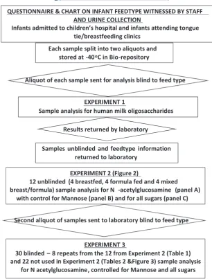
Figure 1 describes the experiments undertaken including sample
collection, splitting samples into two aliquots, initially sending one
aliquot blinded to feed type to the laboratory for analysis, unblinding
of those samples, establishing normal ranges using residual samples
of those initial aliquots, and sending the second aliquot of samples
from the biorepository to the laboratory blind to feed type for analysis
against normal ranges.
2.6
|Reagents used
Acetonitrile and sodium hydroxide were obtained from Fisher
Scien-tific, Hampton, NH, USA. Sodium acetate and trifluoracetic acid
(TFA) were obtained from Sigma‐Aldrich, Saint Louis, MO, USA.
Isopropyl alcohol was obtained from Decon Labs Inc, King of Prussia,
PA, USA.
2.7
|Sample analysis
2.7.1
|Monosaccharide composition analysis of
urine samples
Glycans were isolated from 50μl of urine. Peptides and salts were
removed by solid phase extraction over Sep‐Pak C18 and porous
graphitised carbon (Carbograph) microcolumns (both ThermoFisher
Scientific, Waltham, MA, USA). Glycans were eluted with 40%
aceto-nitrile and 0.1% trifluoroacetic acid. One fifth of the eluate was
lyophilised and then hydrolysed using 2 N trifluoroacetic acid at
100°C for 4 hr. Acid hydrolysed samples were cooled to room
tem-perature and centrifuged at 2,000 rpm for 2 min. Excess acid was
removed by dry nitrogen flush followed by coevaporation using
100 μl of 50% isopropyl alcohol twice. The dried samples were
resuspended in 200μl of MilliQ water, and 25% was injected onto
Dionex ICS‐3000, high‐performance anion exchange chromatography
attached with pulsed amperometric detector (HPAEC‐PAD; Dionex,
now ThermoScientific, USA, Waltham, MA, USA). Monosaccharide
(4 mm × 250 mm) with CarboPac‐BioLC guard column (4 mm × 50 mm;
both ThermoScientific, USA). An isocratic mixture of 19 mM sodium
hydroxide with 0.95 mM sodium acetate was used at a flow rate of
1 ml/min with total run time of 20 min per sample. Monosaccharide
data were collected using the standard Quad waveform for
carbohy-drate as supplied by Chromeleon software version 6.8 (Dionex, now
ThermoScientific USA, Waltham, MA, USA; Hardy, Townsend, & Lee,
1988). All neutral and amino sugars were identified and quantified
by comparison with known amount of authentic monosaccharide
stan-dard mixture consisting ofL‐fucose,D‐galactosamine,D‐glucosamine,
D‐galactose,D‐glucose, andD‐mannose.
2.8
|Statistical analysis
Sensitivity and specificity was used to develop classification rules for
the three assays proposed in Experiment 2. These were then tested
on further samples to determine the diagnostic ability for determining
if an infant was receiving any breast milk (i.e., breast or breast/formula
vs. formula only). All analyses were performed using MedCalc (www.
medcalc.org) version 16 (MedCalc Software, Acacialaan 22, 8400,
Ost-end, Belgium).
3
|R E S U L T S
3.1
|Experiment 1
In Experiment 1, after unblinding 5/12 (42%) of the exclusively
breastfeeding and 7/11 (64%) of the mixed feeders had been classified
as not breastfed, whereas 2/9 (22%) of the solely formula fed had
been classified as breastfed.
3.2
|Experiment 2
Twelve of these initial samples were retested (now unblinded) forN‐
acetylglucosamine (GlcNAc), which is a component of human milk
oligosaccharides and is measured as GlcNH2 after hydrolysis
(Figure 2). Four urines were from exclusively breastfed infants, four
from exclusively formula fed infants, and four from breast and formula
[image:5.595.56.363.41.445.2]fed infants (Mix; Figure 2).
FIGURE 1 Diagram showing the collection
and analysis of the infant urine samples
Figure 2a shows GlcNH2 concentrations alone demonstrating a
difference between groups, but these were not specific or sensitive.
This was thought to reflect difficulty in standardising urine
concentra-tions. To distinguish exclusive formula feeding from any breastfeeding
(exclusive breast and mixed breast and formula), sensitivity was 75%
and specificity 28% with the cut‐off 0.28.
Figure 2b shows GlcNH2 relative to mannose (Man), which is a
sugar that is present in manyN‐glycans that are breakdown products of glycoproteins and other substances that occur in regular
metabo-lism. In other words, everyone has mannose in the urine, so mannose
was used to standardise other sugar concentrations. This produced a
better separation between breast and formula fed infant urine. To
distinguish exclusive formula feeding from any breastfeeding
(exclu-sive breast and mixed breast and formula), sensitivity was 100% and
specificity 100% with a cut‐off of 1.5 (mean for formula feeding plus
2 standard deviations).
Figure 2c shows GlcNH2 percentage relative to other sugars
mea-sured in the samples (mannose, glucose, galactose, fructose, etc.). A
good separation is seen between breast and formula fed infant urine.
To distinguish exclusive formula feeding from any breastfeeding
(exclusive breast and mixed breast and formula), sensitivity was
100% and specificity 100% with a cut‐off of 8.6 (mean for formula
feeding plus 2 standard deviations).
3.3
|Experiment 3
The eight retested specimens showed some variation from their
original estimates, and one of the mixed feeding samples was now
within the exclusive infant formula feeding range (Table 1). For the
22 samples tested blind (Table 2 and Figure 3), the most discerning
test was Panel (b), the GlcNH2/Man ratio, where a cut‐off of >1.5
GlcNH2/Man identified all the children receiving breast milk and none
of the exclusive infant formula feeders. A cut‐off of >8.6 GlcNH2[%]
identified all the children receiving breast milk but also 1/5 of the
exclusive infant formula feeders.
4
|D I S C U S S I O N
This study aimed to develop a simple sample collection and
biochem-ical assay that could be used as a biomarker of intake that would
differentiate between infants receivingany breast milk from those exclusively receiving infant formula. We decided to use urine that is
passed regularly by all infants into a nappy many times each day
because this is a non‐invasive method perfectly suitable for
babies. The aim was to develop a test to detect breastfeeding in
pop-ulation studies.
Examining human milk oligosaccharides as a group in infant urine
(Experiment 1) was not very discerning.
The quantitative assay forN‐acetylglucosamine (GlcNAc, measured as GlcNH2) as a molecule that is a highly abundant building block of
human milk oligosaccharides was then used. This alone was not very
accurate as a method to define feed type. Concentrations of individual
urine components are highly dependent on urine volume, but ratios of
different urine components are often independent of volume. We
therefore used the monosaccharide mannose as a denominator for
urine GlcNAc levels. Mannose is not part of human milk
oligosaccha-rides but is commonly found as part of endogenous glycoproteins.
When the results for GlcNAc were corrected for mannose or when
expressed as a percentage of all sugars in the urine the accuracy
improved. The cut‐off ratio > 1.5 GlcNH2/mannose (>2SD above the mean for exclusively formula fed infants) gave a sensitivity and
[image:6.595.48.286.50.147.2](a) (b) (c)
FIGURE 2 Diagram showing assay of 12 known feed type samples
[image:6.595.46.551.570.721.2](four exclusive breast, four mixed breast and formula, and four formula only) looking for (a) the human milk oligosaccharide building blockN‐acetylglucosamine (GlcNAc, measured as GlcNH2 after hydrolysis), (b) with correction for endogenous mannose, and (c) correction for all other sugars in the urine. (Error bars show mean plus or minus 2SD)
TABLE 1 Ranked individual results for GlcNH2/mannose ratio and GlcNH2[%] from Experiment 2 and results of repeat testing in Experiment 3
Panel (b) GlcNH2/mannose ratio Panel (c) GlcNH2[%]
Sample number Known feed type First result Repeat testing Sample number Known feed type First result Repeat testing
7 Mixed 4.7 5 7 Mixed 17.4 14.9
25 Excl breast 4.2 5.3 22 Mixed 14.4 15.9
22 Mixed 3.8 4 25 Excl breast 12.6 13.3
9 Excl breast 3.4 1.8 6 Excl breast 12.5 12.9
6 Excl breast 1.7 3.1 9 Excl breast 12.5 11.3
21 Mixed 1.7 1.5 21 Mixed 9.2 7.3
60 Formula 1.4 1.3 60 Formula 7.6 5.1
specificity of 100% when differentiating any breastfeeding (exclusive
breast plus breast and formula) from exclusive formula feeding.
GlcNH2 as a percentage of all sugars >8.6% (>2SDabove the mean for exclusively formula fed infants) gave 100% sensitivity and 85%
specificity. To our knowledge, this is the only study that has used
infant urine and human milk oligosaccharide constituents to define
infant feed type. At the present time, this assay system would be
suf-ficiently accurate to be used as a biochemical assay to confirm the
pro-portion of women who misreport feed type asanybreastfeeding when in fact their infant is receiving exclusively infant formula, in the UK.
We developed a test that has great potential as a clinical or a
research assay. A strength of this approach is that the sample
collec-tion method is easy, painless, and non‐invasive, very important
charac-teristics for use in paediatric settings. We found urine collection into a
nappy with cotton wool balls to be acceptable to parents and efficient,
as has been described by others (Ahmad et al., 1991; Gebreegziabher
& Stoecker, 2017). This method of sample collection would work for
clinical and research purposes if a successful assay system to
differen-tiate breastfeeding from formula feeding was established.
We did not test the cotton wool balls for contamination that might
have affected the assay system, but there is no reason to believe that
GlcNAc or mannose are part of cotton balls. This is backed up by a
study that looked at contamination among cotton wool balls from
major U.K. outlets. The saccharides we have used as biomarkers were
not seen among the contaminants (Jackson et al., 2016). We also see a
clear differentiation between breastfed and formula fed groups
[image:7.595.48.552.73.410.2]not-withstanding any possible contamination.
TABLE 2 Experiment 3: Ranked individual results for samples measured blind for GlcNH2/mannose ratio and GlcNH2[%], against feed type
defined by parent
Original sample number Known feed type Panel B GlcNH2/mannose ratio Original sample number Known feed type Panel (c) GlcNH2[%]
23 Mixed 4.2 47 Mixed 16.2
41 Excl breast 4.1 23 Mixed 14.7
13 Excl breast 3.9 43 Excl breast 14.5
51 Mixed 3.5 41 Excl breast 13.8
27 Excl breast 3.4 13 Excl breast 12.8
46 Excl breast 3.4 15 Excl breast 12.7
29 Excl breast 3.3 50 Mixed 12.4
43 Excl breast 3.2 10 Excl breast 12.3
15 Excl breast 3.1 46 Excl breast 12.3
34 Excl breast 3.1 30 Mixed 12.2
10 Excl breast 2.9 51 Mixed 12.2
47 Mixed 2.8 29 Excl breast 11.7
50 Mixed 2.7 27 Excl breast 11.3
30 Mixed 2.6 34 Excl breast 10.4
49 Mixed 2.4 49 Mixed 10.3
20 Mixed 2.1 52 Infant formula 10.3
55 Infant formula 1.5 20 Mixed 9.4
57 Infant formula 1.4 61 Infant formula 5.4
52 Infant formula 1.1 57 Infant formula 5.4
61 Infant formula 1.1 55 Infant formula 5.2
24 Infant formula 1 59 Infant formula 4.3
59 Infant formula 0.8 24 Infant formula 3.6
Note. Cut‐off for any breastfeeding from Experiment 2, GlcNH2/mannose ratio > 1.5; GlcNH2 > 8.6%.
Bold letters are to differentiate babies fed any breastmilk versus babies fed only infant formula milk.
(a) (b) (c)
FIGURE 3 Diagram showing assay of 22 unknown feed type
samples (nine exclusive breast, seven mixed breast and formula, and six formula only) looking for (a) the human milk oligosaccharide building blockN‐acetylglucosamine (GlcNAc, measured as GlcNH2 after hydrolysis), (b) with correction for endogenous mannose, and (c) correction for all other sugars in the urine
[image:7.595.48.287.461.559.2]The assays used in our study may not be easy to establish in
many laboratories. A larger study is warranted using the current
system as a gold standard to develop and translate the concept into
simpler methods—for instance, immunoassays—for widespread use.
Our study has some limitations, due to its initial exploratory
nature, we relied on a small number of samples. From the 34
partic-ipants, 12 were used to create the Panels (a), (b), and (c) and to
calculate cut‐off values. Therefore, only 22 samples were used to
test the accuracy of the analysis algorithm and cut‐offs developed.
Other studies of constituents of urine have used creatinine to
provide correction for urine concentration. Although a similar
correc-tion was made using mannose, correccorrec-tion using creatinine may have
been useful as it is a standard technique (Gowans & Fraser, 1987).
However, the 22 samples tested against the normal range cut‐offs
developed in Experiment 2 showed good agreement with the known
feed type information collected from women at the time of urine
collection corroborated by nursing staff using cut‐offs > 1.5 for
GlcNH2/mannose and >8.6 for GlcNH2[%]. The most accurate
marker was GlcNH2/mannose ratio.
Currently few infant formula companies add human milk
charides to their formula feeds. The potential to add useful
oligosac-charides in an attempt to mimic the effects of breast milk on colonic
microbiota has been described (Vandenplas, 2002; Vandenplas,
Zakharova, & Dmitrieva, 2015). Adding synthetic human milk
oligosac-charides has been studied (Coulet, Phothirath, Allais, & Schilter, 2014).
One infant formula company has started to produce an infant formula
with added human milk oligosaccharides that would have N‐
acetylglucosamine (GlcNAc) as a breakdown product. No other
companies produce infant formula with this human milk
oligosaccha-ride added, and currently, this product is not used for young infants
in the United Kingdom. However, that is likely to change in the future.
How this formula product would affect the assay system we have
developed will need further research if this or similar infant formulas
are increasingly used in the United Kingdom in the future.
Chromato-graphs of urine from infants receiving breast milk contain the full array
of about 150 human milk oligosaccharides. Infant formula companies
are not likely to add all of them to their formulae at least in the medium
term; therefore, differentiation between “any breastfeeding” and
exclusive formula feeding might still be possible if the assays are
refined to detect specific human milk oligosaccharides rather than
gen-eral human milk oligosaccharide building blocks such as GlcNAc.
5
|C O N C L U S I O N
In order to assess improvements in breastfeeding rates a simple
sys-tem of sample collection and assay is required to confirm that an
infant is receiving breast milk.
Prior to this study, stable isotopes of water fed to the woman that
comes through into breast milk through maternal intake and
metabo-lism was the only feasible method to confirm breastfeeding (Medoua
et al., 2012; Moore et al., 2007).
This study used a simple urine sample collection system and
exam-ined human milk oligosaccharide constituents from breast milk that are
secreted into the urine of breastfed babies. Human milk
oligosaccha-rides are present in large amounts in human milk but not normally in
infant formula, which is generally modified cow's milk.
This study confirmed that assay of human milk oligosaccharide
constituentN‐acetylglucosamine, with correction for level of mannose in the urine, provided a sensitive and specific measure of any breast
milk intake by a baby.
A C K N O W L E D G M E N T
We would like to thank Dr. Sulabha Argade at UC San Diego
Glycoanalytical Core for monosaccharide analyses. All data generated
or analysed during this study are included in this published article.
CO N F L I C T S O F I N TE RE S T
The authors declare that they have no conflicts of interest.
C O N T R I B U T I O N S
RA, DT, and CR designed the study. RA and LH collected all the data
and samples. RA arranged for the samples to be transported for
anal-ysis. LB, BC, and BR undertook all the sample analanal-ysis. DY helped with
design and analysed all the data. CW helped with the design and
editing the paper. AG helped edit the paper. DT wrote the paper
sup-ported by AG, CW, LB, DY, BC, and CR.
O R C I D
Lauren Hill https://orcid.org/0000-0001-9444-4435
Lars Bode https://orcid.org/0000-0002-9914-9571
Charlotte Wright https://orcid.org/0000-0001-6256-6315
Clare Relton https://orcid.org/0000-0001-8530-5011
Ada L. Garcia https://orcid.org/0000-0002-3526-2380
David M. Tappin https://orcid.org/0000-0001-8914-055X
RE FE RE NC ES
Ahmad, T., Vickers, D., Campbell, S., Coulthard, M. G., & Pedler, S. (1991). Urine collection from disposable nappies. Lancet, 338, 674–676. https://doi.org/10.1016/0140‐6736(91)91242‐M
Autran, C. A., Kellman, B. P., Kim, J. H., Asztalos, E., Blood, A. B., Hamilton Spence, E. C.,…Bode, L. (2018). Human milk oligosaccharide
composi-tion predicts risk of necrotising enterocolitis in preterm infants.Gut,67, 1064–1070. https://doi.org/10.1136/gutjnl‐2016‐312819
Coppa, G. V., Gabrielli, O., Giorgi, P., Catassi, C., Montanari, M. P., Varaldo, P. E., & Nichols, B. L. (1990). Preliminary study of breastfeeding and bacterial adhesion to uroepithelial cells. Lancet, 335, 569–571. https://doi.org/10.1016/0140‐6736(90)90350‐E
Coulet, M., Phothirath, P., Allais, L., & Schilter, B. (2014). Pre‐clinical safety evaluation of the synthetic human milk, nature‐identical, oligosaccharide
2′‐O‐Fucosyllactose (2′FL).Regulatory Toxicology and Pharmacology,68, 59–69. https://doi.org/10.1016/j.yrtph.2013.11.005
the urine and feces of infants.Analytical and Bioanalytical Chemistry,
405, 4089–4105. https://doi.org/10.1007/s00216‐013‐6817‐1 Gebreegziabher, T., & Stoecker, B. J. (2017). Comparison of two sources of
iodine delivery on breast milk iodine and maternal and infant urinary iodine concentrations in southern Ethiopia: A randomized trial.
Food Science & Nutrition,5, 921–928. https://doi.org/10.1002/fsn3.477
Goehring, K. C., Kennedy, A. D., Prieto, P. A., & Buck, R. H. (2014). Direct evidence for the presence of human milk oligosaccharides in the circu-lation of breastfed infants.PLoS ONE,9, 1–11, e101692–n/a. https:// doi.org/10.1371/journal.pone.0101692
Gowans, E. M., & Fraser, C. G. (1987). Biological variation in analyte con-centrations in urine of apparently healthy men and women. Clinical Chemistry,33(6), 847–850.
Hardy, M. R., Townsend, R. R., & Lee, Y. C. (1988). Monosaccharide analy-sis of glycoconjugates by anion exchange chromatography with pulsed amperometric detection.Analytical Biochemistry,170(1), 54–62.
Hatsu, I. E., McDougald, D. M., & Anderson, A. K. (2008). Effect of infant feeding on maternal body composition.International Breastfeeding Jour-nal,3, 18. https://doi.org/10.1186/1746‐4358‐3‐18
Jackson, F., Georgakopoulou, N., Kaluarachchi, M., Kyriakides, N., Andreas, N., Przysiezna, N.,…Holmes, E. (2016). Development of a pipeline for exploratory metabolic profiling of infant urine. Journal of Proteome Research, 15(9), 3432–3440. https://doi.org/10.1021/acs.jproteome. 6b00234
Jantscher‐Krenn, E., Aigner, J., Reiter, B., Köfeler, H., Csapo, B., Desoye, G.,…
van Poppel, M. N. M. (2019). Evidence of human milk oligosaccharides in maternal circulation already during pregnancy: A pilot study.American Journal of Physiology. Endocrinology and Metabolism, 316(3), E347–E357. https://doi.org/10.1152/ajpendo.00320.2018
Lafferty, L., O'Regan, Z., O'Shea, F., McAuliffe, F., & O'Sullivan, A. (2016). Infant feeding practices, growth and metabolic development.The Pro-ceedings of the Nutrition Society, 75, E84. https://doi.org/10.1017/ S0029665116000999
Mazariegos, M., Slater, C., & Ramirez‐Zea, M. (2016). Validity of
Guatema-lan mother's self‐reported breast‐feeding practices of 3‐month‐old infants. Food and Nutrition Bulletin, 37, 494–503. https://doi.org/ 10.1177/0379572116654644
McAndrew, F., Thompson, J., Fellows, L., Large, A., Speed, M., & Renfrew, M. J. (2010) Infant feeding survey. Retrieved from http://content.digi-tal.nhs.uk/article/3895/Infant‐Feeding‐Survey‐2010.
Medoua, G. N., Sajo Nana, E. C., Ndzana, A. C. A., Makamto, C. S., Etame, L. S., Rikong, H. A., & Oyono, J. L. E. (2012). Breastfeeding practices of Cameroonian mothers determined by dietary recall since birth and the dose‐to‐the‐mother deuterium‐oxide turnover technique.Maternal & Child Nutrition, 8, 330–339. https://doi.org/10.1111/j.1740‐8709. 2011.00293.x
Moore, S. E., Prentice, A. M., Coward, W. A., Wright, A., Frongillo, E. A., Fulford, A. J., … Kabir, I. (2007). Use of stable‐isotope techniques
to validate infant feeding practices reported by Bangladeshi women receiving breastfeeding counseling.American Journal of Clinical Nutrition,85(4), 1075–1082. https://doi.org/10.1093/ajcn/85.4.1075 Oakley, L. L., Renfrew, M. J., Kurinczuk, J. J., & Quigley, M. A. (2013).
Fac-tors associated with breastfeeding in England: An analysis by primary
care trust.BMJ Open,3, e002765. https://doi.org/10.1136/bmjopen‐
2013‐002765
Relton, C. (2017) Financial incentives may increase breastfeeding rates. Retrieved from https://www.sheffield.ac.uk/news/nr/financial‐ incen-tives‐increase‐breastfeeding‐rates‐1.751381
Relton, C., Strong, M., Renfrew, M. J., Thomas, K., Burrows, J., Whelan, B., … Walters, S. (2016). Cluster randomised controlled trial of a financial incentive for mothers to improve breast feeding in areas with low breastfeeding rates: The NOSH study protocol.BMJ Open,
6, e010158. https://doi.org/10.1136/bmjopen‐2015‐010158 Relton, C., Strong, M., Thomas, K. J., Whelan, B., Walters, S. J., Burrows, J.,
… Renfrew, M. J. (2017). Do conditional cash transfers improve 6‐8 week breastfeeding prevalence? The NOurishing Start for Health (NOSH) cluster randomised trial. JAMA, 172, 1–7, e174523–n/a.
https://doi.org/10.1001/jamapediatrics.2017.4523
Shoji, H., Oguchi, S., Shimizu, T., & Yamashiro, Y. (2003). Effect of human breast milk on urinary 8‐hydroxy‐2′‐deoxyguanosine excretion in infants.Pediatric Research,53(5), 850–852. https://doi.org/10.1203/ 01.PDR.0000058924.30819.17
Vandenplas, Y. (2002). Oligosaccharides in infant formula.British Journal of Nutrition,87(Suppl2), S293–S296.
Vandenplas, Y., Zakharova, I., & Dmitrieva, Y. (2015). Oligosaccharides in infant formula: More evidence to validate the role of prebiotics.British Journal of Nutrition, 113, 1339–1344. https://doi.org/10.1017/
S0007114515000823
Victora, C. G., Bahl, R., Barros, A. J., Franca, G. V., Horton, S., Krasevec, J.,
…Group TL (2016). Breastfeeding in the 21st century: Epidemiology, mechanisms, and lifelong effect. Lancet, 387, 475–490. https://doi. org/10.1016/S0140‐6736(15)01024‐7
Vinter, C. A., Jensen, D. M., Ovesen, P., Beck‐Nielsen, H., Tanvig, M., Lamont, R. F., & Jørgensen, J. S. (2014). Postpartum weight retention and breastfeeding among obese women from the randomized controlled Lifestyle in Pregnancy (LiP) trial. Acta Obstetricia et Gynecologica Scandinavica,93, 794–801. https://doi.org/10.1111/aogs.12429 West, R., Hajek, P., Stead, L., & Stapleton, J. (2005). Outcome criteria in
smoking cessation trials: Proposal for a common standard. Addic-tion, 100(3), 299–303. https://doi.org/10.1111/j.1360‐0443.2004. 00995.x
Wise, A., Robertson, B., Choudhury, B., Rautava, S., Isolauri, E., Salminen, S., & Bode, L. (2018). Infants are exposed to human milk oligosaccha-rides already in utero. Frontiers in Pediatrics, 6, 1–4. Article 270. https://doi.org/10.3389/fped.2018.00270
World Health Organization. (2001). The optimal duration of exclusive breastfeeding. Report of an expert consultation. Geneva: World Health Organization. Retrieved from http://www.who.int/nutrition/publica-tions/infantfeeding/WHO_NHD_01.09/en/
How to cite this article: Addison R, Hill L, Bode L, et al. Development of a biochemical marker to detect current breast
milk intake.Matern Child Nutr. 2019;e12859.https://doi.org/ 10.1111/mcn.12859
![TABLE 1Ranked individual results for GlcNH2/mannose ratio and GlcNH2[%] from Experiment 2 and results of repeat testing in Experiment 3](https://thumb-us.123doks.com/thumbv2/123dok_us/1763822.130046/6.595.46.551.570.721/ranked-individual-results-mannose-experiment-results-testing-experiment.webp)

