Predominance of PCR-ribotypes, 018 (smz)
and 369 (trf) of
Clostridium difficile
in Japan:
a potential relationship with other global
circulating strains?
Mitsutoshi Senoh,
1Haru Kato,
1Tadashi Fukuda,
1Akiko Niikawa,
2Yoshiko Hori,
3Hideharu Hagiya,
4Yoichiro Ito,
5Hiroshi Miki,
6Yoshifumi Abe,
6Kiyoshi Furuta,
7Hideki Takeuchi,
7Hirokazu Tajima,
8Harumi Tominaga,
9Hideyuki Satomura,
10Hideaki Kato,
11Sayuri Morita,
11Ai Tanada,
11Toshinori Hara,
12Miki Kawada,
13Yuka Sato,
13Masahiko Takahashi,
14Akiko Higuchi,
14Tomoko Nakajima,
15Yukiko Wakamatsu,
15Masahiro Toyokawa,
16Akiko Ueda,
16Paul Roberts,
17Fabio Miyajima
18and Keigo Shibayama
1Correspondence
Haru Kato [email protected]
1Department of Bacteriology II, National Institute of Infectious Diseases, Tokyo, Japan
2Ishikawa Prefectural Central Hospital, Ishikawa Japan
3Okayama Saiseikai General Hospital, Okayama, Japan
4Tsuyama Chuo Hospital, Okayama, Japan
5Japanese Red Cross Gifu Hospital, Gifu, Japan
6NHO Sendai Medical Center, Miyagi, Japan
7NHO Matsumoto Medical Center, Nagano, Japan
8Aichi Cancer Center Hospital, Aichi, Japan
9NHO Kure Medical Center, Hiroshima, Japan
10Chiba Cancer Center, Chiba, Japan
11Toyokawa City Hospital, Aichi, Japan
12Hiroshima University Hospital, Hiroshima, Japan
13Saitama City Hospital, Saitama, Japan
14NHO Tokyo Medical Center, Tokyo, Japan
15Tsuruoka Municipal Shonai Hospital, Yamagata, Japan
16Osaka University Hospital, Osaka, Japan
17Liverpool Clinical Laboratories, Royal Liverpool and Broadgreen University Hospitals NHS Trust,
Liverpool, UK
18Department of Molecular and Clinical Pharmacology, University of Liverpool, Liverpool, UK
Global spread and evolutionary links of an epidemicClostridium difficilestrain (PCR-ribotype 027) have been noted in recent decades. However, in Japan, no outbreaks caused by type 027 have been reported to date. A total of 120C. difficileisolates from patients at 15 hospitals during non-outbreak seasons between 2011 and 2013 as well as 18 and 21 isolates collected from two hospitals in 2010 and 2009, respectively, in outbreak periods in Japan, were
resistance against gatifloxacin, moxifloxacin, erythromycin and clindamycin were observed in the PCR-ribotype 018 isolates. Interestingly, all trf isolates were toxin-A-negative, toxin-B-positive, but they did not correspond to PCR-ribotype 017, thus being assigned a new ribotype (PCR-ribotype 369). In conclusion, PCR-(PCR-ribotypes 018 (smz) and 369 (trf) were identified as major circulating strains in both outbreak and non-outbreak settings in Japan. Given their
epidemiological relevance, molecular investigations are warranted to clarify potential evolutionary links with related strains found elsewhere, such as PCR-ribotypes 018 and 017 from Europe and North America.
Received 8 May 2015 Accepted 30 July 2015
INTRODUCTION
Clostridium difficileis well known as the leading cause of healthcare-associated infectious diarrhoea. The global spread of a hypervirulent strain, PCR-ribotype 027 (BI/ NAP1/027), that is resistant to fluoroquinolones has been reported in recent decades, and its influence on both the
incidence of C. difficile infection (CDI) and the severity
of CDI has widely been acknowledged (Miller et al.,
2010; Wilcox et al., 2012; He et al., 2013; Davies et al.,
2014). Similarly, PCR-ribotype 078 was reported to be another hypervirulent strain and linked with a severe
disease (Goorhuis et al., 2008; Walker et al., 2013).
A surveillance study in 34 European countries indicated that infections by PCR-ribotypes 018 and 056 were signifi-cantly associated with complicated disease outcome (Bauer
et al., 2011). PCR-ribotype 018 and ribotype 356, of which the fingerprinting profile is closely related to that of 018, were reported to be predominant and associated with
mul-tiple antimicrobial resistance in Italy (Freemanet al., 2015;
Spigagliaet al., 2010). Also, PCR-ribotype 018 was reported
to be a predominant strain in Korea (Han et al., 2014).
On the other hand, the emergence of toxin-A-negative,
toxin-B-positive (A2B+)C. difficilehas been noted in the
Netherlands, Ireland, Poland, Korea, China and Argentina
(Kuijperet al., 2001; Drudyet al., 2007; Pituchet al., 2011;
Shinet al., 2008; Goorhuiset al., 2009; Hawkeyet al.2013), and in some of these reports the resistance of PCR-ribotype
017 (A2B+) to clindamycin was documented (Kuijper
et al., 2001; Goorhuis et al., 2009; Pituch et al., 2011). It was reported that differences in strain types have an impact not only on epidemiology but also on course of
treatment and laboratory diagnosis (Louieet al., 2011;
Ten-over et al., 2010). Hence, a number of reports have
emphasized the importance of local surveillance of both endemic and epidemic strains.
In Japan, we have reported that a specific C. difficiletype
strain, PCR-ribotype smz, has been dominant at numerous
hospitals since the 1990s (Katoet al., 2001a, 2010; Sawabe
et al., 2007; Iwashima et al., 2010). In addition, frequent
isolation of A2B+C. difficile has been noted at some
Japanese hospitals (Komatsu et al., 2003; Sato et al.,
2004; Katoet al., 2010). In the present study, we analysed
C. difficile isolates collected from 15 different hospitals between 2011 and 2013 in non-outbreak settings, and from outbreaks that occurred independently in 2009 and 2010 at two hospitals in Japan.
METHODS
Bacterial strains.A total of 120C. difficileisolates recovered from sporadic CDI cases at 15 medical facilities in 13 prefectures in Japan (hospitals A to O) in non-outbreak settings during a two-year period from April 2011 to March 2013, were examined. Between five and 10 CDI patients were chosen randomly at each hospital, and informed consent was obtained from each of them. Among the 15 hospitals investigated, hospitals L and N were previously reported as outbreak settings in 2010 and 2009, respectively, based on the incidence figures of new cases, which was more than double the averages in the pre-vious period at these sites. In total, 21 and 18 isolates from hospital L and hospital N in outbreak periods, respectively, were available for this study. This study was approved by the Ethics Board of National Institute of Infectious Diseases (NIID), Tokyo, Japan.
PCR detecting the toxin genes and typing analysis.The presence of the non-repeating sequences of the toxin B gene (tcdB) and the repeating sequences of the toxin A gene (tcdA) was examined by PCR as described previously (Katoet al., 2005). Detection of the gene encoding the binding component of binary toxin (CDT) was per-formed as described by Stubbset al.(2000). Typing analysis by PCR ribotyping was performed as described by Stubbset al.(1999) with minor modifications (Katoet al., 2010). A new PCR ribotype was identified when two or more band differences were found from previously identified patterns (Katoet al., 2010). Isolates identified as types smz, ysmz and trf by the Japan ribotyping scheme in NIID were subject to PCR ribotyping at the Liverpool Clinical Laboratories (LCL, Royal Liverpool and Broadgreen University Hospitals, Liver-pool, UK).slpA sequence typing was carried out as described pre-viously (Kato et al., 2010). When compared with the reference libraries, isolates were assigned to distinct major groups if they had 20 or more differing amino acids, but were considered as subtypes when such differences were restricted to less than 20 (Katoet al., 2010). Abbreviations: CDI, Clostridium difficile infection; CDT, binary toxin;
LCL, Liverpool Clinical Laboratories
Antimicrobial susceptibility testing.Isolates were tested for sus-ceptibility to gatifloxacin, moxifloxacin, erythromycin, clindamycin, rifampicin, vancomycin and metronidazole by using Etest strips (SYSMEX-bioMe´rieux) according to the manufacturer’s instructions. The breakpoints used were 8mg ml21for gatifloxacin, moxifloxacin, erythromycin and clindamycin, 16mg ml21 for vancomycin, and 32mg ml21 for rifampicin and metronidazole. Breakpoints for the interpretation of susceptibility test results of moxifloxacin, clin-damycin and metronidazole were available from the Clinical and Laboratory Standards Institute (CLSI, 2014). The CLSI breakpoint for anaerobes against moxifloxacin was used for the breakpoint against gatifloxacin (CLSI, 2014). The MIC results for erythromycin, rifam-picin and vancomycin were interpreted as previously described (Spigagliaet al., 2011; Tenoveret al., 2012).
RESULTS
Typing results of isolates recovered from non-outbreak settings
Of the 120 isolates which came from 15 hospitals in
non-outbreak settings, 96 (80.0 %) isolates were A+B+CDT2
and 19 (15.8 %) were A2B+CDT2. The remaining 5
(4.2 %) isolates were identified as A-positive,
toxin-B-positive, CDT-positive (A+B+CDT+). These 120 isolates
were assigned to 24 PCR ribotypes, and to 19slpAsequence
major types and 29slpAsequence subtypes (Table S1
avail-able in the online Supplementary Material). PCR-ribotype
smz (A+B+CDT2) was most disseminated and accounted
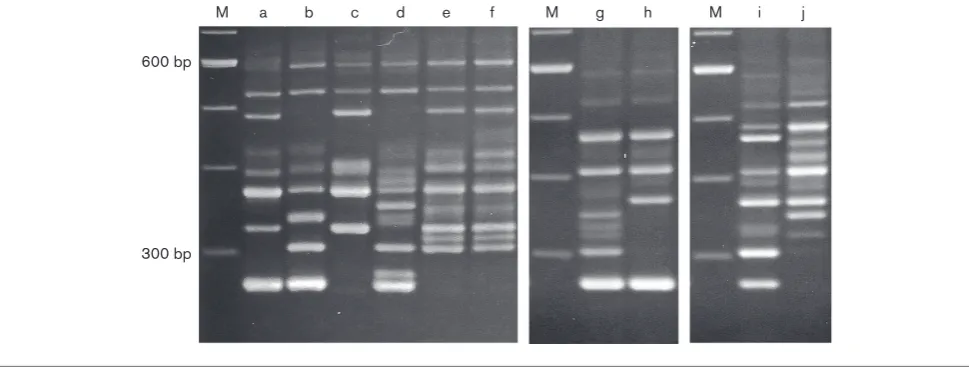
for 34.2 % of isolates (41/120) (Table 1); at least one isolate of this type was found in each of the 15 hospitals examined. We found a variant of PCR-ribotype smz, whose ribotype pattern was nearly identical with that of type smz but different in more than one band (Fig. 1). Thus, we termed it type ysmz. Isolates of both smz and ysmz
ribo-types were classified into the same major slpA sequence
type. The PCR-ribotype ysmz was recovered only at hospi-tal N in a non-outbreak setting in 2013, and during the
outbreak period in 2009 (Table 1). Of the 120 isolates, 47 (39.2 %) were PCR-ribotype smz or ysmz, and these isolates
were assigned to three slpA sequence subtypes (smz-01,
smz-02 and smz-03), with subtype smz-01 being most
common. All 19 A2B+CDT2 isolates were PCR-ribotype
trf (Fig. 1) andslpAsequence major type fr, and were further
assigned to six different slpA sequence subtypes. Sixteen
(13.3 %) and 13 (10.8 %) isolates were PCR-ribotypes 002 and 014, respectively. PCR-ribotypes smz, ysmz, trf, 002 and 014 together accounted for 79.2 % of isolates (95/120), and the remaining 25 isolates were typed into 19 and 16
different PCR ribotypes andslpAsequence types, respectively
(Tables S1 and 1). Among five A+B+CDT+ isolates, five
different PCR ribotypes including types 019, 027 and 078 were identified. The PCR ribotype pattern of one isolate was nearly identical with that of the reference strain (strain CA8), characterized previously as PCR-ribotype 056 (Kill-goreet al., 2008), but was different in more than one band
(PCR-ribotype c056 in Table S1). This isolate had theslpA
gene, the sequence of which was identical to that of strain
CA8 (slpAsequence type y02-01).
Typing results of isolates recovered in outbreak settings
The 21 isolates from hospital L in an outbreak period were assigned to four PCR ribotypes (014, smz, trf and sc1026)
and sixslpAsequence types (Tables S1 and 1). Of those
iso-lates recovered from CDI patients hospitalized in six wards during a four-week period, 13/21 (61.9 %) came from CDI patients at one ward. Out of the 13 isolates from the ward, eight, four and one were classified into PCR-ribotype smz/
slpAsequence type smz-01, trf/fr-12 and trf/fr-01,
respect-ively. TheslpA sequence type fr-12 was not found among
[image:3.595.51.557.542.704.2]the 120 isolates obtained at the 15 hospitals, including hos-pital L, in non-outbreak settings. CDI due to PCR-ribotype
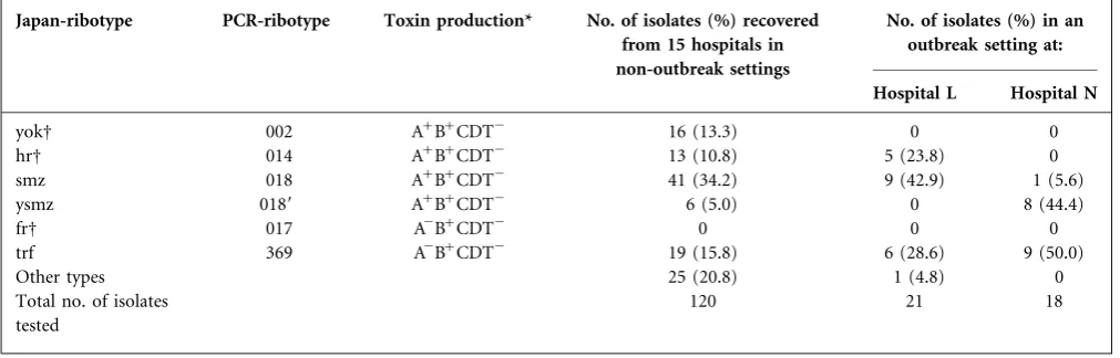
Table 1.Distribution of prevalent PCR ribotypes recovered from 15 hospitals in non-outbreak settings and 2 hospitals in outbreak settings
Japan-ribotype PCR-ribotype Toxin production* No. of isolates (%) recovered from 15 hospitals in non-outbreak settings
No. of isolates (%) in an outbreak setting at:
Hospital L Hospital N
yokD 002 A+B+CDT2 16 (13.3) 0 0
hrD 014 A+B+CDT2 13 (10.8) 5 (23.8) 0
smz 018 A+B+CDT2 41 (34.2) 9 (42.9) 1 (5.6)
ysmz 0189 A+B+CDT2 6 (5.0) 0 8 (44.4)
frD 017 A2B+CDT2 0 0 0
trf 369 A2B+CDT2 19 (15.8) 6 (28.6) 9 (50.0)
Other types 25 (20.8) 1 (4.8) 0
Total no. of isolates tested
120 21 18
*A+B+CDT2, toxin-A-positive, toxin-B-positive, binary-toxin-negative; A2B+CDT2, toxin-A-negative, toxin-B-positive, binary-toxin-negative.
014 was found in the wards other than the one where smz/ smz-01 and trf/fr-14 were epidemic.
Among the 18C. difficileisolates recovered from six wards
of hospital N during an outbreak of CDI, there were two
predominant types, PCR-ribotype trf/slpA sequence type
fr-01 (9 isolates) and type ysmz/smz-01 (8 isolates) (Tables S1 and 1). The remaining one isolate was found to be type smz/smz-01. Both trf/fr-01 and ysmz/smz-01 isolates were disseminated across four and five distinct wards, respectively. Isolation of PCR-ribotype ysmz was restricted to hospital N and this isolate was observed in six of 10 isolates recovered during an endemic period later on.
Analysis of isolates identified as types smz, ysmz and trf by the Japan ribotyping scheme
Since PCR-ribotypes smz/ysmz and PCR-ribotype trf accounted for a significant proportion of clinical isolates
in this and previous studies in Japan (Kato et al., 2001a,
2010; Sawabe et al., 2007; Iwashima et al., 2010), they
were further analysed at LCL. We later identified Japan-ribotype smz as being PCR-Japan-ribotype 018 and replicated a minor band variation between ribotypes smz and ysmz,
which was termed PCR-ribotype 0189
;
. Since Japan-ribotype trf did not match any known reference strains from the LCL library, further investigation of the trf strain was conducted by Dr V. Hall (Public Health Wales, Cardiff, UK); a novel ribotype was assigned, here designated PCR-ribotype 369.
Antimicrobial susceptibility results
The MIC results of gatifloxacin, moxifloxacin,
erythromy-cin, clindamycin, rifampicin, vancomycin and
metronidazole for C. difficile stratified according to PCR
ribotype in non-outbreak settings and in outbreak settings are shown in Tables 2 and 3, respectively. PCR-ribotypes smz and ysmz are presented together in Tables 2 and 3. Resistance to gatifloxacin, moxifloxacin, erythromycin and clindamycin was observed in 100 %, 95.7 %, 100 % and 97.9 % of PCR-ribotype smz/ysmz isolates collected from 15 hospitals, respectively. All PCR-ribotype smz/ ysmz isolates from two hospitals in outbreak settings were resistant to gatifloxacin, moxifloxacin, erythromycin and clindamycin. Among 19 trf isolates from 15 hospitals in non-outbreak settings, 17 (89.5 %) and 13 (68.4 %) were resistant to gatifloxacin and moxifloxacin, respect-ively. These 19 trf isolates were all resistant to erythromycin and clindamycin. All of the 15 trf isolates (6 from hospital L and 9 from hospital N) recovered from outbreak settings were resistant to gatifloxacin. Among six trf isolates from hospital L in an outbreak period, MICs of moxifloxacin
were 6mg ml21 and 8mg ml21 in four and two isolates,
respectively. Of nine trf isolates from hospital N in an out-break period, eight were resistant to moxifloxacin; the MIC
of the remaining one was 6mg ml21. High resistance to
both erythromycin and clindamycin was present in the 15 trf isolates prevalent at hospitals L and N; the MIC
value of all 15 isolates was more than 256mg ml21.
Resist-ance to gatifloxacin, moxifloxacin, erythromycin and clin-damycin was observed sporadically in 002 isolates examined in this study. In PCR-ribotype 014 isolates recov-ered in both non-outbreak settings and outbreak settings, resistance to gatifloxacin, moxifloxacin, erythromycin and clindamycin was less common. Rifampicin resistance
(MIC w32mg ml21) was observed in only one isolate,
which was typed as PCR-ribotype og39/slpA sequence
type og39-01. This isolate was highly resistant to erythro-mycin and clindaerythro-mycin, but susceptible to gatifloxacin
and moxifloxacin. All 159 isolates examined had
600 bp
300 bp
[image:4.595.50.535.67.253.2]M a b c d e f M g h M i j
vancomycin MIC values distributed over a narrow range
(0.19–1mg ml21). The metronidazole MIC was
j0.5mg ml21for all the isolates. One isolate was identified
as PCR-ribotype 027 and was susceptible to all seven anti-microbial agents tested. The PCR-ribotype 078 isolate recovered in this study was highly resistant to erythromy-cin, but susceptible to the other six agents.
CDI due to predominant PCR ribotypes
The mean age of CDI patients due to PCR-ribotypes smz/ smz, trf, 014 and 002 was 77.4, 77.6, 71.9, and 74.0 years old, respectively, and the difference in the four types was not significant. The data for CDI therapy was available in 153 of the 159 patients. While 31 (20.3 %) patients had no antibiotic treatment, 74 (48.4 %), 43 (28.1 %) and 5 (3.3 %) were treated with vancomycin, metronidazole and both, respectively. No significant correlation was found between the CDI treatments and PCR-ribotypes (data not shown). Ten of the 159 patients underwent an endoscopic examination and pseudomembranous colitis (PMC) was found in three patients, who were infected by PCR-ribotypes smz, 002 or 001. Two patients suffered from CDI with severe complications; one patient with type smz died of CDI and a pathological autopsy revealed PMC, and another patient with type 002 survived after emergency colectomy and was diagnosed with PMC during the surgery.
DISCUSSION
PCR-ribotype smz designated by the Japan typing scheme was found to correspond to PCR-ribotype 018 in the
pre-sent study. PCR-ribotypes 018 (smz)/0189 (ysmz) were
identified as the most common circulating strains (39.2 %), and were disseminated across 15 hospitals
exam-ined. Also, types 018 (smz) and 0189 (ysmz) caused
outbreaks at two hospitals, highlighting a major epidemio-logical role of them in both epidemic and endemic CDI in Japan. Of 365 isolates obtained from 26 European countries in 2008, 23 (6 %) were assigned as PCR-ribotype
018, 19 of which were recorded in Italy (Baueret al., 2011).
In a more recent study from 20 European countries, PCR-ribotype 018 was one of the ten most common types, although distribution of 018 was not mentioned (Davies
et al., 2014). In Italy, PCR-ribotype 018C. difficilewas pre-dominant in 2007 and 2008, superseding an other type (PCR-ribotype 126), which was the most predominant
strain until 2005 (Spigaglia et al., 2010); from 2012 to
2014, 20 % of isolates from Italy were identified as type
018 (Freeman et al., 2015). A recent prospective study
showed that PCR-ribotype 018 was also the predominant strain (48.1 %) at three hospitals in Korea from 2011 to
2012 (Han et al., 2014). Conversely, it was documented
that type 018 was not prevalent in England (Wilcox
et al., 2012) and the USA (Tickler et al., 2014). In Japan, there have been historical reports suggesting a high
prevalence of PCR-ribotype 018 since the 1990s, when it was already identified as the most prevalent strain at three Japanese hospitals between 1996 and 1999 (Kato
et al., 2001a) and at four other sites between 2003 and
2007 (Katoet al., 2010). An independent epidemiological
study at another hospital in Japan showed that PCR-ribo-type 018 replaced PCR-riboPCR-ribo-type 014 as the most predomi-nant strain over a five-year period from 2000 to 2004
(Sawabeet al., 2007). A similar shift was observed at a
hos-pital in Korea, where PCR-ribotype 018 has been identified since 2006, and became more prevalent than PCR-ribotype 001 over a 10-year period between 2000 and 2009 (Lee
et al., 2014). Interestingly, in a healthy volunteer study in Japan, 1234 individuals were examined and 94 (7.6 %)
were found to be colonized by C. difficile, but none of
them carried type 018 isolates in their intestinal tract
(Kato et al., 2001b). Isolation of PCR-ribotype 018
C. difficile from edible bivalve molluscs (Pasquale et al.,
2012) and poultry (Janezicet al., 2014) has been reported,
suggesting that food intake may constitute an important
route of C. difficile infection. Further molecular studies
on community-acquired CDI and food surveillance are required in Japan.
In the present study, we identified PCR-ribotype 0189
(ysmz), which had a ribotype pattern shared by most
bands of type 018 (smz), and all type 0189 isolates tested
in the present study displayedslpAsequence type smz-01.
The type 0189strain was exclusive to hospital N and was
recovered in both an outbreak period (2009) and a
non-outbreak setting (2012–2013), suggesting that the 0189
might have persisted within CDI inpatients, asymptomatic carriers and/or in inanimate environments at hospital L for years. More recently, the emergence of PCR-ribotype 356 has been noted in Italy, and it was postulated that this is likely a strain subtype that may have evolved from the
main PCR-ribotype 018 lineage (Freeman et al., 2015).
Albeit ribotype profiles of types 0189 (ysmz) and 356
have not been compared directly, it is epidemiologically significant that strain variants similar to ribotype 018 emerged in independent areas where this strain was predominant.
An early report showed that PCR riboype 018 has been an
endemic strain since the 1990s in Japan (Katoet al., 2001a),
suggesting that 018 and its variants might have spread from Asia, including Japan, to other countries. More compre-hensive studies at a whole genome sequencing level on
types 018/ 0189 in Asia and type 018/356 in Europe are
required to unveil its evolution pattern and potential of acquired functional mechanisms in order to understand their global spread and epidemiology.
High rates of resistance to gatifloxacin, moxifloxacin, ery-thromycin and clindamycin in both PCR-ribotype 018
and 0189 isolates were observed, although they both
remained susceptible to rifampicin. In Italy, resistance against fluoroquinolones in type 018 isolates has been
In a Korean study, eight isolates belonging to type 018 were tested and all of them had high overall resistance against several antibiotics including moxifloxacin, erythromycin
and clindamycin (Lee et al., 2014). Sawabe et al. (2007)
reported that all type 018 isolates at their hospital in Japan were resistant to gatifloxacin and moxifloxacin, as well as clindamycin, and that the noticeable shift in ende-mic strain from type 014 to type 018 may have been related to the introduction of gatifloxacin to their hospital in 2002. Heet al.(2013) documented that the acquisition of resist-ance to commonly used antibiotics (fluoroquinolones) is a major feature of the continued evolution and persistence of
C. difficile BI/NAP1/027 in healthcare settings. In this study, we found only one type 027 isolate, which was sus-ceptible to new fluoroquinolones. In Japan, while some reports have shown sporadic cases including one with
ful-minant colitis due to PCR-ribotype 027 C. difficile (Kato
et al., 2007; Sawabe et al., 2007; Nishimura et al., 2014), no outbreaks associated with type 027 have been reported so far. It is unknown why the nosocomial spread of 027
C. difficile resistant to fluoroquinolones has not been found in Japan, where type 018 resistant to fluoroquino-lones has been distributed to a number of hospitals.
Simi-larly, PCR-ribotype 078C. difficilewas isolated from only
one patient in the present study, while this strain has been reported to be recovered more frequently from
ani-mals in Japan (Niwa et al., 2013; Usui et al., 2014). The
lower frequency of isolation, either of PCR-ribotype 027 or 078 from CDI in human in Asian countries including
Japan (Cheng et al., 2011; Han et al., 2014), may reflect
differences in the intestinal ecosystem and/or immunologi-cal responses between Asian populations and those of countries where 027 or 078 have become predominant.
Freeman et al. (2015) documented that all ribotype 018
and 356 isolates obtained from Italy were resistant to rifampicin, but rifampicin resistance was not observed in
type 018/0189isolates examined in our study. Also,
Free-man et al. (2015) showed that vancomycin MICs were higher among 018 and 356 with geometric mean MICs of
2.00mg ml21 and 2.28mg ml21, respectively, compared
with those of the remaining common ribotypes; geometric mean MICs of metronidazole were elevated in ribotype 027
(1.42mg ml21) and ribotype 356 (0.61mg ml21) isolates
(Freeman et al., 2015). Tickler et al. (2014) observed
reduced susceptibility to vancomycin in 39.2 % of 027 iso-lates collected from US hospitals. In our study, none of the
isolates tested including type 018/0189showed reduced
sus-ceptibility to vancomycin and metronidazole. The potential emergence of increasing resistance to vancomycin and metronidazole may warrant further monitoring of MICs
of the agents especially in type 018/0189isolates in Japan.
In contrast, the emergence of A2B+ isolates, which have
1.8 kbp deletions in the repeating sequences in tcdA and
belong to toxinotype VIII, has been reported in Japan as well as the Netherlands, Ireland, Poland, Korea, China
and Argentina (Kuijperet al., 2001; Komatsuet al., 2003;
Drudy et al., 2007; Shin et al., 2008; Goorhuis et al.,
2009, Katoet al., 2010; Pituch et al., 2011; Hawkeyet al.,
2013). The reports from all these countries except Japan
documented that the vast majority of these A2B+isolates
were found to be of PCR-ribotype 017 (Kuijper et al.,
2001; Drudy et al., 2007; Goorhuis et al., 2009; Pituch
et al., 2011; Hawkeyet al., 2013; Lee et al., 2014). PCR-ribotype 017 was identified in 4 % of isolates from 34
Euro-pean countries (Davieset al., 2014) and 2.2 % of isolates
from 32 hospitals in the USA (Tickler et al., 2014).
Although we have reported the isolation of PCR-ribotype
017 from sporadic cases (Katoet al., 2001b, 2010; Iwashima
et al., 2010) in Japan, notably, all 34 A2B+isolates collected in the present study were typed as Japan-ribotype trf, which corresponded neither to type 017 nor 047, and was newly assigned as PCR-ribotype 369. In this study, type 369 was the second most frequent ribotype (15.8 %) among 120 isolates from 15 hospitals in non-outbreak settings, and was also prevalent at two hospitals during outbreak periods. The type 369 strain was reported to be blamed for outbreaks that occurred at Japanese hospitals in 2000
and 2001 (Komatsuet al., 2003; Satoet al., 2004); ribotype
369 has been identified as an epidemic strain since the early 2000s in Japan. Both PCR-ribotype 017 and 369 isolates tested had identical deletions at the repeating sequences
in tcdA and a nonsense mutation introducing a stop
codon at amino acid position 47 of tcdA (data not
shown), and were classed asslpA sequence major type fr.
This suggests that types 017 and 369 are not phylogeneti-cally distant to each other and they may share a common recent ancestor, though deep genome sequencing has not been performed yet. Evolutionary comparative analysis and determination of exclusive factors to each strain will allow understanding of the preferential establishment of one or another, such as of 369 in Japan and type 017 in Europe. High resistance against erythromycin and clinda-mycin was observed in all type 369 isolates tested, which is consistent with the previous reports on type 017 strains
(Kuijperet al., 2001; Pituch et al., 2011; Leeet al., 2014;
Freeman et al., 2015). In addition, 94.1 % (32/34) and
67.6 % (23/34) of type 369 isolates examined by this study were resistant to gatifloxacin and moxifloxacin, respectively. Resistance against new fluoroquinolones in PCR-ribotype 017 was also reported from other countries
(Pituchet al., 2011; Spigagliaet al., 2011; Lee et al., 2014;
Freeman et al., 2015), indicating the need of escalating
attention to isolates belonging to toxinotype VIII, which have been showing multi-resistance tendencies.
It was reported that PCR-ribotype 002 constituted approxi-mately 5 % of isolates from European countries in 2008, and 3.5 % of isolates from US hospitals between 2011
and 2013 (Baueret al., 2011; Tickleret al., 2014). The
sig-nificant increase in the prevalence of type 002 from 3 % (2007–2008) to 6 % (2009–1010) was documented in
Eng-land (Wilcox et al., 2012). Moreover, PCR-ribotype 002
was reported to be the most prevalent type (10.1 %) in a
healthcare region in Hong Kong (Cheng et al., 2011).
found (13.3 %) in the present study, and a three-year investigation at a university hospital in Japan showed that type 002 was one of the three most prevalent types
(19.7 %) (Iwashima et al., 2010). In addition, Tenover
et al. (2010) reported
<
that the sensitivity of the enzymeimmunoassay (EIA) was significantly lower for detecting toxins from specimens of CDI infected by ribotype 002, suggesting that these cases may be overlooked when diag-nosed by EIA for toxins only. More studies of worldwide variation and differences in prevalence and disease severity by type 002 are required.
PCR-ribotype 014 has been reported to be one of the most
common PCR ribotypes in the world (Kato et al., 2001a,
2010; Iwashima et al., 2010; Bauer et al., 2011; Cheng
et al., 2011; Wilcoxet al., 2012; Davieset al., 2014; Freeman
et al., 2014; Han et al., 2014; Tickler et al., 2014). In the present study, 10.8 % (13/120) of the isolates recovered in non-outbreak settings and
=
19.0 % (4/21) of the isolatesrecovered in an outbreak period at hospital L were typed as PCR-ribotype 014. Moreover, a previous study showed that PCR-ribotype 014 was in fact the most frequently iso-lated strain from healthy Japanese individuals; of 94
indi-viduals positive for C. difficile-culture, 17 of them
(18.0 %) had this type (Kato et al., 2001b). Janezic et al.
(2014) documented that PCR-ribotype 014 was the second most prevalent strain following type 078 among animal isolates and also had a broader range of animal hosts. In our study, the resistance of PCR-ribotype 014 to gatifloxacin, moxifloxacin, erythromycin and clindamycin was less common, consistent with the results of other
reports (Tickleret al., 2014; Freemanet al., 2015). The
abil-ity to colonize a variety of hosts including humans may be responsible for its relatively high nosocomial and
commu-nity prevalence (Janezicet al., 2014).
Our study showed no difference in the mean age of CDI patients due to strains 018, 369, 002 or 014. Also, we found no significant correlation between the CDI treat-ments and PCR ribotypes. Since only limited data about clinical features of patients, such as presence of comorbid-ities, symptoms and outcomes including recurrences was available, neither potential risk factors for CDI due to pre-dominant types nor specific correlation between outcomes and type differences could be clarified in this study. It was reported that PCR-ribotype 018 was significantly associated
with complicated disease outcome (Baueret al., 2011).
Pro-spective studies are needed to elucidate the involvement of these types with CDI progression and clinical outcomes.
In summary, PCR-ribotypes 018 (smz)/0189 (ysmz), 369
(trf), 002 and 014 were identified as the major types
circu-lating in Japan. Particularly, types 018/ 0189and 369 were
found to be prevalent and associated with both epidemic and endemic CDI in Japan. A European study demon-strated that increased awareness of CDI and the use of opti-mal testing methods could reduce the dissemination of
epidemic strains (Davieset al., 2014). The high epidemicity
of ribotypes 018 and 369 and a lower diversity of ribotypes
observed in the present study may reflect the insufficient awareness of CDI and a suboptimal test-density in Japan. Further studies are warranted to understand the epidemio-logical relevance and the role of these strains in relation to their spread and prevalence nationwide and globally.
ACKNOWLEDGEMENTS
We thank Dr V. Hall (Public Health Wales, Cardiff, UK) for analysing ribotype 369 (trf) isolate and Dr S. Suzuki (National Institute of Infectious Diseases, Tokyo, Japan) for her helpful comments. The technical assistance of S. Yonamine, K. Kai, Y. Taki, and Y. Yoshimura is gratefully acknowledged. This work was supported by a grant (Research Program on Emerging and Re-emerging Infec-tious Diseases) from Japan Agency for Medical Research and Devel-opment, AMED. The authors declare no conflicts of interest.
REFERENCES
Bauer, M. P., Notermans, D. W., van Benthem, B. H., Brazier, J. S., Wilcox, M. H., Rupnik, M., Monnet, D. L., van Dissel, J. T. & Kuijper, E. J. (2011). ECDIS Study Group Clostridium difficile infection in Europe: a hospital-based survey.Lancet377, 63–73.
Cheng, V. C., Yam, W. C., Lam, O. T., Tsang, J. L., Tse, E. Y., Siu, G. K., Chan, J. F., Tse, H., To, K. K. & other authors (2011).Clostridium
difficile isolates with increased sporulation: emergence of PCR
ribotype 002 in Hong Kong. Eur J Clin Microbiol Infect Dis 30, 1371–1381.
CLSI (2014). Performance Standards for AntimicrobialSsusceptibility
Testing; 24th Informational Supplement M100–S24. Wayne, PA:
Clinical and Laboratory Standards Institute.
Davies, K. A., Longshaw, C. M., Davis, G. L., Bouza, E., Barbut, F., Barna, Z., Delme´e, M., Fitzpatrick, F., Ivanova, K. & other authors (2014). Underdiagnosis of Clostridium difficile across Europe: the European, multicentre, prospective, biannual, point-prevalence study ofClostridium difficile infection in hospitalised patients with diarrhoea (EUCLID).Lancet Infect Dis14, 1208–1219.
Drudy, D., Harnedy, N., Fanning, S., O’Mahony, R. & Kyne, L. (2007).
Isolation and characterisation of toxin A-negative, toxin B-positive
Clostridium difficile in Dublin, Ireland. Clin Microbiol Infect 13,
298–304.
Freeman, J., Vernon, J., Morris, K., Nicholson, S., Todhunter, S., Longshaw, C. & Wilcox, M. H. (2015). Pan-European Longitudinal Surveillance of Antibiotic Resistance among Prevalent Clostridium difficile Ribotypes’ Study Group Pan-European longitudinal surveillance of antibiotic resistance among prevalent Clostridium
difficileribotypes.Clin Microbiol Infect21, 248.
Goorhuis, A., Bakker, D., Corver, J., Debast, S. B., Harmanus, C., Notermans, D. W., Bergwerff, A. A., Dekker, F. W. & Kuijper, E. J. (2008). Emergence of Clostridium difficile infection due to a new hypervirulent strain, polymerase chain reaction ribotype 078. Clin
Infect Dis47, 1162–1170.
Goorhuis, A., Legaria, M. C., van den Berg, R. J., Harmanus, C., Klaassen, C. H., Brazier, J. S., Lumelsky, G. & Kuijper, E. J. (2009).
Application of multiple-locus variable-number tandem-repeat analysis to determine clonal spread of toxin A-negativeClostridium
difficile in a general hospital in Buenos Aires, Argentina. Clin
Microbiol Infect15, 1080–1086.
clinical features of toxigenic culture-confirmed hospital-onset
Clostridium difficile infection: a multicentre prospective study in
tertiary hospitals of South Korea.J Med Microbiol63, 1542–1551.
Hawkey, P. M., Marriott, C., Liu, W. E., Jian, Z. J., Gao, Q., Ling, T. K., Chow, V., So, E., Chan, R. & other authors (2013). Molecular epidemiology of Clostridium difficile infection in a major Chinese hospital: an underrecognized problem in Asia?J Clin Microbiol 51, 3308–3313.
He, M., Miyajima, F., Roberts, P., Ellison, L., Pickard, D. J., Martin, M. J., Connor, T. R., Harris, S. R., Fairley, D. & other authors (2013). Emergence and global spread of epidemic healthcare-associatedClostridium difficile.Nat Genet45, 109–113.
Iwashima, Y., Nakamura, A., Kato, H., Wakimoto, Y., Wakiyama, N., Kaji, C. & Ueda, R. (2010). A retrospective study of the epidemiology of Clostridium difficile infection at a University Hospital in Japan: genotypic features of the isolates and clinical characteristics of the patients.J Infect Chemother16, 329–333.
Janezic, S., Zidaric, V., Pardon, B., Indra, A., Kokotovic, B., Blanco, J. L., Seyboldt, C., Diaz, C. R., Poxton, I. R. & other authors (2014).
InternationalClostridium difficile animal strain collection and large diversity of animal associated strains.BMC Microbiol14, 173–183.
Kato, H., Kato, N., Watanabe, K., Yamamoto, T., Suzuki, K., Ishigo, S., Kunihiro, S., Nakamura, I., Killgore, G. E. & Nakamura, S. (2001a).
Analysis ofClostridium difficileisolates from nosocomial outbreaks at three hospitals in diverse areas of Japan. J Clin Microbiol 39, 1391–1395.
Kato, H., Kita, H., Karasawa, T., Maegawa, T., Koino, Y., Takakuwa, H., Saikai, T., Kobayashi, K., Yamagishi, T. & Nakamura, S. (2001b). Colonisation and transmission of Clostridium difficile in healthy individuals examined by PCR ribotyping and pulsed-field gel electrophoresis.J Med Microbiol50, 720–727.
Kato, H., Yokoyama, T., Kato, H. & Arakawa, Y. (2005).Rapid and simple method for detecting the toxin B gene ofClostridium difficile in stool specimens by loop-mediated isothermal amplification.
J Clin Microbiol43, 6108–6112.
Kato, H., Ito, Y., van den Berg, R. J., Kuijper, E. J. & Arakawa, Y. (2007). First isolation of Clostridium difficile 027 in Japan. Euro
Surveill12, E070111.3.
Kato, H., Kato, H., Ito, Y., Akahane, T., Izumida, S., Yokoyama, T., Kaji, C. & Arakawa, Y. (2010).Typing ofClostridium difficileisolates endemic in Japan by sequencing of slpA and its application to direct typing.J Med Microbiol59, 556–562.
Killgore, G., Thompson, A., Johnson, S., Brazier, J., Kuijper, E., Pepin, J., Frost, E. H., Savelkoul, P., Nicholson, B. & other authors (2008).
Comparison of seven techniques for typing international epidemic strains of Clostridium difficile: restriction endonuclease analysis, pulsed-field gel electrophoresis, PCR-ribotyping, multilocus sequence typing, multilocus variable-number tandem-repeat analysis, amplified fragment length polymorphism, and surface layer protein A gene sequence typing.J Clin Microbiol46, 431–437.
Komatsu, M., Kato, H., Aihara, M., Shimakawa, K., Iwasaki, M., Nagasaka, Y., Fukuda, S., Matsuo, S., Arakawa, Y. & other authors (2003).High frequency of antibiotic-associated diarrhea due to toxin A-negative, toxin B-positiveClostridium difficilein a hospital in Japan and risk factors for infection.Eur J Clin Microbiol Infect Dis22, 525–529.
Kuijper, E. J., de Weerdt, J., Kato, H., Kato, N., van Dam, A. P., van der Vorm, E. R., Weel, J., van Rheenen, C. & Dankert, J. (2001).
Nosocomial outbreak of Clostridium difficile-associated diarrhoea due to a clindamycin-resistant enterotoxin A-negative strain.Eur J
Clin Microbiol Infect Dis20, 528–534.
Lee, J. H., Lee, Y., Lee, K., Riley, T. V. & Kim, H. (2014).The changes of PCR ribotype and antimicrobial resistance ofClostridium difficilein a tertiary care hospital over 10 years.J Med Microbiol63, 819–823.
Louie, T. J., Miller, M. A., Mullane, K. M., Weiss, K., Lentnek, A., Golan, Y., Gorbach, S., Sears, P., Shue, Y. K. & OPT-80-003 Clinical Study Group (2011). Fidaxomicin versus vancomycin for Clostridium
difficileinfection.N Engl J Med364, 422–431.
Miller, M., Gravel, D., Mulvey, M., Taylor, G., Boyd, D., Simor, A., Gardam, M., McGeer, A., Hutchinson, J. & other authors (2010).
Health care-associated Clostridium difficile infection in Canada: patient age and infecting strain type are highly predictive of severe outcome and mortality.Clin Infect Dis50, 194–201.
Nishimura, S., Kou, T., Kato, H., Watanabe, M., Uno, S., Senoh, M., Fukuda, T., Hata, A. & Yazumi, S. (2014). Fulminant pseudomembranous colitis caused byClostridium difficilePCR ribotype 027 in a healthy young woman in Japan.J Infect Chemother20, 729–731.
Niwa, H., Kato, H., Hobo, S., Kinoshita, Y., Ueno, T., Katayama, Y., Hariu, K., Oku, K., Senoh, M. & other authors (2013).Postoperative
Clostridium difficileinfection with PCR ribotype 078 strain identified
at necropsy in five Thoroughbred racehorses.Vet Rec173, 607–613.
Pasquale, V., Romano, V., Rupnik, M., Capuano, F., Bove, D., Aliberti, F., Krovacek, K. & Dumontet, S. (2012).Occurrence of toxigenic
Clostridium difficile in edible bivalve molluscs. Food Microbiol 31,
309–312.
Pituch, H., Obuch-Woszczatyn´ski, P., Wultan´ska, D., Nurzyn´ska, G., Harmanus, C., Banaszkiewicz, A., Radzikowski, A., Łuczak, M., van Belkum, A. & Kuijper, E. (2011).Characterization and antimicrobial susceptibility of Clostridium difficile strains isolated from adult patients with diarrhoea hospitalized in two university hospitals in Poland, 2004-2006.J Med Microbiol60, 1200–1205.
Sato, H., Kato, H., Koiwai, K. & Sakai, C. (2004).[A nosocomial outbreak of diarrhea caused by toxin A-negative, toxin B-positive
Clostridium difficile in a cancer center hospital]. Kansenshogaku
Zasshi78, 312–319 (in Japanese).
Sawabe, E., Kato, H., Osawa, K., Chida, T., Tojo, N., Arakawa, Y. & Okamura, N. (2007).Molecular analysis ofClostridium difficile at a university teaching hospital in Japan: a shift in the predominant type over a five-year period.Eur J Clin Microbiol Infect Dis26, 695–703.
Shin, B. M., Kuak, E. Y., Yoo, H. M., Kim, E. C., Lee, K., Kang, J. O., Whang, D. H. & Shin, J. H. (2008). Multicentre study of the prevalence of toxigenic Clostridium difficile in Korea: results of a retrospective study 2000–2005.J Med Microbiol57, 697–701.
Spigaglia, P., Barbanti, F., Dionisi, A. M. & Mastrantonio, P. (2010).
Clostridium difficile isolates resistant to fluoroquinolones in Italy: emergence of PCR ribotype 018.J Clin Microbiol48, 2892–2896.
Spigaglia, P., Barbanti, F., Mastrantonio, P., Ackermann, G., Balmelli, C., Barbut, F., Bouza, E., Brazier, J., Delmee, M. & other authors (2011). Multidrug resistance in European Clostridium
difficileclinical isolates.J Antimicrob Chemother66, 2227–2234.
Stubbs, S. L., Brazier, J. S., O’Neill, G. L. & Duerden, B. I. (1999).PCR targeted to the 16S-23S rRNA gene intergenic spacer region of
Clostridium difficile and construction of a library consisting of 116
different PCR ribotypes.J Clin Microbiol37, 461–463.
Stubbs, S., Rupnik, M., Gibert, M., Brazier, J., Duerden, B. & Popoff, M. (2000). Production of actin-specific ADP-ribosyltransferase (binary toxin) by strains of Clostridium difficile. FEMS Microbiol Lett186, 307–312.
Tenover, F. C., Novak-Weekley, S., Woods, C. W., Peterson, L. R., Davis, T., Schreckenberger, P., Fang, F. C., Dascal, A., Gerding, D. N. & other authors (2010).Impact of strain type on detection of toxigenicClostridium difficile: comparison of molecular diagnostic and enzyme immunoassay approaches.J Clin Microbiol48, 3719–3724.
Tenover, F. C., Tickler, I. A. & Persing, D. H. (2012). Antimicrobial-resistant strains of Clostridium difficile from North America.
Tickler, I. A., Goering, R. V., Whitmore, J. D., Lynn, A. N., Persing, D. H., Tenover, F. C. & Healthcare Associated Infection Consortium (2014). Strain types and antimicrobial resistance patterns of Clostridium difficile isolates from the United States, 2011 to 2013.Antimicrob Agents Chemother58, 4214–4218.
Usui, M., Nanbu, Y., Oka, K., Takahashi, M., Inamatsu, T., Asai, T., Kamiya, S. & Tamura, Y. (2014). Genetic relatedness between Japanese and European isolates of Clostridium difficile originating from piglets and their risk associated with human health. Front
Microbiol5, 513–521.
Walker, A. S., Eyre, D. W., Wyllie, D. H., Dingle, K. E., Griffiths, D., Shine, B., Oakley, S., O’Connor, L., Finney, J. & other authors (2013).Relationship between bacterial strain type, host biomarkers, and mortality in Clostridium difficile infection. Clin Infect Dis 56, 1589–1600.
Wilcox, M. H., Shetty, N., Fawley, W. N., Shemko, M., Coen, P., Birtles, A., Cairns, M., Curran, M. D., Dodgson, K. J. & other authors (2012).
Pleasefind enclosed a proof of your article for checking.
When reading through your proof, please check carefully authors’names, scientific data, data in tables, any math-ematics and the accuracy of references. Please do not make any unnecessary changes at this stage. All necess-ary corrections should be marked on the proof at the place where the correction is to be made; please mark up the correction in the PDF and return it to us (see instructions on marking proofs in Adobe Reader).
Any queries that have arisen during preparation of your paper for publication are listed below and indicated on the proof.
Please provide your answers when returning your proof.
Please return your proof by email ([email protected]) within 2 days of receipt of this message.
Query no Query