Viral delivery of C9ORF72 hexanucleotide repeat expansions in mice lead to repeat length dependent neuropathology and behavioral deficits.
Saul Herranz-Martin1#, Jayanth Chandran1#, Katherine Lewis1, Padraig Mulcahy1, Adrian Higginbottom1, Callum Walker1,2, Isabel Martinez-Pena y Valenzuela3, Ross A. Jones4, Ian Coldicott1, Tommaso Iannitti1, Mohammed Akaaboune3, Sherif F. El-Khamisy2, Thomas H. Gillingwater4, Pamela J. Shaw1§ and Mimoun Azzouz1§*.
1Department of Neuroscience, Sheffield Institute for Translational Neuroscience (SITraN), The University of Sheffield, 385A Glossop Road, Sheffield S10 2HQ, UK
2Department of Molecular Biology and Biotechnology, Krebs and Sheffield Institute for Nucleic Acids, Firth Court, University of Sheffield, S10 2TN, Sheffield, UK
3Molecular, Cellular and Developmental Biology, University of Michigan, 830 North University, Ann Arbor, MI 48109-1048, USA
4Centre for Integrative Physiology & Euan MacDonald Centre for Motor Neurone Disease Research,, Hugh Robson Building, The University of Edinburgh, 15 George Square, Edinburgh
EH8 9XD, UK
# Authors contributed equally
§ Senior authors
* Correspondence should be addressed to Mimoun Azzouz Email: [email protected]
Keywords: C9orf72, neurodegeneration, ALS/FTD, mouse model, c9RAN aggregates
http://dmm.biologists.org/lookup/doi/10.1242/dmm.029892 Access the most recent version at
Summary statement
We have created new C9orf72 linked motor neuron disease models through viral mediated
expression of GGGGCC repeat expansions in mice.
Abstract
Intronic GGGGCC repeat expansions in C9orf72 are the most common genetic cause of
amyotrophic lateral sclerosis (ALS) and frontotemporal dementia (FTD). Two major
pathologies stemming from the hexanucleotide RNA expansions (HREs) have been identified
in postmortem tissue: intracellular RNA foci and repeat-associated non-ATG dependent (RAN)
dipeptides, though it is unclear how these and other hallmarks of disease contribute to the
pathophysiology of neuronal injury. Here we generated two novel lines of mice that
overexpress either 10 pure or 102 interrupted G4C2 repeats mediated by adeno-associated virus
(AAV) and characterized relevant pathology and disease-related behavioral phenotypes.
Similar levels of intracellular RNA foci developed in both lines of mice, but only mice
expressing 102 repeats generated c9-RAN pathology, neuromuscular junction (NMJ)
abnormalities, dispersal of the hippocampal CA1, enhanced apoptosis, and deficits in gait and
cognition. Neither line of mice, however, showed extensive TAR DNA-binding protein 43
(TDP-43) pathology or neurodegeneration. Our data suggests that RNA foci pathology is not a
good predictor of c9-RAN dipeptide formation, and that RAN dipeptides and NMJ dysfunction
are drivers of c9-disease pathogenesis. These AAV-mediated models of C9orf72 ALS/FTD
will be useful tools for studying disease pathophysiology and developing new therapeutic
Introduction
Hexanucleotide repeat expansions (HREs) in C9orf72 are regarded as the most common
genetic cause of the progressive neurodegenerative diseases, amyotrophic lateral sclerosis
(ALS) and frontotemporal dementia (FTD). ALS primarily affects motor neurons and leads to
progressive failure of the neuromuscular system with muscle wasting and paralysis, while
frontotemporal dementia (FTD) is caused by the degeneration of neurons in the frontal and
temporal lobes leading to cognitive deficits. G4C2 repeat expansions in the first intron of
C9orf72 are present in up to 40% of familial ALS and 25 % of familial FTD patients, and are
also present in 5-20% of sporadic ALS patients (DeJesus-Hernandez et al., 2011, Renton et al.,
2011, Majounie et al., 2012, Renton et al., 2014). Normal healthy C9orf72 alleles contain
between 2-30 repeats, while disease-associated alleles vary in size and can exceed several
thousand HREs (DeJesus-Hernandez et al., 2011, Renton et al., 2011). Three different
pathological alterations providing clues to the etiology of C9orf72-related disease can be found
in post-mortem tissue from patients with HREs: first, a reduction in all three mRNA transcripts
generated from C9orf72, suggesting disease may arise by a loss-of-function mechanism
(DeJesus-Hernandez et al., 2011, Belzil et al., 2013, Donnelly et al., 2013); second, formation
of intracellular foci by aggregation of RNA expansions; and third, dipeptide repeat proteins
(DPRs) which are generated from long expansions via repeat-associated non ATG-dependent
(RAN) translation (Cooper-Knock et al., 2014, Schludi et al., 2015).
Recent studies suggest that G4C2 expansions and DPRs may induce cell toxicity by
sequestration of RNA binding proteins (Cooper-Knock et al., 2014, Lee et al., 2016) and
disruption of nucleocytoplasmic transport (Zhang et al., 2015, Freibaum et al., 2015). Models
developed to identify the combination of factors inducing disease relevant pathology. While
C9orf72 knockout mice display motor and cognitive deficits along with heightened
neuroinflammation, these mice do not exhibit any significant neuronal pathology, suggesting
that a simple loss-of-function mechanism is unlikely to be the sole cause of C9orf72-linked
neurodegeneration (Koppers et al., 2015, O'Rourke et al., 2016). Four different toxic
gain-of-function mouse models developed through expression of patient derived C9orf72 containing
hundreds of HREs consistently displayed intracellular RNA foci and DPRs in the CNS, though
they differed in the generation of ALS/FTD disease phenotypes (Jiang et al., 2016, Liu et al.,
2016, O'Rourke et al., 2015, Peters et al., 2015, Chew et al., 2015). In contrast, acute
AAV-mediated, actin-promoter-driven expression of 66 repeats in mice recapitulates ALS-like
pathology and induces cognitive and behavioral deficits, despite lacking any flanking C9orf72
sequence (Chew et al., 2015).
Interruptions in repeat expansions are found in alleles for spinocerebellar ataxia (SCA) (Chung
et al., 1993, Pearson et al., 1998a, Matsuura et al., 2006), Myotonic dystrophy type 1 (Musova
et al., 2009), Friedreich ataxia (Stolle et al., 2008) and Fragile X syndromes (Eichler et al.,
1994, Pearson et al., 1998a), although the consequences of the interruptions on disease etiology
remain unclear. Interrupting the expression of pure glycine-alanine (GA) dipeptide repeats with
proline residues in mice prevented the formation of dense ubiquitin-positive inclusions and the
development of behavioral phenotypes and pathological hallmarks consistent with
C9orf72-ALS/FTD (Zhang et al., 2016). An interruption in the expression of DPRs created by inserting
stop codons between G4C2 repeats in Drosophila blocked DPR formation and
neurodegeneration but preserved RNA foci generation demonstrating that DPRs are toxic
(Mizielinska et al., 2014). Here, we generated mice expressing HREs, utilizing AAV serotype
postnatal day 1 (P1) pups. We compared mice expressing 10 HRE expansions to mice
expressing 102 repeats interrupted at regular intervals to examine the effects of interrupted
expansions on disease etiology. We show that both AAV-(G4C2)10 and AAV-(G4C2)102 repeats lead to the formation of a similar number of intranuclear RNA foci in the CNS in vivo, but only
(G4C2)102 transgenic mice generate DPRs and p62 inclusions, have Purkinje cell apoptosis, exhibit neuromuscular junction pathology, and display gait and cognitive deficits.
Materials and Methods
Antibodies and Reagents
Rabbit AAV VP1, VP2, VP3 (1:800, #03-61084, American Res Prod, USA), rabbit
anti-NeuN (1:500, Cell Signalling (USA)- clone D3S3I), rabbit anti-GFAP (1:2000, Dako - Z0334),
rabbit anti-TDP-43 (1:500, Proteintech - catalog #18-280-1-AP), mouse anti-TDP43-phospho
ser409/410-1 (1:4000, Cosmo Bio – cat#:CAC-TIP-PTD-M01), mouse anti-Calbindin (1:1000,
Sigma C9848), mouse anti-p62 lck ligand (1:1000 (WBlot), 1:250 (ICC), BD Biosciences,
clone 3, cat#:610833), rabbit anti-α-tubulin (1:5000, Abcam, cat #4074),
rabbit-cleaved-caspase 3 (1:1000, Cell Signalling #9661), mouse anti-cleaved PARP (1:250, Cell Signalling
#9548), mouse anti-GAPDH (1:5000, Millipore), mouse anti-poly(Glycine-Alanine (GA),
1:500 for western blot; 1:5000 for ICC, clone 5F2 (Mackenzie et al., 2013) kindly provided by
Cell lines and culture
HEK 293T cells were maintained in growth media (GM) consisting of Dulbecco’s modified
Eagle’s medium (DMEM, D5796, Sigma (USA)) supplemented with 10% foetal bovine serum
(Sigma, USA), penicillin (100U/ml), and streptomycin (100U/ml) at 37°C and 5% CO2.
Generation of Repeat Expansion Constructs
Sense (TCGAC (G4C2)10) and antisense (ACGT(G4C2)10) oligonucleotides (Sigma-Aldrich) with SalI/XhoI overhangs were annealed by denaturing at 95ºC for 5 min and cooling at
0.5ºC/min to 25ºC. The dsDNA (G4C2)10 was ligated into SalI and XhoI digested pcDNA6.2-GW/EmGFP-miR (Invitrogen) to generate pcDNA6.2-GW-(G4C2)10. Further (G4C2)n repeats where n varied between 10-17 repeats were subcloned into the XhoI site to generate the longer
pcDNA6.2-(G4C2)102 construct. The (G4C2)10 and (G4C2)102 constructs were subsequently sub-cloned into a pAV2-CMV (cytomegalovirus) plasmid and authenticity was confirmed by
sequencing. Transformations of plasmids containing the (G4C2)n repeat constructs were performed using recombination-deficient β-10 E.coli (NEB) to minimise any rearrangements.
Large scale scAAV9 production
Sixty 15 cm plates containing HEK293 cells at 80% confluence were transfected using
polyethylenimine (MW ~ 25K) with a mixture of three plasmids required to generate an
infectious scAAV9 viral particle: (1) A plasmid providing helper genes isolated from
adenovirus that enhance viral infectivity (pHelper); (2) An ITR-containing plasmid carrying
the gene of interest (pAV2-CMV-(G4C2)n; we used the pAV2-CMV-GFP consisting of two
proteins (pAAV2/9). Each 15cm plate was transfected with a total of 40µg of DNA comprised
of a 2:1:1 molar ratio of pHelper: pAV2-CMV-(G4C2)n: pAAV2/9. Four days after transfection, the AAV enriched media was collected, incubated at 37°C for 2 hours with 3,750 units of
benzonase-nuclease (Sigma, USA), filtered through a 0.22 µm filter, and concentrated to a
volume of 1ml using Amicon spin filter units (Millipore, USA). The virus was then washed
with 50 ml of phosphate buffered saline (PBS, pH 7.3) in the same Amicon spin filter units,
and concentrated to a final volume of 0.5 ml. The viral sample volume was expanded to 14ml
with PBS and separated through a discontinuous iodixanol (D1556, Sigma, USA) gradient (4ml
of 54%, 9 ml of 40%, 9 ml of 25%, 5 ml of 15%), and centrifuged at 69,000 rpm for 1.5 hours
at 18°C. Phenol red was added to the 54% and 25% iodixanol layers to help with identifying
the virus enriched layer. The purified virus was found as a white layer between the 54% and
40% iodixanol gradient and subsequently removed in 0.5ml fractions using a syringe, with 10µl
of each fraction mixed at an equal ratio with 2X reducing sample SDS-PAGE buffer, heated to
75°C for 20 minutes, separated on a 4-20% precast TGX mini-gel (Biorad, USA), and stained
with Sypro-Ruby according to the manufacturer’s protocol (Life Techologies, USA). Fractions
that showed a pure virus composed solely of the VP1, VP2 and VP3 bands were combined,
and washed against 5 full volumes (15ml each) of PBS with an Amicon spin filter, before
collecting in a final volume of between 300-500µl.
Quantitative PCR to measure viral titres
Viral titres were determined with the Quantifast SyBR Green PCR Kit (Qiagen, Cat 204054)
on a BioRad CFX96 thermal cycler, following the manufacturer's instructions. The number of
copies in three dilutions of a purified AAV9 virus (1/100, 1/1000, 1/10,000) were compared to
TTC AGG GGG AGG TG-3’), (PolyA, Reverse: 5’-GCG CAG AGA GGG AGT GGA CTA
GT-3’).
RNA Fluorescent in-situ hybridization (FISH)
RNA FISH was performed as described previously (Cooper-Knock et al., 2014) with a few
modifications. Briefly, a 5’ TYE-563-labelled LNA (16-mer fluorescent)-incorporated DNA
probe was used against the sense RNA hexanucleotide repeat (Exiqon, Inc., batch number
607323). Coronal brain sections (20 µm) directly cut onto slides were air dried, and then
incubated in ice cold acetone for 10 minutes before washing in PBS. Slides were blocked with
a hybridization solution [50% formamide, 2× saline sodium citrate (SSC), 100 mg/ml dextran
sulphate, 50 mM sodium phosphate pH 7.0] for 1 h at 66°C and then incubated with 400 ng/ml
of denatured probe in hybridization solution overnight at 66°C. After hybridization, slides were
washed once in 2×SSC/0.1% Tween-20 at room temperature and three times in 0.1× SSC at
65°C and counterstained with Hoechst 33342. All solutions were made with DEPC-treated
water.
Generation of AAV9-mediated mouse model
Mice were kept in a 12-hour dark/12-hour light cycle, with free access to food and water and a
standardized room temperature, based on the UK Home Office Code of practice for the housing
and care of animals used in scientific Act 1986. All in vivo experiments carried out in this study
were performed according to the Animal (Scientific Procedures) Act 1986, under the Project
License 40/3739, and were approved by the University of Sheffield Ethical Review
Viruses (4.5x10¹⁰ vg; scAAV9-CMV-(G4C2)10 or scAAV9-CMV- (G4C2)102) were injected into the cerebrospinal fluid (CSF) of postnatal 1 (P1) C57BL/6J wild-type mice, via the cisterna
magna, under general anaesthesia. Pups from the same breeding pair were allocated randomly
to the experimental groups. Viral delivery was carried out following the protocol described in
previously (Lukashchuk et al., 2016). Briefly, pups were anaesthetized in an induction chamber
using 5% isoflurane and oxygen at 3 L/min before placing the animal over a red
transilluminator in the prone position. Viruses were loaded in a Hamilton syringe attached to a
peristaltic pump and injected into the cisterna magna, using a stereotaxic apparatus, at a flow
rate of 1 µl/min to a maximum volume of 5 µl. Anaesthesia was maintained with 2% isoflurane
and oxygen at 0.3 L/min during the injection.
Immunohistochemistry
Twelve months after injection, animals were sacrificed under terminal anaesthesia and
transcardially perfused using a solution of PBS-heparin followed by 4% paraformaldehyde
(PFA) in PBS. Brain and spinal cords were harvested and further post-fixed overnight in 4%
PFA. After fixation, tissue was washed with PBS, cryoprotected in 30% of sucrose at 4°C,
embedded in OCT (Cell Path®), and snap frozen in liquid nitrogen. Brains and spinal cords
were cut in 20 μm thick coronal sections on a cryostat (Leica), placed directly onto silane
treated slides, and then air dried. Sections were incubated with gentle agitation for 2 hours at
room temperature in blocking buffer (PBS with 5% normal goat serum, 3% BSA and 0.2%
Triton-X-100) before incubating overnight at 4°C with the appropriate antibody diluted in PBS
with 3% BSA. Following three washes with PBS, sections were incubated with an appropriate
alexa fluorophore conjugated secondary antibody (1:1000; Invitrogen, CA) for 1 hour at room
(Sigma). For visualizing DPR pathology, slides were immersed in sodium citrate buffer (10mM
Sodium Citrate, 0.05% Tween 20, pH 6.0) for 30 minutes at 60°C before the blocking step. We
used a Nissl stain (0.1% Cresyl Fast Violet; BDH, Lutterworth, UK) on spinal cord sections
for counting motor neurons, and in cerebellar brain sections for visualizing p62 expression.
Neuromuscular junction (NMJ) pathology
NMJ pathology was assessed on whole-mount immunohistochemically-labelled preparations
of hindpaw lumbrical muscles from 1 year old mice, as described previously (Powis et al.,
2016, Wishart et al., 2014). Images were captured using a Nikon A1R confocal system
combined with a Ti:E inverted microscope (×60 objective). Individual NMJs were assessed
and abnormalities quantified, by eye, for evidence of neuromuscular pathology (presence of
axonal blebbing/thinning or abnormal neurofilament accumulations in distal motor axons
and/or motor nerve terminals). NMJ analysis was performed by investigators blinded to the
treatment group.
Quantitative Fluorescence Imaging
To assess the effect of CG repeats on the postsynaptic receptor density (marker of synapses),
muscle cells were labeled with a saturating dose of fluorescent BTX (5 ug/ml, ~1 hour) and
images of NMJs were collected using IPLAB software as described previously (Akaaboune et
al., 1999, Martinez-Pena et al., 2015). The fluorescence intensity of labeled AChR was
analyzed with algorithms for Matlab (The Mathworks). Background fluorescence was
determined by manual selection of a boundary region around the each NMJ and subtracting it
from the original image, and the mean of the total fluorescence intensity (which corresponds
to total fluorescence of that synapse divided by the area of that given synapse) was measured
Immunoblotting
Adult mice were sacrificed using an overdose of sodium pentobarbitol, and brains were rapidly
dissected and snap frozen with liquid nitrogen. Brains were then homogenised in RIPA buffer
using a glass dounce homogenizer (150mM NaCl, 50mM Tris pH8, 0.5% sodium
deoxycholate, 0.1% sodium dodecyl sulphate (SDS), 1% NP-40 supplemented with 1X
EDTA-free protease inhibitor cocktail (Roche)) for 30 minutes on ice and centrifuged at 17,000 g at
4°C for 10 minutes. Supernatant was then collected as a cytoplasmic fraction and used to
examine p62 reactivity. To examine poly (GA) DPRs, the membrane enriched pellet was
further solubilized in a buffer containing 1% Triton-X-100 and 0.5M NaCl in 10mM Tris-HCl
(pH 7.4) for 45 minutes at 4°C with constant agitation and centrifuged at 17,000g at 4°C for 10
minutes. Supernatant was collected as the triton-soluble fraction and the pellet was
re-suspended in a buffer containing 2% SDS in 10mM Tris-HCl (pH 7.4) and stored as the Triton
insoluble fraction. Protein lysates were mixed with a 4X Laemmli buffer with 5% β
-mercaptoethanol, heated to 95°C for 5 minutes, separated with a 10% SDS-PAGE and
transferred onto a PVDF membrane (Millipore). Membranes were blocked with either 5% BSA
or 5% nonfat dry milk in TBS with 0.05% Tween (TBST) for 30 minutes, before incubating at
4°C overnight with agitation with the appropriate antibody diluted in 5% BSA in TBST.
Following three washes in TBST for 10 minutes each, membranes were incubated at room
temperature with mild agitation with an appropriate secondary HRP antibody (Sigma Aldrich,
1:5000) diluted in 5% BSA in TBST, washed three times with TBST for 10 minutes each, and
Mouse Behavioural Tests
All mouse behavioural experiments were performed with the experimenter blind to the mouse
cohort groups.
Open Field
The open field was a 60cm x 40cm x 25cm semi-transparent plastic box with a grid of 3x5
squares drawn on the underside of the box with black paint. Open field activity was measured
in a room with minimal light for 10 minutes with the number of squares crossed recorded. A
square crossing was defined as the passage of all four mouse paws over one of the boundaries
of a square.
Object Recognition
Mice were tested for their ability to remember novel objects using a paradigm described by
others (Leger et al., 2013). Briefly, on day 1 mice were habituated in a rectangular 60cm x
40cm x 25cm open field for three minutes. On the second day, mice were placed in the same
box with two identical objects, and allowed to explore until a total of 20 seconds of exploration
between the two objects had elapsed. Exploration was defined as mice actually sniffing, biting,
touching with paws or examining the object. On the third day, one of the objects was replaced
with a novel object, and the time measured at each object was recorded until a total of 20
seconds had elapsed. To make sure that the objects were interesting enough to entice mice to
C7231) filled with a layer of purple dye treated agarose, while the second object was a stack
of red slide boxes taped together.
Gait analysis
To analyse the gait pattern, 6 and 12 month old mice were placed on the Catwalk XT (Noldus®, Netherlands), where they had to traverse a narrow chamber in the dark. Their footprints were
automatically recorded with the Catwalk 7.1 software from Noldus®. Six runs were performed per animal and the three best (chosen for mice walking along the chamber path as opposed to
repeatedly stopping) were selected for analysis. All results are expressed as the average of 3
runs ± SEM. The following parameters were measured and defined as:
Stand. The time the paws spends in contact with the Catwalk surface.
Base of support (BOS). Distance between either the front or hind paws.
Diagonal support. Time spent on the Catwalk surface by a front paw and its opposing
hind paw simultaneously (Right front paw – rear hind paw).
Three support. Time spent on the Catwalk surface by three paws at the same time to
support animal.
Swing. Time between two consecutive placements in which the paw is not in contact
with the Catwalk surface.
Swing speed. This parameter is calculated by dividing the stride length by the swing
Hind limb splay
Twelve month old mice were suspended by the tail and the splay defects were observed and
scored for right and left hind-limbs by using the following scale: 0:normal splay; 1:mild defect;
2: moderate defect; 3: strong defect; 4: paralysed as previously described (Mead et al., 2011)
The results show the average of each group ± SEM.
Rotarod
Performance on the rotarod (Ugo Basile 7650) was assessed every 2 weeks, twice per day,
beginning 2 months after injection. Mice were initially trained for 3 consecutive days before
the first trial. The rotarod was set to accelerate from 3 to 37 rpm in 300 seconds and the
maximum time spent on the rotarod in a single trial out of two trials tested was recorded.
Statistical Analysis
Results are shown as mean ± standard error (S.E.M.). Student's unpaired t-test with a Welch’s
correction for two group comparisons was used to assess differences between HRE-10 and
HRE-102 mice. For comparisons between three groups (PBS injected, HRE-10 and HRE-102
mice), a one-way analysis of variance (ANOVA) was used with Tukey's multiple comparison
post-hoc test when p < 0.05. For assessing the effects of age on the gait and activity in each
mouse cohort, we used a two-way ANOVA with a Sidak’s test for multiple comparisons when
p<0.05. For all tests, a P-value < 0.05 was taken as statistically significant, and Graphpad Prism
Results
Injection of AAV9-(G4C2)10 and AAV9-(G4C2)102 generate intranuclear RNA foci in vivo
In these studies, we used self-complementary AAV9, due to its enhanced CNS transduction
efficiency compared to single stranded AAV9 (McCarty et al., 2001) and its ability to achieve
transduction in multiple regions of the mouse brain (Lukashchuk et al., 2016). To generate
mice expressing HREs, we first constructed two separate AAV9 vectors containing either 10
or 102 HREs (Figure 1A). The AAV9-(G4C2)102 (referred to as HRE-102) construct was made by ligating between 10-17 HREs separated by a short TCGAG linker sequence, which
interrupts the sequence without introducing a stop codon, resulting in mixed dipeptide proteins.
The advantages to using this approach are: firstly, the interruptions potentially stabilized the
sequence, reducing the risk of DNA rearrangements between the flanking inverted terminal
repeats (ITRs) of the AAV; secondly, we could test whether c9-pathological hallmarks, such
as DPRs, p62 inclusions, and TDP-43 aggregation, still persist in the presence of interrupted
repeat sequences.
Mice expressing repeat expansions were generated by delivering AAV9-HREs to early
postnatal (P1) mice via the cisterna magna, which resulted in widespread transduction of the
adult mouse brain as described in our previous work (Lukashchuk et al., 2016). Twelve months
after AAV9 delivery, mice were sacrificed and different brain and spinal cord tissue was
analysed using RNA fluorescent in situ hybridization (FISH) to see whether intracellular RNA
foci were present. We found that the regions with the highest number of RNA foci per area
were the brain stem, Purkinje cell layer of the cerebellum, CA1 and hilus of the hippocampus,
though they were less numerous in this region (Figure 1E). The majority of RNA foci were
intranuclear (Fig 1D-E, white arrowhead), but we also detected perinuclear foci (Figures
1F-G, arrow) and occasionally observed cytoplasmic foci (Figure 1F, yellow arrowhead). There
were no significant differences in the number of RNA foci between the HRE-10 mice and
HRE-102 in any of the regions examined (Figure 1H).
HRE-102 mice exhibit extensive DPR pathology, enhanced p62 expression, and dispersed
CA1 neurons
To test whether the AAV9-engineered mouse model develop other pathological hallmarks of
ALS/FTD such as c9RAN protein aggregates, TDP-43 inclusions, and neuronal loss, we used
a poly-GA antibody (Mackenzie et al., 2013) to probe for glycine-alanine rich DPRs in mouse
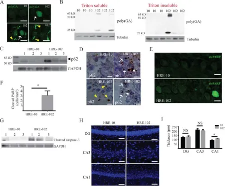
brains. As shown in Figure 2, we detected either a homogenous nuclear expression (arrow) or
an intense, discrete staining (arrowhead) within and outside of the nucleus (Figure 2A). These
poly-GA pathologies were most widely distributed in the cerebellum and brain stem, and
visible only in HRE-102 treated mice (poly-GA detected in 4 out of 6 mice), and absent in
HRE-10 expressing mice (poly-GA detected in 0 out of 6 mice). Analysis of triton-insoluble
and soluble cerebellar/brain stem lysates isolated from a different group of 10 and
HRE-102 mice identified several different sized poly-GA peptides ranging from 25-63 kilodaltons
in both triton-soluble and insoluble fractions solely in a subset of HRE-102 mice (Figure 2B).
Additionally, the expression of p62, a marker of cargo destined for autophagic degradation
(Bjorkoy et al., 2005, Komatsu et al., 2007) and c9RAN pathology (Mori et al., 2013a, Mori et
al., 2013b, Schludi et al., 2015), was enhanced in the cerebellum of HRE-102 mice (Figure
2C), and was detected in dense intracellular (arrow) and extracellular (arrowhead) aggregates
Next, we examined the expression of cleaved poly (ADP-ribose) polymerase (PARP) and
cleaved caspase-3 to see whether there was evidence of cells undergoing apoptosis. We focused
our analysis on the cerebellum, due to the heightened DPR and p62 expression we observed in
the HRE-102 mice (Figure 2A-D) that have been correlated to cell toxicity (Lee et al., 2016,
Lopez-Gonzalez et al., 2016, Tran et al., 2015). As shown in Figures 2E-G, we found that
cleaved PARP in cerebellar purkinje cells (Figures 2E-F) and cleaved caspase-3 expression
(Figure 2G) in the cerebellum were significantly increased in HRE-102 mice, indicating that
these areas were in the early stages of cell death. Interestingly, despite the presence of RNA
foci and c9RAN pathology, neither astrogliosis (Figure S1) nor neurodegeneration in the
cerebellum (Figure S2C-D) were observed. This absence of repeat-length dependent
neurodegeneration was also observed in other regions such as the cortex (Figure S2A-B), spinal
cord (Figure S2E-F) and hippocampus (Figure 2H-I), though there was a significant dispersal
in the CA1 (Figure 2H-I) that is characteristic of reeler mutant mice (Howell et al., 1997,
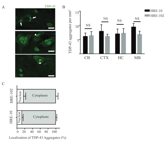
Trommsdorff et al., 1999). Additionally, although we were able to detect infrequent TDP-43
aggregates (Figure S3A-B) in the cerebellum, cortex, midbrain and hippocampus of HRE-10
and HRE-102, there was no significant difference between mouse groups with respect to
TDP-43 pathology. We also did not detect any differences between the groups with respect to
phosphorylated TDP-43 pathology (unpublished observations, JSC). In contrast to the largely
nuclear TDP-43 pathology reported by Chew et al (2015), the majority of TDP-43 aggregates
in both groups of AAV-HRE injected mice were cytoplasmic (Figure S3C).
HRE-102 mice develop disease-related neuromuscular junction (NMJ) pathology
In the absence of significant loss of neuronal cell bodies in the CNS in both mouse cohorts and
Frey et al., 2000, Murray et al., 2010, Bennett et al., 2014), we asked whether peripheral
neuromuscular junctions were altered in the HRE-102 expressing mice. To do this, muscles
from twelve-month old mice were doubly labelled with an anti-neurofilament antibody to label
nerve terminals and fluorescent bungarotoxin to label acetylcholine receptors (AChRs), and
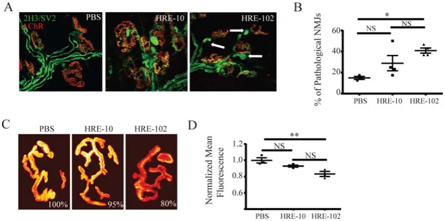
NMJs were imaged with a confocal microscope. A close inspection of NMJs revealed a
significant increase in pathological NMJs in HRE-102 mice, defined as having abnormal
pre-synaptic neurofilament accumulations and/or nerve terminal blebbings (Figure 3A,B),
indicating that the NMJ may be an early target of c9-related toxicity. It is well established that
the density of postsynaptic AChR is significantly reduced in mouse models of motor neurons
disease and myasthenia gravis (Losen et al., 2005, Kong et al., 2009). Here we asked whether
the density of AChR at NMJs of HRE-102 mice is also altered. AChRs were labeled with
BTX-594 and the density of AChR at NMJs of HRE-102, HRE-10 and PBS were compared (Figure
3C-D). The total fluorescence of AChRs (which measured fluorescence of a synapse per area
of the synapse) was decreased by 20% in muscles expressing HRE-102 compared with NMJs
of PBS injected mice. These results indicate that HRE-102 expression reduces the density of
AChRs at postsynaptic sites. Collectively, HRE-102 mice exhibit a loss in NMJ integrity at 12
months of age that is consistent with the early stages of motor neuron degeneration.
HRE-102 mice have progressive gait and behavioural deficits
To assess whether the c9RAN and NMJ pathology have any consequences on mouse
behaviour, we analysed motor and cognitive tasks. We used a Catwalk system to examine
different parameters measuring motor coordination and gait in ambulatory mice, and found that
HRE-102 mice had gait abnormalities, with significantly irregular gait patterns compared to
parameters (Figure 4A). When we assessed the hindlimb splay in the two cohorts (HRE-10 and
HRE-102), we found that there was a significant defect in the right hindlimb in the HRE-102
mice (Figure 4B). These deficits did not appear to affect the exploratory activity of the mice as
HRE-102 mice were not significantly different to HRE-10 mice in the open field at 12 months
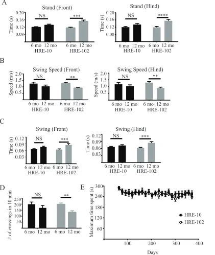
of age (Figure 4C). When we analyzed data between 6 and 12 months of age from several of
the same tasks, we noted that most of the gait parameters including ones for measuring stride
irregularities (stand, swing, and swing speed) in addition to activity in the open field showed
significant deterioration with age in the HRE-102 mice (Figure S4A-D), demonstrating that the
behavioural impairments in HRE-102 mice were progressive. There were, however, no
differences between groups in the accelerating rotarod over the ten months of testing (Figure
S4E). We next used the object recognition paradigm to determine whether the HRE-102 mice
had any cognitive deficits which could correlate with the CA1 dispersal demonstrated in Figure
2D. This behavioural task exploits the innate interest of mice to explore a novel object by
forcing mice to discriminate between a novel object and an object previously explored. Both
sets of mice explored two novel objects without any significant difference in the familiarization
phase (Figure 4D), but only HRE-102 mice were unable to discriminate between a novel object
and one they had previously explored, indicating the presence of a memory deficit.
Discussion
Current mouse models of C9orf72 ALS/FTD have supported a toxic gain of function
mechanism by which long G4C2 hexanucleotide RNA expansions (HREs) induce the formation
of intracellular RNA foci and RAN protein aggregates leading to toxicity and behavioural
dysfunction. Here we used AAVs to express either pure 10 HREs or 102 HREs containing
regularly spaced interruptions in mice, and demonstrate that while both lines of mice have
c9RAN pathology, exhibit NMJ pathology, hippocampal CA1 dispersion, early markers of
apoptosis, and display gait and cognitive deficits.
To date, four independent mouse models expressing either a full length or truncated human
C9orf72 with hundreds of HREs have been generated along with an AAV-mediated model
expressing a shorter (66) number of HREs. All of these mouse models have detectable RNA
foci and c9RAN aggregates in the brain, although the minimum size necessary to induce RNA
foci formation differs widely. Expression of a full length C9orf72 bacterial artificial
chromosome (BAC) transgene in mice was unable to induce RNA foci formation with 36-37
HREs (Liu et al., 2016, O'Rourke et al., 2015), and in mice expressing a truncated C9orf72,
expression of 100 G4C2 expansions yield limited RNA foci (Jiang et al., 2016). In contrast,
AAV mediated expression of 66 HREs with a constitutive actin promoter generated significant
ALS/FTD pathology which was absent when only 2 HREs were expressed (Chew et al., 2015).
We chose a higher minimum expansion length of 10 HREs to determine whether we could
cross the minimal threshold for generating RNA foci without forming DPRs. Surprisingly, our
mice expressing HRE-10 yield similar numbers of RNA foci in the CNS (Figure 1H) to our
mice expressing HRE-102 despite detecting DPRs only in the HRE-102 mice. We therefore
conclude that RNA foci density is not an accurate predictor of DPR formation.
The presence of poly-GA and p62 aggregates (Figures 2A-D) in our HRE-102 mice indicates
that the interruptions we introduced between every 10-17 GGGGCC repeats do not prevent
DPR formation. In contrast, interrupting AAV mediated poly-GA expression in mice with
proline residues (Zhang et al., 2016) or inserting stop codons between extended HREs
102 construct shifted the reading frame of the translated sequence without introducing a stop
codon resulting in the formation of mixed dipeptides as opposed to single extended tracts of
poly-GA, poly-GR, or poly-GP. We were unable to detect robust poly-GR or poly-GP
expression in our mice, although that is more likely due to the cross-reactivity of our antibody
to endogenous proteins in mouse tissue, as a cell culture model expressing an inducible version
of our HRE-102 construct detected poly-GA, poly-GP and poly-GR DPRs (Stopford et al.,
2017).
Interruptions in C9orf72 expansions have not been described yet, but interrupted repeat
expansions have been frequently detected in other neurological disorders including
spinocerebellar ataxia (SCA) (Chung et al., 1993), Fragile X syndrome (Eichler et al., 1994),
Myotonic dystrophy type I (Musova et al., 2009) and Friedreich ataxia (Stolle et al., 2008).
There is no consensus on the general effects of the interruptions on disease progression,
although there is strong evidence that interruptions can stabilize DNA sequences and prevent
the formation of slipped DNA structures in CAG and CGG repeat disorders (Eichler et al.,
1994, Pearson et al., 1998a, Pearson et al., 1998b, Menon et al., 2013). The more rapid
development of ALS/FTD phenotypes in the uninterrupted HRE-66 mice described by Chew
et al (2015) compared with our interrupted HRE-102 may reflect a stabilizing presence of our
interruptions on the expansion sequence.
In large GAA expansions in Friedreich ataxia, interruptions can stabilize the DNA sequence
(Pollard et al., 2004) without necessarily having any effect on the age of onset (Stolle et al.,
2008). In contrast, interruptions in the CTG expansions in Myotonic dystrophy type I delay
disease onset and suppress the muscular dystrophic phenotype (Musova et al., 2009),
premature ovarian failure (Bodega et al., 2006), and CAT insertions between CAG expansions
in the ataxin-1 gene prevent the development of the full SCA1 phenotype. There is also
evidence that interruptions can lead to the presentation of new phenotypes as disruptions in the
ATTCT repeats in SCA10 are associated with a higher incidence of seizures (Matsuura et al.,
2006) and CAG repeats that normally cause SCA2 can lead to parkinsonism when CAA triplets
interrupt the CAG expansion (Charles et al., 2007). As a result, it is difficult to draw
conclusions to the effects of the interruptions on disease progression in our AAV-HRE-102
mice, although it is notable that these mice develop some of the pathologies and behavioral
phenotypes consistent with ALS/FTD suggesting that it is less likely that the interruptions led
to a novel phenotype. It also seems unlikely that the interruptions in our HRE-102 mice disrupt
protein aggregation like the mice described by Zhang et al (2016) which harbor proline
interruptions between DPRs, as we detected clear poly-GA and p62 depositions in the tissue
(Figure 2A-D).
We did not observe extensive TDP-43 pathology in either the HRE-10 or HRE-102 mice and
the majority of TDP-43 depositions were cytoplasmic (Figure S3) which contrasts with the
nuclear TDP-43 pathology in the HRE-66 mice described by Chew et al (2015). The relative
paucity of TDP-43 aggregates in the nucleus of our HRE-102 mice may be due to the
interruptions disrupting the G-quadruplexes formed by pure G4C2 repeats and preventing the
reported association between the G-quadruplexes and mRNA export proteins SRSF1 (Reddy
et al., 2013, Zamiri et al., 2014) and hnRNPs (Conlon et al., 2016, Haeusler et al., 2014, Zamiri
et al., 2014). We can also not rule out the possibility that an uninterrupted RNA sequence
influences TDP-43 deposition due to increased toxicity. Future investigations on the effects of
As progressive deficits in spatial learning and memory and have been reported in C9orf72 BAC
transgenic mice expressing 450 repeats (Jiang et al., 2016), we carefully examined
hippocampal pathology in the AAV-mediated mouse models since the HRE-102 mice failed to
discriminate between familiar and novel objects, demonstrating the presence of a cognitive
deficit (Figure 4D). Previously, DPRs in the hippocampal CA1 of patients with C9-repeats
have been reported (Pletnikova et al., 2014), but while we were unable to detect DPRs
aggregates in the hippocampus, we did observe a dispersal of the CA1 (Figure 2H-I) in the
HRE-102 mice that was reminiscent of the aberrant neuronal migration phenotype of
Reeler/Disabled-1 mouse mutants that occurs during early development (Trommsdorff et al.,
1999, Howell et al., 1997). Interestingly knockdown of Reelin, the glycoprotein absent in
Reeler mutant mice, impairs rats in the object recognition task, while Reelin overexpression
can rescue deficits in the object recognition task in mouse models of Alzheimer's disease
(Brosda et al., 2011, Pujadas et al., 2014). Further analysis will be necessary to determine
whether any link between Reelin and C9orf72 exists.
In summary, these data support a repeat-expansion length dependent gain of toxicity, with
c9RAN formation being the likely driver of pathogenesis as the gait, motor and cognitive
deficits precede any major TDP-43 pathology. AAV mediated expression of 102 GGGGCC
repeats in mice leads to significant disease related pathology despite the presence of
interruptions leading to a mixture of translated DPRs. Our model provides an interesting
comparison to similar AAV-mediated models expressing pure repeat expansions, and may help
Acknowledgements
We thank Dieter Edbauer for the use of the poly-GA antibody and Joseph Scarrot for critical
reading of the manuscript. scAAV and pAAV2/9 plasmids were gifts from J. Gray and J.
Wilson, respectively. The authors have declared that no conflict of interest exists
Funding
This work was supported by the European Research Council grant (ERC Advanced Award no.
294745) to MA. M.A. is also supported by MRC DPFS Award (129016). S.H.M, K.E.A.L.,
J.C., P.M., I.C. and CW were supported by ERC Advanced Award (294745). CW is half funded
by the Wellcome Trust Investigator Award (103844) to S.F.E-K. P.J.S is supported as an NIHR
Senior Investigator and by the Motor Neurone Disease Association (grant Apr16/846-791) and
the Medical Research Council (MRC) (grant MR/M010864/1 in collaboration with Professor
Jun Xu (Projects of International Cooperation and Exchanges NSFC:81261130318) at Tongji
University in China). S.F.E-K is funded by a Wellcome Trust Investigator Award (103844), a
Lister Institute of Preventative Medicine Fellowship, and a European Union British Council
References
AKAABOUNE, M., CULICAN, S. M., TURNEY, S. G. & LICHTMAN, J. W. 1999. Rapid and reversible effects of activity on acetylcholine receptor density at the neuromuscular junction in vivo. Science, 286, 503-7.
BELZIL, V. V., BAUER, P. O., PRUDENCIO, M., GENDRON, T. F., STETLER, C. T., YAN, I. K., PREGENT, L., DAUGHRITY, L., BAKER, M. C., RADEMAKERS, R., BOYLAN, K., PATEL, T. C., DICKSON, D. W. & PETRUCELLI, L. 2013. Reduced C9orf72 gene expression in c9FTD/ALS is caused by histone trimethylation, an epigenetic event detectable in blood. Acta Neuropathol, 126, 895-905.
BENNETT, E. J., MEAD, R. J., AZZOUZ, M., SHAW, P. J. & GRIERSON, A. J. 2014. Early detection of motor dysfunction in the SOD1G93A mouse model of Amyotrophic Lateral Sclerosis (ALS) using home cage running wheels. PLoS One, 9, e107918.
BJORKOY, G., LAMARK, T., BRECH, A., OUTZEN, H., PERANDER, M., OVERVATN, A., STENMARK, H. & JOHANSEN, T. 2005. p62/SQSTM1 forms protein aggregates degraded by autophagy and has a protective effect on huntingtin-induced cell death. J Cell Biol, 171, 603-14.
BODEGA, B., BIONE, S., DALPRA, L., TONIOLO, D., ORNAGHI, F., VEGETTI, W., GINELLI, E. & MAROZZI, A. 2006. Influence of intermediate and uninterrupted FMR1 CGG expansions in premature ovarian failure manifestation. Hum Reprod, 21, 952-7.
BROSDA, J., DIETZ, F. & KOCH, M. 2011. Impairment of cognitive performance after reelin knockdown in the medial prefrontal cortex of pubertal or adult rats. Neurobiol Dis, 44, 239-47.
CHARLES, P., CAMUZAT, A., BENAMMAR, N., SELLAL, F., DESTEE, A., BONNET, A. M., LESAGE, S., LE BER, I., STEVANIN, G., DURR, A., BRICE, A. & FRENCH PARKINSON'S DISEASE GENETIC STUDY, G. 2007. Are interrupted SCA2 CAG repeat expansions responsible for parkinsonism? Neurology, 69, 1970-5.
CHEW, J., GENDRON, T. F., PRUDENCIO, M., SASAGURI, H., ZHANG, Y. J.,
CASTANEDES-CASEY, M., LEE, C. W., JANSEN-WEST, K., KURTI, A., MURRAY, M. E., BIENIEK, K. F., BAUER, P. O., WHITELAW, E. C., ROUSSEAU, L., STANKOWSKI, J. N., STETLER, C., DAUGHRITY, L. M., PERKERSON, E. A., DESARO, P.,
JOHNSTON, A., OVERSTREET, K., EDBAUER, D., RADEMAKERS, R., BOYLAN, K. B., DICKSON, D. W., FRYER, J. D. & PETRUCELLI, L. 2015. Neurodegeneration.
CHUNG, M. Y., RANUM, L. P., DUVICK, L. A., SERVADIO, A., ZOGHBI, H. Y. & ORR, H. T. 1993. Evidence for a mechanism predisposing to intergenerational CAG repeat instability in spinocerebellar ataxia type I. Nat Genet, 5, 254-8.
CONLON, E. G., LU, L., SHARMA, A., YAMAZAKI, T., TANG, T., SHNEIDER, N. A. & MANLEY, J. L. 2016. The C9ORF72 GGGGCC expansion forms RNA G-quadruplex inclusions and sequesters hnRNP H to disrupt splicing in ALS brains. Elife, 5.
COOPER-KNOCK, J., WALSH, M. J., HIGGINBOTTOM, A., ROBIN HIGHLEY, J., DICKMAN, M. J., EDBAUER, D., INCE, P. G., WHARTON, S. B., WILSON, S. A., KIRBY, J., HAUTBERGUE, G. M. & SHAW, P. J. 2014. Sequestration of multiple RNA recognition motif-containing proteins by C9orf72 repeat expansions. Brain, 137, 2040-51.
DEJESUS-HERNANDEZ, M., MACKENZIE, I. R., BOEVE, B. F., BOXER, A. L., BAKER, M., RUTHERFORD, N. J., NICHOLSON, A. M., FINCH, N. A., FLYNN, H., ADAMSON, J., KOURI, N., WOJTAS, A., SENGDY, P., HSIUNG, G. Y., KARYDAS, A., SEELEY, W. W., JOSEPHS, K. A., COPPOLA, G., GESCHWIND, D. H., WSZOLEK, Z. K., FELDMAN, H., KNOPMAN, D. S., PETERSEN, R. C., MILLER, B. L., DICKSON, D. W., BOYLAN, K. B., GRAFF-RADFORD, N. R. & RADEMAKERS, R. 2011. Expanded GGGGCC hexanucleotide repeat in noncoding region of C9ORF72 causes chromosome 9p-linked FTD and ALS. Neuron, 72, 245-56.
DONNELLY, C. J., ZHANG, P. W., PHAM, J. T., HAEUSLER, A. R., MISTRY, N. A., VIDENSKY, S., DALEY, E. L., POTH, E. M., HOOVER, B., FINES, D. M.,
MARAGAKIS, N., TIENARI, P. J., PETRUCELLI, L., TRAYNOR, B. J., WANG, J., RIGO, F., BENNETT, C. F., BLACKSHAW, S., SATTLER, R. & ROTHSTEIN, J. D. 2013. RNA toxicity from the ALS/FTD C9ORF72 expansion is mitigated by antisense intervention. Neuron, 80, 415-28.
EICHLER, E. E., HOLDEN, J. J., POPOVICH, B. W., REISS, A. L., SNOW, K.,
THIBODEAU, S. N., RICHARDS, C. S., WARD, P. A. & NELSON, D. L. 1994. Length of uninterrupted CGG repeats determines instability in the FMR1 gene. Nat Genet, 8, 88-94.
FREIBAUM, B. D., LU, Y., LOPEZ-GONZALEZ, R., KIM, N. C., ALMEIDA, S., LEE, K. H., BADDERS, N., VALENTINE, M., MILLER, B. L., WONG, P. C., PETRUCELLI, L., KIM, H. J., GAO, F. B. & TAYLOR, J. P. 2015. GGGGCC repeat expansion in C9orf72 compromises nucleocytoplasmic transport. Nature, 525, 129-33.
FREY, D., SCHNEIDER, C., XU, L., BORG, J., SPOOREN, W. & CARONI, P. 2000. Early and selective loss of neuromuscular synapse subtypes with low sprouting competence in motoneuron diseases. J Neurosci, 20, 2534-42.
HOWELL, B. W., HAWKES, R., SORIANO, P. & COOPER, J. A. 1997. Neuronal position in the developing brain is regulated by mouse disabled-1. Nature, 389, 733-7.
IGUCHI, Y., KATSUNO, M., NIWA, J., TAKAGI, S., ISHIGAKI, S., IKENAKA, K., KAWAI, K., WATANABE, H., YAMANAKA, K., TAKAHASHI, R., MISAWA, H., SASAKI, S., TANAKA, F. & SOBUE, G. 2013. Loss of TDP-43 causes age-dependent progressive motor neuron degeneration. Brain, 136, 1371-82.
JIANG, J., ZHU, Q., GENDRON, T. F., SABERI, S., MCALONIS-DOWNES, M., SEELMAN, A., STAUFFER, J. E., JAFAR-NEJAD, P., DRENNER, K., SCHULTE, D., CHUN, S., SUN, S., LING, S. C., MYERS, B., ENGELHARDT, J., KATZ, M., BAUGHN, M., PLATOSHYN, O., MARSALA, M., WATT, A., HEYSER, C. J., ARD, M. C., DE MUYNCK, L., DAUGHRITY, L. M., SWING, D. A., TESSAROLLO, L., JUNG, C. J., DELPOUX, A., UTZSCHNEIDER, D. T., HEDRICK, S. M., DE JONG, P. J., EDBAUER, D., VAN DAMME, P., PETRUCELLI, L., SHAW, C. E., BENNETT, C. F., DA CRUZ, S., RAVITS, J., RIGO, F., CLEVELAND, D. W. & LAGIER-TOURENNE, C. 2016. Gain of Toxicity from ALS/FTD-Linked Repeat Expansions in C9ORF72 Is Alleviated by Antisense Oligonucleotides Targeting GGGGCC-Containing RNAs. Neuron, 90, 535-50.
KOMATSU, M., WAGURI, S., KOIKE, M., SOU, Y. S., UENO, T., HARA, T.,
MIZUSHIMA, N., IWATA, J., EZAKI, J., MURATA, S., HAMAZAKI, J., NISHITO, Y., IEMURA, S., NATSUME, T., YANAGAWA, T., UWAYAMA, J., WARABI, E.,
YOSHIDA, H., ISHII, T., KOBAYASHI, A., YAMAMOTO, M., YUE, Z., UCHIYAMA, Y., KOMINAMI, E. & TANAKA, K. 2007. Homeostatic levels of p62 control cytoplasmic inclusion body formation in autophagy-deficient mice. Cell, 131, 1149-63.
KONG, L., WANG, X., CHOE, D. W., POLLEY, M., BURNETT, B. G., BOSCH-MARCÉ, M., GRIFFIN, J. W., RICH, M. M. & SUMNER, C. J. 2009. Impaired synaptic vesicle release and immaturity of neuromuscular junctions in spinal muscular atrophy mice. J Neurosci, 29, 842-51.
KOPPERS, M., BLOKHUIS, A. M., WESTENENG, H. J., TERPSTRA, M. L., ZUNDEL, C. A., VIEIRA DE SA, R., SCHELLEVIS, R. D., WAITE, A. J., BLAKE, D. J., VELDINK, J. H., VAN DEN BERG, L. H. & PASTERKAMP, R. J. 2015. C9orf72 ablation in mice does not cause motor neuron degeneration or motor deficits. Ann Neurol, 78, 426-38.
LEE, K. H., ZHANG, P., KIM, H. J., MITREA, D. M., SARKAR, M., FREIBAUM, B. D., CIKA, J., COUGHLIN, M., MESSING, J., MOLLIEX, A., MAXWELL, B. A., KIM, N. C., TEMIROV, J., MOORE, J., KOLAITIS, R. M., SHAW, T. I., BAI, B., PENG, J.,
KRIWACKI, R. W. & TAYLOR, J. P. 2016. C9orf72 Dipeptide Repeats Impair the
Assembly, Dynamics, and Function of Membrane-Less Organelles. Cell, 167, 774-788 e17.
LIU, Y., PATTAMATTA, A., ZU, T., REID, T., BARDHI, O., BORCHELT, D. R.,
YACHNIS, A. T. & RANUM, L. P. 2016. C9orf72 BAC Mouse Model with Motor Deficits and Neurodegenerative Features of ALS/FTD. Neuron, 90, 521-34.
LOPEZ-GONZALEZ, R., LU, Y., GENDRON, T. F., KARYDAS, A., TRAN, H., YANG, D., PETRUCELLI, L., MILLER, B. L., ALMEIDA, S. & GAO, F. B. 2016. Poly(GR) in C9ORF72-Related ALS/FTD Compromises Mitochondrial Function and Increases Oxidative Stress and DNA Damage in iPSC-Derived Motor Neurons. Neuron, 92, 383-391.
LOSEN, M., STASSEN, M. H., MARTÍNEZ-MARTÍNEZ, P., MACHIELS, B. M., DUIMEL, H., FREDERIK, P., VELDMAN, H., WOKKE, J. H., SPAANS, F., VINCENT, A. & DE BAETS, M. H. 2005. Increased expression of rapsyn in muscles prevents
acetylcholine receptor loss in experimental autoimmune myasthenia gravis. Brain, 128, 2327-37.
LUKASHCHUK, V., LEWIS, K. E., COLDICOTT, I., GRIERSON, A. J. & AZZOUZ, M. 2016. AAV9-mediated central nervous system-targeted gene delivery via cisterna magna route in mice. Mol Ther Methods Clin Dev, 3, 15055.
MACKENZIE, I. R., ARZBERGER, T., KREMMER, E., TROOST, D., LORENZL, S., MORI, K., WENG, S. M., HAASS, C., KRETZSCHMAR, H. A., EDBAUER, D. & NEUMANN, M. 2013. Dipeptide repeat protein pathology in C9ORF72 mutation cases: clinico-pathological correlations. Acta Neuropathol, 126, 859-79.
MAJOUNIE, E., RENTON, A. E., MOK, K., DOPPER, E. G., WAITE, A., ROLLINSON, S., CHIO, A., RESTAGNO, G., NICOLAOU, N., SIMON-SANCHEZ, J., VAN SWIETEN, J. C., ABRAMZON, Y., JOHNSON, J. O., SENDTNER, M., PAMPHLETT, R., ORRELL, R. W., MEAD, S., SIDLE, K. C., HOULDEN, H., ROHRER, J. D., MORRISON, K. E., PALL, H., TALBOT, K., ANSORGE, O., CHROMOSOME, A. L. S. F. T. D. C., FRENCH RESEARCH NETWORK ON, F. F. A., CONSORTIUM, I., HERNANDEZ, D. G.,
AREPALLI, S., SABATELLI, M., MORA, G., CORBO, M., GIANNINI, F., CALVO, A., ENGLUND, E., BORGHERO, G., FLORIS, G. L., REMES, A. M., LAAKSOVIRTA, H., MCCLUSKEY, L., TROJANOWSKI, J. Q., VAN DEERLIN, V. M., SCHELLENBERG, G. D., NALLS, M. A., DRORY, V. E., LU, C. S., YEH, T. H., ISHIURA, H., TAKAHASHI, Y., TSUJI, S., LE BER, I., BRICE, A., DREPPER, C., WILLIAMS, N., KIRBY, J., SHAW, P., HARDY, J., TIENARI, P. J., HEUTINK, P., MORRIS, H. R., PICKERING-BROWN, S. & TRAYNOR, B. J. 2012. Frequency of the C9orf72 hexanucleotide repeat expansion in patients with amyotrophic lateral sclerosis and frontotemporal dementia: a cross-sectional study. Lancet Neurol, 11, 323-30.
MARTINEZ-PENA, Y. V. I., AITTALEB, M., CHEN, P. J. & AKAABOUNE, M. 2015. The knockdown of alphakap alters the postsynaptic apparatus of neuromuscular junctions in living mice. J Neurosci, 35, 5118-27.
MCCARTY, D. M., MONAHAN, P. E. & SAMULSKI, R. J. 2001. Self-complementary recombinant adeno-associated virus (scAAV) vectors promote efficient transduction independently of DNA synthesis. Gene Ther, 8, 1248-54.
MEAD, R. J., BENNETT, E. J., KENNERLEY, A. J., SHARP, P., SUNYACH, C., KASHER, P., BERWICK, J., PETTMANN, B., BATTAGLIA, G., AZZOUZ, M., GRIERSON, A. & SHAW, P. J. 2011. Optimised and rapid pre-clinical screening in the SOD1(G93A) transgenic mouse model of amyotrophic lateral sclerosis (ALS). PLoS One, 6, e23244.
MENON, R. P., NETHISINGHE, S., FAGGIANO, S., VANNOCCI, T., REZAEI, H., PEMBLE, S., SWEENEY, M. G., WOOD, N. W., DAVIS, M. B., PASTORE, A. &
GIUNTI, P. 2013. The role of interruptions in polyQ in the pathology of SCA1. PLoS Genet, 9, e1003648.
MIZIELINSKA, S., GRONKE, S., NICCOLI, T., RIDLER, C. E., CLAYTON, E. L., DEVOY, A., MOENS, T., NORONA, F. E., WOOLLACOTT, I. O., PIETRZYK, J.,
CLEVERLEY, K., NICOLL, A. J., PICKERING-BROWN, S., DOLS, J., CABECINHA, M., HENDRICH, O., FRATTA, P., FISHER, E. M., PARTRIDGE, L. & ISAACS, A. M. 2014. C9orf72 repeat expansions cause neurodegeneration in Drosophila through arginine-rich proteins. Science, 345, 1192-4.
MORI, K., ARZBERGER, T., GRASSER, F. A., GIJSELINCK, I., MAY, S., RENTZSCH, K., WENG, S. M., SCHLUDI, M. H., VAN DER ZEE, J., CRUTS, M., VAN
BROECKHOVEN, C., KREMMER, E., KRETZSCHMAR, H. A., HAASS, C. & EDBAUER, D. 2013a. Bidirectional transcripts of the expanded C9orf72 hexanucleotide repeat are translated into aggregating dipeptide repeat proteins. Acta Neuropathol, 126, 881-93.
MORI, K., WENG, S. M., ARZBERGER, T., MAY, S., RENTZSCH, K., KREMMER, E., SCHMID, B., KRETZSCHMAR, H. A., CRUTS, M., VAN BROECKHOVEN, C., HAASS, C. & EDBAUER, D. 2013b. The C9orf72 GGGGCC repeat is translated into aggregating dipeptide-repeat proteins in FTLD/ALS. Science, 339, 1335-8.
MURRAY, L. M., TALBOT, K. & GILLINGWATER, T. H. 2010. Review: neuromuscular synaptic vulnerability in motor neurone disease: amyotrophic lateral sclerosis and spinal muscular atrophy. Neuropathol Appl Neurobiol, 36, 133-56.
MUSOVA, Z., MAZANEC, R., KREPELOVA, A., EHLER, E., VALES, J., JAKLOVA, R., PROCHAZKA, T., KOUKAL, P., MARIKOVA, T., KRAUS, J., HAVLOVICOVA, M. & SEDLACEK, Z. 2009. Highly unstable sequence interruptions of the CTG repeat in the myotonic dystrophy gene. Am J Med Genet A, 149A, 1365-74.
O'ROURKE, J. G., BOGDANIK, L., MUHAMMAD, A. K., GENDRON, T. F., KIM, K. J., AUSTIN, A., CADY, J., LIU, E. Y., ZARROW, J., GRANT, S., HO, R., BELL, S.,
C9orf72 BAC Transgenic Mice Display Typical Pathologic Features of ALS/FTD. Neuron, 88, 892-901.
O'ROURKE, J. G., BOGDANIK, L., YANEZ, A., LALL, D., WOLF, A. J., MUHAMMAD, A. K., HO, R., CARMONA, S., VIT, J. P., ZARROW, J., KIM, K. J., BELL, S., HARMS, M. B., MILLER, T. M., DANGLER, C. A., UNDERHILL, D. M., GOODRIDGE, H. S., LUTZ, C. M. & BALOH, R. H. 2016. C9orf72 is required for proper macrophage and microglial function in mice. Science, 351, 1324-9.
PEARSON, C. E., EICHLER, E. E., LORENZETTI, D., KRAMER, S. F., ZOGHBI, H. Y., NELSON, D. L. & SINDEN, R. R. 1998a. Interruptions in the triplet repeats of SCA1 and FRAXA reduce the propensity and complexity of slipped strand DNA (S-DNA) formation. Biochemistry, 37, 2701-8.
PEARSON, C. E., WANG, Y. H., GRIFFITH, J. D. & SINDEN, R. R. 1998b. Structural analysis of slipped-strand DNA (S-DNA) formed in (CTG)n. (CAG)n repeats from the myotonic dystrophy locus. Nucleic Acids Res, 26, 816-23.
PETERS, O. M., CABRERA, G. T., TRAN, H., GENDRON, T. F., MCKEON, J. E., METTERVILLE, J., WEISS, A., WIGHTMAN, N., SALAMEH, J., KIM, J., SUN, H., BOYLAN, K. B., DICKSON, D., KENNEDY, Z., LIN, Z., ZHANG, Y. J., DAUGHRITY, L., JUNG, C., GAO, F. B., SAPP, P. C., HORVITZ, H. R., BOSCO, D. A., BROWN, S. P., DE JONG, P., PETRUCELLI, L., MUELLER, C. & BROWN, R. H., JR. 2015. Human C9ORF72 Hexanucleotide Expansion Reproduces RNA Foci and Dipeptide Repeat Proteins but Not Neurodegeneration in BAC Transgenic Mice. Neuron, 88, 902-9.
PLETNIKOVA, O., SLOANE, K. L., RENTON, A. E., TRAYNOR, B. J., CRAIN, B. J., REID, T., ZU, T., RANUM, L. P., TRONCOSO, J. C., RABINS, P. V. & ONYIKE, C. U. 2014. Hippocampal sclerosis dementia with the C9ORF72 hexanucleotide repeat expansion. Neurobiol Aging, 35, 2419.e17-21.
POLLARD, L. M., SHARMA, R., GOMEZ, M., SHAH, S., DELATYCKI, M. B., PIANESE, L., MONTICELLI, A., KEATS, B. J. & BIDICHANDANI, S. I. 2004. Replication-mediated instability of the GAA triplet repeat mutation in Friedreich ataxia. Nucleic Acids Res, 32, 5962-71.
POWIS, R. A., KARYKA, E., BOYD, P., COME, J., JONES, R. A., ZHENG, Y., SZUNYOGOVA, E., GROEN, E. J., HUNTER, G., THOMSON, D., WISHART, T. M., BECKER, C. G., PARSON, S. H., MARTINAT, C., AZZOUZ, M. & GILLINGWATER, T. H. 2016. Systemic restoration of UBA1 ameliorates disease in spinal muscular atrophy. JCI Insight, 1, e87908.
REDDY, K., ZAMIRI, B., STANLEY, S. Y., MACGREGOR, R. B., JR. & PEARSON, C. E. 2013. The disease-associated r(GGGGCC)n repeat from the C9orf72 gene forms tract length-dependent uni- and multimolecular RNA G-quadruplex structures. J Biol Chem, 288, 9860-6.
RENTON, A. E., CHIO, A. & TRAYNOR, B. J. 2014. State of play in amyotrophic lateral sclerosis genetics. Nat Neurosci, 17, 17-23.
RENTON, A. E., MAJOUNIE, E., WAITE, A., SIMON-SANCHEZ, J., ROLLINSON, S., GIBBS, J. R., SCHYMICK, J. C., LAAKSOVIRTA, H., VAN SWIETEN, J. C.,
MYLLYKANGAS, L., KALIMO, H., PAETAU, A., ABRAMZON, Y., REMES, A. M., KAGANOVICH, A., SCHOLZ, S. W., DUCKWORTH, J., DING, J., HARMER, D. W., HERNANDEZ, D. G., JOHNSON, J. O., MOK, K., RYTEN, M., TRABZUNI, D.,
GUERREIRO, R. J., ORRELL, R. W., NEAL, J., MURRAY, A., PEARSON, J., JANSEN, I. E., SONDERVAN, D., SEELAAR, H., BLAKE, D., YOUNG, K., HALLIWELL, N.,
CALLISTER, J. B., TOULSON, G., RICHARDSON, A., GERHARD, A., SNOWDEN, J., MANN, D., NEARY, D., NALLS, M. A., PEURALINNA, T., JANSSON, L., ISOVIITA, V. M., KAIVORINNE, A. L., HOLTTA-VUORI, M., IKONEN, E., SULKAVA, R.,
BENATAR, M., WUU, J., CHIO, A., RESTAGNO, G., BORGHERO, G., SABATELLI, M., CONSORTIUM, I., HECKERMAN, D., ROGAEVA, E., ZINMAN, L., ROTHSTEIN, J. D., SENDTNER, M., DREPPER, C., EICHLER, E. E., ALKAN, C., ABDULLAEV, Z., PACK, S. D., DUTRA, A., PAK, E., HARDY, J., SINGLETON, A., WILLIAMS, N. M.,
HEUTINK, P., PICKERING-BROWN, S., MORRIS, H. R., TIENARI, P. J. & TRAYNOR, B. J. 2011. A hexanucleotide repeat expansion in C9ORF72 is the cause of chromosome 9p21-linked ALS-FTD. Neuron, 72, 257-68.
SCHLUDI, M. H., MAY, S., GRASSER, F. A., RENTZSCH, K., KREMMER, E., KUPPER, C., KLOPSTOCK, T., GERMAN CONSORTIUM FOR FRONTOTEMPORAL LOBAR, D., BAVARIAN BRAIN BANKING, A., ARZBERGER, T. & EDBAUER, D. 2015.
Distribution of dipeptide repeat proteins in cellular models and C9orf72 mutation cases suggests link to transcriptional silencing. Acta Neuropathol, 130, 537-55.
STOLLE, C. A., FRACKELTON, E. C., MCCALLUM, J., FARMER, J. M., TSOU, A., WILSON, R. B. & LYNCH, D. R. 2008. Novel, complex interruptions of the GAA repeat in small, expanded alleles of two affected siblings with late-onset Friedreich ataxia. Mov Disord, 23, 1303-6.
STOPFORD, M. J., HIGGINBOTTOM, A., HAUTBERGUE, G. M., COOPER-KNOCK, J., MULCAHY, P. J., DE VOS, K. J., RENTON, A. E., PLINER, H., CALVO, A., CHIO, A., TRAYNOR, B. J., AZZOUZ, M., HEATH, P. R., ITALSGEN CONSORTIUM, N. C., KIRBY, J. & SHAW, P. J. 2017. C9ORF72 hexanucleotide repeat exerts toxicity in a stable, inducible motor neuronal cell model, which is rescued by partial depletion of Pten. Hum Mol Genet, 26, 1133-1145.
TROMMSDORFF, M., GOTTHARDT, M., HIESBERGER, T., SHELTON, J.,
STOCKINGER, W., NIMPF, J., HAMMER, R. E., RICHARDSON, J. A. & HERZ, J. 1999. Reeler/Disabled-like disruption of neuronal migration in knockout mice lacking the VLDL receptor and ApoE receptor 2. Cell, 97, 689-701.
TURNEY, S. G., CULICAN, S. M. & LICHTMAN, J. W. 1996. A quantitative fluorescence-imaging technique for studying acetylcholine receptor turnover at neuromuscular junctions in living animals. J Neurosci Methods, 64, 199-208.
WISHART, T. M., MUTSAERS, C. A., RIESSLAND, M., REIMER, M. M., HUNTER, G., HANNAM, M. L., EATON, S. L., FULLER, H. R., ROCHE, S. L., SOMERS, E., MORSE, R., YOUNG, P. J., LAMONT, D. J., HAMMERSCHMIDT, M., JOSHI, A., HOHENSTEIN, P., MORRIS, G. E., PARSON, S. H., SKEHEL, P. A., BECKER, T., ROBINSON, I. M., BECKER, C. G., WIRTH, B. & GILLINGWATER, T. H. 2014. Dysregulation of ubiquitin homeostasis and beta-catenin signaling promote spinal muscular atrophy. J Clin Invest, 124, 1821-34.
ZAMIRI, B., REDDY, K., MACGREGOR, R. B., JR. & PEARSON, C. E. 2014. TMPyP4 porphyrin distorts RNA G-quadruplex structures of the disease-associated r(GGGGCC)n repeat of the C9orf72 gene and blocks interaction of RNA-binding proteins. J Biol Chem, 289, 4653-9.
ZHANG, K., DONNELLY, C. J., HAEUSLER, A. R., GRIMA, J. C., MACHAMER, J. B., STEINWALD, P., DALEY, E. L., MILLER, S. J., CUNNINGHAM, K. M., VIDENSKY, S., GUPTA, S., THOMAS, M. A., HONG, I., CHIU, S. L., HUGANIR, R. L., OSTROW, L. W., MATUNIS, M. J., WANG, J., SATTLER, R., LLOYD, T. E. & ROTHSTEIN, J. D. 2015. The C9orf72 repeat expansion disrupts nucleocytoplasmic transport. Nature, 525, 56-61.
ZHANG, Y. J., GENDRON, T. F., GRIMA, J. C., SASAGURI, H., JANSEN-WEST, K., XU, Y. F., KATZMAN, R. B., GASS, J., MURRAY, M. E., SHINOHARA, M., LIN, W. L., GARRETT, A., STANKOWSKI, J. N., DAUGHRITY, L., TONG, J., PERKERSON, E. A., YUE, M., CHEW, J., CASTANEDES-CASEY, M., KURTI, A., WANG, Z. S., LIESINGER, A. M., BAKER, J. D., JIANG, J., LAGIER-TOURENNE, C., EDBAUER, D.,
CLEVELAND, D. W., RADEMAKERS, R., BOYLAN, K. B., BU, G., LINK, C. D.,
Figures
Figure 1. AAV9-HREs can generate RNA foci in the mouse CNS. (A) Two different HREs were designed and subcloned into AAV9 vectors with the (G4C2)102 expansions constructed by a mixture of (G4C2)10-17 oligonucleotides with an intervening linker sequence. (B-G) RNA fluorescent in situ hybridization was used to detect RNA foci, which were abundant in the (B)
hippocampus (HC), (C) cortex (CTX), (D) cerebellum (CB) and sparse in the (E) spinal cord
(SC, *motor neuron). The majority of the foci were intranuclear (D-E, white arrowhead),
(H) There were no significant differences in the number of foci detected per mm2 in either the HC, CTX, or CB between HRE-10 and HRE-102 mice as assessed using an unpaired two-tailed
Student’s t-test for each region. Scale bar is 10µm. Nuclei counterstained with Hoechst 33342
Figure 2. Dipeptide protein repeat (DPR) pathology, hippocampal CA1 dispersal, and upregulation of cell death markers are observed in HRE-102 mice. (A) Cytoplasmic and nuclear DPRs adopt several morphologies in a subset of HRE-102 mice using a poly-(GA)
antibody in cerebellar tissue. (B) Immunoblots of cerebellar lysates show that both triton
soluble and triton insoluble proteins contain GA-rich DPRs in HRE-102 mice, and were never
observed in HRE-10 mice. (C) HRE-102 mice show an increased p62 expression compared to
HRE-10 mice in cerebellar tissue; arrowhead shows 60 kD isoform that was consistently absent
in HRE-10 brains. (D) p62 inclusions in the cerebellum are evident in both the nucleus (white
arrow) and cytoplasm (yellow arrowhead) of HRE-102 mice, and are absent in HRE-10 mice.
of early apoptosis reflected by cleaved PARP immunoreactivity (n=4 for both, p=0.046). (G)
Enhanced cleaved-caspase-3 expression is observed in cerebellar lysates from HRE-102 mice.
(H-I) There were no differences in the number of neurons as determined by NeuN staining in
the dentate gyrus (DG, 10 (n=4), 100 (n=6); p=0.66) or CA3 (10 (n=4),
HRE-100 (n=6); p=0.69) in the hippocampus between HRE-10 and HRE-102, but there was a
significant dispersal of CA1 neurons in HRE-102 mice (HRE-10 (n=4), HRE-100 (n=6);
p=0.046). Nuclei in (H) counterstained with Hoechst 33342 and stained blue. All comparisons
were made with an unpaired two-tailed Student's t-test with Welch’s correction; mean ± SEM.
Figure 3. Neuromuscular junction (NMJ) defects are present in the HRE-102 mice. (A-B) Abnormal neurofilament accumulation and nerve terminal blebbings (arrows) were prominent
in HRE-102 mice (PBS:n=3, HRE-10:n=4, HRE-102:n=4; n=individual muscles per genotype;
(F(2,8)=6.47, *p=0.02, post-hoc Tukey: HRE-10 vs PBS (p=0.19); HRE-10 vs HRE-102
(p=0.23); HRE-102 vs PBS (p=0.02)); scale bar 30 µm. (C-D) HRE-102 mice have a significant
decrease in acetylcholine receptor density compared to PBS injected controls (F(2,7)=10.79,
**p=0.007, post-hoc Tukey: 10 vs PBS (p=0.16); 10 vs 102 (p=0.056);
HRE-102 vs PBS (p=0.006)). The fluorescence intensity (total fluorescence of that synapse divided
by the area of that given synapse) of a saturating dose of α-bungarotoxin-alexa fluor 594 was
measured and normalized to PBS controls. All comparisons were made with a one-way
Figure 4. Gait and cognitive deficits observed in HRE-102 mice are absent in HRE-10 mice in 12 month old mice. (A) Gait abnormalities such as the front and hind stand index, the hind base of support (BOS), the diagonal support and the three-legged support were observed
using a Catwalk system in HRE-102 mice (HRE-10 (n=11), HRE-100 (n=13) for each; front
stand:*p=0.04, hind stand:*p=0.04; front BOS:p=0.19, hind BOS:*p=0.02; diagonal
support:*p=0.01, three-legged support:**p=0.006). (B) HRE-102 mice (n=13) had a moderate
splay in the right (*p=0.04) but not left (p=0.59) hindlimb compared to HRE-10 mice (n=11).
(C) There were no significant differences in activity (p=0.15) between HRE-10 (n=11) and
HRE-102 (n=13) in the open field. (D) An object recognition test identified a cognitive deficit
in HRE-102 mice (n=13) as they failed to discriminate in the testing phase (HRE-102:
familiarization phase: p=0.43, testing phase: p=0.09) between a novel object and an object
previously explored in the familiarization phase unlike HRE-10 mice (n=11, familiarization
phase:p=0.57, testing phase:*p=0.02). All comparisons were made with an unpaired two-tailed
10
102-r epeat
0 50 100 150 200
NS 200
150
100
50
GF
AP
intensity units per cell
[image:39.648.153.412.243.417.2]HRE-10 HRE-102
Figure S1. No differences in astrogliosis between HRE-10 and HRE-102 mice. GFAP immunoreactivity in twelve-month cerebellar tissue was similar between HRE-10 and HRE-102 mice (n=3 for each; p=0.79, unpaired two-tailed Student's t-test; mean ± SEM).
Disease Models & Mechanisms 10: doi:10.1242/dmm.029892: Supplementary information
10-re p 102-r ep 100 150 200 250 300
(G4C 210)
(G4C 2)10
2 40 45 50 55 0 250 500 750 1000 1250 NS NeuN
+ cells /µm
( 0 15 30 45 60 NS
C
A
Calbindin+ cells /µm
B
D
HRE-10
HRE-10 HRE-102
HRE-10 HRE-102
HRE-10
HRE-1022
HRE-102
NeuN NeuN
Calbindin Calbindin
MNs per section
10 102-r epeat 0 5 10 15 20
[image:40.648.55.535.91.602.2]HRE-10 HRE-102 NS
E
F
HRE-102 HRE-10 55 50 45 40 20 15 10 05 300 250 200 150 100Figure S2. No evidence of loss of neuronal cell bodies in the cortex or cerebellum of HRE-102 mice.
(A) NeuN immunoreactivity in the cortex in twelve-month cortical tissue was not significantly different (B) between repeat expansion mice (n=5 for each; p=0.15, unpaired two-tailed Student's t-test with Welch’s
correction; mean ± SEM). (C) Calbindin immunoreactivity, used to label Purkinje cells in the cerebellum, was not significantly different (D) between HRE-10 and HRE-102 mice (n=5 for each; p=0.37, unpaired two-tailed Student's t-test with Welch’s correction; mean ± SEM). (E) Nissl staining of spinal cord sections show that there are no significant differences (F) between HRE-10 and HRE-102 in the number of motor neurons in the spinal cord (n=3 for each;
p=0.87, unpaired two-tailed Student's t-test with Welch’s correction; mean ± SEM). Scale bar for (A): 10µm, (C): 20µm, (E): 20 µm.
Disease Models & Mechanisms 10: doi:10.1242/dmm.029892: Supplementary information
0 20 40 60 80 100 10 102 0 102-r epea t 10 102-r epea t 10 1 2-r epea t 10 1 2-r epea t 0 5 10 15 NS
TDP-43 aggregates per
mm
2
NS NS NS
15
10
5
CB CTX HC MB
0 20 40 60 80 100 Localization of TDP-43 Aggregates (%)
[image:41.648.40.598.107.596.2]A
B
PC
HRE-102 HRE-10 HRE-10 HRE-102 TDP-43 Cytoplasm Cytoplasm Nuclear NuclearFigure S3. No differences in presence of TDP-43 aggregates between HRE-10 and HRE-102 mice. (A) Infrequent TDP-43 aggregates were identified in the nucleus (arrowhead) and cytoplasm (arrow) of brain tissue in (B) multiple regions, though there were no significant differences detected between the HRE-10 and HRE-102 mice in either the cerebellum (CB, n=3 for each; p=0.89), cortex (CTX, n=6 for each; p=0.48), hippocampus (HC, n=6 for each; p=0.93), or midbrain (MB, n=6 for each; p=0.15). (C) The majority of TDP-43 aggregates in both sets of mice were found in the cytoplasm (HRE-10:n=6, HRE-102:n=5; p=0.87) with a minority in the nucleus (HRE-10: n=6, HRE-102: n=5; p=0.52). An unpaired two-tailed Student's t-test was used to assess data between the two mouse groups for all comparisons. Scale bar for (A): 10µm.
Disease Models & Mechanisms 10: doi:10.1242/dmm.029892: Supplementary information