0022-538X/93/031620-07$02.00/0
Copyright X) 1993,AmericanSocietyforMicrobiology
Deletion
Analysis
of the
Capsid
Protein of Sindbis
Virus:
Identification of the
RNA
Binding
Region
UTEGEIGENMULLER-GNIRKE, HANSNITSCHKO,t ANDSONDRASCHLESINGER*
Department ofMolecular Microbiology, Washington UniversitySchoolofMedicine, St. Louis, Missouri 63110-1093
Received 9October 1992/Accepted 6 December 1992
The capsid protein of Sindbisvirus hasmultiplefunctions in the lifecycleof the virus. One essentialfunction istointeract withthegenomicRNAof the virustoformthe nucleocapsid. Theexperimentsdescribed inthis article define a region ofthe protein that isrequired forbinding toSindbis virusRNA. The assayweused measuredthe bindingof in vitro-translated proteins toRNA on the basis of theirmigrationwith theRNA during electrophoresis inanagarosegel. BindingtoRNAshowedspecificity;moreproteinbound toanRNA
containing thepreviouslydefinedpackaging signalin Sindbis virus RNAs thantoasimilar RNAlackingthis
sequence.Wewereabletoproduceavarietyof deleted forms of thecapsid protein by constructingcDNAs with
in-frame deletions throughoutthecoding region ofthecapsidproteingene.ThesecDNAswerethen transcribed
intomRNAs and translated invitro. C-terminal deletions in thecapsid protein wereobtained by preparing
transcripts from cDNAs linearized atsiteswithin the codingregion. Our studies identified a32-amino-acid regionthatisessential forthespecificityinRNAbinding,andtheydefineda68-amino-acid minimalsequence
which displays almostthecomplete specific RNAbindingactivity of the intact Sindbis virus capsid protein containing264 amino acids.
Sindbis virus is a positive-strand RNA-enveloped virus andtheprototypicalmember of theAlphavirusgenusof the
Togavindae family (reviewed inreferences 21 and23). The genomic 49S RNAserves asthe mRNA for translation ofa
polyproteinprecursorfor the nonstructuralproteinsencoded
inthe5' two-thirds of the viralgenome. These proteins are
required for the replication and transcription of viral RNA. The viral structuralproteinsarealsotranslatedas a
polypro-tein, but from a subgenomic 26S mRNA identical in
se-quence to the 3'-terminal one-third of the 49S RNA. The
viral capsid protein is translated first and cleaves itself autoproteolytically fromthenascentpolyprotein, leavingthe
precursor for the envelope proteins (1). These envelope proteins are synthesized and processed in the rough
endo-plasmic reticulum and are transported through the Golgi
networkto theplasma membrane ofthe infectedcell. Theviral capsid protein plays several important roles in the life cycle of the virus. In addition to its proteolytic activity, it interacts with the genomic RNA to form the icosahedral nucleocapsid. This interaction is specific; only genomic RNA, not the 26S subgenomic RNA, is encapsi-dated. Inananalysis ofthebinding of Sindbisvirus-derived
RNAsto thecapsid protein, Weisset al. showed that there isaregion locatedbetween nucleotides 746 and 1226 in the
genomicRNAthat is requiredforthebindingof RNAtothe capsid protein (29). They also showed that this region is important for the encapsidation of viral RNAs in infected cells.DefectiveinterferingRNAsof Sindbisvirus containing these sequences (the packaging signal) are packaged into
extracellular virions to a much greater extent than similar RNAs whichlack thesesequences. The capsid polypeptide
also undergoes protein-protein interactions in forming the nucleocapsid. Inthecontextof thenucleocapsid,the capsid
*Corresponding author.
t Present address: Max von Pettenkofer Institute, D-8000 Mu-nich, Germany.
protein must interact with the cytoplasmic tail of the viral glycoprotein E2 in the final step in assembly: the budding and release ofmaturevirions from the plasmamembraneof the infectedcell.
Theseactivities of thecapsid proteinareessential forvirus
assembly, but thealphavirus capsidmay alsoplayarolein
the initialstepsof infection. Thecapsidbindstoribosomes, both in vitro and in vivo, and it has been proposed
previ-ouslythat thisinteraction is involved in thedisassemblyof the nucleocapsid (24, 25, 31, 32). The capsid protein can
interfere with the binding of host mRNA to translation initiation complexes, suggesting that it is a factor in the
shutoff of host cell protein synthesis (26).Theobservations that the capsid protein of Semliki Forest virus can have diverse effects on host cell protein synthesis and that it is transported to the nuclei of cells suggest that the capsid proteinmayhavesome additionalfunction in thedisruption
of host cell functions (4, 14).
Thealphavirus capsidpolypeptidecanbedivided intotwo
domainsonthebasis of its amino acidsequence(6, 17).The
N-terminal domain of about 113 residues isverybasic,and it
containsmanyproline residues. The C-terminaldomainhas
a more conventionalamino acid compositionwhich is
con-served among alphaviruses. Previous analysis of
tempera-ture-sensitive mutants of Sindbis virus and site-directed mutagenesis of the capsid protein gene identified His-141, Asp-147, and Asp-163 as the amino acids essential for autoprotease activity (7, 8). Thesequence information and
mutagenesis laid the foundation for the proposal that the capsid protein is a chymotrypsin-like serine protease, a
proposalwhich hasnowbeenconfirmedby X-raydiffraction
data(3).Thecapsidexists inthecrystalstructureas adimer with the monomer-monomer contacts made up ofresidues
185to190 ina 13-strand andresidue222. Thestructureof the C-terminal domain(aminoacids 114to264)wasdetermined,
and it is similar to the structure of mammalian serine
proteasesofthechymotrypsin family.Incontrast,the N-ter-minal 113 residues remain largelyunstructured in the
crys-1620
on November 9, 2019 by guest
http://jvi.asm.org/
DELETION ANALYSIS OF CAPSID PROTEIN OF SINDBIS VIRUS 1621
tals. The basic nature of the N-terminal arm and its apparent
flexibility are reminiscent of the coat proteins of several
plantRNAviruses (9, 10, 18) and suggest that it is this region that interacts with the genomic RNA in the nucleocapsid.
Inpreviousstudies, we identified sequences in the Sindbis virus genomic RNA required for the RNA to bind to the
capsid protein (29). In the studies described here, we exam-ined the amino acids involved in this binding. We were able toproduceavariety of deleted forms of the capsid protein by
constructing cDNAs with in-frame deletions throughout the
coding
regionof thecapsid protein gene. These cDNAs werethen transcribed into mRNAs and translated in vitro. We alsoobtained C-terminal deletions in the capsid protein by
preparing transcripts from cDNAs linearized at sites within the coding region. The binding of the in vitro-translated
proteins to RNA was detected by their migration with the Sindbis virus RNA during electrophoresisin an agarose gel. Ourstudiesidentified a 32-amino-acid region that is essential for the specificity in RNA binding, and they defined a 68-amino-acid minimal sequence which displayed almost the
complete specific RNAbinding activity of the Sindbis virus
capsid protein.
MATERIALS AND METHODS
Plasmid constructions. (i) sr(cap.stop). The first 974 bp of the Sindbis virus 26S cDNA, containing the nontranslated5'
end, the capsid protein gene, and part of the E3 gene, were put under the control of the SP6 DNA-dependent RNA
polymerase
promoter and inserted into the 7rAN7 vector(13).
The rrAN7 vector contains relatively few restrictionsites, somany of the sites within the capsid protein coding sequence were unique. In the original construction, suffi-cient sequences downstream of the C terminus of the capsid
protein were included to retain its autoproteolytic activity. Since deletions in the capsid protein would probably inter-fere with the protease activity, weinserted two stop codons downstream of the capsid protein cDNA by means of
polymerase
chain reaction (PCR) (AmpliTaq; Perkin-ElmerCetus, Norwalk, Conn.). The upstream primer was within thecapsid protein codingregion, and the downstream primer contained the sequences complementary to the 3' 20 nucle-otides of thecapsid proteingene, two opal stop codons, and
a
polylinker
site. The PCR product was cut withNruI withinthe
capsid
protein codingregion and withHindlIl at the 3'end of the polylinkerand ligated into the NruI-HindIII-cut
7r(capsid)
vector.ThePCR-derived sequence in the resultingsr(cap.stop)
DNA was confirmed by dideoxy nucleotidesequencing
(20)with theSequenase version 2.0 kit from U.S.BiochemicalCorp.,Cleveland, Ohio, and the upstream PCR
primer.
(ii)Capsiddeletionmutants. In-frame deletions within the
capsid protein
wereobtainedby cuttingm(cap.stop)DNA atthe restrictionsites indicated in Fig. 2A, blunting the termini withT4DNApolymerase, and religating the DNA. For the
construction of A2, the reading frame was restored by insertion ofa10-bpSalI linkerinto thedeletionsite. A13 and A18 contain out-of-frame deletions from the blunt-ended DraIIItothe
NnuI
sites. Inthesemutants, a stop codon was moved into the reading frame 1 amino acid downstream of thedeletion.Alldeletion mutants were sequenced across the deletionjunctions.(iii) cDNAs with Sindbis virus capsid protein-BMV coat
protein fusions. cDNAs containing the sequences of the Sindbis viruscapsid protein-bromemosaic virus(BMV) coat
protein
fusionproteins (Fig.
2D) were constructed from'r(cap.stop) DNA that was cut with
NarI
orNcoI,
blunt ended, and then cut withHindIII. An intermediate contain-ing a 10-bp Sall linker in thefilled-inAflII
site ofTr(cap.stop) DNA was cut with Sal, filled in, and cut withHindIlI.
A 589-bpAvaII-HindIII fragment of pB3RS25 (18) containing the coding sequence of amino acids 27 to 190 of the coat protein of BMV was then inserted into the rr(cap.stop) DNA vector fragments described above. All three fusion protein cDNA clones were sequenced across the fusion junction. pB3RS25 was provided by Paul Ahlquist, University of Wisconsin, Madison.(iv) CTS-1 and CTS-14. The two cDNAs, CTS-1 and CTS-14, have been described previously; CTS-14, but not CTS-1, contains the packaging signal for Sindbis virus RNA (29). The original cDNAs were downstream of an SP6 DNA-dependent RNA polymerase promoter which was re-placed by theT7 polymerase promoter.
(v) KS-G1. The packaging signal for the Sindbis virus genomeis contained within a 572-bp region of Sindbis virus cDNA(29). KS-G1 was constructed by inserting a
HindIII-cut 582-bp fragment generated by PCR corresponding to positions 721 to 1302 of
Totoll02
(16) into aSmaI-HindIII-cut Bluescript vector (Stratagene, La Jolla, Calif.).
In vitro transcription. T7 RNA polymerase-dependent in vitro runoff transcription of SspI-cut T7CTS-1,
DraI-cut
T7CTS-14, or HindIII-cut KS-G1 was performed under standard conditions (15). These RNAs were not capped.
T7
RNA polymerase was a gift from Birgit Lewicki, Max-Planck-Institut fur Molekulare Genetik, Berlin, Germany.
The original ir(cap.stop) DNA was linearized with HindIll downstream of the capsid protein coding sequence. Capsid protein-BMV coat protein fusion constructs were cut with EcoRI in the vector sequence. Truncated capsid protein mRNAs for the synthesis of C-terminal deletion mutants (Fig. 2B and C) were generated by cutting the cDNA within the coding region of the capsid protein at anAflII, AatII, NcoI, orHincII site. Runoff transcriptions from a DNA cut withAatII or DraIII, which leave a 3' overhang, resulted in multiple RNA species, probably because of nonspecific initiation at the 3' overhang. Removal of the 3' overhang by treatment with T4 DNA polymerase prior to transcription eliminated these multiple RNA bands. For
DraIII
runoff transcriptions, the problem was circumvented by using the out-of-frame deletion mutantsA13and A18 cut at theHindIIIsite. The linearized fragments of those DNAs cut within the coding region of the capsid protein were gel purified. The transcription reactions with SP6 DNA-dependent RNA poly-merase were performed by following the protocol described by the manufacturer. All transcripts were analyzed for size and intactness by gel electrophoresis under denaturing con-ditions. RNAs larger than 300 nucleotides were denatured by treatment with glyoxal (2) and run on a 1% agarose gel in 10 mM sodium phosphate buffer. Transcripts smaller than 300 nucleotides were denatured in 40%formamide-8 mM EDTA and separated on a 6% polyacrylamide-9 M urea gel in 50 mM Tris-borate-EDTA (TBE).
In vitro translation. In vitro-transcribed (and -capped) mRNAs coding for intact capsid protein and the various deletion and fusion mutants were translated in vitro in a wheat germ translation system (Promega, Madison, Wis.) according to themanufacturer'sprotocol, except that 20
p.Ci
of [35S]methionine and 1 ,ug of in vitro-transcribed RNA were used per 25 ,ulof reaction mixture. The incorporation of labeled amino acid, determined by acid precipitation after alkaline hydrolysis of the charged tRNA, was about20% for the full-length (wild-type [wt]) capsid protein. The valuesfor VOL.67, 1993
on November 9, 2019 by guest
http://jvi.asm.org/
1622 GEIGENMULLER-GNIRKE ET AL.
percent incorporation differed reproducibly for the various capsid protein mutants. Translation products were analyzed on sodium dodecyl sulfate (SDS)-polyacrylamide gels (10, 12, or 15% acrylamide) (12) and generally showed one defined band (see Fig. 3). When the invijzo translation was performed with a rabbit reticulocyte system, the capsid protein mRNAs gave rise to several1bandson theacrylamide gel which may be due to ubiquitination of the proteins.
Assay to detect binding of the capsid protein to RNA. The capsid protein or mutated forms of the protein were incu-bated with CTS-1 or CTS-14 RNA in a 10-,ul solution containing 10 mM Tris-HCl (pH 7.0), 100 mM NaCl, 1 mM EDTA, 1 mMdithiothreitol, 5.3 ,ug of HB101 RNA, 10 U of RNasin, 0.4 ,ug of CTS-1 or CTS-14 RNA, and 0.5 to 2
pl
of the total in vitro translation reaction mixture. The exact amount of translation mix added was determined on the basis of the extent of incorporation of [35S]methionine into protein during the in vitro translation reaction. The percent acid-insoluble counts per minute was corrected for the number of Met molecules per capsid protein, and equal molar amounts of capsid protein molecules were used. After the addition of the translation reaction mixture containing the capsid protein, the binding reaction mixture was not vortexed. Samples were incubated for 20 min at room temperature, 1[lI
of dye (20% Ficoll-0.05% DEPC (dieth-ylpyrocarbonate)-0.05% bromophenol blue in 25 mM TBE) wasadded, and the entire sample (11 ,ul) was loaded onto a 1% agarose gel in 25 mM TBE which had been prerun in this buffer for at least 20 min at 40 mA. The gel was then run for 3to 4h at 40 mA, soakedovernight in methanol, and dried. Thebinding to RNA was analyzed with a Betascope 603 blot analyzer (Betagen, Waltham, Mass.). Comparisons between the wt and deleted capsid proteins were usually made at more than oneconcentration of protein. Varying the protein concentration had little or no effect on the relative binding values of the different capsid proteins.The extent of binding to RNAof the various altered forms of the capsid protein in different experiments was normal-izedtothat of the binding obtained for the wt capsid protein analyzed in the same experiment, after correction for the number of Met residues in the different samples. The re-ported binding values were calculated as the means of at least 3 and up to 12 individual binding assays. The range of binding values is reflected in the standard error. For most of the altered capsid proteins (all except All, A15, A16, and A18), products from at least two different translation reac-tionswere used forbinding experiments.
RESULTS
Binding of the Sindbisvirus capsid protein to Sindbis virus RNA.We made a series of deletions in the capsid protein to test their effect on thebinding of the protein to viral RNA. The deletions were made in cDNAs containing the genefor the capsid protein. RNAs transcribed in vitro from these DNAs were translated in vitro. This approach made it possible to prepare many different deleted forms of the capsid protein and to generate C-terminal deletions of the protein by runoff transcription of the RNA followed by translation. The specific binding of the capsid protein was detected by incubating the [35S]methionine-labeled protein with RNAand then analyzing the amount of35Slabel that migrated with the RNA in an agarose gel. The binding of protein did not lead to a shift in the migration of the RNA in the gel, but this assay does not require a gel shift of the RNA. Two different RNAs were used in the assay. Both
A I3--_
..d
i :i:...
S S-seS.;
w w wksx -::SR. ,lis:
.::. 4}:.e::> bJeLt
>::: =
[image:3.612.350.531.73.273.2]:.==|
..|4--...^.:
-.4.
* * - :. *
2 3i 3 1;
#
,::W
f:
(I'S- 4 I 4 - (''1'S- 1.X 14 14
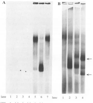
FIG. 1. Binding of capsid protein to RNA. (A) Comparison of the binding of thecapsid protein to CTS-14 and CTS-1 RNAs. Binding conditions were as described in Materials and Methods except that
CTS-1andCTS-14 RNAs werelabeled with[3H]UTP at aspecific
activity 10-fold higher than that in the other experiments. One microliter of mock translation reaction mixture was first added to CTS-1(lane 1) or CTS-14 (lane 2) RNA. CTS-1 (lane 3) and CTS-14 (lane 4) RNA in the absence of the in vitrotranslation extract are also shown. The bands in lanes 3 and 4 are indicated by asterisks. Although it may bedifficult to see the bands in this figure, they were clearlyvisible on the autoradiogram and were equal in intensity. In all of the other lanes, thebands represent35S-labeledprotein. The level of3H-labeledRNA was toolow to be detected. In other lanes, 1 ,ul oftranslation reaction mixture with full-length capsid protein was added to CTS-1 RNA (lane 5), CTS-14 RNA (lane 6), or a binding assay mixture containing only HB101 RNA (lane 7). (B)
Competitionbetweenthe packaging signal from Sindbis virus RNA and CTS-14 RNA. Binding conditions were as described in Materi-alsand Methods except that the amounts of CTS-1 and CTS-14 RNA were reduced to 0.2pgper reactionmixture. Lanes: 1 and 2, CTS-1 RNAand CTS-14 RNA, respectively, with no competitor RNA; 3, an equimolar amount of competitor added to CTS-14 RNA; 4, a 10-fold molar excess of competitor added to CTS-14 RNA. The competitor RNA was KS-Gl-HindIII RNA. It contains only the sequences from nucleotides 721 to 1302 in Sindbis virus 49S RNA and 55nucleotides from the Bluescript vector. This RNA migrates as two bands(indicated by arrows) in agarose gels unless it is first denatured.
were derived from a defective interfering RNA of Sindbis virus. CTS-14 contains the 572 nucleotides defined as the packaging signal required for encapsidation of the RNA, and CTS-1 lacks these sequences (29). CTS-14, but not CTS-1, binds to thecapsid protein and is packaged into extracellular particles in infected cells. For the experiments described here, the cDNAs were linearized upstream of the 3'terminus sothat the size of the CTS-14 RNA was 849nucleotides and that of the CTS-1 RNA was 831 nucleotides. The data in Fig. 1A show thedifference in binding of the capsid protein to the two RNAs (lanes 5 and 6). The in vitro-translated capsid protein migrated as a high-molecular-weight aggregate (Fig. 1A, lane7), which may be because of nonspecific binding to RNAs present in the in vitro translation mix. This high-molecular-weight band decreased when the intact capsid boundtoCTS-14RNA(lane 6).
To demonstrate that the difference in binding between
J. VIROL.
on November 9, 2019 by guest
http://jvi.asm.org/
DELETION ANALYSIS OF CAPSID PROTEIN OF SINDBIS VIRUS 1623
A Internal in-frame deletions in the capsid protein sites residues
used for aminoacids deleted
mutant deletion deleted
Lys Arg
wt
A1 EagI-Sacil 11-23
A2 Sacil-AlwNI 24-44 3
4
A3 AlwNI-BanI 44-74 2 4 A4 Eagl-Narl 11-74 2 1 2 05 Banl-Afili 76-107 12 2
A6 Afill-AatlI 110-118 1 1
A7 Aatil-Ball 117-130 1 A8 Ncol-Pmll 133-141 2 09 Pmll-Dralll 142-149 1
B C-terminal deletions in the capsid protein
run-off amino acids residues
mutant site deleted deleted
Lys Arg wt
AI0 Afl1 109-264 7 1_
All AatlII 117-264 0 6 A12 Ncol 133-264 9 6 A13 Dra111* 150-264 6 5
A14 Hincll 172-264 3 6
C Combined N-terminal and C-terminal deletions A15 AflIl 109-264 13 9
A16 Aatl 117-264 12 18 _ _
A17 Ncol 133-264 1 1 18 =
018 Dralll1* 150-264 8 17 = D Sindbis capsid protein-BMV coat protein fusion proteins
capsid protein
fusion derived
protein fusion site aminoacids
14
Wt
F Narl 1-75
F2 Afll* 1-108
F3 Ncol 1-132
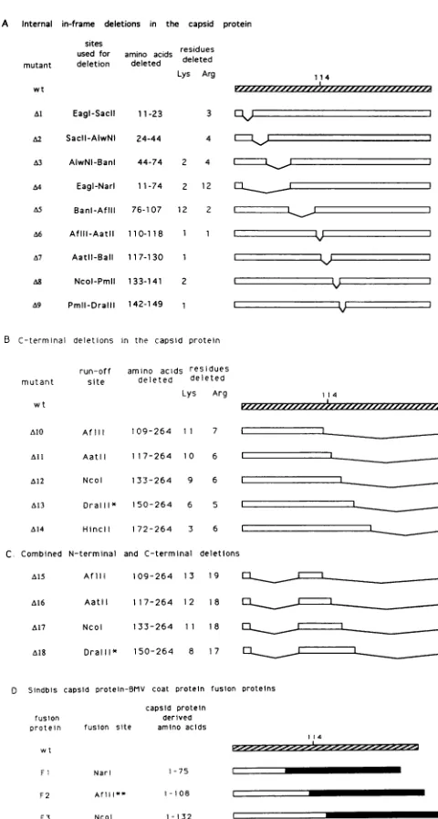
FIG. 2. Diagrams of capsid protein showing locations of the deletions. Open boxessignify the amino acidregionsretained in the
capsid protein,solid lines show the deletedregions,and thewtisthe
full-lengthcapsid protein.Amino acid 114 of thewtcapsid proteinis
highlighted inpanelsA,B,and Dtoindicate thejunctionbetween the unstructured N-terminal and the structuredC-terminalpartsof theprotein. (A)Internal in-frame deletions in thecapsidprotein. (B)
Deletions in the C terminus of thecapsidprotein.TheDraIll runoff mutant, indicatedbyasterisks,wasmade fromaHindIll-cut
DraIII-NruI internal out-of-frame deletion mutant which moved a stop codon into thereadingframe 1amino acid(arginine)downstream of the deletion. (C) CombinedN- and C-terminal deletions. Mutants
A15toA18contain the sameinternal deletionas A4(Eagl-NarI) in addition tovarious C-terminal deletions. (D) Sindbis virus capsid protein-BMVcoatfusionproteins. Solid boxes represent BMVcoat
protein-derived sequences. TheAflll fusion protein, indicated by
doubleasterisks,wasconstructedbyinsertion ofa10-bp Salllinker into thefilled-inAflllsite followedby fusion of the Sall-cut capsid proteingene tothe deleted BMVcoat protein gene. Three amino acids(Asp,Gly,andArg)wereadded between the Sindbis virus and theBMV-derived amino acids.
TABLE 1. Quantitative results from Betascopeanalysisa
Capsid Total Nonspecific Specific binding %of mutant binding binding Initial Correctedb wtb
wt 1.47 0.34 1.13 1.13 100
Al 1.29 0.40 0.89 0.99 88
A2 2.21 0.71 1.50 1.66 147
A3 2.18 0.80 1.38 1.38 122
A4 1.15 0.47 0.68 0.86 76
A5 0.12 0.18 -0.6 -0.7 0
aDataanalyzed wereobtained from the experiment shown in Fig. 4. Data inthe table, except as indicated, are the35Scounts(104)collected in a 30-min scanwith the Betascope blotanalyzer.Totalbindingis thebindingtoCTS-14 RNA;nonspecificbinding is the binding to CTS-1 RNA;specificbinding is the difference between the total (CTS-14-bound)andnonspecific(CTS-1-bound) counts.
bThe specificbinding was corrected for the number of Met residues per capsid mutant polypeptide(10 Met residues per full-lengthcapsid protein). Corrected specific binding values obtained for capsid mutants were then expressedasthepercentageof correctedspecific bindingobtained with the full-length (wt)capsid protein.
CTS-14andCTS-1was duetothe 572-nucleotide sequence, we carried out the binding assay in the presence of a
competitor RNA that contained only the 572-nucleotide sequencederived fromSindbis virusRNA(andanadditional 55 nucleotides derived from the vector). The competitor RNA was added at concentrations equal to or in 10-fold excessof those ofCTS-14RNA. SpecificbindingtoCTS-14
[image:4.612.66.306.77.526.2]was reduced 2- and 10-fold, respectively (Fig. 1B, lanes 3
TABLE 2. SpecificRNAbindingactivities ofcapsid
protein mutants
Characteristics and
muaratersstlcs
Amino acids No. Bindingofmutant antAmmoetelds
of as%of wtbind-mutant deleted ext in( ±f)
expts ing(xco+)' Internal deletions
Al 11-23 4 80 ± 20
A2 24-44 6 154 53
A3 44-74 6 170 + 59
A4 11-74 12 101 ± 42
A5 76-107 6 1.5 ± 2
A6 110-118 8 14 ± 8
A7 117-130 8 17 ± 10
A8 133-141 7 11 + 5
A9 142-149 7 13 + 5
C-terminal deletions
AlO 109-264 6 11 + 8
All 117-264 5 37 ± 21
A12 133-264 6 50 ± 8
A13 150-264 10 48 ± 17
A14 172-264 6 12± 9
Combined N- and
C-terminaldeletions
A15 11-74, 109-264 4 4 ± 3
A16 11-74, 117-264 7 38 ± 21
A17 11-74, 133-264 10 70 + 25
A18 11-74, 150-264 3 38 ± 14
Fusionproteins
Fl 76-264 6 3 3
F2 109-264 9 17± 10
F3 133-264 9 31 ± 16
a , mean ofn experiments (given in previous column); Wr, standard deviation.
VOL. 67, 1993
11 4
rl.,)
4
on November 9, 2019 by guest
http://jvi.asm.org/
[image:4.612.324.565.89.178.2] [image:4.612.322.563.411.707.2]wt Al A? ; A4 A( A7 A8 Al.)I)_A I 4
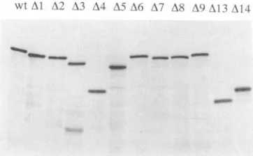
FIG. 3. Analysis of in vitro translation products. mRNAs that code for the capsid protein and various deleted forms of thisprotein weretranslated in vitro by using wheat germ extracts. Samplesof
the proteins were analyzed on an SDS-10% polyacrylamide gel. Approximately 5 x 10⁵ acid-precipitable cpm was loaded in each lane.
and 4). Withoutdenaturation, the572-nucleotide RNA tran-script migratesastwobands inanagarose gel(arrows in Fig.
iB, lane4). Thiscomplicated the analysisof the agarosegel and made it difficult to use this smaller RNA in binding
assays.
We had originally planned to use the [3H]uridine-labeled
RNA as an internal standard, but incubation of the RNA
with amock translation reaction mixture containing every-thingbut35S-labeled capsid protein ledtodegradation ofthe RNA (Fig. 1A; compare lanes 1 and 2 with lanes 3 and 4).
Theintact capsidprotein seemedto protectbothRNAsfrom degradation. Because the protection could be variable
be-tween experiments, we did not normalize the data to the
amount of[3H]uridine-labeled RNA recovered. The lack of internal standards mayhave contributed totheconsiderable variation between experiments, as seen in the standard deviations (see Table 2). The experimentswere all repeated multiple times, with the intact capsid protein included as a control inevery experiment.
Theeffect of deletions in the N-terminal half ofthe capsid
protein on bindingto RNA. The internal in-frame deletions thatweconstructed inthe amino-terminal half of the capsid
protein aredepictedinFig. 2A. The[35S]methionine-labeled
proteins produced by translation in vitrowere analyzed on
acrylamide gels (Fig. 3), and the results of a gel-binding
assay using these proteins are shown in Fig. 4. Only the
35S-labeled protein bands were detected in this
autoradio-gram. Bindingwasquantifiedbyanalysis of the dried gelon
a Betascopeblot analyzer (Tables 1 and2).
Deletion of amino acids 11 to 23 (Al) reduced only
Capsi(i
protcin kvt A I - '3-l \A
-("'S- 1 14 14 1I 14
FIG. 4. Agarosegel analysis ofthe bindingofcapsid proteins
withN-terminalin-framedeletionstoSindbisvirusRNAs.Binding
conditionswereasdescribed in Materialsand Methods. Numbers
obtainedfrom theBetascopeblotanalysisaregiveninTable1.
marginally thespecific binding of the capsidproteintoRNA, anddeletion ofamino acids 24to44(A2)or44to74(A3)may have enhanced binding (Table 2). The latter two peptides showed anincrease bothin the nonspecific binding toCTS-1 and the specific binding to CTS-14 (Table 1). A deletion covering the entire region from amino acids 11 to 74(A4)had nosignificanteffectonthebinding activity, eventhough this deletion eliminated 14 positive charges (12 arginines and 2 lysines)(Tables1and2). In contrast,deletion of amino acids 76 to 107, which removed an identical number of positive charges (12 lysines and 2 arginines), decreased nonspecific binding to CTS-1 (Table 1) and almost completely abolished specific binding (residual binding of 1.5%) (Table 2).
The effect of deletions in the C-terminal part of the capsid protein on binding to RNA. Two types of deletions were introduced into this region: internal in-frame deletions (Fig. 2A) and C-terminal truncations (Fig. 2B). The mutant (M10) lacking the C-terminal 156 amino acids retained only 11%of the specific binding activity (Table 2). A truncation of only 148of these amino acids (All) restored the activity to 37%. Two mutants, one(A12)lacking half of the C-terminal amino acids and the other(A13)lacking 115 of the C-terminal amino acids, retained about 50% of the specific binding activity of the full-length capsid protein (Table 2). Surprisingly, re-moval of the C-terminal 92 amino acids (A14) reduced the binding activity to only 12% that of the complete protein. Similarly, small internal deletions (A8 and A9) within this region also led to reductions in binding activity more severe than those that remove the entire region.
The effect of deletions in both the N-terminal and the C-terminal domains of the capsid protein on binding to RNA. The capsid protein with a large deletion in the amino-terminal half (A4) retained all of the binding activity of the intact capsid protein. C-terminal deletions of this protein were made by in vitro translation of truncated RNA tran-scripts (Fig. 2C). One of these mutants, A17,which contains amino acids 1 to 10 and 75 to 132 of the capsid protein, was almost fully active (70% residual binding activity) (Table 2). A16, which retained amino acids 1 to 10 and 75 to 116, still showed 38% of the original binding activity, but only 4% of the activity was retained by 15(amino acids 1 to 10 and 75 to 108).
These results taken together with those obtained from the internal and the C-terminal deletions show that the amino acidsbetween residues 75 and 117 were the most important for binding to the packaging signal in Sindbis virus RNA. They also reveal that in some instances, small internal deletions (A8 and A9) or partial deletions (M14) were more detrimental than more extensive deletions ( All andA12or A16andA17).
Binding of Sindbis virus capsid protein-BMV coat protein fusion proteins to Sindbis virus RNA. Another approach to examiningthe abilities of fragments of the capsid protein to bindto Sindbis virus RNA which we took was to examine fusion proteins in which different amounts of the N-terminal domain ofthe capsid protein had been fused to the BMV coat protein. We chose the coat protein of BMV because it is similar in size to the Sindbis capsid protein and it also encapsidates a genomic RNA into an icosahedral capsid. The region important for specific RNA binding in the BMV coat protein has been assigned to the N terminus (19). Several different fragments of the Sindbis virus capsid pro-tein cDNA were fused to nucleotide 79 of the BMV coat proteingene, eliminating the proposed RNA binding region of the latter. The Sindbisvirus capsid-derived fragments in the fusion proteins contained the N-terminal 75 (Fl), 108
on November 9, 2019 by guest
http://jvi.asm.org/
[image:5.612.71.281.590.678.2]DELETION ANALYSIS OF CAPSID PROTEIN OF SINDBIS VIRUS 1625 bindingasS of
wtcapsid protein binding
60-
120-802
40-unstructured domain structureddomain
N i'&us II
[image:6.612.67.295.69.363.2]regionsdeleted Incapsidprotein
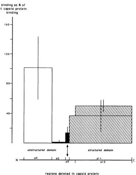
FIG. 5.
Specific binding
of deleted formsofthecapsid proteintoSindbisvirus RNAs. Eachrectangle symbolizesoneofthedeleted forms of the
capsid protein.
Width reflects theamino acid regiondeleted,
andheightrepresentsthespecificRNAbindingnormalizedto that of the full-length capsid protein. Binding values are the
meansofatleastfiveexperiments (Table 2).Barsatthetop of each
rectanglereflectstandarddeviations. Mutantsaregroupedintofour classes
according
totheirbindingactivitiescomparedwiththat ofthe
full-length capsid
protein: (i) noreduction, (ii) 100-foldreduc-tion,
(iii)
5tolOxreduction, and(iv)2to3x reduction.(F2),
or132(F3)
aminoacids(Fig. 2D).
Fl showedverylittleRNA
binding activity (3%
that of thefull-length
Sindbisviruscapsid protein) (Table 2),
butthe activities of F2(17%)
and F3(31%)
weresignificant.
These resultsagain point
to theimportance
of amino acids 76to108 inparticular
but alsotothat of amino acids 109 to 132 forthe
specific
RNAbinding
activity
of thecapsid protein.
Inaddition,
the low-levelbinding activity
ofFl arguesagainst
amajor
role of amino acids 1to10inRNAbinding
(this region
wasnotcovered in the deletionanalysis).
DISCUSSION
We have describedan assay for
analyzing
thebinding
ofan in vitro-translated
protein
to RNA on the basis of itsmigration
with theRNAduring electrophoresis
inanagarosegel.
This method of detection isavariationonthe theme of thegel
shift,
in which it is theprotein
that shifts when it bindsto the nucleic acid.Using
this assay, weanalyzed
a series of deletions in the Sindbis viruscapsid protein
and identifiedaregion
in theprotein
thatbindstothepreviously
defined
packaging signal
in Sindbis virus RNA.Figure
5 illustrates four different classes of deletion mutantsthat do notoverlap,
inspite
of thelarge
standard errors. The firstclass, consisting
ofmutantslocated betweenamino acids11and74, had littleornoeffect on the ability of the protein to bind RNA. In contrast, the second class, composed of the deletion from amino acids 76to107, defined a region of the
protein that is essential forbinding. The third class,
exem-plifiedinFig. 5 by the deletion between amino acids 110 and
118,hadonly 10% the binding activity of the wt capsid, but the activity was 10-fold higher than that obtained with the class ii mutant. The fourth class lacked almost all of the C-terminal half of the protein, and it had intermediate activity.
The determination of the structure of the Sindbis virus
capsid proteinby Choietal. (3) provides further insight into our results. The residues from Arg-114 to the C-terminal
tryptophan arewell ordered andarefolded into two similar ,-barrel domains with the
connecting
loops
fromapproxi-matelyresidue 170 to residue 187. One C-terminal deletion
(A14) from amino acid 172tothe C terminus removed the second domain, causing a 90% reduction in the binding
activity of theprotein. A significant fraction of the activity wasrestored, however,withmoreextensivedeletions in the C terminus (All, A12, and A13). Deletion of only the C-terminal 93 residues may lead to a distortion in the structureof theprotein,which is correctedbytheremoval of additional amino acid residues.
Deletions between amino acids 117 and 149 (A7, A8, and A9) were similar to the deletion from amino acid 172 to the C terminus (A14) in their abilities to decrease the binding
activity about 90%. Deletion of the entire C terminus,
including these residues, reduced the activity only about 50%. This contradiction suggests that deletions inthis region of theprotein also affectstructureand canbecompensated for
by
more extensive deletions.Our data support the conclusion that the actual binding
regionlies between amino acids 76 and 116. The presence of amino acids 75 to 116 in the combined N-terminal- and C-terminal-deleted protein (A16) stimulated binding to 40% that of thecomplete capsid.Deletion of amino acids 76 to 107 almost completely eliminated binding, and the presence of this stretch of amino acids in theproteinfused to the BMV
coatproteinincreased thebinding activityof thatproteinfrom 3to 17%.Thecapsid proteinexists as adimerinthecrystal
structure.Theregion containingthe amino acidsinvolved in the monomer-monomer contacts (residues 185 to 190 and residue 222)canbe deleted from thecapsid proteinwithout
affecting binding
toRNA,indicatingthat theoligomericstateof the
protein
isnotinvolved in its abilitytobind RNA. Several different types of studies have shown previously that thecapsid protein
ofonealphaviruscanencapsidatethe RNAofadifferentalphavirus (11, 30).This suggests that the amino acidsinvolved inbindingto genomicRNAwould be similar in the differentcapsid proteins.
Thus, a potentialbinding
motif is the stretch of 10 amino acids in this part ofthe
protein
that ishighly
conserved amongalphaviruses.Theaminoacid sequence in the Sindbis viruscapsid proteinfrom residues 97to106 isLgProLgPw
.fly
LysAkgGlnArg
Met. The underlined residues are invariant among eight
differentalphaviruses.
Thedetermination of the structure of several RNAplant
viruses
provides
adescription of the domains of their coatproteins (9, 10, 18).
The N-terminal residues of many ofthese
proteins
containalarge number ofpositively chargedamino acids thatare
presumed
tobeimportant
inneutraliz-ing
thenegatively
chargedRNA. Removal of theNterminusfrom thecoatproteinof several of these virusesby
proteo-lytic cleavage destroyed
theability
oftheprotein
tointeractwithRNAand ledtotheassemblyof RNA-freeparticles (5,
VOL. 67,1993
on November 9, 2019 by guest
http://jvi.asm.org/
22, 27, 28). More recent evidence for the importance of the N-terminal domain in assembly comes from the studies of
Sacher and Ahlquist with BMV (19). They constructed a coat protein mutant lacking the first 25 N-terminal amino acids and showed that the proteinwasunabletointeract with RNA to form virus either in plant protoplasts or in a
systemic infection of barley plants. The interaction of the Sindbis virus capsid protein with the packaging signal in Sindbis virus RNAcannotbesolely duetothe high density of basic residues in the N-terminal domain of the protein. Ourresults show that the highly basic region from residues 11to74playsnorole in thespecificbinding of the proteinto
Sindbis virusRNA. Wesuggestthat the interaction between the capsid protein and Sindbis virus RNA described here
represents the nucleation event that confers specificity on
theassemblyprocess. Otherregions of the protein maythen
function in the subsequent steps in the process that lead to theformation of thenucleocapsids.
ACKNOWLEDGMENTS
We thank Birgit Lewicki for providing the T7 polymerase and Paul Ahlquist for sendingusthepB3RS25 DNA used for preparing the fusion clones. Wealso thank Barbara Weiss forcommentsand discussionsthroughout thecourseof thiswork.
Thisresearchwassupported bygrantA111377 from theNational Institute of Allergy and Infectious Diseases. U.G.-G. received fellowships from the scientific committee of NATO and the Keck Foundation.
REFERENCES
1. Aliperti, G., and M. J. Schlesinger. 1978. Evidence for an
autoproteaseactivity ofSindbis virus capsidprotein. Virology 90:366-369.
2. Carmichael, G. G., and G.K. McMaster. 1980. The analysis of nucleic acidsingels using glyoxal and acridineorange.Methods Enzymol.65:380-389.
3. Choi, H.-K., L. Tong, W. Minor, P. Dumas, U. Boege, M. G. Rossmann, and G. Wengler. 1991. Structure of Sindbis virus coreprotein revealsachymotrypsin like serineproteaseand the organization of thevirion.Nature (London) 354:37-43. 4. Elgizoli, M., Y. Dai, C. Kempf, H. Koblet, and M. R. Michel.
1989. Semliki Forestviruscapsid protein acts as apleiotropic
regulator of host cellular protein synthesis. J. Virol. 63:2921-2928.
5. Erickson, J. W., and M. G. Rossmann. 1982. Assembly and crystallization of a T= 1 icosahedral particle from trypsinized
southern beanmosaic viruscoatprotein. Virology 116:128-136. 6. Garoff, H., A.-M. Frischauf, K. Simons, H. Lehrach, and H.
Delius. 1980. The capsid protein of Semliki Forest virus has clustersof basicamino acids andprolines in its amino terminal region. Proc. Natl. Acad. Sci. USA 77:6376-6380.
7. Hahn, C. S., E. G. Strauss,and J. H. Strauss. 1985. Sequence analysisofthreeSindbisvirusmutantstemperature-sensitive in thecapsid protein autoprotease. Proc. Natl. Acad. Sci. USA 82:4648-4652.
8. Hahn, C.S., and J.H.Strauss.1990.Site-directedmutagenesis oftheproposed catalytic amino acids of the Sindbis viruscapsid proteinautoprotease. J.Virol. 64:3069-3073.
9. Harrison, S. C. 1983. Virusstructure: high resolution perspec-tives.Adv. VirusRes.28:175-240.
10. Harrison, S. C. 1990. Common features in the structures of someicosahedral viruses: apartlyhistorical overview. Semin. Virol 1:387-403.
11. Kuhn, R. J., S. Lopez, and J. H. Strauss. 1992. Personal communication.
12. Laemmli, U. K. 1970. Cleavage of structural proteins during the assembly of the head of bacteriophage T4. Nature (London) 227:680-685.
13. Lutz, C. T., W. C. Hollifield, B.Seed, J. M.Davie,and H. V. Huang. 1987.Syrinx2A: animproved X phage vector designed for screening DNA libraries by recombinationin vivo. Proc. Natl. Acad.Sci. USA 84:4379.
14. Michel, M. R., M. Elgizoli,Y. Dai, R. Jakob, H. Koblet, and A.-P. Arrigo. 1990. Karyophilic properties of Semliki Forest virusnucleocapsid protein.J.Virol. 64:5123-5131.
15. Milligan, J. F., D. R. Groebe, G. W. Witherell, and 0. C. Uhlenbeck. 1987. Oligoribonucleotide synthesis usingT7 RNA polymerase andsyntheticDNAtemplates. NucleicAcidsRes. 15:8783-8798.
16. Rice, C. M., R. Levis, J. H. Strauss, and H. V. Huang. 1987.
Production of infectious RNA transcripts from Sindbis virus cDNAclones:mappingoflethalmutations,rescueofa
temper-ature-sensitive marker, and in vitro mutagenesis to generate
definedmutants. J.Virol. 61:3809-3819.
17. Rice, C. M., and J. H. Strauss. 1981.Nucleotidesequenceof the 26S mRNA of Sindbis virus and deduced sequence of the encoded virus structuralproteins.Proc. Natl. Acad. Sci. USA 78:2062-2066.
18. Rossman, M. G., and J. Johnson. 1989. Icosahedral RNAvirus
structure. Annu.Rev. Biochem. 58:533-573.
19. Sacher, R., and P. Ahlquist. 1989. Effect of deletions in the
N-terminal basic arm of brome mosaic virus coat proteinon RNApackagingandsystemicinfection. J. Virol. 63:4545-4552. 20. Sanger, F. S.Nicklen, and A. R. Coulson. 1977. DNA
sequenc-ing with chain-terminating inhibitors. Proc. Natl. Acad. Sci. USA74:5463-5467.
21. Schlesinger,S., and M. Schlesinger. 1990.Replication of Toga-viridae andFlaviviridae,p.697-711. In B. N. Fields and D. M.
Knipe(ed.), Virology. RavenPress,NewYork.
22. Sorger, P. K., P. G. Stockley, and S. C. Harrison. 1986. Structure andassemblyofturnipcrinkle virus II mechanism of reassembly in vitro. J. Mol. Biol. 191:639-658.
23. Strauss, E. G., and J. H. Strauss. 1986. Structure andreplication
of thealphavirusgenome, p.35-82.InS.Schlesingerand M. J.
Schlesinger (ed.), The Togaviridae and Flaviviridae. Plenum
Press, New York.
24. Ulmanen, I., H. Soderlund, and L. Kaariainen. 1976. Semliki
Forest virus capsid protein associates with the 60S ribosomal subunit in infected cells. J. Virol. 20:203-210.
25. Ulmanen, I., H. Soderlund, and L. Kaariainen. 1979. Role of
protein synthesis in the assembly of Semliki Forest virus nucleocapsid. Virology99:265.
26. Van Steeg, H., M. Kasperaitis, H. 0. Voorma, and R. Benne. 1984. Infection of neuroblastoma cellsby SemlikiForestvirus. Theinterferenceof viralcapsidproteinwith thebindingof host messenger RNAs into initiationcomplexes is the cause of the shut-off of hostprotein synthesis.Eur. J. Biochem.138:473-478. 27. Vriend,G., M.A.Hemminga, B. J. M. Verduin, J. L.DeWit, and T. J. Schaafsma. 1981. Segmental mobility involved in
protein-RNA interaction in cowpea chlorotic mottle virus. FEBS Lett. 134:167-171.
28. Vriend, G., B. J. M. Verduin, and M. A. Hemminga. 1986. Role of the N-terminal part of the coatprotein in the assembly of cowpea chlorotic mottle virus. A 500 MHz proton nuclear
magnetic resonance study and structural calculations. J. Mol.
Biol. 19:453-460.
29. Weiss, B., H. Nitschko, I. Ghattas, R. Wright, and S. Schle-singer. 1989. Evidence for specificity in the encapsidation of Sindbis virus RNAs. J. Virol. 63:5310-5318.
30. Weiss, B.,andS. Schlesinger. 1981. Defectiveinterfering
parti-cles of Sindbis virus donotinterfere with thehomologousvirus obtained frompersistently infected BHK cells but do interfere with Semliki Forest virus. J. Virol.37:840-844.
31. Wengler, G. 1987. The mode ofassemblyofalphaviruscores
implies a mechanism for the disassembly ofthe cores in the earlystagesof infection. Arch. Virol. 94:1-14.
32. Wengler,G.,andG. Wengler. 1984. Identificationofatransfer of viralcoreproteintocellular ribosomesduringtheearlystages ofalphavirusinfection.Virology 134:435-442.