STUDY OF EXPRESSION OF CD 117 AND CD 34 IN
PHYLLODES TUMOR OF BREAST AND ITS
CORRELATION WITH HISTOPATHOLOGICAL
GRADE
Dissertation submitted in
Partial fulfillment of the regulations required for the award of M.D.Degree
in PATHOLOGY-BRANCH III April 2017
DECLARATION
I solemnly declare that this dissertation entitled “STUDY OF EXPRESSION OF CD 117 AND CD 34 IN PHYLLODES TUMOR
OF BREAST AND ITS CORRELATION WITH
HISTOPATHOLOGICAL GRADE” was done by me in the Department of Pathology, Coimbatore Medical College, Coimbatore
during the period of June 2014 to July 2016 under the guidance and
supervision of DR.C.LALITHA, MD., Professor and Head, Department of Pathology, Coimbatore Medical College, Coimbatore.
This dissertation is submitted to The Tamilnadu Dr.M.G.R. Medical University, Chennai towards the partial fulfillment of the requirement for the award of M.D., Degree in Pathology.
Place:
Date: Dr. R. ARTHI
CERTIFICATE
This is to certify that the dissertation entitled “STUDY OF EXPRESSION OF CD 117 AND CD 34 IN PHYLLODES TUMOR
OF BREAST AND ITS CORRELATION WITH
HISTOPATHOLOGICAL GRADE” is a record of bonafide work done by Dr.R.Arthi, Post Graduate student in the Department of Pathology, Coimbatore Medical College and Hospital, Coimbatore
under the guidance and supervision of Dr.C.Lalitha, M.D., Professor
and Head, Department of Pathology, Coimbatore Medical College and
Hospital, Coimbatore in partial fulfillment of the regulations of
Tamilnadu Dr.M.G.R. Medical University, Chennai towards the award
of M.D.Degree (Branch III) in Pathology.
Dr. Edwin Joe, M.D.,B.L., Dr. C. Lalitha, M.D.,
Dean, Coimbatore Medical College, Professor and Head,
Coimbatore. Department of Pathology,
Coimbatore Medical College,
ACKNOWLEDGEMENT
To begin with, I thank the almighty God for bestowing his
blessing on me in this dissertation a successful one.
I wish to thank the beloved Dean Dr.A. EDWIN JOE,M.D,B.L, Coimbatore Medical College and hospital, Coimbatore for permitting me to conduct this study.
It’s a great pleasure to express my humble gratitude to the most
respectable teacher and my guide Dr. C.Lalitha. M.D., Professor and Head of the Department, Department of Pathology, Coimbatore Medical College ,Coimbatore for her guidance and support. This dissertation bears her valuable suggestions and highly professional
advice.
I wish to express my gratitude and sincere thanks to
Dr. A. Arjunan. M. D., for his guidance and support.
I also thank all Associate Professors, all Assistant professors and
all Tutors, who were a constant force behind me in encouraging to
accomplish this wonderful thesis.
I thank Department of Surgery and Department of Surgical
clinical cases, valuable support and guidance which made this
dissertation possible.
I thank my family, especially my husband Dr.P.Satheesh, for his support and encouragement.
I thank all lab technicians working in Department of Pathology,
TABLE OF CONTENTS
S.NO TITLE PAGE NO
1. INTRODUCTION 1
2. AIM 3
3. OBJECTIVES 4
4. REVIEW OF LITERATURE 5
5. MATERIALS AND METHODS 42
6. OBSERVATIONS AND RESULTS 49
7. DISCUSSION 67
8. SUMMARY 72
9. CONCLUSION 75
10. BIBLIOGRAPHY
11. ANNEXURES:
I.PROFORMA
II.GLOSSARY
III.CONSENT FORM
LIST OF TABLES
S.NO TITLE PAGE NO
1
Distribution of Phyllodes Tumor of Breast
According to Different Age Group
49
2 Association of Age With Histopathological Grade 51
3
Mean Age of Occurrence of Tumor in Different
Histopathological Categories
53
4
Percentage of Tumors in Each Histopathological
Grade
55
5 Overall Stromal Expression of CD117 57
6 Overall Epithelial Expression of CD117 58
7 Overall Stromal Expression of CD34 59
8
Association of Stromal Expression of CD117 With
Histopathological Grade
60
9
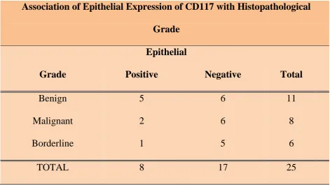
Association of Epithelial Expression of CD117
With Histopathological Grade
62
10
Association of Stromal Expression of CD34 With
Histopathological Grade
64
11
Frequency of CD34/CD117 Immuno profiles in the
3 Morphological Categories of Phyllodes Tumors
LIST OF CHARTS
S.NO TITLE PAGE NO
1
Distribution of Phyllodes Tumor of Breast
According to Different Age Group
50
2 Association of Age With Histopathological Grade 52
3
Mean Age of Occurrence of Tumor in Different
Histopathological Categories
54
4
Percentage of Tumors in Each Histopathological
Grade
56
5 Overall Stromal Expression of CD117 57
6 Overall Epithelial Expression of CD117 58
7 Overall Stromal Expression of CD34 59
8
Association of Stromal Expression of CD117 With
Histopathological Grade
61
9
Association of Epithelial Expression of CD117
With Histopathological Grade
63
10
Association of Stromal Expression of CD34 With
Histopathological Grade
LIST OF COLOUR PLATES
PLATE NUMBER
TITLE
1
IMMUNOHISTOCHEMISTRY OF STROMAL CD117
IN MALIGNANT PHYLLODES TUMOR
2
IMMUNOHISTOCHEMISTRY OF STROMAL CD34 IN
BENIGN PHYLLODES TUMOR
3
IMMUNOHISTOCHEMISTRY OF STROMAL CD34
AND CD117 IN BORDERLINE PHYLLODES TUMOR
4
HEMATOXYLIN AND EOSIN STAINING OF
MALIGNANT PHYLLODES TUMOR
5
HEMATOXYLIN AND EOSIN STAINING OF
BORDERLINE PHYLLODES TUMOR
6
HEMATOXYLIN AND EOSIN STAINING OF BENIGN
PHYLLODES TUMOR
7
IMMUNOHISTOCHEMISTRY OF EPITHELIAL CD117
IN BENIGN PHYLLODES TUMOR
8
IMMUNOHISTOCHEMISTRY OF VASCULAR CD34
INTRODUCTION
1
INTRODUCTION
Phyllodes tumor of breast includes a group of lesions of varying
malignant potential. They can be completely benign tumors to fully
malignant sarcomas. Malignant Phyllodes tumor is the most aggressive
mesenchymal tumor of the breast, with a five year survival rate of
50-60%. It has a tendency for local recurrence (65%) and distant metastasis
(10%) as well if treated conservatively. Current clinical and pathological
variables have limited ability to accurately predict the nature of the
tumor and differentiating the different grades of phyllodes tumor often
possess difficulties. So there is clearly a need for definitive markers for
differentiating benign from malignant tumors so that appropriate
management protocol can be followed.
The c-kit is a protooncogene that encodes a tyrosine kinase
receptor (CD 117), which plays a role in cell proliferation and survival.
Its overexpression can lead to increased cell proliferation and
malignancy.
CD 34 is a transmembrane glycoprotein involved in signal
transduction, cellular proliferation and differentiation. It also determines
2
Both of these markers can act as diagnostic and prognostic
indices.
With the need to differentiate benign from malignant tumors and
also with the advent of specific therapy targeted at CD117, this study
was conducted to explore the expression of CD117 and CD34 in
3
AIM OF THE STUDY
To study the immunoexpression of CD 34 and CD 117 in
4
OBJECTIVES
1. To assess the expression of CD34 and CD 117 in phyllodes tumor
of breast
2. To correlate the expression of these markers with the
histopathological grading
3. To assess the usefulness of these markers in differentiating benign
from malignant phyllodes tumor of breast so that appropriate
5
REVIEW OF LITERATURE
ANATOMY AND PHYSIOLOGY OF THE BREAST
The structure of human breast is a reflection of its special
function, which is production of milk. The epithelial component of
breast tissue is constituted by lobules, which is the site for milk
production and it connects to ducts that open onto the nipple. These
lobules and ducts are found to lie in a background of fibrous and adipose
tissue. Adipose tissue makes up the main breast mass. The structure of
the male breast is similar to that of the female breast except that it lacks
the specialized lobules since there is no need for milk production.
Anatomically the adult breast lies on top of the pectoralis muscle
over the ribcage. The breast tissue extends horizontally from the edge of
the sternum to the midaxillary line. It is to be noted that a portion of
breast tissue called the axillary tail of Spence extends into the axilla.
The breast tissue is surrounded by a thin layer of connective tissue
known as fascia. The deep layer of the fascia lies immediately over the
pectoralis muscle and the superficial layer lies immediately under the
6
The skin covering the breast is just the same as the skin anywhere
else on the body and has got sweat glands, hair follicles etc.
The blood supply for the breast is primarily from internal
mammary artery which courses beneath the breast tissue proper. The
lymphatic vessels of the breast course in a direction opposite to that of
the blood vessels and drain into the lymph nodes. Most lymphatic
vessels drain into the axillary lymph nodes while a few of the lymphatics
drain into internal mammary lymph nodes, which are present deep to the
breast tissue. Knowledge of this lymphatic drainage is important because
when a breast cancer metastasizes it usually involves the first lymph
node in the chain of lymph nodes. This is called the "sentinel lymph
node‖ and a surgeon may remove this lymph node to check for
metastases in a patient with breast tumor.
Physiologically the breast is an organ specialized for milk
formation (lactation). Many additional changes are seen in the breast
tissue during pregnancy and lactation due to the changes in hormones
7 PHYLLODES TUMOR
Phyllodes tumor of breast was first fully described in 1838 by
Johannes Müller1. It was given the name cystosarcoma phyllodes to
highlight the leaf-like pattern and fleshy gross appearance of the tumor.
The other name commonly used for the tumor is periductal stromal
tumor because of its origin from periductal stroma. PT is sub classified
into three groups benign, borderline and malignant based on the
histologic characteristics of the tumor.
CLINICAL PRESENTATION
Patients usually present with a firm to hard mass. No clinical
feature can accurately distinguish between fibroadenoma, benign
Phyllodes tumor, and malignant Phyllodes tumor2. Features favouring
diagnosis of PT are tumor size more than 4cm and history of rapid
growth of tumor. When there is sudden enlargement of a tumor that was
stable for many years the possibility of tumor originating from a
preexisting fibroadenoma or malignant transformation of a benign
phyllodes tumor can be considered. Clonal analysis of a few tumors that
were originally diagnosed as fibroadenomas but later recurred as
phyllodes tumors suggest that phyllodes tumor can arise from a
8
It was found that the same allele of the X chromosome linked AR
gene was inactivated in the fibroadenoma and PT samples from each
patient.
Phyllodes tumors usually present as solitary unilateral masses. It
rarely involves both breasts or can occur as multicentric tumor 4,5,6,7,8.
Coexistent fibroadenomatoid lobular hyperplasias are often noted in the
surrounding breast tissue.
Phyllodes tumors can affect any age group from 10 to 86 years
9,10,11,12
. Most commonly affects women in the fourth decade, the median
age being 45 years.
It is only rarely seen in women younger than 30 years. A few cases
have been reported in adolescent females and most of the cases were
benign, only occasional cases of malignant phyllodes have been reported
in the patients of this age group 16, 17,18,19,20,21,22.
The tumors can range in size from 1-20 cms but they are mostly
around 4 cms in size. Malignant tumors are generally larger in size but
malignancy has been reported in lesions as small as 2 cms in size and
some of the large tumors have proved to be benign. Large tumors can
9
Mammography mostly reveals a lobulated and well-defined
opaque mass. A few cases have indistinct border. In ultrasound the
tumor appears well circumscribed but not homogenous due to the
presence of cystic spaces and epithelium-lined clefts 26, 27.
Calcifications can be rarely seen in both benign and malignant
lesions 27,28. Ultrasonography and mammography are not very useful in
distinguishing benign from malignant phyllodes tumor. MRI is more
reliable for distinguishing benign from malignant tumor 29,30.
The role of flow cytometry analysis of ploidy and S-phase in the
classification of PTs is uncertain. S phase and ploidy analysis by
flowcytometry is only of limited help in the classification of phyllodes
tumor.
Complex karyotypic abnormalities have been observed in
malignant phyllodes tumor via Cytogenetic studies 35. Recurrent tumors
have also been found to have the same genomic abnormality as the
primary 39.
Biochemical analysis has revealed expression of PR in the stroma
of phyllodes tumors but only a few of the tumors show expression of ER
40
10
associated with hypoglycemia in one case 41. IGF-II has also been
occasionally detected immunohistochemically in tumor stromal cells.
GROSS PATHOLOGY
The tumors are generally encapsulated circumscribed single or
multinodular masses. Even Phyllodes tumors with microscopically
invasive borders may appear circumscribed grossly.
The cut surface of the tumor is firm, bulging, gray to tan tissue.
Areas of hemorrhage and necrosis can be noted which are most
commonly seen in malignant tumors. Rarely large benign tumors may
also show these changes. Cysts containing keratotic material are rarely
seen. Very rarely phyllodes tumor can have a cystic component
mimicking a cystic papilloma (42)
MICROSCOPIC PATHOLOGY
The tumor originates from periductal stroma. Histologically most
Phyllodes tumors have a heterogeneous appearance and only a few
tumors have the structure of an intracanalicular fibroadenoma with
exaggerated stromal cellularity. In many cases there is ductal epithelial
11
Many features help in distinguishing a fibroadenoma from a
benign Phyllodes tumor. Unlike fibroadenomas, Phyllodes tumors have
increased cellularity of the stromal component. In some cases stromal
cellularity is more intense in areas adjacent to epithelial components
(periductal stroma). Mitotic activity is also increased in the same pattern;
on the other hand fibroadenomas do not reveal any stromal mitoses. PTs
may have a nodular structure with prominent periductal stromal
proliferation. These are called periductal stromal tumors and further classified as periductal stromal hyperplasia or periductal stromal
sarcoma (43). These tumors are highly prone for recurrence with features
of phyllodes tumor being noted in the recurrent tumor. Yet a significant
number of PTs show no zonal stromal distribution.
The presence of epithelial-lined clefts is a feature seen in PTs and
rarely these clefts are dilated and condensation of the stroma
immediately surrounding it can be seen. These clefts can also be seen in
fibroadenomas. The intracanalicular pattern of fibroadenomas may
mimic the clefted architecture of PTs and sometimes differentiating the 2
tumors can be difficult. This difficulty is particularly common giant
fibroadenomas where tumor size and clefts that are made out grossly
suggest PT. Microscopically the stroma in intracanalicular
12
Myxoid change in stroma can be seen in both fibroadenomas and
PTs. It is mostly homogeneous in fibroadenomas and is patchy with
degenerative changes in PTs. Pseudoangiomatous stromal hyperplasia
(PASH) can occur in PT and in some cases PASH can be a predominant
feature. Occasionally multinucleated giant cells are found in stroma of a
PT with PASH features. These giant cells can show
lymphophagocytosis. They may express markers such as CD68 which is
histiocytic marker as well as p53 and Ki67.
Stromal cellularity in phyllodes tumor can be heterogeneous with
areas indistinguishable from fibroadenoma that juxtapose sharply on
areas with increased cellularity. These features can make one to
conclude that the PT arises from a fibroadenoma when actually these
features are a part of some PTs. In certain tumors sampled by fine needle
aspiration or needle core biopsy, variations in pattern and stromal
cellularity may create difficulties in the classification of the tumor.
Inappropriate classification and inadequate sampling may be the causes
for malignant behavior and metastases from a benign tumor. Excisional
13
The PT is sub classified into three groups. Distinguishing benign
PT from low-grade malignant PT is important because the benign tumors
do not metastasize, they have a low risk for local recurrence, the time
interval to recurrence is longer, and initial recurrences are histologically
benign most of the time. Low-grade malignant PTs have very early local
recurrence and the recurrences are mostly histologically high grade.
A benign PT has very few mitoses - usually one to two per 10 hpf,
moderate to marked cellular overgrowth and moderate cytologic
pleomorphism141. Stromal expansion and cellularity are maintained
uniform throughout the lesion but these characteristics can be
heterogeneous141. There is a correlation between the degree of epithelial
proliferation and the appearance of the stroma. Epithelial hyperplasia
which is not very conspicuous in an ordinary benign PT can occasionally
be well identified in some cases. Although the border is usually well
defined invasion can be present , at times in the form of nodules of
benign tumor around the main tumor. Lipomatous and osseous
metaplasia can be seen in the stroma of a benign PT. Multinucleated
giant cells with hyperchromatic nuclei can occasionally noted in the
stroma of PT. These cells have been found to be immune positive for
14
PT. Pseudoangiomatous hyperplasia of stroma can be seen in both
benign and malignant PTs.
A malignant PT features a high degree of hypercellular stromal
overgrowth141. In many cases this can lead to significant separation of
epithelial elements, high proliferative activity in the stroma, more than
five mitoses per 10 hpf and an invasive tumor border141. Stromal
overgrowth and cellular atypia is common in these tumors. Stromal
overgrowth has been defined as marked stromal proliferation to the point
where the epithelial component is absent in at least 1 low-power field
(×40)140. Rarely the stroma can contain heterologous sarcomatous
elements like angiosarcoma, liposarcoma, chondrosarcoma etc. (45,46,47,48).
Borderline tumors show a circumscribed or invasive border, two
to five mitoses per 10 hpf, and moderate amount of stromal cellularity
that is mostly heterogeneously distributed admixed with hypocellular
areas. The stroma containing spindle cells may mimic fibromatosis or
low-grade fibrosarcoma or it may show pseudoangiomatous hyperplasia.
Occasionally cartilaginous, osseous or lipomatous metaplasia have been
found in borderline PTs. Epithelial hyperplasia is noted in many tumors.
This is seen as varying increase in the thickness of the epithelium lining
the slit-like space and the epithelium is usually cuboidal or columnar.
15
myoepithelial cells is often found. This may progress to papillary and
cribriform hyperplasia. The severity of epithelial hyperplasia correlates
with cellularity of the stroma and mitotic count but this is not always the
rule. Atypical epithelial hyperplasia can be extensive in some cases
leading to mistaken diagnosis of intraductal carcinoma. Sometimes the
stromal component can be misdiagnosed as reactive and the diagnosis of
phyllodes tumor can be overlooked. Tumors in which phyllodes pattern
of growth is obscured by an uncommon epithelial distribution usually are
misinterpreted as papillary neoplasms or as adenosis tumors.
Very rarely the epithelial abnormality can reach a level that can be
considered as intraductal carcinoma and finding intraductal or invasive
duct carcinoma in phyllodes tumor is very uncommon 49,50. In situ and
invasive duct and lobular carcinoma can also be seen PTs 51.
Squamous metaplasia, which can occur in benign and malignant
PTs, can be seen in about 10% of cases. Aspiration from a cystic area of
squamous metaplasia can lead to a mistaken diagnosis of squamous cyst
52
. Apocrine metaplasia has also been reported occasionally in PTs 53,54.
Lobules showing proliferative changes like sclerosing adenosis can
sometimes be seen in PTs. The presence of lobules especially with
hyperplasia can lead to mistaken diagnosis of fibroadenoma when the
16
epithelium in the form of adenosis and papillary hyperplasia can be so
extreme that it masks the underlying PT which may go unrecognized and
recur.
Very rarely stromal cells in benign phyllodes tumor can show
intracytoplasmic inclusion bodies like in infantile digital fibromatosis 55.
Electron microscopy in such cases showed a combination of fibroblasts
and myofibroblasts. The intracytoplasmic inclusions were associated
with cytoplasmic microfilaments that formed tadpole-like structures.
These cells stained very weakly for actin by immunohistochemical
method and the inclusion bodies did not show any reactivity. Post
pretreatment with potassium hydroxide in 70% ethanol and also 0.1%
trypsin both the cells and inclusions stained strongly for actin.
Ductal elements can be seen in locally recurrent PTs in the breast
or chest wall. With some rare exceptions most of the metastatic PTs at
distant sites have entirely the stromal component only. Few case reports
have shown epithelial component in lung metastases 56,57. One of these
represents inclusion of pulmonary tissue in the metastatic deposit 58.
Exceptional primary malignant PTs exhibiting liposarcomatous
differentiation with adenosis-like glandular component are on record.
17
components in the primary tumor and in the metastases were
immunopositive for gross cystic disease fluid protein 15 (GCDFP-15).
The cells in these glandular structures were surrounded by myoepithelial
cells, which were immunopositive for actin.
Because most of the malignant PTs are high-grade spindle cell
lesions with fibrosarcoma like pattern, it is the most common pattern
seen in metastatic lesions. Rarely locally recurrent tumor or the
metastatic tumor may exhibit heterologous differentiation that was not
noted in the primary tumor 58. Rare heterologous sarcomatous
components in the primary tumor like lipo- , osteo- 60, chondro- 59, and
leiomyosarcoma can be seen in metastases. Rhabdomyosarcoma has
been observed in the lung metastases from a malignant PT that had
rhabdomyosarcomatous element (45). Metastases from a PT with
liposarcomatous element had mainly immature lipoblasts and very few
adipocytes 62.
Adequate sampling is important with at least 1 block for every 1
18 IMMUNOHISTOCHEMISTRY
The stroma is vimentin-positive. Actin, CD34, and desmin
positivity are present in the stroma of cases that exhibit myoid or
pseudoangiomatous stromal differentiation of myofibroblasts 63,64.
Stromal cells are occasionally positive for S-100. Expression of c-kit, a
protooncogene that codes for tyrosine kinase receptor (CD117) has been
observed in the stroma of Phyllodes tumors 62. A significantly higher
frequency of CD117 immunoreactivity has been observed in high-grade
malignant tumors than in benign tumors . It has also been shown that
vascular endothelial growth factor (VEGF) is also expressed in
phyllodes tumors. Expression of p53 in more than 10% of stromal cells
has been observed more in malignant PTs than in benign PTs 68.
Expression of CD10 in fibroepithelial tumors has also been studied.
CD10 positivity has been found in fibroadenomas, benign PTs,
borderline phyllodes tumors and malignant phyllodes tumors. But the
reactivity is found in more number of malignant phyllodes tumors
compared to others.
Immunomarker of stromal proliferative activity can be helpful in
distinguishing fibroadenomas from phyllodes tumors and also in the
classification of phyllodes tumors. The immunohistochemical expression
19
malignant and benign phyllodes tumors 71. The proportion of
Ki67-positive cells was found to be higher in malignant phyllodes tumors
compared to the benign ones. Ki67 immunopositivity in more than 10%
of stromal cells has been reported in many malignant PT compared to
benign tumors68. The Ki-67 index has also proved to be helpful in
distinguishing fibroadenomas from benign phyllodes tumors in females
25 years of age or younger. A significant difference in the Ki67
reactivity in the stroma and epithelium of phyllodes tumors and
fibroadenomas has been found with the Ki67 indices being lower in
fibroadenomas. There was also a significant correlation between the
stromal and epithelial Ki67 activity. Further evaluation is needed to find
out about the heterogeneity of Ki67 expression in Phyllodes tumor and
to determine whether a needle core biopsy specimen is reliable for
assessing the expression of this immunomarker.
It has been found that PTs contain a much higher concentration of
endothelin-1 compared to fibroadenomas 73. This vasoactive peptide
stimulates synthesis of DNA in vascular smooth muscle cells and breast
stromal cells 74. Immunohistochemical studies have revealed that
endothelin-1 is found in the epithelium of PTs and that it is absent in the
stromal cells of these tumors 73. This feature suggests that endothelin-1
20
stimulating the proliferation of stromal cells. Routine histologic and
morphometric studies have shown that mitotic activity tends to be higher
in stroma close to the epithelium in Phyllodes tumors rather than at sites
distant from epithelium and this is also suggests the paracrine function
of PT epithelium 75.
Tenascin an extracellular matrix glycoprotein that inhibits
interactions between cells and between cells and stroma has been found
in some fibroepithelial tumors, restricted subepithelial zone of stroma in
normal breasts and in cases of fibroadenomas but it is more diffusely
seen in the stroma of PT 76.
ELECTRON MICROSCOPY
Ultrastructurally the stroma of PT is made of cells with features of
fibroblasts and myofibroblasts that are similar to the usual cellular
constituents of the breast stroma. Electron dense cytoplasmic bodies at
times with a crescent shape have been described as a unique feature by
some authors 77. These structures are of lysosomal origin, and they are
found in higher number in malignant tumors. Various other types of
cytoplasmic inclusions have also been described 78,79. Intermediate
21
Electron microscopy has not revealed much of unusual features in the
epithelial component of PTs.
CYTOLOGY:
A cytologic diagnosis of PT can be made when the aspirate has an
epithelial component like a fibroepithelial neoplasm and in addition has
excess bipolar stromal cells. It’s the stromal cells with cytoplasm rather
than the naked bipolar nuclei that are typical of Phyllodes tumors 80.
Cellular stromal fragments are useful in differentiating Phyllodes tumor
from fibroadenoma 81,82. Aspiration cytology is not a very reliable
procedure for diagnosing PT in certain situations 83.
Tumors with prominent epithelial hyperplasia may yield an
aspirate or biopsy sample with obscured stromal element and this can
lead to mistaken diagnosis of a carcinoma 84 or a fibroadenoma, in case
the sample is obtained from a tumor, which is heterogenous and has
bland epithelium with sparse stromal cells. Scarce single epithelial cells,
cohesion of epithelial cells and polarity are epithelial features seen in PT
rather than carcinoma in those specimens 84. The aspirate from malignant
PT mostly contains cellular stromal fragments made of atypical cells
22
differentiation may be seen in the cytologic specimen from a PT having
adipose or liposarcomatous differentiation 85.
TREATMENT AND PROGNOSIS
Classifying PT into benign, borderline and malignant gives an
estimate of the clinical course based on the histologic features of the
tumor. Benign phyllodes tumors do not metastasize and have a low
probability for recurrence after excision86. Low-grade malignant and
borderline PTs have a minimal probability of metastasis; such tumors
however are more likely to recur locally than a benign Phyllodes tumor.
Metastasis occurs in about 25% of high-grade malignant tumors and
these tumors also have a very high probability of local recurrence.
Recurrences tend to occur earlier with high-grade malignant tumors than
benign or borderline tumors post initial treatment. Axillary lymph node
metastasis is noted in less than 1% of high grade tumors 87. One case of
Rotter lymph node showing metastatic PT has been reported 88.
Classification of PT as benign, borderline and malignant correlates with
local recurrence in women who did not undergo surgery.
The basis of therapy is complete excision to prevent any local
recurrence 91,92,93,94. Features that favour local recurrence are incomplete
23
at periphery. Primary tumor size could be a factor involved in the
success of local excision because a wider margin resection is possible
when tumor size is small 91. Local recurrence is dangerous especially
because of the tendency of some tumors to become a higher-grade lesion
during recurrence than the corresponding primary tumor and there is risk
of chest wall invasion during recurrence.
Systemic metastases are not necessarily preceded by local
recurrence in patients with malignant PT. However very rare instances of
a benign or a low-grade malignant PT giving rise to metastases have
almost always had local recurrences with higher grade malignant
features before the appearance of systemic lesions. Almost half of the
patients with malignant PT who develop metastases do not develop a
local recurrence before systemic spread 95,96.
As the diagnosis of PT is not predicted clinically in a lot of cases,
surgical excision may be incomplete initially and re-excision may be
required. Both the primary excision and re-excision specimens have to
be inked and margins should be thoroughly examined histologically.
Mastectomy is indicated in cases of malignant PT. Axillary lymph node
dissection is done if there is concurrent carcinoma in PT or in some other
location in the same breast or when the lymph nodes appear to be
24
Tumors with different types of stromal differentiation appear to
have differences in prognosis as well. Many patients with osteogenic or
chondrosarcomatous differentiation have developed systemic metastases
59,60
but most of the patients with liposarcomatous differentiation have
had good prognosis 46,62,97,98.
Metastasis most commonly occurs in the lungs, bone, and
heart 99. One case has been reported with an uncommon instance of
surgically resected metastatic PT in lung with intravascular dumbbell
extension through the pulmonary vein into left atrium 100. Virtually
metastasis can occur in any organ but many of these metastatic sites are
not evident antemortem. Exceptional sites with clinically detectable
metastasis are the mandible 101, maxilla 102 and brain 103,104.
The 5-year survival rate for phyllodes tumor is around 90% 104.
Local recurrences, which are seen in about 30% of cases and metastases
that occur in about 10% of cases, are usually found within 3 years of
primary treatment 105. Although occasionally instances of late recurrence
have also been reported. Most of the deaths due to metastatic PT occur
within first 5 years of diagnosis 106. Almost all deaths occur in patients
who have high-grade primary tumors or who develop high grade
recurrences. These high-grade tumors are typically characterized by
25
pleomorphism 107,108. It has been reported that all PTs that resulted in
metastases have mitotic rates of at least 15 per 50 hpf either in the
primary tumor or in the recurrences.
Metastatic PTs do not respond to most of the currently
available chemotherapy and radiotherapy 108. Prolonged remission has
been reported in few patients treated with ifosfamide 107 and palliation
has been reportedly achieved with combination chemotherapy and
radiation in few cases 109.
The c-kit expression correlates with the grade of phyllodes tumor 66,67,110. It has been reported that stromal c-kit immunoreactivity is an effective predictor of recurrence. The detection of c-kit expression in some Phyllodes tumors may be useful for
the treatment of these phyllodes tumors with drugs that inhibit tyrosine
kinase receptors 65.
PHYLLODES AND FIBROADENOMA
Phyllodes tumors were once taken to be the same as giant
fibroadenomas as they grow rapidly, are larger in size than
fibroadenomas and can have the same intracanalicular pattern as
fibroadenomas. Often benign phyllodes tumor's slight increase in stromal
26
It is morphologically very difficult to distinguish between the two in
limited tissues like core needle biopsies 111, 113. In one study
fibroadenomas occurred simultaneously with phyllodes tumors in
approximately 3% of cases. Hence the molecular profiles of phyllodes
tumor and fibroadenoma are quite often compared and studied to get the
explanation as to how their behaviours are so different in spite of being
morphologically similar in many ways 115, 116.
Clonal analysis of the fibroadenoma as well as phyllodes tumor
via polymerase chain reaction proved that fibroadenomas were
polyclonal in both epithelium and stroma whereas phyllodes tumors
were polyclonal only in epithelial cells and were monoclonal in stromal
cells 115. It has been suggested that the histogenesis of the two tumors is
similar and in case of phyllodes tumor the neoplastic component is the
stroma 115. It has been further said that when monoclonal stromal cell
proliferation is not likely, phyllodes tumor would start as a fibroadenoma
with stromal cell mutations leading to phyllodes tumor 115. Studies have
lead to the idea that the phyllodes tumor is mainly a tumor of the stroma
with the epithelial component not taking part in the tumorigenic process
as the stroma keeps proliferating. In addition it is said that the
fibroadenoma could be precursor lesion of the phyllodes tumor 116 a
27
Many phyllodes tumors in addition show features of epithelial
hyperplasia and ductal carcinomas 117, this has raised the question of how
innocent is the epithelial component in reality. Comparative genomic
hybridization (CGH) has shown gain of 1q and loss of 3p as the most
frequent chromosomal abnormalities 117. As this genetic profile
suggested that the pathogenesis is similar in both phyllodes tumor and
breast carcinoma, allelic imbalance (AI) assessments were done using
microsatellites on chromosomes 1q and 3p. This study revealed that the
most frequent allelic imbalance is in 1q telomere in both the stroma as
well as epithelium of phyllodes tumors and carcinoma of breast, which
suggests that in some phyllodes tumors the stroma as well as the
epithelium both are neoplastic 117.
Contradictory findings between the epithelial and stromal
components in phyllodes tumors in studies (imbalance at D3S1300 in
stroma and at D3S1293 in epithelium) has led to doubts as to whether
these two components have different clonalities or they arise from one
clone but gain different mutations during the process of tumour
progression 7. Most studies have concluded the later to be a more
28
IDENTIFYING GENETIC CHANGES OF PHYLLODES TUMORS
Some studies concurred with the finding of gain of 1q but did not
reveal any allelic loss at the 3p loci 118. 3p14 harbours the FHIT (fragile
histidine triad) gene, a tumor suppressor gene 118. The DNA mismatch
repair gene homologue (hMLH1) is also present in this location 118. With
the absence of consistent losses in these 3p loci, these abnormalities
might not be significant in phyllodes tumors.
Studies have further investigated the genetic imbalances
characteristic of phyllodes tumors mostly to analyse whether these will
be helpful in evaluating the malignant potential of the tumor119. Results
revealed that the most frequent gain was found in chromosome 1q as
well as chromosomes 5 and 18 119. 13q, 6q, 10p and 12q were the
common sites where loss of chromosome occurred 119. Chromosome
13q14.2 has the RB1 gene which is a tumor suppressor that is the most
common target for deletion. Gain of chromosome 1q and/or loss of 13q
were found to be the trademark alterations in phyllodes tumors. These
recurrent chromosome imbalances were found in almost 100% of
malignant cases 119. Analysis of number of chromosome imbalances help
in separating benign from borderline tumors and malignant ones 119.
Benign tumors in general showed a median of change in one
29
showed a median of changes in 6 chromosomes 119. Statistically there
was no difference between borderline tumors and malignant ones 119.
Yet another finding is tumors with few or nil chromosomal imbalances
had nuclear size of <50um3, mitotic rate of <3/10hpf, cellularity
<100nuclei/1hpf. FISH studies revealed MDM2 and MYC
amplifications rarely 119. MYC amplification has been found in many
epithelial tumors 119. MDM2 has been found to negatively regulate p53.
These amplifications along with frequent 13q loss indicates a connection
between PTs and sarcomas 119.
While genetic expression forms the basis for oncogenesis, many
extracellular factors play a role in the initiation and progression of these
tumors. In phyllodes tumors the interaction between the epithelium and
the stroma plays an important role.
EPITHELIAL-STROMAL INTERACTIONS
Histopathological features can give clues about epithelial
involvement in the stromal proliferation of phyllodes tumor. Perithelial
accentuated stromal proliferation is an important clue for this interaction
113
. Increased mitotic activity is seen in the perithelial stroma but not in
the stroma distant from the epithelium . This suggests that the
30
perithelial stromal mitotic activity has been proposed to be due the
ability of the epithelium to produce a humoral factor, which has a range
of action of approximately 200um 120. The breast epithelium is said to
promote estrogen-dependent stimulation of fibroblast DNA synthesis
during normal breast development. This is achieved by interaction
between the epithelium and stroma, which is mediated by growth factors.
A similar interaction probably occurs during tumorigenesis as well.
The Wnt pathway is a cell signal transduction pathway that causes
beta-catenin stabilization and translocates it to the nucleus to activate
certain genes 121. Most tumors show stromal nuclear staining by
beta-catenin with a periductal accentuation, which suggests the
epithelium-dependent proliferation of stromal cells in benign phyllodes tumors
121
.Malignant tumors in general show weak or nil staining of stroma 121.
Beta-catenin positivity in the nucleus of stromal cells was found to be
associated with expression of epithelial Wnt5a mRNA in excess 121. This
suggests that Wnt5a overexpression in the epithelium can cause stromal
growth in phyllodes tumor of benign category 121. During tumor
progression, stromal growth becomes independent of the Wnt pathway
121
.
As not all PTs overexpress epithelial Wnt , other possible
31
looked for by studying Insulin-like Growth Factor I and II ( IGF I and
II) on PTs 122. IGF-I activates the beta catenin pathway whereas IGF-II
translocates beta-catenin to the nucleus 122. FISH technique has revealed
that most phyllodes tumors show widespread stromal overexpression of
IGF-II and few cases show stromal overexpression of IGF-I 122. The
latter was associated with beta-catenin positivity in the nucleus of
stromal cells and with expression of Wnt5a 122. This showed that the
IGF-I and Wnt signals probably have complementary effects and do not
act alternatively to cause beta-catenin overexpression in stromal cell
nuclei of PTs 122. As increased Insulin like Growth Factor expression is
found in the stroma away from the epithelium it has been suggested that
IGFs may be cause expression of beta-catenin in the stroma distant from
epithelium. And WNT5a signal from epithelium could be the reason for
expression of beta-catenin in the stroma of subepithelial region122. Both
Insulin like Growth Factor I and beta-catenin are only minimally
expressed in malignant phyllodes, this proves that the pathway does not
play a role in stromal overgrowth in malignant PTs and the stromal
growth is malignant PTs is autonomous. Stromal beta-catenin positivity
in fibroadenomas and PTs tumors shows a link between the two at
32
There is inverse association between expression of ER in the
epithelium and mitotic count in the stroma. ER shows diminished
expression in the borderline and malignant PTs as compared to benign
ones 123. This epithelial expression of ER proves its paracrine action on
stromal growth in PTs.
Endothelin 1, a vasoactive peptide has similar role in epithelial
stromal interaction.
MALIGNANT PROGRESSION OF PHYLLODES TUMORS
P53 is a tumor suppressor gene present in chromosome
17p13.1125. Stromal p53 expression has been consistently reported to
increase with phyllodes tumour grade126, which is represented, by
stromal hypercellularity and overgrowth125. Strong p53 staining was seen
in perithelial stroma in malignant PTs127. No correlation has been found
with recurrent disease in most studies125, 126 marked increase in p53
expression was found between benign and malignant tumors 125, 127. The
correlation between p53 epithelial and stromal staining hints about
epithelial-stromal interaction in phyllodes tumors.
Ki-67 is a proliferation marker, which is useful in predicting
33
cytometry shows a marked increase from benign to malignant PTs 126.
And it helpful in predicting prognosis.
Epidermal growth factor receptor (EGFR) 129 is a marker which
plays a role in tumor progression via PI3-K/AKT, phospholipase C
pathways that play a role in cell motility, adhesion and proliferation 130.
EGFR is seen to be expressed in the stroma of PTs. Staining is seen to
progressively increase with increasing tumor grade 129. EGFR has also
been found to be associated with stromal overgrowth, nuclear atypia,
mitotic activity, invasive margins and tumor size 129.FISH technique has
shown amplifications of EGFR in stroma of PTs and PCR has revealed
EGFR amplifications in intron 1 PTs 131. EGFR overexpression and
whole-gene amplifications have not been observed in fibroadenomas 131.
Assessment of microvessel density in stroma of PTs using CD31
has shown marked increase in the number of blood vessels / hpf from
benign to malignant PTs. No significant difference exists between
borderline phyllodes and malignant phyllodes 135. VEGF - Vascular
endothelial growth factor is a peptide that causes endothelial cell
proliferation 136. VEGF positivity in stroma increases markedly with
increase in grade 136. VEGF activates macrophages that in addition
34
Heparan sulfate is yet another protein that is believed to be
responsible for the invasive and metastatic potential of phyllodes tumors
137
. It is needed for intercellular and extracellular matrix adhesion and is
also essential for stabilizing growth factor binding (fibroblast growth
factor to their receptors) 137.10E4 antibody detect heparan sulfate in
tissues. Perithelial stroma and basement membrane showed strong
expression in approximately 10% of PTs 137. Strong 10E4 expression
was seen in stroma of high-grade phyllodes tumors 137.
No association has been found between 10E4 stromal positivity
and individual histological parameters in different grades of PTs137.
Heparan sulfate is now considered as one of the molecules contributing
to proliferation of stroma in PTs. CD10 - CALLA (common acute
lymphoblastic leukemia antigen) is a member of a family of
metalloproteases that also shows increased stromal expression with
increasing grade in PTs 137.
Marked increases in c-kit expression in the stroma from benign to
malignant phyllodes is seen 136 it has been localized to subepithelial
stroma 132. The c-kit is hence an important contributor to stromal growth
in PTs 125,126,132,133. It is presumed to participate in cell cycle progression.
There is a significant correlation between p53 immuno staining and c-kit
35
with grade of phyllodes tumor and recurrence 125. c-kit was also been
found to show moderate to strong positivity in the epithelial component
of benign phyllodes tumors in contrast with the negative staining of
epithelial component of the malignant ones 133 implying
autocrine/paracrine activation and interdependence of stroma and
epithelium.
CD34 is predominantly expressed in stroma of benign phyllodes
tumors 134. On the other hand only few malignant tumors showed stromal
staining 134. There is inverse correlation between CD34 expression and
actin expression which demonstrates myofibroblastic differentiation in
most malignant tumors and only few benign ones 134.
CD117
Proto-oncogene
Also known as c-kit , stem cell factor receptor
Gene at 4q11-21
It’s the receptor for kit protein- a 145 kD tyrosine kinase growth
factor receptor protein which is important for development and
survival of mast cells, hematopoietic stem cells, melanocytes,
36
It has activating or gain of function mutations most often at exon
11 and less often at exons 9 and 13. Tyrosine kinase activity of
c-kit has been found to be inhibited by Imatinib mesylate (Gleevec,
STI571) a tyrosine kinase inhibitor used to treat c-kit positive
tumors.
INTERPRETATION
Should be strong and diffuse cytoplasmic staining, like the
positive control.
Investigation of CD117 status in phyllodes tumors first begun in
the year 2000. Two point mutations Q556X and N564S involving the
juxtamembrane domain, which is exon 11, were found. Mutations of this
domain affect the autoinhibitory action of the receptor and can lead to
activation of the receptor even when there is no ligand binding to it.
However some studies revealed no such mutations neither in exon 11 nor
in exons 9, 13, and 17 which are other commonly reported regions of
mutations. Several other studies have reported scant findings of silent
mutations or mutations of unknown significance. One study has
revealed a point mutation of L510M in exon 10, which is of unknown
significance. It has also been reported that a silent mutation of
37
Despite the lack of activating mutations involving the KIT gene, overexpression of CD117 has been reported in phyllodes tumors. Many
reports have shown an association between CD117 protein expression
and increasing grade with increased expression of CD117 being noted in
the malignant tumors. Toluidine blue staining is routinely performed to
rule out the possibility of the confounding effect of mast cells, as it has
been suggested that the associations observed might be caused by mast
cell phenomenon. Toluidine blue has been found to be negative in cases
that are CD117 positive.
The variable results obtained from different groups can be because
of the variable antibodies, different staining protocols and scoring
criteria that are used. There is no universal agreement currently achieved
as to which protocol and scoring criteria are best suited. Standardization
of protocols in the various laboratories has been a challenging task and
all the antibodies used have to be optimized and validated individually.
For scoring criteria 1% cutoff is used because of the fact that CD117 is not expressed normally in breast stromal cells 23 and even a low percentage of CD117 expression can be an indicator of an abnormal state. This is exemplified by most of the studies done so far in which, CD117 positivity noted on immunohistochemistry showed only a low
38
It has been observed that CD117-positive cases show a shorter
period of recurrence free survival. This correlates with other study
findings that showed association between CD117 stromal positivity and
tumor recurrence. Some studies have shown a higher percentage of
CD117-positive tumors among cases that showed metastasis.
A significant poor survival outcome has also been observed in
patients with tumors showing stromal CD117-positivity. These findings
point to a poorer clinical outcome among tumors expressing CD117,
which suggests that these tumors are of aggressive nature.
Investigations about CD117 protein mutation and expression were
largely motivated by the successful use of tyrosine kinase inhibitors in
patients with Gastro Intestinal Stromal Tumors. It has been suggested
that the stromal component of phyllodes tumors and Gastro Intestinal
Stromal Tumors have some similarities like their spindle cell nature and
their spectrum of behavior from benign to malignant. Recent insights
into the roles played by CD117 in cancer have shed light on the different
types of tumors expressing CD117. On a broader scale
CD117-expressing tumors have been classified into two main categories: (1)
those having activating (gain-of-function) CD117 mutations and derived
from cells that are normally found to express CD117. In such cases
39
(2) those in which CD117 mutations occur only rarely, here tumors are
made of cells that normally do not express CD117. CD117 has got a
passive role in these neoplasms and its expression is acquired only
during the process of tumor progression. This explains the lack of
mutations seen in phyllodes tumors in comparison to GISTs. GISTs arise
due to neoplastic transformation of interstitial cells of Cajal that are
normally found to express high levels of CD117. On a comparative note
the stromal component of phyllodes tumors has been found to arise from
breast mesenchymal tissue which, does not usually express CD117 under
the normal circumstances.
CD117 protein expression correlates with borderline/malignant
tumors and with worse pathologic parameters. Immunohistochemically
CD117 positivity may be seen in tumors without the presence of
activating mutations.
CD34
CD34 is a type I transmembrane glycoprotein expressed in
hemopoietic stem cells, endothelial cells, some fibroblast and bone
40 Terminology
It is also called hematopoietic progenitor cell antigen CD34
CD34+ stromal cells are called dendritic interstitial cells
Pathophysiology
Intercellular adhesion protein and cell surface glycoprotein and
the ligand is CD62L (L-selectin)
Mediates attachment of hematopoietic stem cells to bone
marrow extracellular matrix or to stromal cells
CD34 staining defines adult hematopoietic stem cells but
CD34+ cells can also differentiate into neural cells
CD34 is a transmembrane glycoprotein which is believed to be involved in modulation of signal transduction and cell adhesion and it is expressed by mesenchymal cells at various sites including the stroma of breast. Loss of CD34 in mesenchymal cells has been found in several cases where there is malignant transformation of mesenchymal cells. Malignant phyllodes tumours of breast have been found to exhibit lower levels of CD34 expression than the benign tumors.
41
CD34 expression is persistently lost in malignant phyllodes tumors. Loss of expression is also seen in invasive ductal carcinoma, some cases of ADH but not around glandular structures showing LCIS. This is to be noted because ADH and DCIS are considered to be premalignant lesions on the other hand LCIS is said to confer an increased risk for the development of carcinoma yet the risk relates to the development of carcinoma in both breasts and not to the site of the LCIS.40 This suggests that loss of CD34 may have correlation with invasive potential. The loss of CD34 expression has been found to be very localised with loss being seen predominantly around ducts showing DCIS changes with expression around adjacent normal breast glands being retained. This strongly implicates the epithelial–mesenchymal interactions in the control of expression of this marker. Which factors determine the loss of CD34 is of interest because not all cases show loss and this points to different functional states of the tumor cells. Alterations in the stroma seen in malignancy are increased hyaluronic acid ,increased expression of ED-A fibronectin and vascular endothelial growth factor.
42
MATERIALS AND METHODS
STUDY DESIGN
The present study is a retroprospective study. Retrospective study
period is june 2014-june 2015 and prospective study period is july
2015-july 2016.
Ethical clearance for the study was obtained from the Ethics
Committee of Coimbatore Medical College, Coimbatore.
A total sample of 25 cases of phyllodes tumor of breast were
analyzed.
PLACE OF STUDY
The study is undertaken in the Department of Pathology,
Coimbatore Medical College and hospital, Coimbatore
STUDY PERIOD
43 SELECTION CRITERIA
(a) Inclusion Criteria
Breast lumpectomy/mastectomy specimens diagnosed as
phyllodes tumor histopathologically
Age group: 20-70years
( b) Exclusion Criteria
Overfixed specimens
specimen not sent in formalin/ill fixed specimens
The study includes mastectomy/lumpectomy specimens received
from surgery and surgical oncology departments and diagnosed as
phyllodes tumor histopathologically . Specimens fixed in 10% formalin,
routinely processed and embedded in paraffin blocks , sectioned at 5
microns thickness and stained with haematoxylin and eosin were taken,
then histological grading of tumor was assessed based on stromal
cellularity, nuclear pleomorphism, stromal overgrowth, mitotic rate,
margin of tumor. The threshold for number of mitoses required for
classification into each subgroup were <2 mitoses/10 HPFs for benign
PTs, 2-5 mitoses/10 HPFs for borderline PT, and >5 mitoses/10 HPFs
44
stromal proliferation to the point where the epithelial component is
absent in at least 1 low-power field (×40). Immunohistochemical
staining was used for demonstration of CD117 and CD34.
IHC SCORING CRITERIA FOR CD 117
Staining pattern of CD 117 is both cytoplasmic and membranous.
Intensity of staining of stromal cells for CD 117 will be assessed using
cytoplasmic staining of breast epithelium as internal control.
Staining is graded based on intensity of staining and percentage of
spindle cells that took up the stain.
45
INTERPRETATION OF CD 34 EXPRESSION
Membranous positivity
Endothelium acts as positive internal control
Staining is graded based on intensity of staining and percentage of
stained spindle cells.
CD34-positive tumors were defined as those with more than 10%
of the tumor cells staining positive for CD34142.
REAGENTS USED IN IMMUNOHISTOCHEMISTRY
1. Peroxide- block
2. Power- block
3. Chromogen - Diaminobenzidine
4. DAB substrate-liquid
5. Super- enhancer
6. Poly HRP reagent
7. Hematoxylin which acts as counter stain
46 BUFFERS USED
1. TRIS EDTA : pH- 9.0
TRIS buffer salt - 6.05 grams
Disodium EDTA- 0.744 grams
Distilled water - 1000ml
2. TRIS BUFFER pH - 8
TRIS buffer salt - 6.05 grams
Sodium chloride -8 grams
Distilled water - 1000ml
1N Hydrochloric acid - 3 ml
3. CITRATE BUFFER pH-6
Trisodium citrate - 2.94 grams
Distilled water : 1000ml
47
IMMUNOHISTOCHEMISTRY PROCEDURE
1. Incubate the slides overnight in incubator at 600C
2. Deparaffinise the tissue sections in xylene for 30 minutes
3. Wash using absolute alcohol for five minutes - two changes
4. Wash with tap water for ten minutes
5. Rinse using distilled water for five minutes
6. Antigen retrieval - done by placing the slides in a microwave
4. with appropriate buffers for 20 minutes
7. Cool it in room temperature and then rinse in distilled water
8. Washing is done in TBS buffer for five minutes - two changes
9. Apply peroxide block for ten minutes
10. Washing is done in TBS buffer for five minutes - two changes
11. Power block is applied on sections for ten minutes
12. Drain the slide and add primary antibody which is followed by
incupation at room temperature in a moisture chamber for 1 hour
13. TBS buffer wash for five minutes - two changes.
14. Slides are covered with superenhancer for thirty minutes
48
16. Reagent of poly HRP is applied for thirty minutes.
17. Washing is done in TBS buffer for five minutes - two changes.
18. DAB chromogen is applied for five to eight minutes.
19. Washing is done in TBS buffer for five minutes - two changes.
20. Tap water wash is done for five minutes.
21. Counterstaining is done with Mayers hematoxylin for one
minute.
22. Tap water wash is done for five minutes.
23. Air dried and mounted in DPX.
STATISTICAL ANALYSIS
Statistical correlation between stromal expression of CD34, CD117 and
histopathological grade were analysed using Chi square test. p values of
less than 0.05 were taken as significant.
Statistical Analysis:
The data are reported as the mean +/- SD or the median, depending on their distribution. Frequencies are expressed in percentages.
The differences in quantitative variables between groups were assessed by means of the
un paired t test. Comparsion between groups was made by the Non parameteric Mann - whitney test Comparison between groups were assesed by ANOVA
The chi square test was used assess differences in categoric variables between groups. A p value of <0.05 using a two-tailed test was taken as being
49
[image:66.595.134.499.257.513.2]OBSERVATION AND RESULTS
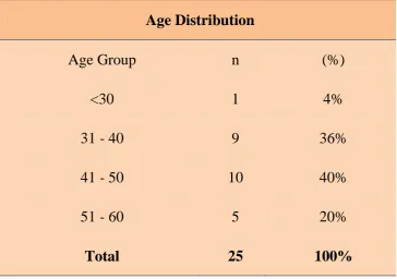
TABLE.1 DISTRIBUTION OF PHYLLODES TUMOR OF BREAST ACCORDING TO DIFFERENT AGE GROUP
Age Distribution
Age Group n (%)
<30 1 4%
31 - 40 9 36%
41 - 50 10 40%
51 - 60 5 20%
Total 25 100%
Most of the tumors around (76%) belonged to the age group
50
CHART.1 DISTRIBUTION OF PHYLLODES TUMOR OF BREAST ACCORDING TO DIFFERENT AGE GROUP
Most of the tumors around (76%) belonged to the age group
51
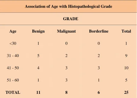
TABLE 2: ASSOCIATION OF AGE WITH HISTOPATHOLOGICAL GRADE
Association of Age with Histopathological Grade
GRADE
Age Benign Malignant Borderline Total
<30 1 0 0 1
31 - 40 5 2 2 9
41 - 50 4 3 3 10
51 - 60 1 3 1 5
TOTAL 11 8 6 25
Most of the benign tumors belonged to the age group between 30
and 50 years(80%) and the majority of malignant tumors belonged to the
52
CHART 2: ASSOCIATION OF AGE WITH HISTOPATHOLOGICAL GRADE
Most of the benign tumors belonged to the age group between 30
and 50 years(80%) and the majority of malignant tumors belonged to the
age group between 40 and 60 years(76%) 0%
5% 10% 15% 20% 25% 30% 35% 40% 45% 50%
<30 31 - 40 41 - 50 51 - 60
Benign 9% 45% 36% 9%
Malignant 0% 25% 38% 38%
Borderline 0% 33% 50% 17%
53
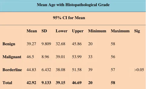
TABLE 3: MEAN AGE OF OCCURRENCE OF TUMOR IN DIFFERENT HISTOPATHOLOGICAL CATEGORIES
Mean Age with Histopathological Grade
95% CI for Mean
Mean SD Lower Upper Minimum Maximum Sig
Benign 39.27 9.809 32.68 45.86 20 58
Malignant 46.5 8.96 39.01 53.99 33 56
Borderline 44.83 6.432 38.08 51.58 39 57 >0.05
Total 42.92 9.133 39.15 46.69 20 58
Mean age for benign tumors-39.27
Mean age for borderline tumors-44.83