Structural Basis for the Broad, Antibody-Mediated
Neutralization of H5N1 Influenza Virus
Qingshan Lin,
aTingting Li,
aYixin Chen,
a,bSiu-Ying Lau,
cMinxi Wei,
aYuyun Zhang,
bZhenyong Zhang,
aQiaobin Yao,
aJinjin Li,
bZhihai Li,
aDaning Wang,
bQingbing Zheng,
bHai Yu,
bYing Gu,
a,bJun Zhang,
bHonglin Chen,
b,cShaowei Li,
a,bNingshao Xia
a,baState Key Laboratory of Molecular Vaccinology and Molecular Diagnostics, School of Life Sciences, Xiamen
University, Xiamen, China
bNational Institute of Diagnostics and Vaccine Development in Infectious Disease, School of Public Health,
Xiamen University, Xiamen, China
cState Key Laboratory for Emerging Infectious Diseases, Department of Microbiology, Li Ka Shing Faculty of
Medicine, The University of Hong Kong, Hong Kong SAR, China
ABSTRACT
Human infection with highly pathogenic avian influenza A viruses
causes severe disease and fatalities. We previously identified a potent and broadly
neutralizing antibody (bnAb), 13D4, against the H5N1 virus. Here, we report the
co-crystal structure of 13D4 in complex with the hemagglutinin (HA) of A/Vietnam/
1194/2004 (H5N1). We show that heavy-chain complementarity-determining region 3
(HCDR3) of 13D4 confers broad yet specific neutralization against H5N1, undergoing
conformational rearrangement to bind to the receptor binding site (RBS). Further,
we show that mutating four critical residues within the RBS—Trp153, Lys156, Lys193,
and Leu194 — disrupts the binding between 13D4 and HA. Viruses bearing Asn193
instead of Lys/Arg can evade 13D4 neutralization, indicating that Lys193
polymor-phism might be, at least in part, involved in the antigenicity of recent H5 genotypes
(such as H5N6 and H5N8) as distinguished from H5N1. BnAb 13D4 may offers a
tem-plate for therapeutic RBS inhibitor design and serve as an indicator of antigenic
change for current H5 viruses.
IMPORTANCE
Infection by highly pathogenic avian influenza A virus remains a threat
to public health. Our broadly neutralizing antibody, 13D4, is capable of neutralizing all
representative H5N1 viruses and protecting mice against lethal challenge. Structural
analysis revealed that 13D4 uses heavy-chain complementarity-determining region 3
(HCDR3) to fit the receptor binding site (RBS) via conformational rearrangement. Four
conserved residues within the RBS are critical for the broad potency of 13D4.
Impor-tantly, polymorphism of Lys193 on the RBS may be associated with the antigenicity shift
from H5N1 to other newly emerging viruses, such as H5N6 and H5N8. Our findings may
pave the way for highly pathogenic avian influenza virus vaccine development and
ther-apeutic RBS inhibitor design.
KEYWORDS
avian influenza virus, antibody, broad-specific neutralization, receptor
binding site
H
ighly pathogenic avian influenza viruses (HPAI) are responsible for serious
out-breaks in poultry, leading to huge economic loss worldwide, and for occasional
human infections since 1997 (1–4). The current H5N1 viruses are considered to be
derived from influenza virus A/Goose/Guangdong/1/96, which emerged in 1996
through genetic reassortment (5, 6). It is believed that genetic reassortment of H5N1
has generated H5N6 and H5N8, among others, which are emerging in south China and
parts of North America as the most recent development (3, 7–9). Currently, there is a
lack of effective antivirals for treatment of human infections with H5N1 or its derivatives
Received31 March 2018Accepted7 June 2018
Accepted manuscript posted online20 June 2018
CitationLin Q, Li T, Chen Y, Lau S-Y, Wei M, Zhang Y, Zhang Z, Yao Q, Li J, Li Z, Wang D, Zheng Q, Yu H, Gu Y, Zhang J, Chen H, Li S, Xia N. 2018. Structural basis for the broad, antibody-mediated neutralization of H5N1
influenza virus. J Virol 92:e00547-18.https://doi
.org/10.1128/JVI.00547-18.
EditorAdolfo García-Sastre, Icahn School of Medicine at Mount Sinai
Copyright© 2018 American Society for
Microbiology.All Rights Reserved.
Address correspondence to Shaowei Li, [email protected], or Ningshao Xia, [email protected].
Q.L., T.L., and Y.C. contributed equally to this work.
crossm
September 2018 Volume 92 Issue 17 e00547-18 Journal of Virology jvi.asm.org 1
on November 6, 2019 by guest
http://jvi.asm.org/
(10–12). Vaccines and therapeutics (13–17) that target the H5 subtype are thus urgently
needed.
Hemagglutinin (HA) is one of the three membrane proteins on the surface of the
influenza virus responsible for receptor recognition and virus entry. The head of the
HA1 subunit harbors the major targeting sites of neutralizing antibodies to influenza
virus (18, 19). The HA2 subunit is an
␣
-helix-rich “stem” region of the HA trimer that
functions to anchor the HA trimer onto the viral envelope membrane (20). We and
others have described several broadly neutralizing antibodies (bnAbs) that target either
the HA1 receptor binding site (RBS) or the HA2 stem region of H5N1 virus (21–23).
Structure analysis of HA complexes with antibodies has provided insight into the molecular
basis for the properties of broadly neutralizing antibodies (24–27). Canonically, heavy-chain
complementarity-determining region 3 (HCDR3) is most diverse in both length and amino
acid sequence and is often the major determinant of the specificity of antibody-antigen
recognition (28–30). Upon binding to the RBS of influenza virus, bnAbs commonly use
their HCDR loops to plug or fit the structural contour of the narrow pocket of the RBS
by mimicking binding of the sialoglycan receptor (31, 32). For example, the monoclonal
antibody (MAb) CH65 extends the HCDR3 loop into the pocket and is thus capable of
neutralizing 30 representative seasonal H1 viruses (33, 34). In contrast, MAb C05 has an
extraordinarily long HCDR3 loop that covers the entire RBS (58), facilitating broad
neutralization against H1, H2, H3, H9, and H12. Other MAbs have different actions:
HCDR3 of S139/1 recognizes various RBSs of H1, H2, H3, and H13 (59), whereas HCDR3
of F045-092 recapitulates the binding of the
␣
-2,6 of the sialoglycan receptor,
neutral-izing almost all H3 viruses (60). Recently, a human antibody, AVFluIgG03, was identified,
with broad-spectrum activity against most H5N1 clades, except for clades 1, 4, and 8,
which were isolated between 1997 and 2008 (22, 35). The crystal structure of
AVFlu-IgG03 in complex with A/Anhui/1/2005 HA highlights its significant neutralization
breadth through HCDR3 targeting of the RBS.
Previously, we generated a panel of 14 anti-H5N1 broadly neutralizing MAbs, among
which MAb 13D4 presented as the most efficacious in terms of its therapeutic potential,
with the widest neutralization capacity. Here, we solve and characterize the crystal
structures of the 13D4 Fab and its complex with the A/Vietnam/1194/2004 (VN1194) HA
ectodomain. We show that HCDR3 of 13D4 targets the RBS of the HA head region
(HAhr) by conformational rearrangement. Using structure-guided mutagenesis, we
identify the key binding sites that are specifically conserved among the H5N1 isolates.
Furthermore, we show that 13D4 can neutralize the most recent H5N1 strains. To our
knowledge, 13D4 might represent one of the most broad-spectrum H5N1 neutralizing
antibodies. Intriguingly, the antibody loses its neutralization activity against other H5N1
genetic derivatives, such as H5N6 and H5N8, which emerged in recent years,
presum-ably under evolutionary pressure caused by massive application of poultry vaccine. We
used structural comparisons among the HA crystal structures bearing polymorphisms in
key 13D4 binding sites to identify the determinants of antigenic shift from H5N1 to
other H5 genotypes. Collectively, our findings offer molecular insight into the broad yet
specific antibody-mediated neutralization of H5N1, which might be partially related to
the antigenic shift from H5N1 to other genotypes, and provide a strategy for the design
and optimization of antiviral therapeutics and broad-spectrum vaccines against H5N1
infection.
RESULTS
Overall structure of 13D4:HAhr.
To elucidate how the 13D4 MAb neutralizes all of
the representative H5N1 isolates, we solved the crystal structures of the 13D4 Fab alone
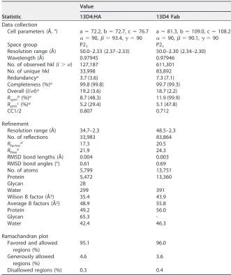
and in complex with the VN1194 HA head region (13D4:HAhr) to 2.30-Å and 2.33-Å
resolution, respectively, and refined to
R
work/
R
freevalues of 20.5% and 24.3%, and 17.3%
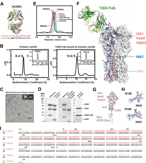
and 21.9%, respectively (Table 1). Four 13D4 Fab molecules are found in the asymmetric
unit and display consistent conformation of the CDR loops, albeit HCDR3 is involved in
crystal packing (Fig. 1A). For crystallization of the immune complex, HA was generated
in baculovirus-infected insect cells, as described by Stevens et al. (36). One trimeric HA
on November 6, 2019 by guest
http://jvi.asm.org/
can bind three 13D4 Fabs in solution, as shown in the continuous molar mass
distribution [c(M)] profiles of the analytical ultracentrifugation (AUC) assay (Fig. 1B) and
two-dimensional (2D) average figures of cryo-electron microscopy (EM) imaging
(Fig. 1C). Purified HA0 was digested with thrombin and trypsin and mixed with the
13D4 Fab for crystallization (Fig. 1D and E). However, in the asymmetric unit of the
HA:13D4 Fab complex, one 13D4 Fab binds with only the head region of HA1 (amino
acids [aa] 47 to 309; H3 numbering), denoted HAhr (Fig. 1F, G, and I). HA might have
degraded due to the length (
⬃
3 months) of the crystallization process. However, we
found that the structure of the HA1 head region is congruent with the corresponding
moiety of the full-length Viet04 HA trimer structure (Protein Data Bank [PDB] accession
no.
2IBX
) (superimposition with a root mean square deviation [RMSD] of 0.37 Å for all
HA1 C-
␣
atoms) (Fig. 1G). The glycan chains linked to Asn158 and Asn169 in our HA1
[image:3.585.39.370.91.485.2]structure are well resolved in the electron density map (Fig. 1F and H) and superimpose
with HA1 of trimeric HA (Fig. 1F). Overall, the 13D4 interaction interface in HA1 is
located on the RBS; this is consistent with our previous result for the escape mutation
TABLE 1Data collection and refinement statistics for 13D4:HA complex and 13D4 Fab alone
Statistic
Value
13D4:HA 13D4 Fab
Data collection
Cell parameters (Å, °) a⫽72.2, b⫽72.7, c⫽76.7 a⫽81.3, b⫽109.0, c⫽108.2 ␣⫽90,⫽93.4,␥⫽90 ␣⫽90,⫽90.1,␥⫽90
Space group P21 P21
Resolution range (Å) 50.0–2.33 (2.37–2.33) 50.0–2.30 (2.34–2.30)
Wavelength (Å) 0.97945 0.97946
No. of observed hkl (I⬎) 127,187 611,301
No. of unique hkl 33,998 83,892
Redundancya 3.7 (3.6) 7.3 (7.1)
Completeness (%)a 99.8 (99.8) 99.7 (99.3)
Overall (I/I)a 19.2 (3.6) 18.7 (2.2)
Rsymb(%)a 8.7 (48.3) 11.9 (99.9)
Rpimc(%)a 5.2 (29.4) 5.1 (47.8)
CC1/2 0.807 0.712
Refinement
Resolution range (Å) 34.7–2.3 48.5–2.3
No. of reflections 33,983 83,864
Rfactord 17.3 20.5
Rfreee 21.9 24.3
RMSD bond lengths (Å) 0.004 0.003
RMSD bond angles (°) 0.61 0.69
No. of atoms 5,799 13,751
Protein 5,472 13,360
Glycan 28
Water 299 391
Wilson B factor (Å2) 35.4 43.9
Average B factors (Å2) 48.9 55.8
Protein 49.2 56.0
Glycan 65.3
-Water 42.4 46.3
Ramachandran plot Favored and allowed
regions (%)
95.1 96.0
Generously allowed regions (%)
4.6 3.6
Disallowed regions (%) 0.3 0.4
aThe numbers in parentheses refer to the highest resolution shell. bR
sym⫽ ⌺h⌺i|I1(h)¡I(h)|/⌺h⌺i I1(h). cR
pim⫽ ⌺hSQRT[1/(n⫺1)]⌺i|I(1h)¡I(h)|/⌺h⌺i I(1h). dR
factor⫽ ⌺hkl||Fobs|⫺k|Fcalc||/⌺hkl|Fobs|. eR
freeis calculated using the same equation as forRfactor, but 5.0% of the reflections were chosen randomly and omitted from the refinement.
September 2018 Volume 92 Issue 17 e00547-18 jvi.asm.org 3
on November 6, 2019 by guest
http://jvi.asm.org/
FIG 113D4 binds to the RBS on the HAhr. (A) Overlay of four 13D4 Fab molecules in the asymmetric unit and the HA-bound 13D4 Fab. The structure superimposition shows that 13D4 Fabs in the crystal have a consistent loop conformation for CDR loops in the crystal packing. The HCDR3 loop undergoes a substantial conformational change. (B) Sedimentation coefficient measurements of HA alone and in complex with 13D4 Fab. (Left) HA exists as single species in c(s) profile, with an 8.4S sedimentation coefficient and 205-kDa apparent molecular mass; this indicates that HA mainly exists as a trimer in solution. (Right) The HA:13D4 complex presents major species in c(s) profile, 10.6S and 320 kDa, corresponding to one HA trimer binding with 3 Fabs. recHA, recombinant HA. (C) Cryo-EM micrograph of unstained, vitrified HA:13D4 complex. Discernible particles were manually boxed within 20-nm diameter and subjected to 2D classification with Relion software. Two representative classes (morphology) are shown in the insets, corresponding to a top view and a side view of three Fabs binding to the HA trimer. The brightness of the original image was enhanced by 50% to make the trimer shape clearer. (D) Protease treatment of HA. Before protease treatment, HA resolved as a full-length HA0 and separate HA1 and HA2 in SDS-PAGE. The foldon facilitating HA trimerization was removed with thrombin, and the thrombin-treated sample was further catalyzed with trypsin, demonstrating that HA0 was completely cleaved to HA1 and HA2. The purified immune complex sample was further analyzed by SDS-PAGE. (E) HPLC profiles of protease-treated HA and its complex with 13D4 Fab. (F) Overall structure of
(Continued on next page)
on November 6, 2019 by guest
http://jvi.asm.org/
[image:4.585.41.539.67.622.2]on 13D4 neutralization. 13D4 Fab in the complex has a well-defined electron density
map and shares similar overall structure with the free 13D4 Fab (RMSD, 0.85 Å for all of
the C-
␣
atoms), except for a significant change in HCDR3 (Fig. 1A).
Interactions between HA1 and 13D4 Fab.
13D4 Fab binds to the membrane-distal
region of the globular head of HA at nearly 45° to the 3-fold axis of the HA trimer (Fig.
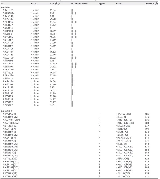
1F). The HAhr-13D4 interaction buries 857 Å
2of surface area on HA1, as calculated by
PISA (37) (Table 2). The heavy chain mediates most of the HAhr-13D4 contacts (75.6%
of the total buried surface area of HA1, i.e., 648 Å
2out of 857 Å
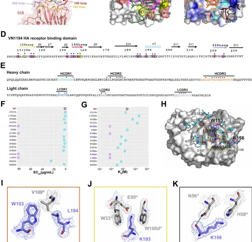
2). All three loops of the
CDR on the 13D4 heavy chain (HCDR) bind the RBS, which is framed by four structural
elements: the 130 loop, the 150 loop, the 190 helix, and the 220 loop (Fig. 2A). We
found that HCDR1 binds to the ridge between the 130 loop and the 150 loop, HCDR2
binds to the 150 loop, and HCDR3 binds deeply into the negatively charged RBS core
region. In contrast, the light-chain CDR (LCDR) loops mostly make contact with the
residues outside the RBS, i.e., LCDR1 to -3 bind to the regions near the 190 helix and
the 220 loop (Fig. 2A to C). Together, these CDR recognitions define a conformational
epitope comprising four stretches of amino acids: Glu131 to Ser137, Trp153 to Ser159,
Asn186 to Leu194, and Lys222 to Gln226. These amino acid stretches are highly
conserved (83%) among all H5 isolates (
n
⫽
5,693; data collected before 2016 from the
NCBI Influenza Virus Resource) (Fig. 2B and Table 3). Our observations are also
consis-tent with our previous data on the neutralization breadth and potency of MAb 13D4.
In this complex structure, extensive interactions are established, with 16
hydrogen-bonding contacts and 6 salt-bridging linkages (Table 2) formed between the side chains
of the epitope residues (Fig. 2D) and the residues of 13D4 Fab (Fig. 2E).
Structure-based mutational analysis of the 13D4 neutralization sites.
In the
HAhr:13D4 complex structure, 16 HA residues at the interface interact with the 13D4
Fab through their side chains (Table 2). To identify the residues critical for 13D4
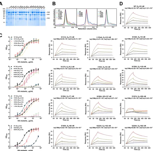
binding, alanine-scanning mutagenesis was carried out on all 16 amino acids. All of the
unprocessed HA mutants were detected in sodium dodecyl sulfate-polyacrylamide gel
electrophoresis (SDS-PAGE) (Fig. 3A) and resolved as trimers in solution, similar to
wild-type (WT) HA (Fig. 3B). We used 50% effective concentration (EC
50) (enzyme-linked
immunosorbent assay [ELISA]) and affinity (surface plasma resonance [SPR])
measure-ments to confirm the importance of each mutation. Notably, 4 mutations (W153A,
K156A, K193A, and L194A) dramatically decreased the binding of 13D4 with HA, with
the EC
50more than 20 times higher for these mutants than for WT HA (Fig. 2F and G
and 3C and D). The association/disassociation SPR responses of the four mutants to
MAb 13D4 were undetectable. The other mutants showed affinities similar to that of WT
HA, and two mutants (L133aA and S137A) showed significantly enhanced binding
affinities to 13D4, with affinity constants (
K
D) of
⬃
53 nM to 5 to 8 nM (Fig. 3D).
The functions of these four strategic residues are structural: Trp153 and Leu194
simultaneously interact with Val100
H, where the superscript H denotes heavy chain,
making hydrophobic interactions and van der Waals attractions (Fig. 2H and I); Lys193
makes contact with Glu50
Hthrough hydrogen bonding and salt bridging, and the stem
of its hydrophobic side chain is stabilized by two flanking benzpyrole rings derived
from Trp33
Hand Trp100d
H(61) residues (Fig. 2H and J); and Lys156 binds to Asn56
Hvia
one hydrogen bond (Fig. 2H and K). Together, Trp153 and Lys156, located on the 150
loop, and Lys193 and Leu194 on the 190 helix are critical for 13D4 neutralization. These
residues are highly conserved (Trp153, 100%; Lys156, 99.3%; Lys/Arg193, 88.4%;
FIG 1Legend (Continued)
the 13D4:HAhr complex. The HAhr of the 13D4:HAhr complex overlaps one protomer of the HA trimer (PDB accession no.2IBX). The HA1 head region of the 13D4:HAhr complex is in salmon; the heavy chain and light chain of 13D4 Fab are in forest green and lime green, respectively; and HA1 and HA2 components of the HAhr-superimposed protomer are in dark blue and pink, respectively. (G) Superimposition of the HA head region on the HA1 monomer in the reported crystal structure (PDB accession no.2IBX). (H) Sample electron density (2Fo-Fc [Fo, true complex structure factors of a crystal structure; Fc, calculated complex structure factors of any model structure]) maps contoured at 1above the mean are shown for N-linked glycans bound to Asn158 and Asn169 of HA. NAG, N-acetylglucosamine. (I) Sequence alignment of the HA head region (aa 47 to 309; H3 numbering) resolved in the 13D4:HAhr crystal and the HA construct (consisting of HA1 [aa 11 to 329] and HA2 [aa 1 to 174]). The dashes denote the corresponding residues that are missing in the crystal structure.
September 2018 Volume 92 Issue 17 e00547-18 jvi.asm.org 5
on November 6, 2019 by guest
http://jvi.asm.org/
TABLE 2Interface identification and interaction analysis of 13D4:HAhr by PISA Program
HA 13D4 BSA (Å2)a % buried areab Typec 13D4 Distance (Å)
Interface
A:GLU131 H chain 19.34 ||||
A:LEU133a H chain 91.94 ||||||||
A:GLY134 H chain 1.81 ||
A:VAL135 H chain 29.28 ||||
A:SER136 H chain 11.94 ||||||||||
A:SER137 H chain 14.12 ||
A:SER145 H chain 14 |||
A:TRP153 H chain 18.69 ||||||||
A:ILE155 H chain 15.75 ||||||||
A:LYS156 H chain 40.75 ||||||||||
A:LYS157 H chain 11.39 ||
A:ASN158 H chain 34.09 |||
A:SER159 H chain 47.19 ||||||
A:ASN186 H chain 4 ||
A:ASP187 H chain 19.52 ||||
A:ALA189 H chain 22.76 ||||
A:GLU190 H chain 35.92 ||||||||
A:TRP192 H chain 9.03 ||
A:LYS193 H chain 132.46 ||||||||||
A:LEU194 H chain 28.12 |||||||||
A:GLN196 H chain 5.88 |
A:LYS222 H chain 16.98 ||
A:GLN226 H chain 13.48 ||||
A:SER227 H chain 9.97 ||||
A:ASN186 L chain 16.54 |||||||
A:ASP187 L chain 25.96 ||||||
A:ALA188 L chain 2.95 |
A:ALA189 L chain 50.32 |||||||
A:THR192 L chain 15.79 |||
A:LYS193 L chain 19.90 ||
A:THR219 L chain 69.97 ||
A:LYS222 L chain 59.27 ||||
A:SER227 L chain 6.15 |||
Interaction
A:LYS156[O] H H:ASN56[ND2] 3.05
A:SER159[OG] H H:ILE57[N] 2.79
A:ASP187 [OD1] H H:ARG100b[NE] 2.75
A:ASP187[OD2] H H:ARG100b[NH2] 2.84
A:LYS193[NZ] H H:GLU50[OE2] 2.77
A:ASN158[N] H H:SER54[O] 2.91
A:SER159[N] H H:GLY55[O] 3.32
A:SER159[OG] H H:GLY55[O] 3.10
A:ASN158[N] H H:ASN56[OD1] 3.11
A:SER159[N] H H:ASN56[OD1] 2.76
A:SER159[OG] H H:ILE57[O] 3.43
A:SER136[OG] H H:GLU100a[OE1] 3.71
A:SER136[OG] H H:GLU100a[OE2] 3.53
A:SER137[N] H H:GLU100a[OE2] 2.83
A:SER137[OG] H H:GLU100a[OE2] 3.27
A:LYS222[NZ] H L:SER50[OG] 3.24
A:ASP187[OD2] S H:ARG100b[NE] 3.25
A:ASP187[OD1] S H:ARG100b[NE] 2.75
A:ASP187[OD2] S H:ARG100b[NE2] 2.84
A:ASP187[OD1] S H:ARG100b[NE2] 3.23
A:LYS193[NZ] S H:GLU50[OE1] 3.54
A:LYS193[NZ] S H:GLU50[OE2] 2.77
aBSA, buried surface area. b|, 10%.
cH, hydrogen bond; S, salt bridge.
on November 6, 2019 by guest
http://jvi.asm.org/
FIG 2Interaction analysis of the 13D4:HAhr immune complex. (A) Enlarged view of the interface of the 13D4:HAhr complex. (B) Sequence conservation of the 13D4 epitope. HAhr is shown in surface representation with epitope residues colored according to their conservation values. (C) The electrostatic potential surface of HA is depicted in gradient color: red, negative,⫺4 kT; blue, positive,⫹4 kT; white, neutral. Contacting residues of the 13D4 CDR loops are in stick form for side chains and cartoonform for main chains. Panels B and C are shown horizontally rotated 90° from panel A. (D and E) Sequences of the RBS domain and 13D4 CDR, respectively. The residues in the interface are marked with asterisks and are depicted with the same color scheme as in panels B and C. (F) Reactivity profiles of HA and its mutants against 13D4 antibody measured through sandwich ELISA. The EC50values were calculated by sigmoidal fitting as shown in Fig. 3C. The EC50is plotted as circles along the horizontal axis for each protein. Purple circles, EC50of⬎80g/ml; cyan,⬍30g/ml; brown, wild-type HA. (G) Binding affinity measurements of HA and its mutants against 13D4 by SPR. The kinetic constants between bnAb 13D4 and W153A, K156A, K193A, and
(Continued on next page)
September 2018 Volume 92 Issue 17 e00547-18 jvi.asm.org 7
on November 6, 2019 by guest
http://jvi.asm.org/
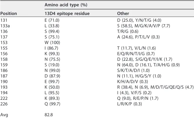
[image:7.585.40.541.164.647.2]Leu194, 95.5%) among all H5 isolates, and the RBS localization (Fig. 2B and Table 3)
determines the broad neutralization specificity of the 13D4 epitope. These results are
also consistent with our previous 13D4 neutralization experiment on virus escape,
where we replaced Lys156 and Lys193 with Glu and Met, respectively. In addition, in the
present study, we found alanine replacement of the other residues, such as Asn158,
Asn186, Asp187, Lys222, and Gln226, could also perturb 13D4 binding to HA to some
extent, with 5-fold lower affinity with respect to WT HA (Fig. 3D). These residues, which
line the outside the RBS (Fig. 2B), might assist in antibody binding but do not appear
to be critical for binding.
13D4 HCDR3 adaptive fitting to the RBS.
Similar to other reported RBS-directed
bnAbs, the HCDR3 loop of 13D4 binds deeply within the RBS groove. Notably, structural
superimposition of HA-bound and unbound 13D4 Fabs packing in the crystals revealed
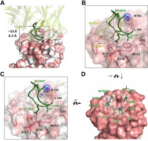
substantial conformational changes at the HCDR3 loop (Fig. 1A). Amino acids 93 to 101
of the HCDR3 loop of the free Fab would clash with the 220 loop of receptor binding
domain (RBD) if 13D4 underwent direct binding to HA (Fig. 4A and B). To avoid this
collision, a subloop, aa 95 to 101, swings from the 220 loop toward the 190 helix at
about 33.6°, with Arg94
Hand Ala99
Hacting as mechanical fulcrums (Fig. 4A; see Movie
S1 in the supplemental material). There, the C-
␣
atoms of Val100
Hand Glu100a
Hmove
8.3 Å and 7.6 Å, respectively, away to create hydrogen bonding and hydrophobic
interactions with Ser136, Ser137, Trp153, Leu194, and Ile155 (Fig. 4B). The
conforma-tional fitting over the RBS may determine the broad yet specific nature of MAb 13D4,
and this mode of binding is rarely observed in other bnAb-mediated binding. Indeed,
in the literature, the HCDR3 loop of MAb H5.3 has been reported to rotate 90°
in situ
between its unliganded and liganded positions (38), which is distinct to the
confor-mational change of MAb 13D4.
Minor conformational changes were also observed in the RBS during HCDR3
bind-ing; most notably, the side chain of Lys193 rotates to accommodate the gap created by
the benzpyrole rings of Trp100d
Hand Trp33
Hand donates a hydrogen bond to Glu50
H.
There is also subtle perturbation to the side chain of Ile155, which constitutes a
FIG 2Legend (Continued)
[image:8.585.41.370.82.283.2]L194A mutants were not determinable and are reported as higher than 1,000 nM. The calculated affinity constants and fitting results are depicted in Fig. 3D. TheKDvalues are plotted as circles along the horizontal axis for each protein. Purple circles,KDof⬎1,000 nM; cyan,⬍500 nM; brown, wild-type HA. (H) The 13D4 epitope sites for HA are depicted in stick form on the molecular surface of 13D4 Fab. The epitope residues were identified by the PISA server (Table 2). (I to K) Enlarged views of the interactions for W153 and L194 (red box), K193 (yellow box), and K156 (black box) from the corresponding boxes in panel H. Sample electron density (2Fo-Fc) maps contoured at 1above the mean are shown for all the residues. Hydrogen bonds are shown as black dashed lines. TABLE 3Sequence conservation of 13D4 epitope residues in all H5 isolatesa
Position
Amino acid type (%)
13D4 epitope residue Other
131 E (71.0) D (25.0), Y/N/T/G (4.0)
133a L (33.8) S (58.5), M/G/K/A/V/P (7.7)
136 S (99.4) T/R/G (0.6)
137 S (75.1) A (24.6), P/T/L/V (0.3)
153 W (100)
155 I (86.7) T (11.7), V/L/N (1.6)
156 K (99.3) E/Q/R/N/T/I/G (0.7)
158 N (75.5) D (22.8), S/G/Q/E/Y/I/K (1.7)
159 S (19.0) N (64.0), D (16.1), T/A/H/G (0.9)
186 N (99.0) S/K/T/A/D/I (1.0)
187 D (87.9) N (11.1), H/G/S/Y (1.0)
190 E (99.7) K/H/A/D/V (0.3)
193 K (50.0) R (38.4), N (6.9), M/D/T/G/QE/Q/S (4.7)
194 L (95.5) I (4.3), V/F/S (0.2)
222 K (89.3) Q (9.0), R/E/P/N (1.7)
226 Q (99.7) L/R/K/P (0.3)
Avg 82.8
aThe cumulative number deposited in the NCBI Influenza Database by 2016 was 5,693.
on November 6, 2019 by guest
http://jvi.asm.org/
hydrophobic cavity, together with Trp153 and Leu194, for the binding of Val100
Hof
HCDR3 (Fig. 4C). Compared to the free Fab structure, the fitted conformation of the
HCDR3 loop against the RBS might be induced energetically by the spatial restriction
of the RBS structural contour (Fig. 4D; see Movie S1 in the supplemental material).
These interactive conformational changes manifest an induced fit during HA-13D4
binding, and, given that W153A and L194A mutations abrogate this HA-13D4 binding,
it is likely that these residues are critical for the HA-13D4 interaction (Fig. 1F and 2A).
FIG 3Characterization of HA mutants with structure-based alanine substitutions in 13D4 binding sites. (A) SDS-PAGE analysis of the purified HA mutants. Notably, most of the N158A mutant proteins were cleaved into HA1 and HA2. (B) HPLC profiles of the purified HA mutants. The mutants were resolved as major components in HPLC curves with the same retention volume as WT HA, indicating the proteins exist mainly as trimers in solution. Abs, absorbance. (C) The interaction of HA and its mutants against MAb 13D4 was tested by double-antibody sandwich ELISA, and EC50s were calculated by sigmoid trend fitting and plotted as in Fig. 2F. OD450, OD at 450 nm. The error bars represent standard deviations of two repeats. (D) Binding curves of HA, HA mutants, and MAb 13D4 in SPR. A total of 5 concentrations (8.93, 17.9, 35.7, 71.4, and 143 nM) of HA and its mutants (serial dilutions) were injected onto a MAb 13D4-bound chip. The kinetic constants between MAb 13D4 and HA/HA mutants are reported.
September 2018 Volume 92 Issue 17 e00547-18 jvi.asm.org 9
on November 6, 2019 by guest
http://jvi.asm.org/
[image:9.585.45.548.68.560.2]Comparison with receptor analogue and other H5 head region antibodies.
We
next sought to compare the 13D4:HAhr structure with the structure of an avian
receptor analogue (LSTa [3
=
-sialyl-
N
-acetyllactosamine]) bound to HA (PDB accession
no.
3ZP0
) (39) and with other immune complexes that contain H5 head region
antibodies. Overall, the tip residues E100a
Hand V100
Hof the 13D4 HCDR3 loop
recapitulate the receptor analogue binding scenario (Fig. 5A to D).
There are two major interactions between LSTa and the RBS, one through hydrogen
bonding and one through hydrophobic interactions. In the first, triple hydrogen
bonding occurs between the sialic acid carboxylate and Ser136 and Ser137 inside the
RBS (Fig. 5A). The carboxylate of E100a
Hcreates four hydrogen-bonding connections
with Ser16 and Ser137, and the hydroxyl group of the side chain of Ser137 receives one
more hydrogen bond than LSTa (Fig. 5B). Although other receptor-mimetic antibodies
against H1 and H3 show similar hydrogen-bonding scenarios (PDB accession no.
4XNM
,
3SM5
,
4M5Z
,
2VIR
,
1KEN
, and
4O58
) (38, 40, 41), 13D4 uses Glu instead of Asp for its
hydrogen-bonding network (Fig. 5A and F). This hydrogen-bonding modality differs
from those of other H5 RBS-directed MAbs: H5.3 exclusively uses the main chain of Asp
for H bonding, whereas AVFluIgG03 does not form hydrogen bonds via its Asp residue
(Fig. 5E). Thus, the use of Glu to recapitulate hydrogen bonding instead of Asp in 13D4
is unique (Fig. 5F). No similar hydrogen bonding between 13D4 Fab and the RBS was
observed, as the LSTa acetamide accepts a hydrogen bond from the main chain of
Val135 (Fig. 5A and B).
FIG 4Conformational change in 13D4 HCDR3 and HA, with HCDR3 adapting to fit into the RBS. (A) The free 13D4 and unbound HA monomer (from PDB accession no.2IBX) were superimposed on correspond-ing components of the 13D4:HAhr complex. Fabs are depicted in cartoon mode, with free Fab in yellow and bound Fab in forest green. HA molecules are shown in surface presentation, with unbound HA in white and bound HA in salmon. (B) Enlarged view of the HCDR3 rearrangement (boxed in panel A). For clarity, other parts of the interaction from panel A have been omitted. Hydrogen bonds are shown as black dashed lines. W100dHand E100aHin the free Fab are shown in transparent sphere dot represen-tation, demonstrating clashing with the 220 loop. (C) Conformational changes to HA while binding with 13D4. For clarity, other parts of the interaction from panel A are omitted. (D) Eventual fine fitting of HCDR3 to the RBS. The non-H atoms of HCDR3 up to 4.5 Å from HA are shown in dot sphere representation.
on November 6, 2019 by guest
http://jvi.asm.org/
[image:10.585.55.355.72.359.2]FIG 5Receptor mimicking of 13D4 and footprint comparison of 13D4 and other H5 neutralizing antibodies. (A) Hydrogen bonding between receptor analogue LSTa and the RBS as shown in the co-crystal structure (PDB no.3ZP0). (B) Hydrogen bonding between the 13D4 HCDR3 tip and the RBS. (C) The carbohydrate group of the LSTa SIA terminus stretches into a hydrophobic cavity enfolded by side chains of Trp153, Leu194, and Ile155. (D) V100Hof HCDR3 mimics the hydrophobic interaction with the RBS, resembling the carboxylate group of the LSTa SIA terminus. (E) Overlay of the 13D4 HCDR tip with representative RBS-directed neutralizing antibodies. The Glu at the tip of 13D4 HCDR3 (green) is different from the Asps of other antibodies; the longer side chain of Glu forms four hydrogen bonds with S136 to S137, more than the hydrogen bond numbers formed by Asp at the HCDR3 tips of other antibodies: H5.3 (PDB accession no.4XNM; orange), CH65 (3SM5; cyan), 5J8 (4M5Z; teal), HC19 (2VIR; white), HC63 (1KEN; purple), Fo45-092 (4O5I; brown), and AVFluIgG03 (5DUP; pink). (F) Sequence alignment of HCDR3 RBS-directed antibodies highlighting the Glu at the tip of 13D4 HCDR3, which is distinct from those of other antibodies. The same color scheme as in panel E was used. (G) Comparison of the binding sites of 13D4 and other H5-specific antibodies that recognize the HA1 globular head domain. The immune complex structures are overlaid in surface mode: 13D4 (green) in AVFluIg03 (PDB accession no.5DUP; pink), H5.3 (4XNM; orange), 65C6 (5DUM; yellow), 100F4 (5DUR; pale cyan), FLD194 (5A3I; cyan), and H5M9 (4MHH; brown). (H) The view is horizontally rotated 90° with respect to panel G. (I) Footprints of three H5N1 RBS-targeting antibodies. The HA head region is shown in surface representation with the overlapping region in salmon and the unique binding residues in green, pink, and orange for 13D4, AVFluIgG03, and H5.3, respectively. (J) Footprints of four H5N1 non-RBS-directed antibodies. Table 4 details the neutralization breadths and evolving germ lines for these antibodies.
September 2018 Volume 92 Issue 17 e00547-18 jvi.asm.org 11
on November 6, 2019 by guest
http://jvi.asm.org/
[image:11.585.38.540.61.568.2]For the second major interaction, the sialic acid of LSTa binds to a hydrophobic
region created by the highly conserved Trp153, Ile155, and Leu194 in almost all H5
isolates (Table 3). The tip residue, Val100
H, of the 13D4 HCDR3 loop binds to this region
via its hydrophobic side chain, which is also observed in many other antibodies, such
as CH65 and 2G1 (33, 42).
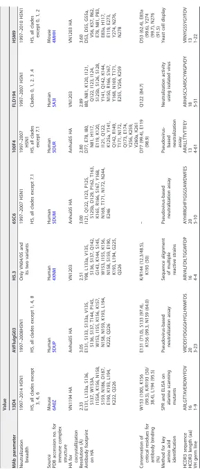
All of the reported structures for HA-H5 bnAbs show targeting of the HA head
region, but they can be further classified into two groups: non-RBS-directed binding
(MAbs 65C6, 100F4, FLD194, and H5M9 [43, 44]), with footprints shifted toward the
distal membrane and twisting counterclockwise about 60° along the HA 3-fold axis with
respect to the RBS (Fig. 5G, H, and J), versus RBS-directed binding, where 13D4,
AVFLuIG03, and H5.3 all share similar binding orientations against HA and define an
overlapping region (Fig. 5G to I). However, the key residues mediating binding of 13D4,
AVFLuIG03, and H5.3 to HA are different, and this may account for the differences noted
in the breadths of neutralization. 13D4 corresponds to sites more conserved than those
of the other two MAbs (Fig. 5I and Table 4).
A key 13D4 binding site associated with antigenicity shift from H5N1 to other
H5 viruses.
In our previous study, we showed that MAb 13D4 conferred broad
neutralization against all representative H5 isolates identified between the years 1997
and 2009 and was verified as a potential therapeutic candidate for curing H5-infected
animals from lethal infection following H5 virus challenge. To confirm the neutralization
breadth since our previous study, we tested the emergent H5 subtype viruses isolated
from 2012 to 2014 using HA inhibition (Fig. 6A). The data showed that our 13D4 MAb
can inhibit hemagglutination of H5N1, but not H5N6 and H5N8, two viral subtypes that
first emerged in 2013 and 2006, respectively (Fig. 6A, table). We then analyzed the
antigenic shifts of the emergent H5N6 and H5N8 from their ancestral H5N1 (62) using
escape neutralization of 13D4. Sequence alignment of the H5 HAs revealed that only 2
residues, D187N and K/R193N, of the 15 13D4 epitope residues are mutated between
H5N1 and H5N6/H5N8, both located on the 190 loop (Fig. 6A). In our structure and
mutagenesis analysis, Asp187 was not involved in 13D4 binding, whereas the side chain
of Lys193 was shown to play a strategic role in the interaction between 13D4 and HA
(Fig. 2F, G, and J). Moreover, 13D4 maintains strong neutralization against H5N1 viruses
that retain Arg193 in HA, such as HK/5923/2012 and Zhejiang/98/2014 isolates, and also
those viruses bearing the Lys193 residue (Fig. 6A). Superimposition of the HA structures
with either Lys193 or Arg193 showed similar interaction with Trp33
H, Glu50
H, and
Trp100d
Hof 13D4 (Fig. 6B). However, Asn193 of H5N6/H5N8 has a shorter side chain
than Lys/Arg and thus abrogates 13D4 recognition, possibly because it fails to establish
the critical interaction for 13D4 binding (Fig. 6C). Polymorphic analysis of residue 193
among 5,693 H5 isolates revealed high conservation of the Lys/Arg residue at this
position, with 88.4% identity (Table 5). Lys/Arg193 is completely conserved in all
13D4-reactive H5 isolates (Table 3).
DISCUSSION
Although H5 subtype influenza viruses still exclusively circulate in avian hosts,
cross-species infection in sporadic human cases led to severe disease, with a
⬎
50%
mortality rate. No effective antiviral is available for the treatment of human infection
with H5N1 or other highly pathogenic H5 subtype viruses. Like other influenza viruses,
H5 subtype virus evolves rapidly in the field. Development of broadly neutralizing
antibodies has been considered one of the therapeutic means to respond to the
potential threat of highly pathogenic avian influenza viruses. We previously
character-ized an H5-specific broadly neutralizing antibody, 13D4, and showed potent
therapeu-tic activity against H5N1 infection in mice (21). Here, we report the structure and
function of this potent neutralizing antibody against H5N1 isolates. We show that
HCDR3 adaptively fits into the shallow groove of the HA RBS and highlight the key
residues involved in this binding.
Comparing sequence conservation and genetic variation, there are three highly
conserved regions on HA that are associated with the bnAbs, including the RBD, the
on November 6, 2019 by guest
http://jvi.asm.org/
TABLE 4 Summary of H5N1 neutralizing antibodies targeting the HA head region MAb parameter Value 13D4 AVFluIgG03 H5.3 65C6 100F4 FLD194 H5M9 Neutralization breadth 1997–2014 H5N1 1997–2008H5N1 Only VN04/05 and its two variants 1997–2007 H5N1 1997–2007 H5N1 1997–2007 H5N1 1997–2010 H5N1 H5, all clades except 3, 4, 6 H5, all clades except 1, 4, 8 H5, all clades except 7.1 H5, all clades except 7.1 Clades 0, 1, 2. 3 .4 H5, all clades except 0, 1, 2 Source Mouse Human Human Human Human Human Mouse PDB accession no. for immune complex structure 6A0Z 5DUP 4XNM 5DUM 5DUR 5A3I 4MHH HA for cocrystallization VN1194 HA Anhui05 HA VN1203 Anhui05 HA Anhui05 HA VN1203 VN1203 HA Resolution (Å) 2.33 3.05 2.51 3.00 2.80 2.89 3.60 Antibody footprint on HA E131, L133a, S136, S137, W153A, I155A, K156, N158, S159, N186, D187, E190, K193, L194, K222, Q226 E131, S133, S133a, V135, S136, S137, T144, P145, W153, I155, K156, K157, N158, N159, K193, L194, K222, Q226 Y98, L133a, V135, S136, S137, Q142, G143, K144, S145, W153, I155, K156, N158, S159, E190, K193, L194, G225, Q226 I121, Q122, I123, P125, S125b, D129, P162, T163, K165, R166, S167, Y168, N169, T171, N172, N244, E246 D77, E78, I80, N81, H117, E119, K120, I121, Q122, K125a, Y141, Q142, R149, T171, N172, Q173, E174, Y256, K259, V260a, K261 I80, N81, K120, I121, Q122, I123, I124, S125b, S126, S128, Y141, Q142, K144, N150, R166, S167, Y168, N169, T171, E255, Y256, K259 D53, D55, G55a, V56, K57, R62, E78, N81, P83, E83a, H117, E119, E273, Y274, N276, N278 Conservation of critical residues for antibody binding (%) W153 (100), K156 (99.3), K/R193 (50/ 38.4), L194 (95.5) E131 (71.0), S133 (97.4), K156 (99.3, N159 (64.0) K/R144 (12.3/48.5), K193 (50) – D77 (99.4), E119 (98.9) Q122 (84.7) D53 (62.4), E83a (99.6), Y274 (99.7), N276 (91.5) Method for key amino acid identification SPR and ELISA on alanine scanning mutants Pseudovirus-based neutralization assay Sequence alignment of multiple reactive strains Pseudovirus-based neutralization assay
Pseudovirus-based neutralization assay
Neutralization activity using isolated virus Yeast cell display HCDR3 sequence ARLGTTAVERDWYFDV VRDDSYDGGGHYGLHNWFDS ARVALFDILTGGWFDP AYHRRGHFYGSGSAWDWFES ARALLTTVTTFEY ARHASCSARSCYWGPVDY VRNYGSSYGYF DV HCDR3 length (aa) 16 20 16 20 13 18 13 VH germ line 1-9 3-23 4-4 5-10 4-61 5-51 1-22
September 2018 Volume 92 Issue 17 e00547-18 jvi.asm.org 13
on November 6, 2019 by guest
http://jvi.asm.org/
[image:13.585.52.341.63.741.2]stem region, and the vestigial esterase region (45). Most isolated bnAbs target the RBD,
such as 5J8, 1F1 (63), and CH65, with single-subtype-specific breadth, and C05, S139/1,
and F045-092, with cross-subtype activity. The globular head domain bears the
con-served RBS, which mediates host specificity and is responsible for the antigenic shift
and antigenic drift in the major antigen determinants, which leads to escape from viral
neutralization. Notably, bnAbs usually demonstrate receptor mimicry through various
binding strategies using the HCDR3 loop. In the case of the H5 subtype, two known
bnAbs, AVFluIgG03 and H5.3, are RBS-directed antibodies, and HCDR3 inserts directly
into the binding groove. These RBS-directed antibodies neutralize all clades of H5
(except 1, 4, and 8), as well as VN/1203 and two H5
rdt
variants only, respectively (Fig.
5I and J). Our 13D4 can neutralize all major H5N1 clades isolated between 1997 and
2014. Structure-guided mutagenesis on 13D4 binding sites has identified four highly
FIG 6Molecular determinants for antigenic shift from H5N1 to other genotypes. (A) RBD sequence alignment of six H5 subtype viruses and HI titer assay of 13D4 or ferret antiserum. The epitope residues of 13D4 are boxed in cyan. Disparate residues between H5N1 and H5N6/H5N8 are indicated by arrowheads (below). Four key sites of 13D4 binding are designated by black stars. The overlapping subset of mutated and key binding residues is designated by the red asterisk. Dots denote that the residue is identical with that of the first VNM sequence. (B) Arg (R) 193 shares a similar side chain rotamer and putative 13D4 interaction mode with Lys (K) 193. Free HA structures of A/duck/Egypt/10185SS/2010 (PDB accession no.5E30) and A/Vietnam/1194/2004 (PDB accession no. 2IBX), bearing R193 and K193, respectively, are superimposed with the 13D4:HAhr complex. (C) N193 differs from K193 and fails to interact with 13D4. Free HA (A/Sichuan/26221/2014; PDB accession no.5HU8) containing N193 was overlaid with the free and bound HAs containing K193, as shown in panel B. Dotted ovals highlight the interaction in 193 position. The dashed lines are hydrogen bonds.
on November 6, 2019 by guest
http://jvi.asm.org/
[image:14.585.45.544.73.498.2]conserved amino acids in its targets, which may account for the wide breadth of
coverage. 13D4 mimics sialic acid-containing receptor binding, similar to other bnAbs,
such as 5J8, CH65, and F045-092. However, we showed that it identifies a unique
Val-Glu motif instead of the known Val-Asp to recapitulate the hydrogen-bonding
network of receptor binding. Glu harbors a C atom side chain that is longer than that
of Asp, which allows it to preferentially donate hydrogen bonds to Ser136 and Ser137
(Fig. 5E). The structural mimicry of the receptor analogue might explain the broad
neutralization achieved by 13D4, in concert with conservation of the key sites.
Structural comparison of the unbound and bound 13D4 Fab highlights the
signifi-cant conformational changes that occur in HCDR3 (Fig. 4A and B), and this
rearrange-ment simultaneously induces subtle side chain rotamer changes on Lys193 and Ile155
to accommodate its fitting (Fig. 4C). Unlike changes invoked by other bnAbs, 13D4
HCDR3 in its original unbound state would bump into the 220 loop, with the other
loops also contacting HA (Fig. 4B). We reason that this physical obstruction would cause
the HCDR3 loop to “flap away” from the 220 loop and then adaptively fit into the RBS
groove. Eventually, the side chains of Ala99
Hand W100d
Hwould almost fill the space
of the RBS rather than the receptor analogue. It is worth noting that fitting is further
optimized when the side chain of Asp100c
Hinserts into the gap instead of extending
outside the RBS (Fig. 4D). Intriguingly, replacement of Ser136 or Ser137 with Ala does
not decrease but exceptionally enhances the binding affinity of 13D4 for HA (Fig. 2G),
as shown in the smoothed dissociation curve of the SPR trace and the decreased
dissociation rate (Fig. 3D); this indicates that the initial stage of conformational
rear-rangement might be related to the final stable binding conformation. We synthesized
several oligopeptides according to the 13D4 HCDR3 sequence but found no activity
against HA in SPR or isothermal titration calorimetry (ITC) (data not shown), implying
that the HCDR3 loop alone is insufficient to undergo the proper conformational
changes required for the action of the 13D4 antibody. Thus, a more rational strategy
should be devised, such as the use of pharmacophore-based designs or sequential
molecular-dynamic simulation (46). Nevertheless, our structures of the two-state HCDR3
loops, the key conserved neutralization sites, and the conformational rearrangement
will aid in the design of efficacious polypeptides or therapeutic drugs against the H5
subtype virus.
[image:15.585.40.547.85.195.2]The threat of HPAI H5 subtype virus spread to humans is still present, given the
recent emergence of new H5N1 variant subclades (2.3.4.4) (47, 48) and other
reassor-tants, including H5N2, H5N3, H5N5, H5N6, and H5N8, in poultry in Asia, Europe, and
North America. Human infections with the H5N6 strain have been observed (49). The
H5N6 virus has undergone more than 17 site mutations (50) in the HA1 primary
sequence compared with the original H5N1 in infected humans (Fig. 6A). Most of these
mutated sites do not reside in the 13D4 binding sites, with only position 193 related to
13D4 neutralization activity (Fig. 6A). Although HA with Lys193 or Arg193 is reactive to
TABLE 5Polymorphisms of residue 193 in H5 isolates that emerged 2004 to 2016
Yr
Conservation (%)a
H5N1 H5N2 H5N3 H5N5 H5N6 H5N8 Total H5
Lys/Arg Asn n Lys/Arg Asn n Lys/Arg Asn n Lys/Arg Asn n Lys/Arg Asn n Lys/Arg Asn n Lys/Arg Asn n
2004 96.3 0.1 640 93.4 0.7 137 100 0 29 100 0 1 100 0 4 100 0 4 93.8 0.8 1,051
2006b 100 0 4 83.3 16.7 6
2008 81.2 0.3 2,564 95.7 2.3 427 98.5 65 100 0 5 100 0 4 85.7 14.3 7 94.0 1.0 3,433 2012 92.6 1.6 3,517 94.1 2.8 556 95.5 90 100 0 12 100 0 4 47.4 52.6 19 91.5 1.2 4,665
2013c 5.8 9.3 18
2014 82.0 0.3 3,971 9.8 86.7 68 14.0 86.0 71 85.9 4.2 5,381
2016 86.6 0.6 4,162 85.6 12 667 95.9 97 97.4 0 39 5.9 80.5 118 11.6 88.4 86 88.4 6.9 5,693
aConservation of the amino acid(s) in all isolate sequences. The statistical result was based on the number of isolate sequences deposited in the NCBI Influenza
Database by the specified year.
bFirst year when N193 emerged as H5N8. cFirst year when N193 emerged as H5N6.
September 2018 Volume 92 Issue 17 e00547-18 jvi.asm.org 15
on November 6, 2019 by guest
http://jvi.asm.org/
13D4, 13D4 lost neutralizing activity against H5N6 and H5N8 viruses, which bear an
Asn193. As shown in the unbound and bound 13D4:HAhr complex structures, Lys193
or Arg193 may change its side chain rotamer to create elaborate hydrophobic and salt
bridge connections with the two benzopyrrole rings of Trp and one negatively charged
Glu; mutation of Lys193 or Arg193 to Asn193 abrogates the interaction and allows the
virus to escape 13D4 neutralization. Thus, acquisition of polymorphisms at aa 193
might be associated with the evasion of antibody immunity of H5N6, which was
considered to have been built up through consistent massive vaccine use against H5N1
virus since 2005 in Asia. It is worthy of note that residue 193 is located at an
acknowledged classic antigenic site (the Sb site for H1 [51, 52] and site B for H3 [53, 54])
on the HA RBD; however, it is quite distinct in polymorphism among H1, H3, and H5
subtypes, i.e., the amino acid types with the highest frequencies are Ser (
⬃
78%) and Thr
(
⬃
12%) for H1; Phe (
⬃
56.5%), Asn (
⬃
17%), and Ser (
⬃
22%) for H3; and Lys (
⬃
50%) and
Arg (38.4%) for H5. Other key residues involved in 13D4 interaction are also attributed
to major antigenic sites of other genotypes, i.e., residues 156 and 194 residing in the
Sa/Sb site for H1 or site B for H3, whereas residue 153 is located in the RBS of all
genotypes. The structural basis characterized from bnAb 13D4 and the HA complex
may provide a molecular basis for vaccine strain selection and design of therapeutic
reagents for pan-H5 protection.
MATERIALS AND METHODS
Cloning, expression, and purification of HA. The HA gene of A/Vietnam/1194/2004 (Viet04; accession no.EF541402), encoding HA1 aa 11 to 329 (H3 sequence numbering) and HA2 aa 1 to 174, was cloned into the baculovirus transfer vector pAcGP67B (BD Biosciences) using the Gibson assembly cloning method for baculovirus preparation. The construct contains an N-terminal gp67 secretion signal peptide, a thrombin cleavage site, a T4 fibritin “foldon” domain to assist in trimerization of the product, and a C-terminal 6-His tag for purification, as previously described. Site-directed HA mutants were constructed using a Mut Express II fast mutagenesis kit V2 (Vazyme). For the expression of recombinant HA proteins, Hi5 suspension cells (Invitrogen) were infected with recombinant baculovirus at a multi-plicity of infection (MOI) of 5 to 8 at 28°C for 72 h. The supernatant was then dialyzed against phosphate-buffered saline (PBS), pH 7.4, and purified with Ni-nitrilotriacetic acid (NTA) resin (GE Health-care) by the elution of 250 mM imidazole. HA proteins for crystallization were treated with thrombin to remove the C-terminal fusion elements and further digested with 200 mU/mg HA TPCK (tosylsulfonyl phenylalanyl chloromethyl ketone)-treated trypsin (Sigma-Aldrich) to cleave HA0 into HA1 and HA2. Trypsin was quenched with 2 mM phenylmethylsulfonyl fluoride (PMSF) (Beyotime). The purified HA proteins were mixed with loading buffer (50 mM Tris, pH 6.8, 2% SDS, 5% 2-mercaptoethanol, 0.01% bromophenol blue, 8% glycerol), boiled for 10 min, and subjected to SDS-PAGE.
Purification, crystallization, and structure determination.The 13D4 Fab was prepared by papain digestion of MAb 13D4 (64) and purified with DEAE-5PW (Tosoh Bioscience, Tokyo, Japan), as described previously (55). HA was mixed with 13D4 Fab in a molar ratio of 1:3.2 and incubated at 37°C for 2 h. The immune complex was purified by gel filtration on a Superdex 200 column (GE Healthcare) in 10 mM Tris, pH 8.0, with 50 mM NaCl. The free 13D4 Fab and its complex with HA were concentrated to⬃20 and ⬃8.5 mg/ml, respectively, for crystallization. The crystallization was implemented by sitting-drop vapor diffusion in the screening stage and hanging drop in microseeding optimization at 20°C. Crystals of 13D4 Fab were grown in 12.5% (wt/vol) polyethylene glycol (PEG) 3350, 150 mM potassium acetate, whereas the complex crystals were grown in 0.1 M Bis-Tris-propane, pH 6.5, 0.2 M sodium acetate, and 18% (wt/vol) PEG 3350. Crystal growth of the immune complex took about 3 months for final data collection. Crystals were cryoprotected in reservoir solution supplemented with 30% glycerol at 100 K. Diffraction data were collected at Shanghai Synchrotron Radiation Facility (SSRF) beamline BL17U using a Quantum-315r charge-coupled-device (CCD) area detector. Data sets were processed using the HKL-2000 program package (HKL Research, Inc.). According to the results from multiple input trials with proposed molecular mass, only a protein of 78 kDa (corresponding to one Fab [47 kDa] plus one HA head region [31 kDa], spanning aa 47 to 309 of HA1 in the final model) in one asymmetry unit gives a Matthews coefficient of 2.58 Å3/Da, related to 52.3% solvent content, with probabilities of 1.00 both for high resolution and overall, which indicates a protein(s) of about 78 kDa is harbored in the crystal (56). Then, we used this permutated complex of partial HA and one Fab for phase searching in molecular replacement (MR), which was implemented with the PHASER suite (65), using Phenix to find the initial phases. The search models for the light chain and heavy chain of 13D4 Fab were PDB accession no. 3CLE and4KI5, respectively. The final 13D4 Fab model and head region of HA (PDB accession no.2IBX) served for the MR search. The resulting models were manually built in COOT (66), refined with PHENIX (67), and analyzed with MolProbity (57). In brief, one round of rigid-body refinement was performed after MR. The refined models were manually modified in COOT; coordinates and individual B factors were refined in reciprocal space. Translation/libration/screw (TLS) refinement was performed in the later stages with autosearched TLS groups in PHENIX, which are listed in the REMARK 3 sections in the deposited PDB files. The data collection and structure refinement statistics are summarized in Table 1.
on November 6, 2019 by guest
http://jvi.asm.org/
Cryo-EM of the HA:13D4 complex.A 3-l sample of HA:13D4 immune complex (4 mg/ml) was deposited onto a glow-discharged holey carbon Cu grid (Quantifoil). After blotting for 6 s, the grids were flash frozen in liquid ethane in an FEI Vitrobot. The grids were examined in an FEI G2 Tecnai F30 transmission electron microscope at 300 keV. The data were recorded on a Falcon II direct electron detector, with an electron dose of 30e⫺/Å2and a nominal magnification of⫻93,000. The defocus ranged from 2 to 3.7m, and the pixel size was 1.128. Because the sample dispersed poorly, the particles were manually boxed from the raw micrographs using Relion/1.4. CTF fitting. Reference-free 2D class averages were calculated using Relion/1.4.
BIAcore biosensor analysis.The anti-mouse IgG Fc antibody was amine coupled to a CM5 sensor chip on a BIAcore 3000 (GE Healthcare). MAb 13D4 was then captured on the sensor surface at a flow rate of 30l/min in HBS buffer (10 mM HEPES, 150 mM NaCl, 3 mM EDTA, 0.005% Tween 20, pH 7.4). Injection of the HBS buffer served as a negative control. The kinetics of MAb 13D4 binding with HA and its mutants were measured at a flow rate of 30 l/min in HBS buffer at 2-fold serially diluted concentrations (8.93, 17.9, 35.7, 71.4, and 143 nM). The flow durations were 200 s for the association stage and 10 min for dissociation. Association rates (ka), dissociation rates (kd), and affinity constants (KD) were calculated using BIAcore evaluation software. All experiments were repeated three times. The mean values of theka,kd, andKDwere reported.
Enzyme-linked immunosorbent assay.The binding profiles of HA and its mutants against MAb 13D4 were evaluated by double-antibody sandwich ELISA. The wells of a 96-well plate were coated with 200 ng of 13D4 MAb prepared in PBS, pH 7.4, and incubated overnight at 4°C. HA proteins in half-serially diluted concentrations (starting from 2g/ml; diluted in PBS) were added to each well and incubated at 37°C for 30 min. This was followed by the addition of horseradish peroxidase (HRP)-conjugated MAb 8G9 (21) and a further 30-min incubation. The color was developed using tetramethylbenzidine substrate. The optical density (OD) was measured using a microplate reader (Sunrise; Tecan) at 450 nm with a reference wavelength of 620 nm. The EC50was calculated at the half-point of the curve by polynomial fitting.
Gel filtration chromatography.TSK Gel PW5000xl 7.8- by 300-mm columns (Tosoh Corporation) were equilibrated in PBS using a Waters high-performance liquid chromatography (HPLC) system. Samples of HA, HA mutants, 13D4 Fab, and the immune complex were dialyzed against PBS and diluted to 1 mg/ml before loading. The flow rate was maintained at 0.5 ml/min, and the absorbance at 280 nm was recorded.
Analytical ultracentrifugation.Sedimentation velocity (SV) experiments were carried out at 20°C in a Beckman XL-A analytical ultracentrifuge equipped with absorbance optics and an An60-Ti rotor. HA and its 13D4 immune complex samples were diluted in PBS to an OD of⬃1.0 at 280 nm in a 1.2-cm light path. The rotor speed was set to 30,000 rpm according to the molecular weights of the control proteins. The sedimentation coefficient was obtained by the continuous sedimentation coefficient distrubution [c(s)] method using Sedfit software, kindly provided by P. Schuck (National Institutes of Health, Bethesda, MD, USA).
Hemagglutinin inhibition assay.Half-serially diluted MAb 13D4 and anti-SZ ferret serum were incubated with 4 HA units of various live viruses in the wells of a 96-well plate. Red blood cells were added to a final concentration of 0.5%, and the plate was incubated at room temperature for 30 to 60 min. Button formation was scored as evidence of HA inhibition. The maximum dilution titers that produced a hemagglutinin inhibition (HI) phenomenon were reported.
Accession numbers.The coordinates and structure factors for the HA:13D4 complex and 13D4 Fab have been deposited in the Protein Data Bank (accession no.6A0Zand6A0X).
SUPPLEMENTAL MATERIAL
Supplemental material for this article may be found at
https://doi.org/10.1128/JVI
.00547-18
.
SUPPLEMENTAL FILE 1,
MOV file, 10.1 MB.
ACKNOWLEDGMENTS
We acknowledge the use of beamline 17U of the SSRF for data collection and also
thank J. Sivaraman and Xuhua Tang (National University of Singapore) for assistance
with X-ray structure determination.
N.X., S.L., and Y.C. acknowledge funding support from the Chinese government:
National Natural Science Foundation (grants 31670934 and 30901077), Fujian Provincial
Science Fund for Distinguished Young Scholars and for Innovative Platform (grants
2017J07005 and 2014Y2004), Xiamen Haicang Science Foundation for Innovative
De-velopment (grant 350205Z20154007), and the Scientific Research Foundation of State
Key Laboratory of Molecular Vaccinology and Molecular Diagnostics (grant 2016ZY005).
H.C. thanks the Research Grants Council of the Hong Kong SAR (grants 7629/13M,
17103214, and 17154516).
S.L. and N.X. designed the study. Q.L., T.L., S.-Y.L., M.W., Y.Z., Z.Z., Q.Y., J.L., Z.L., and
D.W. performed experiments. Q.L., T.L., Y.C., Q.Z., H.Y. Y.G., J.Z, H.C., S.L., and N.X.
analyzed data. Q.L., T.L., and S.L. wrote the manuscript. Q.L., T.L., Y.C., Q.Z., H.Y., Y.G., J.Z.,
September 2018 Volume 92 Issue 17 e00547-18 jvi.asm.org 17
on November 6, 2019 by guest
http://jvi.asm.org/
H.C., S.L., and N.X. participated in discussions and interpretation of the results. We all
contributed to experiment design.
We declare that no competing interests exist.
REFERENCES
1. Shinya K, Ebina M, Yamada S, Ono M, Kasai N, Kawaoka Y. 2006. Avian flu: influenza virus receptors in the human airway. Nature 440:435– 436. https://doi.org/10.1038/440435a.
2. Yamada S, Suzuki Y, Suzuki T, Le MQ, Nidom CA, Sakai-Tagawa Y, Muramoto Y, Ito M, Kiso M, Horimoto T, Shinya K, Sawada T, Kiso M, Usui T, Murata T, Lin Y, Hay A, Haire LF, Stevens DJ, Russell RJ, Gamblin SJ, Skehel JJ, Kawaoka Y. 2006. Haemagglutinin mutations responsible for the binding of H5N1 influenza A viruses to human-type receptors. Nature 444:378 –382. https://doi.org/10.1038/ nature05264.
3. de Vries E, Guo H, Dai M, Rottier PJ, van Kuppeveld FJ, de Haan CA. 2015. Rapid emergence of highly pathogenic avian influenza subtypes from a subtype H5N1 hemagglutinin variant. Emerg Infect Dis 21:842– 846. https://doi.org/10.3201/eid2105.141927.
4. Park AW, Glass K. 2007. Dynamic patterns of avian and human influenza in east and southeast Asia. Lancet Infect Dis 7:543–548.https://doi.org/ 10.1016/S1473-3099(07)70186-X.
5. Xu X, Subbarao Cox NJ, Guo Y. 1999. Genetic characterization of the pathogenic influenza A/Goose/Guangdong/1/96 (H5N1) virus: similarity of its hemagglutinin gene to those of H5N1 viruses from the 1997 outbreaks in Hong Kong. Virology 261:15–19.https://doi.org/10.1006/ viro.1999.9820.
6. Wan X-F, Dong L, Lan Y, Long L-P, Xu C, Zou S, Li Z, Wen L, Cai Z, Wang W, Li X, Yuan F, Sui H, Zhang Y, Dong J, Sun S, Gao Y, Wang M, Bai T, Yang L, Li D, Yang W, Yu H, Wang S, Feng Z, Wang Y, Guo Y, Webby RJ, Shu Y. 2011. Indications that live poultry markets are a major source of human H5N1 influenza virus infection in China. J Virol 85:13432–13438. https://doi.org/10.1128/JVI.05266-11.
7. Verhagen JH, Herfst S, Fouchier RA. 2015. Infectious disease. How a virus travels the world. Science 347:616 – 617.https://doi.org/10.1126/science .aaa6724.
8. Bi Y, Chen Q, Wang Q, Chen J, Jin T, Wong G, Quan C, Liu J, Wu J, Yin R, Zhao L, Li M, Ding Z, Zou R, Xu W, Li H, Wang H, Tian K, Fu G, Huang Y, Shestopalov A, Li S, Xu B, Yu H, Luo T, Lu L, Xu X, Luo Y, Liu Y, Shi W, Liu D, Gao GF. 2016. Genesis, evolution and prevalence of H5N6 avian influenza viruses in China. Cell Host Microbe 20:810 – 821.https://doi .org/10.1016/j.chom.2016.10.022.
9. Yang L, Zhu W, Li X, Bo H, Zhang Y, Zou S, Gao R, Dong J, Zhao X, Chen W, Dong L, Zou X, Xing Y, Wang D, Shu Y. 2017. Genesis and dissemi-nation of highly pathogenic H5N6 avian influenza viruses. J Virol 91: e02199 –16.https://doi.org/10.1128/JVI.02199-16..
10. van der Vries E, Martin S, Pieter F, Charles B, Albert O. 2013. Influenza virus resistance to antiviral therapy. Adv Pharmacol 67:217–246.https:// doi.org/10.1016/B978-0-12-405880-4.00006-8.
11. Kilbourne ED. 2006. Influenza pandemics of the 20th century. Emerg Infect Dis 12:9 –14.https://doi.org/10.3201/eid1201.051254.
12. Webster RG, Govorkova EA. 2006. H5N1 influenza: continuing evolu-tion and spread. N Engl J Med 355:2174 –2177. https://doi.org/10 .1056/NEJMp068205.
13. Stöhr K. 2005. Avian influenza and pandemics: research needs and opportunities. N Engl J Med 352:405– 407.https://doi.org/10.1056/ NEJMe048344.
14. Ungchusak K, Auewarakul P, Dowell SF, Kitphati R, Auwanit W, Puthavathana P, Uiprasertkul M, Boonnak K, Pittayawonganon C, Cox NJ, Zaki SR, Thawatsupha P, Chittaganpitch M, Khontong R, Simmerman JM, Chunsutthiwat S. 2005. Probable person-to-person transmission of avian influenza A (H5N1). N Engl J Med 352:333–340.https://doi.org/10.1056/ NEJMoa044021.
15. Krammer F, Palese P, Steel J. 2015. Advances in universal influenza virus vaccine design and antibody mediated therapies based on conserved regions of the hemagglutinin. Curr Top Microbiol Immunol 386:301–321. https://doi.org/10.1007/82_2014_408.
16. Tan Y, He F, Kwang J. 2015. How can we develop universal H5N1 vaccines. Immunotherapy 7:713–716.https://doi.org/10.2217/imt.15.42. 17. Prabakaran M, Kolpe AB, He F, Kwang J. 2013. Cross-protective efficacy of bivalent recombinant baculoviral vaccine against heterologous
influ-enza H5N1 challenge. Vaccine 31:1385–1392.https://doi.org/10.1016/j .vaccine.2013.01.003.
18. Skehel JJ, Wiley DC. 2000. Receptor binding and membrane fusion in virus entry: the influenza hemagglutinin. Annu Rev Biochem 69:531–569. https://doi.org/10.1146/annurev.biochem.69.1.531.
19. Weis W, Brown JH, Cusack S, Paulson JC, Skehel JJ, Wiley DC. 1988. Structure of the influenza virus haemagglutinin complexed with its receptor, sialic acid. Nature 333:426 – 431. https://doi.org/10.1038/ 333426a0.
20. Barbey-Martin C, Gigant B, Bizebard T, Calder LJ, Wharton SA, Skehel JJ, Knossow M. 2002. An antibody that prevents the hemagglutinin low pH fusogenic transition. Virology 294:70 –74. https://doi.org/10.1006/viro .2001.1320.
21. Chen Y, Qin K, Wu WL, Li G, Zhang J, Du H, Ng MH, Shih JW, Peiris JS, Guan Y, Chen H, Xia N. 2009. Broad cross-protection against H5N1 avian influenza virus infection by means of monoclonal antibodies that map to conserved viral epitopes. J Infect Dis 199:49 –58.https://doi.org/10.1086/ 594374.
22. Zuo T, Sun J, Wang G, Jiang L, Zuo Y, Li D, Shi X, Liu X, Fan S, Ren H, Hu H, Sun L, Zhou B, Liang M, Zhou P, Wang X, Zhang L. 2015. Compre-hensive analysis of antibody recognition in convalescent humans from highly pathogenic avian influenza H5N1 infection. Nat Commun 6:8855. https://doi.org/10.1038/ncomms9855.
23. Sui J, Hwang WC, Perez S, Wei G, Aird D, Chen L-M, Santelli E, Stec B, Cadwell G, Ali M, Wan H, Murakami A, Yammanuru A, Han T, Cox NJ, Bankston LA, Donis RO, Liddington RC, Marasco WA. 2009. Structural and functional bases for broad-spectrum neutralization of avian and human influenza A viruses. Nat Struct Mol Biol 16:265–273.https://doi.org/10 .1038/nsmb.1566.
24. Lee PS, Wilson IA. 2015. Structural characterization of viral epitopes recognized by broadly cross-reactive antibodies. Curr Top Microbiol Immunol 386:323–341.https://doi.org/10.1007/82_2014_413.
25. Velkov T, Ong C, Baker MA, Kim H, Li J, Nation RL, Huang JX, Cooper MA, Rockman S. 2013. The antigenic architecture of the hemagglutinin of influenza H5N1 viruses. Mol Immunol 56:705–719. https://doi.org/10 .1016/j.molimm.2013.07.010.
26. Air GM. 2015. Influenza virus antigenicity and broadly neutralizing epitopes. Curr Opin Virol 11:113–121.https://doi.org/10.1016/j.coviro .2015.03.006.
27. Corti D, Lanzavecchia A. 2013. Broadly neutralizing antiviral antibod-ies. Annu Rev Immunol 31:705–742.https://doi.org/10.1146/annurev -immunol-032712-095916.
28. Tonegawa S. 1983. Somatic generation of antibody diversity. Nature 302:575–581.https://doi.org/10.1038/302575a0.
29. Finn JA, Crowe JE, Jr. 2013. Impact of new sequencing technologies on studies of the human B cell repertoire. Curr Opin Immunol 25:613– 618. https://doi.org/10.1016/j.coi.2013.09.010.
30. Finn JA, Koehler Leman J, Willis JR, Cisneros A, Crowe JE, Meiler J. 2016. Improving loop modeling of the antibody complementarity-determining region 3 using knowledge-based restraints. PLoS One 11:e0154811.https://doi.org/10.1371/journal.pone.0154811. 31. Wu NC, Wilson IA. 2017. A perspective on the structural and functional
constraints for immune evasion: insights from influenza virus. J Mol Biol 429:2694 –2709.https://doi.org/10.1016/j.jmb.2017.06.015.
32. Corti D, Cameroni E, Guarino B, Kallewaard NL, Zhu Q, Lanzavecchia A. 2017. Tackling influenza with broadly neutralizing antibodies. Curr Opin Virol 24:60 – 69.https://doi.org/10.1016/j.coviro.2017.03.002.
33. Whittle JRR, Zhang R, Khurana S, King LR, Manischewitz J, Golding H, Dormitzer PR, Haynes BF, Walter EB, Moody MA, Kepler TB, Liao H-X, Harrison SC. 2011. Broadly neutralizing human antibody that recog-nizes the receptor-binding pocket of influenza virus hemagglutinin. Proc Natl Acad Sci U S A 108:14216 –14221.https://doi.org/10.1073/ pnas.1111497108.
34. Schmidt AG, Xu H, Khan AR, O’Donnell T, Khurana S, King LR, Manisch-ewitz J, Golding H, Suphaphiphat P, Carfi A, Settembre EC, Dormitzer PR, Kepler TB, Zhang R, Moody MA, Haynes BF, Liao H-X, Shaw DE, Harrison
on November 6, 2019 by guest
http://jvi.asm.org/
SC. 2013. Preconfiguration of the antigen-binding site during affinity maturation of a broadly neutralizing influenza virus antibody. Proc Natl Acad Sci U S A 110:264 –269.https://doi.org/10.1073/pnas.1218256109. 35. Sun L, Lu X, Li C, Wang M, Liu Q, Li Z, Hu X, Li J, Liu F, Li Q, Belser JA, Hancock K, Shu Y, Katz JM, Liang M, Li D. 2009. Generation, character-ization and epitope mapping of two neutralizing and protective human recombinant antibodies against influenza A H5N1 viruses. PLoS One 4:e5476.https://doi.org/10.1371/journal.pone.0005476.
36. Stevens J, Corper AL, Basler CF, Taubenberger JK, Palese P, Wilson IA. 2004. Structure of the uncleaved human H1 hemagglutinin from the extinct 1918 influenza virus. Science 303:1866 –1870.https://doi.org/10 .1126/science.1093373.
37. Krissinel HE. 2007. Inference of macromolecular assemblies from crys-talline state. J Mol Biol 372:774 –797.https://doi.org/10.1016/j.jmb.2007 .05.022.
38. Winarski KL, Thornburg NJ, Yu Y, Sapparapu G, Crowe JE, Spiller BW. 2015. Vaccine-elicited antibody that neutralizes H5N1 influenza and variants binds the receptor site and polymorphic sites. Proc Natl Acad Sci U S A 112:9346 –9351.https://doi.org/10.1073/pnas.1502762112. 39. Crusat M, Liu J, Palma AS, Childs RA, Liu Y, Wharton SA, Lin YP, Coombs
PJ, Martin SR, Matrosovich M, Chen Z, Stevens DJ, Hien VM, Thanh TT, Nhu LN, Te NT, Nguyet LA, Ha DQ, van Doorn HR, Hien TT, Conradt HS, Kiso M, Gamblin SJ, Chai W, Skehel JJ, Hay AJ, Farrar J, de Jong MD, Feizi T. 2013. Changes in the hemagglutinin of H5N1 viruses during human infection—influence on receptor binding. Virology 447:326 –337.https:// doi.org/10.1016/j.virol.2013.08.010.
40. Hong M, Lee PS, Hoffman RM, Zhu X, Krause JC, Laursen NS, Yoon S-I, Song L, Tussey L, Crowe JE, Ward AB, Wilson IA. 2013. Antibody recognition of the pandemic H1N1 influenza virus hemagglutinin receptor binding site. J Virol 87:12471–12480. https://doi.org/10 .1128/JVI.01388-13.
41. Fleury D, Wharton SA, Skehel JJ, Knossow M, Bizebard T. 1998. Antigen distortion allows influenza virus to escape neutralization. Nat Struct Biol 5:119.https://doi.org/10.1038/nsb0298-119.
42. Xu R, Krause JC, McBride R, Paulson JCJE, Jr, Wilson IA. 2013. A recurring motif for antibody recognition of the receptor-binding site of influenza hemagglutinin. Nat Struct Mol Biol 20:363–370.https://doi.org/10.1038/ nsmb.2500.
43. Xiong X, Corti D, Liu J, Pinna D, Foglierini M, Calder LJ, Martin SR, Lin YP, Walker PA, Collins PJ, Monne I, Suguitan AL, Santos C, Temperton NJ, Subbarao K, Lanzavecchia A, Gamblin SJ, Skehel JJ. 2015. Structures of complexes formed by H5 influenza hemagglutinin with a potent broadly neutralizing human monoclonal antibody. Proc Natl Acad Sci U S A 112:9430 –9435.https://doi.org/10.1073/pnas.1510816112.
44. Zhu X, Guo Y-H, Jiang T, Wang Y-D, Chan K-H, Li X-F, Yu W, McBride R, Paulson JC, Yuen K-Y, Qin C-F, Che X-Y, Wilson IA. 2013. A unique and conserved neutralization epitope in H5N1 influenza viruses identified by an antibody against the A/Goose/Guangdong/1/96 hemagglutinin. J Virol 87:12619 –12635.https://doi.org/10.1128/JVI.01577-13.
45. Brandenburg B, Koudstaal W, Goudsmit J, Klaren V, Tang C, Bujny MV, Korse HJ, Kwaks T, Otterstrom JJ, Juraszek J, van Oijen AM, Vogels R, Friesen RH. 2013. Mechanisms of hemagglutinin targeted influenza virus neutralization. PLoS One 8:e80034.https://doi.org/10.1371/journal.pone .0080034.
46. Strauch E-MM, Bernard SM, La D, Bohn AJ, Lee PS, Anderson CE, Nieusma T, Holstein CA, Garcia NK, Hooper KA, Ravichandran R, Nelson JW, Sheffler W, Bloom JD, Lee KK, Ward AB, Yager P, Fuller DH