Early Vertebrate Evolution of the Host Restriction Factor Tetherin
Elena Heusinger, Silvia F. Kluge, Frank Kirchhoff, Daniel Sauter
Institute of Molecular Virology, Ulm University Medical Center, Ulm, Germany
ABSTRACT
Tetherin is an interferon-inducible restriction factor targeting a broad range of enveloped viruses. Its antiviral activity depends on an unusual topology comprising an N-terminal transmembrane domain (TMD) followed by an extracellular coiled-coil re-gion and a C-terminal glycosylphosphatidylinositol (GPI) anchor. One of the two membrane anchors is inserted into assembling virions, while the other remains in the plasma membrane of the infected cell. Thus, tetherin entraps budding viruses by physi-cally bridging viral and cellular membranes. Although tetherin restricts the release of a large variety of diverse human and ani-mal viruses, only mamani-malian orthologs have been described to date. Here, we examined the evolutionary origin of this protein and demonstrate that tetherin orthologs are also found in fish, reptiles, and birds. Notably, alligator tetherin efficiently blocks the release of retroviral particles. Thus, tetherin emerged early during vertebrate evolution and acquired its antiviral activity before the mammal/reptile divergence. Although there is only limited sequence homology, all orthologs share the typical topol-ogy. Two unrelated proteins of the slime moldDictyostelium discoideumalso adopt a tetherin-like configuration with an N-ter-minal TMD and a C-terN-ter-minal GPI anchor. However, these proteins showed no evidence for convergent evolution and failed to inhibit virion release. In summary, our findings demonstrate that tetherin emerged at least 450 million years ago and is more widespread than previously anticipated. The early evolution of antiviral activity together with the high topology conservation but low sequence homology suggests that restriction of virus release is the primary function of tetherin.
IMPORTANCE
The continuous arms race with viruses has driven the evolution of a variety of cell-intrinsic immunity factors that inhibit differ-ent steps of the viral replication cycle. One of these restriction factors, tetherin, inhibits the release of newly formed progeny viri-ons from infected cells. Although tetherin targets a broad range of enveloped viruses, including retro-, filo-, herpes-, and arena-viruses, the evolutionary origin of this restriction factor and its antiviral activity remained obscure. Here, we examined diverse vertebrate genomes for genes encoding cellular proteins that share with tetherin the highly unusual combination of an N-termi-nal transmembrane domain and a C-termiN-termi-nal glycosylphosphatidylinositol anchor. We show that tetherin orthologs are found in fish, reptiles, and birds and demonstrate that alligator tetherin efficiently inhibits the release of retroviral particles. Our find-ings identify tetherin as an evolutionarily ancient restriction factor and provide new important insights into the continuous arms race between viruses and their hosts.
V
iruses have most likely existed since the first living cellsemerged and infect species from all three domains of life, i.e.,
archaea, bacteria, and eukaryotes (1). During hundreds of
mil-lions of years, cellular organisms have evolved sophisticated and highly diversified antiviral defense strategies to secure their sur-vival. Even prokaryotic bacteria and archaea are able to defend themselves against invading viruses by degrading viral nucleic
ac-ids using restriction enzymes (2) and clustered regularly
inter-spaced short palindromic repeats (CRISPR) (3). With the
evolu-tion of multicellular eukaryotic organisms more than 600 million years ago (mya), the variety of antiviral defense mechanisms has significantly expanded. The immune system of higher organisms is classically divided into two branches: the innate immune sys-tem, which recognizes and counteracts pathogens in a generic and unspecific way, and the adaptive immune system, which is antigen specific and may confer long-lasting immunity against certain pathogens. Whereas the classical adaptive immune system evolved
in jawed fish and is unique to vertebrates (4), the innate immune
system is substantially older. Effector mechanisms such as the gen-eration of reactive oxygen or nitrogen species, antimicrobial pep-tides, complement-like proteins, or cytokines can already be found in ancient invertebrates such as arthropods, mollusks, and
cnidarians (5–9).
In recent years, several “intrinsic immunity” or “host
restric-tion” factors have been identified (10). These antiviral proteins are
not unambiguously defined but usually share several characteris-tics. For example, their expression is often upregulated by inter-ferons and they represent a first line of defense against viral
infec-tions (11). Furthermore, restriction factors often inhibit specific
steps of the viral replication cycle by targeting conserved viral components. As a result of the continuous evolutionary arms race between viruses and their hosts, most restriction factors show sig-natures of positive selection and act in a species-specific manner
(12).
Received25 August 2015 Accepted17 September 2015
Accepted manuscript posted online23 September 2015
CitationHeusinger E, Kluge SF, Kirchhoff F, Sauter D. 2015. Early vertebrate evolution of the host restriction factor tetherin. J Virol 89:12154 –12165. doi:10.1128/JVI.02149-15.
Editor:G. Silvestri
Address correspondence to Daniel Sauter, [email protected]. E.H. and S.F.K. contributed equally to this article.
Supplemental material for this article may be found athttp://dx.doi.org/10.1128 /JVI.02149-15.
Copyright © 2015, American Society for Microbiology. All Rights Reserved.
on November 7, 2019 by guest
http://jvi.asm.org/
Four well-characterized antiretroviral restriction factors are
TRIM5␣ (tripartite motif 5␣), APOBEC3G (apolipoprotein B
mRNA-editing enzyme, catalytic polypeptide-like 3G), SAMHD1 (sterile alpha motif and histidine/aspartic acid
domain-contain-ing protein 1), and tetherin. TRIM5␣induces the untimely
un-coating of the incoming retroviral capsid (13), APOBEC3G is a
deaminase introducing lethal hypermutations into the viral
ge-nome (14), SAMHD1 inhibits reverse transcription by depleting
intracellular deoxynucleoside triphosphate (dNTP) pools (15,
16), and tetherin restricts the release of newly formed virions from
infected cells (17,18). Whereas the antiviral activities of TRIM5␣,
APOBEC3G, and SAMHD1 are largely restricted to retroviruses, tetherin inhibits diverse enveloped viruses, including retro-, filo-, rhabdo-, herpes-, arena-, flavi-, corona-, and paramyxoviruses (17,19–27). This broad activity can be attributed to its ability to
target viral membranes instead of a specific viral protein (28).
Tetherin has an unusual topology. It comprises two membrane
anchors, an␣-helical transmembrane domain (TMD) and a
C-terminal glycosylphosphatidylinositol (GPI) anchor, which are
linked by an extracellular coiled-coil domain (29). Budding
viri-ons incorporate one of these two membrane anchors, whereas the
other one remains embedded in the membrane of the cell (30).
Thus, tetherin inhibits the release of newly formed virions by
di-rectly linking viral and cellular membranes (28). Although
teth-erin has broad activity against different human and animal vi-ruses, only mammalian orthologs have been described. Here, we investigated the deep evolutionary origin of this antiviral protein to further elucidate its role in intrinsic antiviral immunity.
MATERIALS AND METHODS
Expression plasmids.Tetherinandponticulingenes were cloned into the cytomegalovirus (CMV) promoter-based pCG expression vector via XbaI and MluI (31). An internal ribosome entry site (IRES) enhanced green fluorescent protein (eGFP) or IRES DsRed cassette was inserted via BamHI so that the gene of interest was expressed together with the fluo-rophore from a single bicistronic mRNA. Theponticulin AandBgenes as well as the coelacanth and alligator orthologs oftetherinwere codon op-timized to enhance expression in human cells. In some experiments, FLAG-tagged variants of tetherin or ponticulin were used. The PIG-L expression vector (pMEEB-PIG-L-FLAG) was kindly provided by Taroh Kinoshita (32).
Proviral constructs.Virion release was quantified using a vpu-defi-cient mutant of human immunodeficiency virus type 1 (HIV-1) group M NL4-3 that has been described previously (33). The lack ofvpurenders this virus susceptible to inhibition by human tetherin. The V3 region of 92TH014-12envwas introduced to render this virus R5 tropic (34).
Cell culture and transfections. Human embryonic kidney 293T (HEK293T) cells (obtained from the American Type Culture Collection [ATCC]) were first described by DuBridge et al. (35). They were main-tained in Dulbecco’s modified Eagle medium (DMEM) supplemented with 10% fetal calf serum (FCS) and 2 mM glutamine, streptomycin (120 mg/ml), and penicillin (120 mg/ml). The Chinese hamster ovary (CHO) IIIB2A cell line and its PIG-L-deficient derivative (kindly provided by Taroh Kinoshita) were first described by Nakamura and colleagues (32). These cell lines were generated by stably transfecting CHO-K1 cells with expression vectors for DAF and CD59. They were cultured in a mixture of 75% supplemented DMEM and 25% supplemented Ham’s F12 medium. HEK293T and CHO cells were transfected using the calcium phosphate method and Lipofectamine LTX, respectively.
Western blotting.To monitor expression oftetherinandponticulin, transfected HEK293T cells were lysed in TMPER and M-PER buffer (Thermo Scientific) 2 days posttransfection, respectively. Cell lysates were separated in 4% to 12% Bis-Tris gels (Invitrogen) and transferred to
poly-vinylidene difluoride (PVDF) membranes. Blots were probed with anti-bodies against the FLAG tag (F1804; Sigma). For internal controls, blots were incubated with antibodies specific for-actin (8227; abcam) and GFP (ab290; abcam). Subsequently, membranes were incubated with an-ti-mouse or anti-rabbit IRDye Odyssey antibodies and proteins were de-tected using a Li-COR Odyssey scanner.
Flow cytometry.To determine protein levels at the cell surface, cells were transfected with 5g (HEK293T, calcium phosphate, 6 wells) or 1 g (CHO, Lipofectamine LTX, 12 wells) of vectors coexpressing eGFP and the respective gene. Two days posttransfection, cells were stained extracellularly with an antibody against the FLAG tag (F1804; Sigma) and a secondary allophycocyanin (APC)-conjugated anti-mouse antibody (A-865; Invitrogen). Fluorescence was detected by two-color flow cytometry, and surface expression levels of eGFP-positive cells were calculated.
Immunofluorescence microscopy.To analyze the subcellular local-ization of tetherin, HEK293T or CHO cells were seeded in ibidi 8-well -slides and transfected with 0.25g of vectors expressing the respective gene using Lipofectamine LTX. At 16 h later, cells were stained with an antibody against the FLAG tag (F1804; Sigma) and a secondary Alexa Fluor 647-conjugated anti-mouse antibody (A21237; Life Technologies). Nuclei were stained with Hoechst 33342 (H1399; Life Technologies). HEK293T cells were additionally stained for the trans-Golgi network us-ing an anti-human TGN46 antibody (AHP500GT; Serotec) and a second-ary Alexa Fluor 488-conjugated anti-sheep antibody (A11015; Life Tech-nologies). Cells were analyzed using a confocal laser scanning microscope (LSM 710; Zeiss) with the corresponding software (Zeiss Zen Software [2010]).
Virus release assay.To determine tetherin-mediated restriction of virion release, HEK293T cells were seeded in 6-well plates and transfected with 5g of a proviral construct and increasing amounts of a plasmid coexpressing tetherin or ponticulin. At 40 h posttransfection, cells and supernatants were lysed in Triton X-100 and the relative levels of p24 release were determined using a homemade p24 enzyme-linked immu-nosorbent assay (ELISA).
Topology prediction software.TTMHMM Server v. 2.0 (http://www .cbs.dtu.dk/services/TMHMM/), PSIPRED v3.3 (http://bioinf.cs.ucl.ac .uk/psipred/), COILS/PCOILS (http://toolkit.tuebingen.mpg.de/pcoils/), and PredGPI (http://gpcr.biocomp.unibo.it/predgpi/pred.htm) were used to predict the presence and localization of transmembrane domains, -sheet secondary structures, coiled-coil domains, and GPI anchor addi-tion omega sites, respectively.
Statistical analyses.Statistical calculations were performed with a two-tailed unpaired Student’sttest or a one-samplettest using Graph Pad Prism 5.03.Pvalues of⬍0.05 were considered statistically significant.
Nucleotide sequence accession numbers.The GenBank accession numbers for the tetherin ortholog sequence data reported in this article are listed inTable 1.
RESULTS
Thetetherin gene arose>450 million years ago.To date, or-thologs of tetherin (also called BST2 or CD317) have been identi-fied in a variety of placental mammals, including primates,
ro-dents, ungulates, and carnivorans (36). With the exception of
tetherin from the gray-handed night monkeyAotus lemurinus
gri-seimembra(37), all tetherin orthologs tested are able to inhibit the egress of enveloped viruses from infected cells. While the general topology consisting of an N-terminal TMD, a coiled-coil ectodo-main, and a C-terminal GPI anchor is highly conserved, there is often only minimal sequence homology between different
teth-erin orthologs (36). This is in line with the finding that the overall
protein configuration rather than the primary sequence is critical
for its antiviral activity (28).
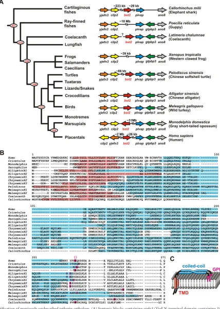
To identify novel tetherin orthologs, we combined sequence
analyses within silicotopology predictions andin vitroassays. An
Tetherin Evolution
on November 7, 2019 by guest
http://jvi.asm.org/
initial BLAST search using known tetherin sequences from
pla-cental mammals led to the identification of potentialtetherin/bst2
orthologs in marsupials (Monodelphis domesticus,Sarcophilus
har-risii), crocodilians (Alligator sinensis), turtles (Chrysemis picta,
Pelodiscus sinensis), and birds (Meleagris gallopavo) (Fig. 1Aand
Table 1). Despite very limited sequence homology (Fig. 1B), all
genes identified share similar exon-intron structures (Table 1)
and are located at a specific locus flanked by genes encoding the plasmalemma vesicle-associated protein (PLVAP) and cartilage
intermediate layer protein 2 (CILP2) (Fig. 1A). This strongly
sug-gests that the identified genes represent true orthologs of tetherin. Furthermore, all of these proteins are predicted to have the typical
topology of this restriction factor (Fig. 1BandCandTable 1). The
TMHMM Server v. 2.0 bioinformatics tool predicted the presence of an N-terminal TMD with a length of 16 to 23 amino acids (aa) (Fig. 1BandTable 1), and, according to the PredGPI analysis software, the probability of the presence of a C-terminal GPI
an-chor was⬎95% for most of these species (Fig. 1BandTable 1).
Finally, the COILS/PCOILS algorithm predicted the presence of
extracellular left-handed coiled-coil domains in all proteins (Fig.
1BandTable 1). Using the MTIDK matrix, probabilities for the presence of coiled coils of above 90% were found for all scanning windows (28, 21, and 14 residues) (see Fig. S1 in the supplemental material). The probabilities determined by the use of weighted and unweighted matrices were similar, indicating that elevated scores were not due to a high incidence of positively charged res-idues but are indicative of the presence of real coiled-coil struc-tures.
Since some tetherin orthologs may have been missed due to low sequence homology between tetherins from different ver-tebrate classes, we also manually searched for genes encoding proteins with a predicted tetherin-like configuration. To this
end, we took advantage of the high conservation of theplvap
andcilp2genes and screened the respective syntenic blocks in various vertebrate and invertebrate species. In addition to the orthologs identified by the BLAST algorithm, we identified
genes in the coelacanth (Latimeria chalumnae) and the elephant
shark (Callorhinchus milii) which very likely represent the tetherin
orthologs of these species. Like the genes of all other tetherin
vari-ants, these piscine genes are adjacent toplvapand encode proteins
with a predicted TMD, a coiled-coil ectodomain, and a GPI
an-chor attachment site (Fig. 1andTable 1; see also Fig. S1 in the
supplemental material). Notably, sequence analyses did not
iden-tify any tetherin orthologs in higher invertebrates such as
Tuni-cata,Echinodermata, orCephalochordata.These findings suggest that a tetherin gene arose early during vertebrate evolution, at least 450 million years ago, before the separation of cartilaginous fish
from bony vertebrates (38).
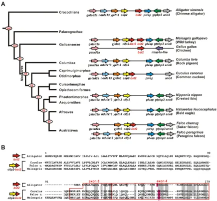
Gene erosion may have resulted in the loss of tetherin in many bird species.Although tetherin emerged before the diver-gence of reptiles and birds from mammals and can been found in
turkeys (Fig. 1), many bird species lack an obvioustetherinopen
reading frame (ORF) and seem to contain deletions in theplvap/
cilp2intergenic region (Fig. 2A). In some bird species (e.g., pere-grine falcons, rock pigeons, bald eagles, and crested ibises), this genetic erosion seems to have resulted in a complete loss of the
tetheringene. Other species such as the common cuckoo or the
saker falcon retained at least exons 3 and 4 (Fig. 2). The latter
species are predicted to express fusion proteins consisting of
CILP2 and the C-terminal part of tetherin (Fig. 2B). Although
these fusion proteins comprise the GPI anchor attachment site encoded by exon 4 of tetherin, the N-terminal CILP2 part is not predicted to form a TMD and it remains to be determined whether these proteins exert any antiviral activity. Interestingly, turkeys
also contain such acilp2-tetherinfusion gene in addition to their
regular tetherin ortholog, suggesting that duplication events
and/or gene rearrangements may have preceded the gene loss in thecilp2-plvaplocus (Fig. 2). Turkeys are morphologically
conser-vative members of the ancient superorderGalloanserae, indicating
that the loss of tetherin occurred after the divergence of turkeys
from modern birds (i.e.,Neoaves). In agreement with this
hypoth-esis, we also identified a putative tetherin ortholog in chickens,
another member of theGalloanseraesuperorder (Fig. 2andTable
1). In chickens, thecilp2/plvapsyntenic block has been disrupted
andtetherinis not flanked by thecilp2andplvapgenes (Fig. 2A). The high incidence of deletions and gene rearrangements is in line with the recent observation that birds have experienced a massive reduction in genome size, including the disruption of more than
100 conserved syntenic blocks (39,40).
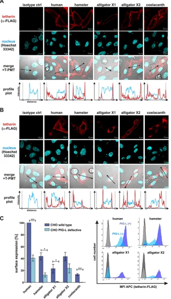
Piscine, reptilian, and mammalian tetherins are GPI-an-chored cell surface proteins.To functionally characterize dis-tantly related tetherin variants, we selected orthologs of humans and coelacanths as well as both isoforms (X1 and X2) of Chinese alligator tetherin. These species represent the groups of mammals,
fish, andSauropsida. Although alligator isoforms X1 and X2 share
exons 3 and 4, they differ substantially in their N-terminal halves, including the short intracellular tail, the TMD, and parts of the ectodomain. Chinese hamster tetherin was also included in the analyses since this ortholog has been suggested to almost
exclu-sively localize to the Golgi apparatus (41), whereas most
previ-ously described tetherin variants are mainly found at the cell sur-face. Western blotting showed that all orthologs are efficiently expressed from CMV promoter-based expression vectors (see Fig. S2A in the supplemental material). The detection of multiple bands ranging from 18 kDa to more than 125 kDa suggested that all tetherins are subject to posttranslational modifications, pre-sumably N-linked glycosylation (see Fig. S2A).
Next, we analyzed the subcellular localization of human, ham-ster, coelacanth, and alligator tetherin in transfected HEK293T and CHO cells. Flow cytometry revealed that all these proteins are
expressed at the cell surface, where virion trapping occurs (17)
(see Fig. S2B in the supplemental material). However, surface lev-els of the coelacanth ortholog and the X1 isoform of alligator tetherin were lower than those of human, hamster, and alligator X2 tetherin (see Fig. S2B). This expression phenotype was con-firmed by immunofluorescence microscopy of transfected
HEK293T (Fig. S2C) and CHO (Fig. 3A) cells. Whereas the
hu-man, hamster, and alligator X2 tetherins were preferentially ex-pressed at the cell surface, substantial amounts of coelacanth and alligator X1 tetherin were also localized to a perinuclear
compart-ment (Fig. 3A; see also Fig. S2C).
To verify the presence of a GPI anchor, we took advantage of the availability of a mutant CHO cell line that lacks a functional
pig-lgene and thus fails to synthesize this membrane anchor (32).
As previously shown (28), the localization of human tetherin was
largely restricted to intracellular compartments in GPI
anchor-deficient cells (Fig. 3B). A similar redistribution from the cell
sur-face to the cytoplasm was observed for hamster, coelacanth, and
both alligator tetherins (Fig. 3B), indicating that all these
or-thologs represent GPI-anchored proteins. Flow cytometric
analy-Heusinger et al.
on November 7, 2019 by guest
http://jvi.asm.org/
FIG 1Identification of previously undescribed tetherin orthologs. (A) Syntenic blocks containingyjefn3(YjeF N-terminal domain-containing 3),cilp2 (Car-tilage Intermediate-Layer Protein 2),bst2/tetherin,plvap(plasmalemma vesicle-associated protein), andano8(anoctamin 8) of diverse vertebrate species are shown on the right. Arrows indicate the direction of the ORFs. Gaps represent larger genome regions containing additional ORFs. A phylogenetic tree of vertebrate evolution (72) is shown on the left. Red numbers indicate divergence time estimates (in millions of years ago) for major nodes that are based on Inoue et al. (42). (B) Protein sequence alignment of tetherin orthologs from mammals, reptiles, and fishes. Dashes indicate gaps that were introduced to improve the alignment. Predicted TMDs are highlighted in red and coiled-coil regions in blue. The GPI anchor attachment site (⍀site) is shown in pink. X1 to X3 designate different tetherin isoforms from one species. (C) Cartoon of tetherin illustrating its typical topology comprising an N-terminal TMD (red), an extracellular coiled-coil domain (blue), and a C-terminal GPI lipid raft anchor (pink).
on November 7, 2019 by guest
http://jvi.asm.org/
[image:4.585.78.506.37.639.2]ses confirmed that tetherin surface levels were reduced in the
ab-sence of a functional GPI anchor (Fig. 3C). Notably, the PIG-L
deficiency specifically affected the plasma membrane localization of GPI-anchored proteins, since surface expression of tetherin but not of the CD4 receptor was rescued by exogenous overexpression of PIG-L (see Fig. S3 in the supplemental material).
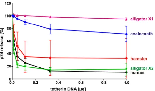
Tetherin’s ability to restrict virus release evolved at least 310 million years ago.To investigate whether the ability to restrict virus release is an evolutionarily ancient function of tetherin, we analyzed the release of the human immunodeficiency virus 1 (HIV-1) from HEK293T cells cotransfected with increasing amounts of tetherin and
avpu-deficient mutant of HIV-1 NL4-3 that fails to antagonize this
restriction factor. Two days posttransfection, p24 levels in the cells and culture supernatants were quantified by ELISA to calculate virus release. Human, hamster, and alligator X2 tetherin efficiently
re-stricted viral particle release in a dose-dependent manner (Fig. 4). In
comparison, coelacanth tetherin showed only marginal inhibitory activity and alligator X1 tetherin had no effect. These results demon-strate that not only mammals but also reptiles and possibly fish carry genes that encode tetherin orthologs with antiviral activity. Thus, the ability to restrict the egress of budding virions evolved at least 310
mya, before the divergence of reptiles from mammals (42).
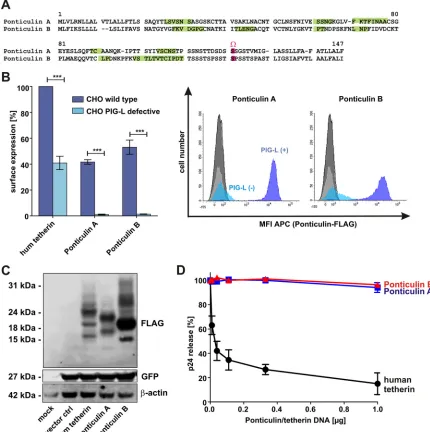
Ponticulins do not restrict virus release, although they share a tetherin-like topology.Since the primary amino acid sequence
is not critical for the ability of tetherin to restrict virus release (28),
we wondered whether completely unrelated proteins with a teth-erin-like topology might also show antiviral activity. Only a very small number of proteins containing an N-terminal TMD and a C-terminal GPI anchor have been described. Among them are ponticulin A and B, encoded by genes carried by the slime mold
Dictyostelium discoideum(43,44) (Fig. 5A). In contrast to
teth-erin, which contains a single␣-helical TMD, ponticulins span the
membrane multiple times via hydrophobic-strand structures
(43,45) (Fig. 5A). Flow cytometric analyses of transfected CHO cells confirmed the presence of a GPI anchor, since surface
expres-sion was completely abrogated in the absence of a functionalpig-l
gene (Fig. 5B). Although both proteins were efficiently expressed
in HEK293T cells (Fig. 5C), they failed to inhibit the release of
retroviral particles (Fig. 5D). Thus, the combination of an
N-ter-minal TMD and a C-terN-ter-minal GPI anchor is not sufficient to con-fer antiviral activity to a protein. Notably, the extracellular loop of ponticulin linking the TMD and GPI anchor is only very short and is not predicted to adopt a coiled-coil structure (see Fig. S4 in the supplemental material). Thus, the lack of a flexible ectodomain may account for the absence of antiviral activity in ponticulin A and B.
DISCUSSION
In the present report, we show that orthologs of the restriction factor tetherin are found in various marsupial, bird, reptile, and
fish species. The most basal species carrying atetheringene was the
elephant shark, or Australian ghost shark (Callorhinchus milii), a
“living fossil” that is evolutionarily even older than the coelacanth. This indicates that tetherin emerged at least 450 mya, before the
divergence of bony vertebrates from the elephant shark (38).
The elephant shark is not only one of the oldest but also among the
most slowly evolving jawed vertebrate species (38). It has
accumu-lated only a very low number of chromosomal rearrangements and has experienced fewer intron gains or losses than any bony
vertebrate (38). We took advantage of this low evolution rate to
[image:5.585.40.547.81.325.2]examine whether tetherin may be the result of a duplication event involving a neighboring gene. Notably, we found some minor sequence homology between tetherin and the plasmalemma vesi-cle-associated protein (PLVAP) of this species (data not shown), indicating that the two proteins may share a common ancestry. According to the TMHMM Server v. 2.0, COILS/PCOILS, and
TABLE 1Tetherin orthologs from mammals, reptiles, and fishesa
Accession no. Species
Exon length (nt) Protein
length (aa)
% GPI anchor probability Exon 1 Exon 2 Exon 3 Exon 4 Exon 5 Exon 6 Exon 7
NP_004326 Homo sapiens(human) 285 67 61 130 180 99.4
NP_001231044 Cricetulus griseus(Chinese hamster) 288 181 40 103 203 99.9
XP_007489270 Monodelphis domestica(gray short-tailed opossum)
246 55 61 100 153 100.00
XP_012399618 Sarcophilus harrisiia(Tasmanian devil) 219 76 61 97 150 100.00
XP_006017475(isoform X1) Alligator sinensis(Chinese alligator) 216 97 49 130 163 99.9
XP_006017476(isoform X2) Alligator sinensis(Chinese alligator) 186 88 49 130 150 99.9
XP_008169758(isoform X1) Chrysemis picta bellii(Western painted turtle)
321 97 82 151 216 100.0
XP_008169759(isoform X2) Chrysemis picta bellii(Western painted turtle)
321 85 49 154 202 99.9
XP_005279003(isoform X3) Chrysemis picta bellii(Western painted turtle)
315 85 49 154 200 100.0
XP_006132368 Pelodiscus sinensis(Chinese soft-shelled turtle)
270 97 53 97 49 174 76 271 95.0
XP_010723297(isoform X1) Meleagris gallopavo(turkey) 330 97 133 81 10 216 81.8
XP_010723300(isoform X2) Meleagris gallopavo(turkey) 330 97 133 66 10 211 87.4
XP_418228 Gallus gallus(chicken) 651 97 133 81 10 323 67.7
XP_006001674 Latimeria chalumnae(West Indian Ocean coelacanth)
342 97 154 126 7 241 37.3
XP_007897024 Callorhinchus milii(elephant shark) 258 97 115 115 194 99.9
aA TMD and a coiled-coil domain were predicted for each of the tetherin orthologs. aa, amino acids; nt, nucleotides. Heusinger et al.
on November 7, 2019 by guest
http://jvi.asm.org/
PredGPI bioinformatics tools, the latter is predicted to have an N-terminal TMD which is followed by a coiled-coil region and a 30% probability of a GPI anchor at its C terminus. However, the overall homology is very low and it remains to be determined whether PLVAP constitutes the true ancestor of tetherin.
The analysis of piscine, reptilian, and mammalian orthologs of tetherin revealed that its ability to restrict virion release evolved at least 310 million years ago. Since tetherin directly targets the
bud-ding process rather than a specific viral protein (28), it restricts the
egress of a large variety of enveloped viruses. Thus, although only HIV was tested in the present study, these findings are most likely applicable to the restriction of other enveloped viruses as well. The specific viruses that have driven the early evolution of tetherin remain unknown. However, paleovirological analysis of endoge-nous virus elements (EVEs) has clearly demonstrated that
verte-brates have been exposed to diverse virus families for millions of
years (46). The most prominent examples of EVEs are
endoge-nous retroviruses that can be found in most vertebrate species
(47). Even the coelacanth and elephant shark genomes contain a
high diversity of endogenous retroviruses as remnants of ancient
infections (48,49). In addition to retroviral elements, many bird
and mammal species also contain EVEs related to hepadna- or
filoviruses (46). The extant members of these virus families have a
broad host range and are known to be restricted by tetherin (19,
50). Thus, the expression of an active tetherin protein has certainly
provided a selection advantage to many vertebrate species. Notably, tetherin also plays an important role in viral cross-species transmission events, since it is often antagonized in a
spe-cies-specific manner (51). For example, the sensitivity of influenza
A viruses to human tetherin is strain specific (52), suggesting that
FIG 2Aviantetherin. (A) Thebst2/tetheringene loci of the indicated bird species are shown. Arrows indicate the direction of the ORFs. Overlapping arrows
representcilp2-bst2fusion genes. A phylogenetic tree of bird evolution (73) is shown on the left. Red numbers indicate divergence time estimates (in millions of years) for major nodes determined on the basis of data from Green et al. (crocodilian-bird divergence) (74) and Jarvis et al. (intra-avian divergence) (73). (B) Amino acid alignment of alligator X2 tetherin with the CILP2-tetherin fusion proteins of the common cuckoo (XP_009566634), the saker falcon (XP_005444407), and the turkey (XP_010723307). Only the C-terminal ends of the fusion proteins are depicted. Dashes indicate gaps that were introduced to improve the alignment. Sequence comparison revealed that exons 3 and 4 of tetherin (highlighted in red) are fused to the C terminus of cuckoo, saker falcon, and turkey CILP2. Identical amino acids are shown in gray. The GPI anchor attachment site (⍀site) is highlighted in pink.
Tetherin Evolution
on November 7, 2019 by guest
http://jvi.asm.org/
[image:6.585.74.509.65.467.2]FIG 3Subcellular localization and GPI anchor dependency of human, hamster, alligator, and coelacanth tetherin. (A and B) Immunofluorescence pictures of CHO wild-type cells (A) or mutant CHO cells lacking a functionalpig-lgene, required for GPI anchor synthesis (B). Two days posttransfection with the indicated tetherin expression vectors, cells were permeabilized and incubated with an anti-FLAG antibody. Nuclei were stained using Hoechst 33342. The regions used to generate profile plots are indicated by black arrows. In the profile plots, the localization of the plasma membrane is indicated by circles and vertical dotted lines. T-PMT, transmission-photomultiplier tube. ctrl, control. (C) Flow cytometric analysis of tetherin levels at the surface of transfected CHO cells (wild type [wt] or PIG-L deficient). Means⫾ standard errors of the means (SEM) of the results of three to five independent experiments are shown on the left (***,P⬍0.001; *,P⬍0.05). Examples of primary fluorescence-activated cell sorter (FACS) data indicating the mean fluorescence intensity (MFI) of allophycocyanin (APC) are shown on the right.
on November 7, 2019 by guest
http://jvi.asm.org/
[image:7.585.118.471.38.655.2]these viruses may differ in their adaptation to the human host. Influenza A viruses infect a broad range of host species, including
mammals, birds, and possibly even reptiles (53, 54). Notably,
members of theGalloanseraesuperorder (e.g., chickens and
tur-keys) are among the most important reservoirs for human-patho-genic influenza A viruses. Since these bird species encode a teth-erin ortholog, it will be interesting to characterize the role of avian and human tetherin in cross-species transmission events involv-ing influenza A viruses.
Despite its long-standing and important role in antiviral
im-munity, some species seem to have lost theirtetheringenes. Most
bird species, for example, showed evidence of gene erosion in the
cilp2-plvapintergenic region that usually contains the tetherin
ORF. These deletions resulted in the complete loss oftetherinor in
the emergence ofcilp2-tetherinfusion genes. The recent
sequenc-ing of 48 bird genomes revealed that birds have experienced a
massive gene loss during evolution (39,40). Thus,tetherinis
prob-ably one of more than 1,000 vertebrate genes that have been lost in
many bird species (40). Similarly, we did not detect any obvious
tetherin orthologs in amphibians or ray-finned fishes, although these vertebrates share an ancestor with tetherin-expressing rep-tiles, birds, and mammals. This suggests that tetherin may have been lost independently several times during evolution. Ray-finned fish such as the zebra fish, however, show high rates of molecular evolution and evolve(d) much faster than the elephant
shark or the coelacanth (38). Thus, we cannot entirely exclude the
possibility that orthologs were missed due to the almost complete lack of sequence homology and to incomplete sequencing and/or because they translocated to another gene locus.
Since viruses exert tremendous selection pressure on the evo-lution of host restriction factors, the respective genes often dupli-cate and neofunctionalize. Antiretroviral TRIM5 and APOBEC3 proteins, for example, are encoded by large gene clusters of
paralo-gous copies (55,56). In stark contrast to the TRIM5 and APOBEC
families, however, most species seem to carry just a singletetherin
gene. The only known exceptions are members of the family of
Bovidaewhich encode two or three paralogs (57,58). Some species
increase their tetherin repertoire by expressing different isoforms from a single gene. Humans, for example, express two alterna-tively translated isoforms that differ in their antiviral activity and
sensitivity toward viral antagonists (59). Similarly, several bird
and reptile species may produce different tetherin isoforms via alternative splicing.
Although tetherin orthologs and isoforms often differ substan-tially in their primary amino acid sequence, most of them share the typical topology that seems to be sufficient for the ability of
tetherin to restrict the release of budding virions (28). In
agree-ment with this, human and alligator X2 tetherin efficiently blocked the egress of HIV-1. Even hamster tetherin restricted vi-rus release, although this ortholog has been reported to primarily localize in Golgi cisternae, where it maintains the structure of the
Golgi apparatus (41). While our microscopic analyses in
trans-fected HEK293T cells confirmed colocalization with the Golgi marker TGN46, a considerable amount of hamster tetherin was also detectable at the cell surface. In contrast to the findings by Li
et al. (41), this was especially pronounced in CHO cells, where
tetherin almost exclusively localized at the plasma membrane. In contrast to human, hamster, and alligator X2 tetherin, the coela-canth ortholog showed only a weak antiviral effect, although it adopts the typical tetherin configuration and was efficiently ex-pressed at the cell surface. Notably, the cell membranes of cold-blooded fish and warm-cold-blooded mammals differ in their lipid
composition and fluidity (60). Thus, piscine tetherins may be
ef-ficient restriction factorsin vivo, and antiviral activities might
have been missed in human HEK293T cells due to species-specific adaptations. Surprisingly, isoform X1 of alligator tetherin had no impact on virus release at all, although it shares exons 3 and 4 with the active X2 isoform. Interestingly, the extracellular coiled-coil domain and the TMD are predicted to overlap in isoform X1, whereas they are separated by 10 amino acids in isoform X2. This structural difference might affect the flexibility of the hinge region at the transition of the transmembrane to the ectodomain. Fur-thermore, isoform X1 contains two sequence stretches (aa 65 to 72 and 96 to 100) in its extracellular region that may interrupt the coiled-coil structure. Whether these structural characteristics and/or other reasons account for the lack of antiviral activity of alligator X1 tetherin remains to be determined.
Only a few cellular proteins containing an N-terminal TMD and a C-terminal GPI anchor have been described. These include
an unusual isoform of the prion protein (61–63), Sm23 from
Schistosoma mansoni(64), and NcSRS2 fromNeospora caninum
(65) as well as ponticulin A and B fromDictyostelium discoideum
(43–45). Although these proteins are all unrelated to tetherin, they may have acquired the ability to inhibit virus release as a result of convergent evolution. Here, we analyzed the antiviral activity of ponticulin A and B since these proteins not only exhibit a tetherin-like configuration but (tetherin-like tetherin) also contain N-linked glyco-sylation sites and conserved cysteine residues that form disulfide
bonds (43). Furthermore, tetherin and ponticulins both bind
ac-tin via their intracellular domains (44,66,67). Although we could
confirm that ponticulin A and B are GPI-anchored cell surface proteins, there was no evidence for convergent evolution and pon-ticulins did not restrict virus release. One possible explanation for the lack of antiviral activity may be the absence of coiled coils in the short extracellular domains of ponticulin A and B.
In summary, our results demonstrate that the host restriction factor tetherin is an evolutionarily ancient protein that is
substan-FIG 4Restriction of HIV-1 release by human, hamster, alligator, and
coela-canth tetherin. HEK293T cells were cotransfected with avpu-deficient proviral construct of HIV-1 NL4-3 and increasing amounts of the indicated tetherin expression vectors. Two days posttransfection, cells and supernatants were harvested and p24 contents were determined by ELISA. Virus release was cal-culated by dividing the amount of viral capsid in the supernatant by the total amount. The means of the results of three to eight independent experiments⫾ SEM are shown.
Tetherin Evolution
on November 7, 2019 by guest
http://jvi.asm.org/
[image:8.585.41.288.65.210.2]tially (⬎450 million years) older and more widespread than, for
example, theAPOBEC3andTRIM5genes, which are unique to
(placental) mammals and evolved about 90 to 180 million years
ago (55,56,68–71). In contrast to most other restriction factors,
tetherin does not interact with viral proteins or nucleic acids but targets viral membranes. This antiviral mechanism provides sev-eral advantages. First, tetherin has broad activity against diverse enveloped viruses. Second, viruses cannot become resistant by simply acquiring evasion mutations. Third, tetherin tolerates
many amino acid substitutions since its antiviral activity does not depend on its primary amino acid sequence but rather on the overall protein configuration. Thus, even if viruses evolve antag-onists that remove it from the sites of budding, tetherin may read-ily mutate the respective interaction sites without losing its anti-viral activity. With regard to the selection advantage that tetherin confers to its host, it may come as a surprise that some vertebrate species seem to have lost their tetherin gene. Whether these species encode unknown paralogs of tetherin or whether unrelated
pro-FIG 5Functional characterization of ponticulin A and B. (A) Amino acid alignment of ponticulin A and B from the slime moldDictyostelium discoideum. Dashes
indicate gaps that were introduced to improve the alignment.-Sheets were predicted using PSIPRED v3.3 and are highlighted in green; the GPI anchor attachment site (⍀site) is shown in pink. (B) Flow cytometric analysis of ponticulin levels at the surface of transfected CHO cells (wt or PIG-L deficient). The means of the results of three to five independent experiments⫾SEM are shown on the left (***,P⬍0.001). Examples for primary FACS data indicating the mean fluorescence intensity (MFI) of allophycocyanin (APC) are shown on the right. hum, human. (C) Western blot analysis of HEK293T cells transfected with expression vectors for human tetherin or ponticulin A or B. An anti-FLAG antibody was used for detection.-Actin and eGFP served as loading and transfection controls, respectively. (D) Virus release from HEK293T cells cotransfected with avpu-deficient proviral construct of HIV-1 NL4-3 and increasing amounts of expression vectors for human tetherin or ponticulin A or B. Two days posttransfection, cells and supernatants were harvested and p24 contents were determined by ELISA. Virus release was calculated by dividing the amount of viral capsid in the supernatant by the total amount. The means of the results of five to seven independent experiments⫾SEM are shown.
Heusinger et al.
on November 7, 2019 by guest
http://jvi.asm.org/
[image:9.585.75.505.69.501.2]teins have also acquired this simple yet very efficient antiviral ac-tivity remains to be investigated.
ACKNOWLEDGMENTS
We thank Susanne Engelhart and Martha Mayer for excellent technical assistance. CHO cell lines and a PIG-L expression vector were kindly provided by Taroh Kinoshita. J. Bernd Helms kindly provided an expres-sion vector for hamster tetherin.
This study was funded by the Deutsche Forschungsgemeinschaft (Ki548/11-1), a European Research Council Advanced Grant to F.K., and the FP7 European Union’s Research and Innovation funding programme. D.S. was supported by a starting grant of the medical faculty of the Uni-versity of Ulm (L.SBN.0080). Parts of this work were supported by the Gottfried-Wilhelm Leibniz award to F.K. The funders had no role in study design, data collection and interpretation, or the decision to submit the work for publication.
REFERENCES
1.Koonin EV, Dolja VV, Krupovic M. 2015. Origins and evolution of
viruses of eukaryotes: the ultimate modularity. Virology479 – 480:2–25.
2.Meselson M, Yuan R.1968. DNA restriction enzyme from E. coli. Nature
217:1110 –1114.http://dx.doi.org/10.1038/2171110a0.
3.Barrangou R, Fremaux C, Deveau H, Richards M, Boyaval P, Moineau
S, Romero DA, Horvath P.2007. CRISPR provides acquired resistance
against viruses in prokaryotes. Science315:1709 –1712.http://dx.doi.org /10.1126/science.1138140.
4.Flajnik MF, Kasahara M.2010. Origin and evolution of the adaptive
immune system: genetic events and selective pressures. Nat Rev Genet
11:47–59.http://dx.doi.org/10.1038/nrg2703.
5.Hahn UK, Bender RC, Bayne CJ.2000. Production of reactive oxygen
species by hemocytes of Biomphalaria glabrata: carbohydrate-specific stimulation. Dev Comp Immunol24:531–541.http://dx.doi.org/10.1016 /S0145-305X(00)00017-3.
6.Hahn UK, Bender RC, Bayne CJ.2001. Involvement of nitric oxide in
killing of Schistosoma mansoni sporocysts by hemocytes from resistant Biomphalaria glabrata. J Parasitol87:778 –785.http://dx.doi.org/10.1645 /0022-3395(2001)087[0778:IONOIK]2.0.CO;2.
7.Nonaka M.2014. Evolution of the complement system. Subcell Biochem
80:31– 43.http://dx.doi.org/10.1007/978-94-017-8881-6_3.
8.Meister M, Lemaitre B, Hoffmann JA.1997. Antimicrobial peptide defense in
Drosophila. Bioessays19:1019–1026.http://dx.doi.org/10.1002/bies.950191112.
9.Detournay O, Schnitzler CE, Poole A, Weis VM. 2012. Regulation of
cnidarian-dinoflagellate mutualisms: evidence that activation of a host TGF innate immune pathway promotes tolerance of the symbiont. Dev Comp Immunol38:525–537.http://dx.doi.org/10.1016/j.dci.2012.08.008.
10. Bieniasz PD.2004. Intrinsic immunity: a front-line defense against viral
attack. Nat Immunol5:1109 –1115.http://dx.doi.org/10.1038/ni1125.
11. Doyle T, Goujon C, Malim MH.2015. HIV-1 and interferons: who’s
interfering with whom? Nat Rev Microbiol13:403– 413.http://dx.doi.org /10.1038/nrmicro3449.
12. Kirchhoff F.2010. Immune evasion and counteraction of restriction
fac-tors by HIV-1 and other primate lentiviruses. Cell Host Microbe8:55– 67.
http://dx.doi.org/10.1016/j.chom.2010.06.004.
13. Stremlau M, Owens CM, Perron MJ, Kiessling M, Autissier P, Sodroski
J.2004. The cytoplasmic body component TRIM5alpha restricts HIV-1 infection in Old World monkeys. Nature427:848 – 853.http://dx.doi.org /10.1038/nature02343.
14. Sheehy AM, Gaddis NC, Choi JD, Malim MH.2002. Isolation of a human
gene that inhibits HIV-1 infection and is suppressed by the viral Vif protein. Nature418:646 – 650.http://dx.doi.org/10.1038/nature00939.
15. Laguette N, Sobhian B, Casartelli N, Ringeard M, Chable-Bessia C, Ségéral
E, Yatim A, Emiliani S, Schwartz O, Benkirane M.2011. SAMHD1 is the
dendritic- and myeloid-cell-specific HIV-1 restriction factor counteracted by Vpx. Nature474:654 – 657.http://dx.doi.org/10.1038/nature10117.
16. Hrecka K, Hao C, Gierszewska M, Swanson SK, Kesik-Brodacka M,
Srivastava S, Florens L, Washburn MP, Skowronski J.2011. Vpx relieves
inhibition of HIV-1 infection of macrophages mediated by the SAMHD1 protein. Nature474:658 – 661.http://dx.doi.org/10.1038/nature10195.
17. Neil SJD, Zang T, Bieniasz PD.2008. Tetherin inhibits retrovirus release
and is antagonized by HIV-1 Vpu. Nature451:425– 430.http://dx.doi.org /10.1038/nature06553.
18. Van Damme N, Goff D, Katsura C, Jorgenson RL, Mitchell R, Johnson
MC, Stephens EB, Guatelli J. 2008. The interferon-induced protein
BST-2 restricts HIV-1 release and is downregulated from the cell surface by the viral Vpu protein. Cell Host Microbe3:245–252.http://dx.doi.org /10.1016/j.chom.2008.03.001.
19. Jouvenet N, Neil SJD, Zhadina M, Zang T, Kratovac Z, Lee Y, McNatt
M, Hatziioannou T, Bieniasz PD.2009. Broad-spectrum inhibition of
retroviral and filoviral particle release by tetherin. J Virol83:1837–1844.
http://dx.doi.org/10.1128/JVI.02211-08.
20. Sakuma T, Noda T, Urata S, Kawaoka Y, Yasuda J.2009. Inhibition of
Lassa and Marburg virus production by tetherin. J Virol83:2382–2385.
http://dx.doi.org/10.1128/JVI.01607-08.
21. Radoshitzky SR, Dong L, Chi X, Clester JC, Retterer C, Spurgers K,
Kuhn JH, Sandwick S, Ruthel G, Kota K, Boltz D, Warren T, Kranzusch PJ, Whelan SPJ, Bavari S.2010. Infectious Lassa virus, but not filoviruses, is restricted by BST-2/tetherin. J Virol84:10569 –10580.http://dx.doi.org /10.1128/JVI.00103-10.
22. Weidner JM, Jiang D, Pan X-B, Chang J, Block TM, Guo J-T.2010.
Interferon-induced cell membrane proteins, IFITM3 and tetherin, inhibit vesicular stomatitis virus infection via distinct mechanisms. J Virol84:
12646 –12657.http://dx.doi.org/10.1128/JVI.01328-10.
23. Sarojini S, Theofanis T, Reiss CS. 2011. Interferon-induced tetherin
restricts vesicular stomatitis virus release in neurons. DNA Cell Biol30:
965–974.http://dx.doi.org/10.1089/dna.2011.1384.
24. Kong W-S, Irie T, Yoshida A, Kawabata R, Kadoi T, Sakaguchi T.2012.
Inhibition of virus-like particle release of Sendai virus and Nipah virus, but not that of mumps virus, by tetherin/CD317/BST-2. Hiroshima J Med Sci61:59 – 67.
25. Blondeau C, Pelchen-Matthews A, Mlcochova P, Marsh M, Milne RSB,
Towers GJ.2013. Tetherin restricts herpes simplex virus 1 and is
antago-nized by glycoprotein M. J Virol87:13124 –13133.http://dx.doi.org/10 .1128/JVI.02250-13.
26. Wang S-M, Huang K-J, Wang C-T. 2014. BST2/CD317 counteracts
human coronavirus 229E productive infection by tethering virions at the cell surface. Virology449:287–296.http://dx.doi.org/10.1016/j.virol.2013 .11.030.
27. Pan X-B, Qu X-W, Jiang D, Zhao X-L, Han J-C, Wei L.2013. BST2/
Tetherin inhibits hepatitis C virus production in human hepatoma cells. An-tiviral Res98:54 – 60.http://dx.doi.org/10.1016/j.antiviral.2013.01.009.
28. Perez-Caballero D, Zang T, Ebrahimi A, McNatt MW, Gregory DA,
Johnson MC, Bieniasz PD. 2009. Tetherin inhibits HIV-1 release by
directly tethering virions to cells. Cell139:499 –511.http://dx.doi.org/10 .1016/j.cell.2009.08.039.
29. Kupzig S, Korolchuk V, Rollason R, Sugden A, Wilde A, Banting G.
2003. Bst-2/HM1.24 is a raft-associated apical membrane protein with an unusual topology. Traffic 4:694 –709. http://dx.doi.org/10.1034/j.1600 -0854.2003.00129.x.
30. Venkatesh S, Bieniasz PD.2013. Mechanism of HIV-1 virion entrapment
by tetherin. PLoS Pathog9:e1003483.http://dx.doi.org/10.1371/journal .ppat.1003483.
31. Tanaka M, Herr W.1990. Differential transcriptional activation by Oct-1
and Oct-2: interdependent activation domains induce Oct-2 phosphoryla-tion. Cell60:375–386.http://dx.doi.org/10.1016/0092-8674(90)90589-7.
32. Nakamura N, Inoue N, Watanabe R, Takahashi M, Takeda J, Stevens
VL, Kinoshita T.1997. Expression cloning of PIG-L, a candidate
N-acetylglucosaminyl-phosphatidylinositol deacetylase. J Biol Chem272:
15834 –15840.http://dx.doi.org/10.1074/jbc.272.25.15834.
33. Rücker E, Grivel J-C, Münch J, Kirchhoff F, Margolis L.2004. Vpr and Vpu
are important for efficient human immunodeficiency virus type 1 replication and CD4⫹T-cell depletion in human lymphoid tissue ex vivo. J Virol78:
12689 –12693.http://dx.doi.org/10.1128/JVI.78.22.12689-12693.2004.
34. Münch J, Ständker L, Pöhlmann S, Baribaud F, Papkalla A, Rosorius O,
Stauber R, Sass G, Heveker N, Adermann K, Escher S, Klüver E, Doms
RW, Forssmann W-G, Kirchhoff F.2002. Hemofiltrate CC chemokine
1[9-74] causes effective internalization of CCR5 and is a potent inhibitor of R5-tropic human immunodeficiency virus type 1 strains in primary T cells and macrophages. Antimicrob Agents Chemother46:982–990.http: //dx.doi.org/10.1128/AAC.46.4.982-990.2002.
35. DuBridge RB, Tang P, Hsia HC, Leong PM, Miller JH, Calos MP.1987.
Analysis of mutation in human cells by using an Epstein-Barr virus shuttle system. Mol Cell Biol7:379 –387.
36. Sauter D.2014. Counteraction of the multifunctional restriction factor teth-erin. Front Microbiol5:163.http://dx.doi.org/10.3389/fmicb.2014.00163.
Tetherin Evolution
on November 7, 2019 by guest
http://jvi.asm.org/
37. Wong SK, Connole M, Sullivan JS, Choe H, Carville A, Farzan M.2009. A New World primate deficient in tetherin-mediated restriction of human immunodeficiency virus type 1. J Virol83:8771– 8780.http://dx.doi.org /10.1128/JVI.00112-09.
38. Venkatesh B, Lee AP, Ravi V, Maurya AK, Lian MM, Swann JB, Ohta
Y, Flajnik MF, Sutoh Y, Kasahara M, Hoon S, Gangu V, Roy SW, Irimia M, Korzh V, Kondrychyn I, Lim ZW, Tay B-H, Tohari S, Kong KW, Ho S, Lorente-Galdos B, Quilez J, Marques-Bonet T, Raney BJ, Ingham PW, Tay A, Hillier LW, Minx P, Boehm T, Wilson RK, Brenner S,
Warren WC.2014. Elephant shark genome provides unique insights into
gnathostome evolution. Nature505:174 –179.http://dx.doi.org/10.1038 /nature12826.
39. Lovell PV, Wirthlin M, Wilhelm L, Minx P, Lazar NH, Carbone L,
Warren WC, Mello CV.2014. Conserved syntenic clusters of protein
coding genes are missing in birds. Genome Biol15:565.http://dx.doi.org /10.1186/s13059-014-0565-1.
40. Zhang G, Li C, Li Q, Li B, Larkin DM, Lee C, Storz JF, Antunes A,
Greenwold MJ, Meredith RW, O¨ deen A, Cui J, Zhou Q, Xu L, Pan H,
Wang Z, Jin L, Zhang P, Hu H, Yang W, Hu J, Xiao J, Yang Z, Liu Y, Xie Q, Yu H, Lian J, Wen P, Zhang F, Li H, Zeng Y, Xiong Z, Liu S, Zhou L, Huang Z, An N, Wang J, Zheng Q, Xiong Y, Wang G, Wang B, Wang J, Fan Y, da Fonseca RR, Alfaro-Núñez A, Schubert M,
Orlando L, Mourier T, Howard JT, Ganapathy G, et al.2014.
Compar-ative genomics reveals insights into avian genome evolution and adapta-tion. Science346:1311–1320.http://dx.doi.org/10.1126/science.1251385.
41. Li X, Kaloyanova D, van Eijk M, Eerland R, van der Goot G, Oorschot
V, Klumperman J, Lottspeich F, Starkuviene V, Wieland FT, Helms JB.
2007. Involvement of a Golgi-resident GPI-anchored protein in mainte-nance of the Golgi structure. Mol Biol Cell18:1261–1271.http://dx.doi .org/10.1091/mbc.E06-03-0236.
42. Inoue JG, Miya M, Lam K, Tay B-H, Danks JA, Bell J, Walker TI,
Venkatesh B.2010. Evolutionary origin and phylogeny of the modern
holocephalans (Chondrichthyes: Chimaeriformes): a mitogenomic per-spective. Mol Biol Evol27:2576 –2586.http://dx.doi.org/10.1093/molbev /msq147.
43. Hitt AL, Lu TH, Luna EJ.1994. Ponticulin is an atypical membrane
protein. J Cell Biol126:1421–1431.http://dx.doi.org/10.1083/jcb.126.6 .1421.
44. Hitt AL, Hartwig JH, Luna EJ.1994. Ponticulin is the major high affinity
link between the plasma membrane and the cortical actin network in Dictyostelium. J Cell Biol126:1433–1444.http://dx.doi.org/10.1083/jcb .126.6.1433.
45. Hitt AL, Iijima-Shimizu M, DuBay MJ, Antonette LL, Urushihara H,
Wilkerson CG.2003. Identification of a second member of the ponticulin
gene family and its differential expression pattern. Biochim Biophys Acta
1628:79 – 87.http://dx.doi.org/10.1016/S0167-4781(03)00115-5.
46. Katzourakis A, Gifford RJ.2010. Endogenous viral elements in animal
ge-nomes. PLoS Genet 6:e1001191. http://dx.doi.org/10.1371/journal.pgen .1001191.
47. Herniou E, Martin J, Miller K, Cook J, Wilkinson M, Tristem M.1998.
Retroviral diversity and distribution in vertebrates. J Virol72:5955–5966.
48. Han G-Z, Worobey M.2012. An endogenous foamy-like viral element in
the coelacanth genome. PLoS Pathog8:e1002790.http://dx.doi.org/10 .1371/journal.ppat.1002790.
49. Han G-Z.2015. Extensive retroviral diversity in shark. Retrovirology12:
34.http://dx.doi.org/10.1186/s12977-015-0158-4.
50. Yan R, Zhao X, Cai D, Liu Y, Block TM, Guo J-T, Guo H.2015. The
interferon-inducible protein tetherin inhibits hepatitis B virus virion se-cretion. J Virol89:9200 –9212.http://dx.doi.org/10.1128/JVI.00933-15.
51. Sauter D, Specht A, Kirchhoff F.2010. Tetherin: holding on and letting
go. Cell141:392–398.http://dx.doi.org/10.1016/j.cell.2010.04.022.
52. Gnirß K, Zmora P, Blazejewska P, Winkler M, Lins A, Nehlmeier I,
Gärtner S, Moldenhauer A-S, Hofmann-Winkler H, Wolff T, Schindler
M, Pöhlmann S.24 June 2015. Tetherin sensitivity of influenza A viruses
is strain specific: role of hemagglutinin and neuraminidase. J Virolhttp: //dx.doi.org/10.1128/JVI.00615-15.
53. Temple BL, Finger JW, Jones CA, Gabbard JD, Jelesijevic T, Uhl EW,
Hogan RJ, Glenn TC, Tompkins SM.2015. In ovo and in vitro
suscep-tibility of American alligators (Alligator mississippiensis) to avian influ-enza virus infection. J Wildl Dis51:187–198.http://dx.doi.org/10.7589 /2013-12-321.
54. Davis LM, Spackman E.2008. Do crocodilians get the flu? Looking for
influenza A in captive crocodilians. J Exp Zool A Ecol Genet Physiol309:
571–580.http://dx.doi.org/10.1002/jez.454.
55. Münk C, Willemsen A, Bravo IG.2012. An ancient history of gene
duplica-tions, fusions and losses in the evolution of APOBEC3 mutators in mammals. BMC Evol Biol12:71.http://dx.doi.org/10.1186/1471-2148-12-71.
56. Johnson WE, Sawyer SL.2009. Molecular evolution of the antiretroviral
TRIM5 gene. Immunogenetics 61:163–176. http://dx.doi.org/10.1007 /s00251-009-0358-y.
57. Arnaud F, Black SG, Murphy L, Griffiths DJ, Neil SJ, Spencer TE,
Palmarini M.2010. Interplay between ovine bone marrow stromal cell
antigen 2/tetherin and endogenous retroviruses. J Virol84:4415– 4425.
http://dx.doi.org/10.1128/JVI.00029-10.
58. Takeda E, Nakagawa S, Nakaya Y, Tanaka A, Miyazawa T, Yasuda J.2012.
Identification and functional analysis of three isoforms of bovine BST-2. PLoS One7:e41483.http://dx.doi.org/10.1371/journal.pone.0041483.
59. Cocka LJ, Bates P.2012. Identification of alternatively translated tetherin isoforms with differing antiviral and signaling activities. PLoS Pathog
8:e1002931.http://dx.doi.org/10.1371/journal.ppat.1002931.
60. Cossins AR, Prosser CL.1978. Evolutionary adaptation of membranes to
temperature. Proc Natl Acad Sci U S A75:2040 –2043.http://dx.doi.org /10.1073/pnas.75.4.2040.
61. Stewart RS, Drisaldi B, Harris DA.2001. A transmembrane form of the
prion protein contains an uncleaved signal peptide and is retained in the endoplasmic reticulum. Mol Biol Cell12:881– 889.http://dx.doi.org/10 .1091/mbc.12.4.881.
62. Hegde RS, Mastrianni JA, Scott MR, DeFea KA, Tremblay P, Torchia
M, DeArmond SJ, Prusiner SB, Lingappa VR.1998. A transmembrane
form of the prion protein in neurodegenerative disease. Science279:827– 834.http://dx.doi.org/10.1126/science.279.5352.827.
63. Hegde RS, Tremblay P, Groth D, DeArmond SJ, Prusiner SB, Lingappa
VR.1999. Transmissible and genetic prion diseases share a common path-way of neurodegeneration. Nature 402:822– 826. http://dx.doi.org/10 .1038/45574.
64. Köster B, Strand M.1994. Schistosoma mansoni: Sm23 is a
transmem-brane protein that also contains a glycosylphosphatidylinositol an-chor. Arch Biochem Biophys310:108 –117.http://dx.doi.org/10.1006 /abbi.1994.1146.
65. Nishikawa Y, Tragoolpua K, Makala L, Xuan X, Nagasawa H.2002.
Neospora caninum NcSRS2 is a transmembrane protein that contains a glycosylphosphatidylinositol anchor in insect cells. Vet Parasitol109:191– 201.http://dx.doi.org/10.1016/S0304-4017(02)00256-X.
66. Chia CP, Hitt AL, Luna EJ.1991. Direct binding of F-actin to ponticulin,
an integral plasma membrane glycoprotein. Cell Motil Cytoskeleton18:
164 –179.http://dx.doi.org/10.1002/cm.970180303.
67. Rollason R, Korolchuk V, Hamilton C, Jepson M, Banting G.2009. A
CD317/tetherin-RICH2 complex plays a critical role in the organization of the subapical actin cytoskeleton in polarized epithelial cells. J Cell Biol
184:721–736.http://dx.doi.org/10.1083/jcb.200804154.
68. Conticello SG, Thomas CJF, Petersen-Mahrt SK, Neuberger MS.2005.
Evolution of the AID/APOBEC family of polynucleotide (deoxy)cytidine deaminases. Mol Biol Evol22:367–377.
69. Tareen SU, Sawyer SL, Malik HS, Emerman M.2009. An expanded clade
of rodent Trim5 genes. Virology385:473– 483.http://dx.doi.org/10.1016 /j.virol.2008.12.018.
70. Sawyer SL, Emerman M, Malik HS.2007. Discordant evolution of the
adjacent antiretroviral genes TRIM22 and TRIM5 in mammals. PLoS Pat-hog3:e197.http://dx.doi.org/10.1371/journal.ppat.0030197.
71. Conticello SG.2008. The AID/APOBEC family of nucleic acid mutators.
Genome Biol9:229.http://dx.doi.org/10.1186/gb-2008-9-6-229.
72. Meyer A, Zardoya R.2003. Recent advances in the (molecular) phylogeny
of vertebrates. Annu Rev Ecol Evol Syst34:311–338.http://dx.doi.org/10 .1146/annurev.ecolsys.34.011802.132351.
73. Jarvis ED, Mirarab S, Aberer AJ, Li B, Houde P, Li C, Ho SY, Faircloth
BC, Nabholz B, Howard JT, Suh A, Weber CC, da Fonseca RR, Li J, Zhang F, Li H, Zhou L, Narula N, Liu L, Ganapathy G, Boussau B, Bayzid MS, Zavidovych V, Subramanian S, Gabaldón T, Capella-Gutiérrez S, Huerta-Cepas J, Rekepalli B, Munch K, Schierup M, Lindow B, Warren WC, Ray D, Green RE, Bruford MW, Zhan X, Dixon A, Li S, Li N, Huang Y, Derryberry EP, Bertelsen MF, Sheldon FH, Brumfield RT, Mello CV, Lovell PV, Wirthlin M, Schneider MP,
Pros-docimi F, et al.2014. Whole-genome analyses resolve early branches in
the tree of life of modern birds. Science346:1320 –1331.http://dx.doi.org /10.1126/science.1253451.
Heusinger et al.
on November 7, 2019 by guest
http://jvi.asm.org/
74. Green RE, Braun EL, Armstrong J, Earl D, Nguyen N, Hickey G, Vandewege MW, St John JA, Capella-Gutiérrez S, Castoe TA, Kern C, Fujita MK, Opazo JC, Jurka J, Kojima KK, Caballero J, Hubley RM, Smit AF, Platt RN, Lavoie CA, Ramakodi MP, Finger JW, Suh A, Isberg SR, Miles L, Chong AY, Jaratlerdsiri W, Gongora J, Moran C, Iriarte A, McCormack J, Burgess SC, Edwards SV, Lyons E,
Williams C, Breen M, Howard JT, Gresham CR, Peterson DG, Schmitz J, Pollock DD, Haussler D, Triplett EW, Zhang G, Irie N, Jarvis ED, Brochu CA, Schmidt CJ, McCarthy FM, Faircloth BC, et al.2014. Three crocodilian genomes reveal ancestral patterns of evo-lution among archosaurs. Science346:1254449.http://dx.doi.org/10 .1126/science.1254449.
Tetherin Evolution