Activates Interleukin-1

Release in Murine Dendritic Cells
Tessa M. Lawrence,aAndrew W. Hudacek,aMarcel R. de Zoete,bRichard A. Flavell,b,cMatthias J. Schnella,d
Department of Microbiology and Immunology, Jefferson Medical College, Thomas Jefferson University, Philadelphia, Pennsylvania, USAa; Department of Immunobiology,
Yale University School of Medicine, New Haven, Connecticut, USAb; Howard Hughes Medical Institute, Yale University School of Medicine, New Haven, Connecticut, USAc;
Jefferson Vaccine Center, Jefferson Medical College, Thomas Jefferson University, Philadelphia, Pennsylvania, USAd
Inflammasome activation is important for the development of an effective host defense against many pathogens, including RNA viruses. However, the mechanism by which the inflammasome recognizes RNA viruses and its role in rabies virus (RABV) patho-genicity and immunopatho-genicity remain poorly defined. To determine the function of the inflammasome in response to RABV in-fection, we infected murine bone marrow-derived dendritic cells (BMDCs) with RABV. Our results indicate that the infection of BMDCs with RABV induces both the production of pro-interleukin-1(pro-IL-1) and its processing, resulting in the secretion of active IL-1through activation of the NLRP3-, ASC-, and caspase-1-dependent inflammasome. As previously shown for the induction of type I interferon by RABV, the induction of pro-IL-1also depends upon IPS-1. We demonstrate that both the pro-duction of pro-IL-1and activation of the inflammasome require viral replication. We also demonstrate that increased viral replication in BMDCs derived from IFNAR-deficient mice resulted in significantly more IL-1release. Additionally, IL-1 recep-tor-deficient mice show an increase in RABV pathogenicity. Taken together, these results indicate an important role of the in-flammasome in innate immune recognition of RABV.
E
arly recognition of viral infection by the innate immune sys-tem is essential for an effective host defense. The innate im-mune response has evolved to recognize common motifs of pathogens by a variety of pattern recognition receptors (PRRs) (for a review, see reference1). Viral motifs recognized by these sensors include distinct nucleic acids such as double-stranded RNA (dsRNA), cytosolic DNA, or uncapped single-stranded RNA. Recognition of these motifs results in the release of type I interferons (IFNs) and inflammatory cytokines, which leads to the control of the pathogen and supports the development of an adap-tive immune response (1). Four main families of PRRs have been described: Toll-like receptors (TLRs), retinoic acid inducible gene I (RIG-I)-like receptors (e.g., RIG-I and MDA5), PYHIN proteins (e.g., AIM2), and nucleotide-binding oligomerization domain (NOD)-like receptors (e.g., NOD1, NOD2, NLRP1, NLRP3, and NLRC4) (1–3).The NOD-like receptor family consists of cytoplasmic recep-tors that function in the regulation of inflammatory responses and contain PRRs that are involved in the assembly of large protein complexes referred to as inflammasomes (for a review, see refer-ence2). The inflammasome is typically composed of a NOD-like receptor, the adaptor protein ASC, and pro-caspase-1. Depending on the danger signal present, distinct inflammasome complexes are formed. This multiprotein complex regulates caspase-1 acti-vation and serves as a scaffold for the proteolytic actiacti-vation of interleukin-1(IL-1) and IL-18. Processed forms of IL-1and IL-18 are potent mediators of the inflammatory response and are involved in a range of activities, including cell proliferation, acti-vation, differentiation, apoptosis, and antibody production (4,5). One of the best-characterized inflammasomes consists of NLRP3/NALP3/cryopyrin (6). NLRP3 recognizes a wide range of pathogen- and danger-associated molecular patterns (2,3). Spe-cifically, this particular inflammasome can recognize both posi-tive- and negaposi-tive-sense RNA viruses, including influenza virus, Sendai virus, encephalomyocarditis virus (EMCV), vesicular
sto-matitis virus (VSV), and West Nile virus (7–15). However, the exact mechanism by which these pathogens activate the inflam-masome is presently unknown.
In contrast to the immediate activation of type I interferons, the activation of IL-1is a two-step process combining two sig-naling pathways found mainly within antigen-presenting cells, such as dendritic cells (DCs) and macrophages (16). The first pathway leads to the nuclear factorB (NF-B)-dependent tran-scription of pro-IL-1and components of the inflammasome, including NLRP3 (2). The second pathway is activated through the assembly of the inflammasome, which results in the activation of caspase-1 and subsequent cleavage of pro-IL-1to active, se-creted IL-1.
Rabies virus (RABV) is a negative-sense single-stranded RNA virus that replicates exclusively in the cytosol (17). Previous re-ports have shown that RABV RNA is recognized by RIG-I and MDA5, which exclusively signal via IPS-1/MAVS/VISA/Cardif to activate both NF-B- and type I IFN-regulated responses (18–20). Like many viruses, RABV has evolved a variety of mechanisms to counteract these host defenses. For example, RABV phosphopro-tein (P) is able to suppress both the induction of type I IFN and the ensuing signaling by IFN-␣/in certain cell types, including fi-broblasts and neuronal cells (21–23). Despite this, RABV infection triggers type I IFN and proinflammatory responses, which con-tribute to an efficient immune response (20,24,25). RIG-I-like receptors have also been shown to activate NF-B pathways, which leads to the production of IL-1and IL-6 (26), but the
Received23 January 2013 Accepted5 March 2013
Published ahead of print13 March 2013
Address correspondence to Matthias J. Schnell, [email protected]. Copyright © 2013, American Society for Microbiology. All Rights Reserved.
doi:10.1128/JVI.00203-13
on November 7, 2019 by guest
http://jvi.asm.org/
mechanism by which RABV activates the inflammatory cytokine IL-1has not yet been elucidated.
Here, we examine the role of the inflammasome-dependent IL-1activation pathway in RABV infection of myeloid dendritic cells. We demonstrate that RABV functionally activates the signal-ing pathway leadsignal-ing to the release of IL-1by primary murine bone marrow-derived dendritic cells (BMDCs). IPS-1, an adaptor for RIG-I and MDA5, is essential for the production of pro-IL-1. An inflammasome composed of NLRP3, ASC, and caspase-1 pro-cesses pro-IL-1into active IL-1, which is then secreted by the cell. The increased pathogenicity of RABV in IL-1 receptor-knockout mice highlights the role of the inflammasome as a reg-ulator of RABV infection.
MATERIALS AND METHODS
Viruses.SPBN, a RABV vaccine strain-based vector, has been described previously (27). Vesicular stomatitis virus expressing green fluorescent protein (VSV-GFP) was obtained from John Hiscott (McGill University) (17). For pathogenicity studies, recombinant BNSP-Cre was used (28).
Mice.Mice used were as follows: BALB/c (NIH), C57BL/6 (NIH), IPS-1 (29), IFNAR⫺/⫺(30), and IL-1R⫺/⫺(Il1r1tm1Imx/J; Jackson Lab-oratories; stock number 003245). Mice deficient in NLRP3 (31), ASC (Pycard/CARD5) (31), and caspase-1/11 (32,33) were provided by Millennium Pharmaceuticals, Lexicon Genetics, and Yale University. The Institutional Animal Care and Use Committee at Thomas Jeffer-son University approved all animal protocols.
For the pathogenicity study, 6- to 8-week-old C57BL/6 and IL-1R⫺/⫺ mice were infected intranasally with 1⫻105focus-forming units (FFU) of BNSP-Cre (28). The mice were monitored daily for weight loss and signs of RABV pathology. Health scoring used the following conventions: 0, no sign of disease; 1, hunched back; 2, motor impairment; 3, unilateral hind limb paralysis; 4, bilateral hind limb paralysis; 5, moribund. Mice with a weight loss of 25% (an indicator of severe RABV infection) were hu-manely euthanized.
BMDC differentiation.BMDCs were differentiated as described pre-viously (20,34,35). Briefly, bone marrow was obtained from the mouse femur and tibia. Red blood cells were lysed using ACK lysing buffer (Lonza). The remaining cells were seeded into 24-well plates at a density of 1⫻106cells per ml in differentiation medium supplemented with 10 ng/ml mouse granulocyte-macrophage colony-stimulating factor (GM-CSF; Shenandoah Biotechnology Inc.). On day 7, cells were washed twice with differentiation medium supplemented with GM-CSF and then im-mature BMDCs were harvested. Differentiation was confirmed by flow cytometry with surface staining using hamster anti-mouse CD11c (Bec-ton Dickinson [BD]) as described below.
BMDC stimulation.BMDCs were plated in 12-well plates at a density of 1⫻106cells per ml of differentiation medium. At 6 h after plating, BMDCs were left uninfected, infected with RABV at a multiplicity of infection (MOI) of 10, or stimulated with UV-inactivated RABV, fol-lowed by incubation at 34°C and 5% CO2. RABV was UV inactivated with a 254-nm UV light source for 15 min. When indicated in the figures, BMDCs were primed by stimulation with 50 ng per ml lipopolysaccharide (LPS) fromEscherichia coliserotype O111:B4 (Invivogen) for 3 h before stimulation (36). As a positive control for NLRP3 inflammasome activa-tion, 5 mM ATP (Sigma-Aldrich) was added to LPS-primed cells for 20 min (37). For caspase inhibitor treatment, cells were preincubated for 30 min with 25M Z-VAD-fmk (benzyloxycarbonyl-Val-Ala-Asp-fluoro-methylketone; pancaspase inhibitor; Calbiochem) before stimulation. At the indicated time after stimulation, both detached and adherent cells were collected and spun down at 510⫻gfor 10 min to separate the cells from supernatant. Cells were evenly divided into two harvests: one for flow cytometry (fluorescence-activated cell sorting [FACS]) as described below and one to prepare cell lysate. Lysate harvests were washed with phosphate-buffered saline (PBS) and lysed in RIPA buffer (10 mM Tris
[pH 7.6], 1% NP-40, 0.4% deoxycholate, 66 mM EDTA) supplemented with HALT protease inhibitor cocktail (Thermo Scientific). Lysates were stored at⫺20°C until analysis.
FACS analysis.BMDCs were collected as described above and blocked at 4°C for 30 min with 2l Fc block (anti-CD16/32; Biolegend) in 100l FACS buffer (2% bovine serum albumin [BSA] in PBS) per 1⫻106cells. Cells were then stained with antibodies against cell surface markers: allo-phycocyanin (APC)-CD11c (clone HL3; BD) and brilliant violet 605-CD86 (clone GL-1; Biolegend) for 30 min at room temperature. After staining, cells were washed with FACS buffer and fixed with 4% paraform-aldehyde for 20 min at 4°C. Cells were incubated in Perm/Wash buffer (BD) overnight at 4°C. Cells were stained with fluorescein isothiocyanate (FITC)-conjugated rabbit anti-RABV-N (FITC-RVN; Centacor) in Perm/Wash buffer for 1 h at room temperature. After staining, cells were washed and resuspended in FACS buffer and analyzed on an LSRII (BD) cytometer; 50,000 events were collected. When analyzing flow cytometry data from BMDCs, the cells were first gated on viability (using forward and side scatter) and then gated for CD11c. Infectivity was analyzed by looking at FITC-conjugated RABV nucleoprotein (FITC-RVN), and ac-tivation was analyzed by looking at the upregulation of CD86. All FACS data were analyzed using FlowJo (version 9.5.3) software.
Intracellular staining.BMDCs were plated and collected as described above. BMDCs were left uninfected or infected with RABV at an MOI of 10 in the presence of GolgiPlug (BD) and GolgiStop (BD) followed by incubation at 34°C and 5% CO2for 6 h. Cells were fixed with 4% paraform-aldehyde for 20 min at 4°C, after which cells were incubated in Perm/ Wash buffer (BD) overnight at 4°C. Cells were stained for tumor necrosis factor alpha (TNF-␣) (BD) in Perm/Wash buffer for 1 h at room temper-ature. FACS analysis was performed as described above.
IFN sensitivity assay.Supernatants were assessed for the ability to inhibit vesicular stomatitis virus (VSV) replication. Cell culture superna-tants were analyzed for type I IFN as described previously (17). Briefly, supernatant collected at 48 h postinfection as described above was UV inactivated. Dilutions of the supernatant in RPMI 1640 were then added to the reporter cell line (3T3 cells). After a 24-h incubation, the reporter cells were then infected with VSV-GFP at an MOI of 5 for 6 h. VSV replication was determined by the presence of GFP as measured by FACS analysis.
IL-1ELISA.Cell-free supernatants and cell lysates collected as de-scribed above were analyzed for the amount of secreted IL-1and intra-cellular pro-IL-1, respectively. Detection was performed by sandwich enzyme-linked immunosorbent assay (ELISA) using a mouse IL-1 ELISA kit (BD OptEIA) according to the manufacturer’s recommenda-tions.O-Phenylenediamine dihydrochloride (OPD) peroxidase substrate (SigmaFast OPD tablets; Sigma-Aldrich) was used for color development. Light absorbance at 450 nm was read on a BioTek EL800 microplate reader (BioTek Instruments, Inc.).
Statistical analysis.All data were analyzed by Prism software (Graph-Pad, version 5.0d). Statistical analysis was performed using Student’sttest to compare two groups and represented as two-tailedPvalue with a con-fidence interval of 95%. Presented results show the mean⫾standard deviation (SD) of measurements within a group. Statistical significance of mouse survival between groups was calculated by log-rank Mantel-Cox test. For all statistics, the following notations are used to indicate signifi-cance between two groups: *,P⬍0.05; **,P⬍0.01; ***,P⬍0.001; ****, P⬍0.0001.
RESULTS
IL-1is secreted after RABV infection.Whereas the interaction of RABV with the innate immune system is well studied, the role of the inflammasome during RABV infection is unknown. Previous results indicate that both murine and human dendritic cells can be infected and activated by RABV (19,20,34), and such cells are widely used to study inflammasome activation. Therefore, we first determined whether IL-1was activated and secreted in BMDCs
on November 7, 2019 by guest
http://jvi.asm.org/
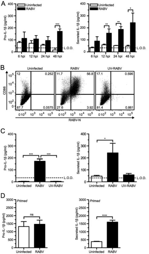
from mice after infection with RABV. For this approach, BMDCs were either infected with RABV at a multiplicity of infection (MOI) of 10 or mock infected for various periods of time. After 48 h, BMDCs infected with RABV produced a significantly larger amount of pro-IL-1than did the uninfected control as deter-mined byttest analysis (Fig. 1A, left panel; ***,P⬍0.001).
More-over, significantly greater amounts of IL-1were released from infected BMDCs than from uninfected cells as early as 12 h postin-fection (hpi) and continued to increase over time (Fig. 1A, right panel; **,P⬍0.01). The production of pro-IL-1as well as the secretion of IL-1was dependent upon infection with replication-competent RABV, secreting between 200 and 400 pg/ml IL-1, and was not observed when cells were treated with UV-inactivated RABV (UV-RABV) (Fig. 1BandC).
Even though RABV is not a cytolytic virus, some cell death of BMDCs has been described at 48 hpi (34). Necrotic cell death can result in the release of intracellular pro-IL-1and IL-1. To de-termine if the IL-1secretion after RABV infection was solely dependent upon the infection or cell death induced over time, pro-IL-1was induced using LPS priming. Priming alone sup-plies a reservoir of immature pro-IL-1but does not result in the secretion of mature IL-1(36). As expected, infected and unin-fected cells showed similar amounts of pro-IL-1 within the BMDCs (Fig. 1D, left panel). However, a significant amount of IL-1was detected in the supernatant of the RABV-infected cells compared to that for uninfected control BMDCs within 6 hpi (Fig. 1D, right panel; ****,P⬍0.0001). Taken together, these results indicate that RABV replication activates the inflammasome and the subsequent secretion of IL-1during infection of murine BM-DCs in a time-dependent manner.
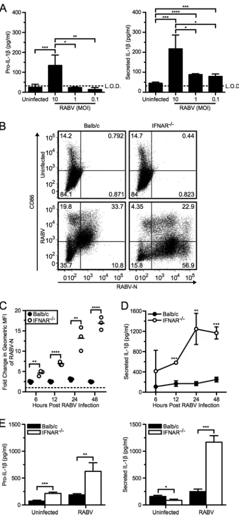
Increased viral infection results in increased IL-1secretion. It has previously been shown that although antigen-presenting cells are susceptible to RABV infection, there is limited viral rep-lication (20). To further investigate the impact of RABV replica-tion on IL-1secretion in BMDCs, we infected cells with a mul-tiplicity of infection (MOI) between 0.01 and 10 (Fig. 2A). We found that increased IL-1release was proportional to the MOI. Based on these results, we next analyzed IL-1secretion in BMDCs derived from IFNAR-deficient mice, as previous studies have shown robust RABV replication within this model (20,38). BMDCs from wild-type BALB/c and IFNAR⫺/⫺mice were in-fected at an MOI of 10 and analyzed 6, 12, 24, and 48 hpi. To assess the level of viral replication within BMDCs and DC activation, cells were stained for both RABV-N and CD86 (Fig. 2BandC). As observed previously (20), within 6 hpi we detected significantly more replication of RABV in IFNAR-deficient BMDCs than in wild-type cells, which further increased over time (Fig. 2C, open symbols; **,P⬍0.01). In contrast, infection in wild-type BMDCs remained constant over time (Fig. 2C, closed symbols). In cells lacking IFNAR, increased RABV replication does not upregulate CD86, a costimulatory molecule on mature DCs, compared to wild type (Fig. 2B). Thus, although RABV infection is sufficient to induce IL-1secretion in IFNAR-deficient BMDCs in a replica-tion-dependent manner (Fig. 2D), RABV-induced DC matura-tion requires stimulamatura-tion by type I IFNs, as has been previously reported (20). The level of RABV replication correlated with the level of released IL-1(Fig. 2CandD). Additionally, we observed that increased virus replication correlated with both increased production and secretion of IL-1at 48 hpi (Fig. 2E). Together, these results demonstrate that increased replication of RABV cor-relates with increased IL-1secretion.
IPS-1 is required for the secretion of IL-1following RABV infection.In order to better characterize the RABV and host cell interaction, we sought to define the activated signaling pathways leading to IL-1secretion by BMDCs in response to viral infec-tion. RABV activates antiviral immune responses in BMDCs via FIG 1RABV infects murine dendritic cells and induces pro-IL-1and the
secretion of IL-1in a time- and replication-dependent manner. (A) Bone marrow-derived DCs (BMDCs) from C57BL/6 mice were left uninfected (open bars) or infected at a multiplicity of infection (MOI) of 10 with RABV (closed bars) for 6, 12, 24, and 48 h postinfection (hpi). ELISA measuring pro-IL-1in cell lysates (left) and secreted IL-1in BMDC supernatants (right). (B and C) BMDCs left uninfected, infected for 48 h with RABV (MOI of 10), or treated for 48 h with UV-inactivated RABV (MOI of 10). (B) BMDCs were stained with an antibody against RABV-N and CD86. Results shown from one representative mouse at 48 hpi. (C) ELISA measuring pro-IL-1in cell lysates (left) and secreted IL-1in supernatants (right). (D) BMDCs primed with 50 ng LPS per ml for 3 h before being left uninfected or infected with RABV (MOI of 10) for 6 hpi. Results shown are representative of four inde-pendent experiments (n⫽12 mice per group). L.O.D., limit of detection. *, P⬍0.05; **,P⬍0.01; ***,P⬍0.001; ****,P⬍0.0001; ns, not significant. All error bars show standard deviations (SD).
on November 7, 2019 by guest
http://jvi.asm.org/
[image:3.585.42.288.67.486.2]the adaptor protein IPS-1 following recognition by RIG-I and MDA5 (20). It has been previously shown that IPS-1-mediated signaling is capable of activating the NF-B signaling cascade fol-lowing infection with RABV (19,20). Furthermore, transcription of pro-IL-1is NF-B dependent (39). Therefore, we hypothe-sized that IPS-1 contributes to the production of pro-IL-1in BMDCs infected with RABV.
BMDCs were collected from IPS-1⫹/⫹(wild-type) and IPS-1⫺/⫺mice, infected with RABV at an MOI of 10, and then ana-lyzed at 6, 12, 24, and 48 hpi. As previously shown (20), IPS-1⫹/⫹ BMDCs express high levels of CD86 on their surface at 48 hpi, whereas on IPS-1⫺/⫺BMDCs there is a reduction of CD86
expres-sion (Fig. 3A). However, in IPS-1⫺/⫺BMDCs, RABV undergoes significantly more replication than it does in the wild-type cells
(Fig. 3B; **,P⬍0.01).
Based on the fact that increased RABV replication in IFNAR⫺/⫺ BMDCs resulted in increased IL-1 secretion, we could also expect to see increased IL-1secretion in the IPS-1⫺/⫺ BMDCs. However, we found that IPS-1⫺/⫺BMDCs infected with RABV had significantly lower pro-IL-1production at early time points following infection (Fig. 3C; *,P⬍0.05). Infection of IPS-1⫺/⫺BMDCs induced much less IL-1release by 24 hpi, whereas infection with RABV in wild-type BMDCs induced robust IL-1 release in the supernatant (Fig. 3D). Therefore, it is likely that IPS-1 mediates synthesis of pro-IL-1as a response to RABV in-fection.
RABV engages an NLRP3-, ASC-, and caspase-1-dependent inflammasome.The processing of pro-IL-1 to mature IL-1 requires the cleavage of pro-caspase-1 to caspase-1, which is the common step for all inflammasomes (2). One of the best-charac-terized inflammasomes is activated by NLRP3, which recognizes a variety of pathogens, including RNA viruses (11); therefore, we investigated whether NLRP3 activation is required for IL-1 se-cretion after RABV infection.
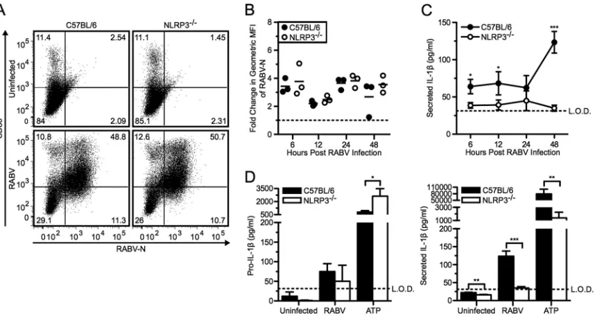
For this approach, we infected BMDCs derived from NLRP3-deficient and C57BL/6 wild-type mice with RABV at an MOI of 10 and followed viral replication, BMDC activation, and production of pro-IL-1as well as IL-1secretion over time. There was no difference in viral replication or activation in NLRP3⫺/⫺or wild-type BMDCs (Fig. 4AandB). However, NLRP3 was required for RABV-triggered release of IL-1in BMDCs, while RABV induced similar levels of pro-IL-1production in NLRP3-deficient and wild-type BMDCs (Fig. 4CandD). ATP can trigger the activation of the NLRP3 inflammasome and served as a control in these experiments (37). Compared to wild-type BMDCs, NLRP3⫺/⫺
BMDCs treated with ATP plus LPS priming secreted significantly less active IL-1(Fig. 4D, right panel; **,P⬍0.01). Taken to-gether, this experiment indicates an essential role of NLRP3 in RABV-induced inflammasome activation.
Many inflammasomes require the adaptor protein ASC to bridge the receptor and pro-caspase-1 (2). We therefore assessed ASC-induced secretion of IL-1 using ASC-deficient BMDCs. Compared to the wild-type BMDCs, the ASC⫺/⫺BMDCs showed no observable difference in viral replication or activation (Fig. 5A
andB). C57BL/6 BMDCs showed robust pro-IL-1production and IL-1release in response to infection with RABV and ATP plus LPS (Fig. 5CandD). However, removing ASC significantly reduced RABV-induced secretion of IL-1by 48 hpi (Fig. 5D, right panel; *,P⬍0.05). Thus, ASC is essential for coupling rec-FIG 2Increased RABV replication amplifies IL-1secretion. (A) BMDCs
from C57BL/6 mice were left uninfected or infected at an MOI of 10, 1, and 0.1 with RABV for 48 hpi. ELISA measuring pro-IL-1in cell lysates (left) and secreted IL-1in supernatants (right). (B to E) BMDCs from BALB/c (wild type; closed symbols and bars) and IFNAR⫺/⫺(open symbols and bars) mice
were left uninfected or infected with RABV (MOI of 10) for 6, 12, 24, and 48 hpi. (B) BMDCs were stained with an antibody against RABV-N and CD86. Results shown from one representative mouse at 48 hpi. (C) For each sample, the fold increase in RABV-N was determined by dividing the geometric mean fluorescence intensity (MFI) of the infected BMDCs by the MFI of the unin-fected BMDCs. Each point is representative of BMDCs from one mouse (n⫽ 3 mice per group). (D) ELISA measuring IL-1in supernatants over time. (E) ELISA measuring pro-IL-1in cell lysates (left) and secreted IL-1in super-natants (right) at 48 hpi. Results shown are representative of two independent experiments (n⫽6 mice per group). *,P⬍0.05; **,P⬍0.01; ***,P⬍0.001; ****,P⬍0.0001. All error bars show SDs.
on November 7, 2019 by guest
http://jvi.asm.org/
[image:4.585.44.281.65.577.2]ognition of RABV infection by NLRP3 to caspase-1-dependent activation of IL-1production.
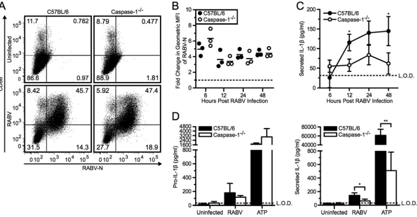
The primary function of the inflammasome complex is to me-diate the conversion of pro-caspase-1 to active caspase-1. Initially, to examine the involvement of caspase activity in secretion of IL-1after RABV infection, we pretreated BMDCs with a pan-caspase inhibitor, Z-VAD-fmk. Adding a pan-caspase inhibitor prior to stimulation with RABV dramatically reduced the level of IL-1 detected in the supernatants of BMDCs (data not shown). There-fore, together with the above knockouts, we studied IL-1release
in BMDCs from caspase-1-deficient mice. As seen for NLRP3⫺/⫺ and ASC⫺/⫺cells, replication of RABV and activation of BMDC were also unaffected in caspase-1⫺/⫺cells compared to the wild-type BMDCs (Fig. 6AandB). However, the lack of caspase-1 re-sulted in significantly less secretion of IL-1after RABV infection compared to wild-type BMDC controls, while pro-IL-1 produc-tion remained unaffected (Fig. 6CandD; *,P⬍0.05).
We also examined whether the effect of these knockouts was limited to the release of IL-1. We thus measured TNF-␣and type I IFN secretion as markers for NF-B and IRF3/7 activation, re-FIG 3RABV-induced pro-IL-1depends on IPS-1-mediated signaling pathways. BMDCs from IPS-1⫹/⫹(wild type; closed bars) and IPS-1⫺/⫺(open bars)
mice were left uninfected or infected with RABV (MOI of 10) for 6, 12, 24, and 48 hpi. (A) BMDCs were stained with an antibody against RABV-N and CD86. Results shown from one representative mouse at 48 hpi. (B) For each sample, the fold increase in RABV-N was determined by dividing the geometric MFI of the infected BMDCs by the MFI of the uninfected BMDCs. Each point is representative of BMDCs from one mouse (n⫽3 mice per group). ELISA measuring pro-IL-1in cell lysates (C) and secreted IL-1in supernatants (D). Results shown are representative of four independent experiments (n⫽12 mice per group). L.O.D., limit of detection. *,P⬍0.05; **,P⬍0.01; ***,P⬍0.001; ****,P⬍0.0001. All error bars show SDs.
FIG 4RABV-induced IL-1secretion is dependent on the NLRP3 inflammasome. (A to C) BMDCs from C57BL/6 (wild type; closed symbols and bars) and NLRP3⫺/⫺(open symbols and bars) mice were left uninfected or infected with RABV (MOI of 10) for 6, 12, 24, and 48 hpi. (A) BMDCs were stained with an
antibody against RABV-N and CD86. Results shown from one representative mouse at 48 hpi. (B) For each sample, the fold increase in RABV-N was determined by dividing the geometric MFI of the infected BMDCs by the MFI of the uninfected BMDCs. Each point is representative of BMDCs from one mouse (n⫽3 mice per group). (C) ELISA measuring secreted IL-1in supernatants over time. (D) C57BL/6 and NLRP3⫺/⫺BMDCs were left uninfected, infected with RABV for
48 hpi, or stimulated with 5 mM ATP for 30 min after priming with LPS. ELISA measuring pro-IL-1in cell lysates (left) and measuring secreted IL-1in supernatants (right). Results shown are representative of two independent experiments (n⫽6 mice per group). L.O.D., limit of detection. *,P⬍0.05; **,P⬍ 0.01; ***,P⬍0.001; ****,P⬍0.0001. All error bars show SDs.
on November 7, 2019 by guest
http://jvi.asm.org/
[image:5.585.45.539.65.207.2] [image:5.585.80.509.421.649.2]spectively (Fig. 7AandB). NLRP3, ASC, and caspase-1 were com-pletely dispensable for TNF-␣and type I IFN secretion. We did not detect any significant differences in either TNF-␣or type I IFN in the absence of NLRP3, ASC, or caspase-1 compared to
wild-type BMDCs following RABV infection. Taken together, these results indicate that release of IL-1from RABV-infected BMDCs is NLRP3, ASC, and caspase-1 dependent, whereas the production of pro-IL-1 is not dependent on the inflammasome components.
FIG 5RABV-induced IL-1secretion requires the adaptor protein ASC. (A to C) BMDCs from C57BL/6 (wild type; closed symbols and bars) and ASC⫺/⫺(open
symbols and bars) mice were left uninfected or infected with RABV (MOI of 10) for 6, 12, 24, and 48 hpi. (A) BMDCs were stained with an antibody against RABV-N and CD86. Results shown from one representative mouse at 48 hpi. (B) For each sample, the fold increase in RABV-N was determined by dividing the geometric MFI of the infected BMDCs by the MFI of the uninfected BMDCs. Each point is representative of BMDCs from one mouse (n⫽3 mice per group). (C) ELISA measuring secreted IL-1in supernatants over time. (D) C57BL/6 and ASC⫺/⫺BMDCs were left uninfected, infected with RABV for 48 hpi, or stimulated with 5 mM ATP for 30
min after priming with LPS. ELISA measuring pro-IL-1in cell lysates (left) and measuring secreted IL-1in supernatants (right). Results shown are representative of two independent experiments (n⫽6 mice per group). L.O.D., limit of detection. *,P⬍0.05; **,P⬍0.01; ***,P⬍0.001; ****,P⬍0.0001. All error bars show SDs.
FIG 6RABV induced IL-1secretion in a caspase-1-dependent manner. (A to C) BMDCs from C57BL/6 (wild type; closed symbols and bars) and caspase-1⫺/⫺(open
symbols and bars) mice were left uninfected or infected with RABV (MOI of 10) for 6, 12, 24, and 48 hpi. (A) BMDCs were stained with an antibody against RABV-N and CD86. Results shown from one representative mouse at 48 hpi. (B) For each sample, the fold increase in RABV-N was determined by dividing the geometric MFI of the infected BMDCs by the MFI of the uninfected BMDCs. Each point is representative of BMDCs from one mouse (n⫽3 mice per group). (C) ELISA measuring secreted IL-1in supernatants over time. (D) C57BL/6 and caspase-1⫺/⫺BMDCs were left uninfected, infected with RABV for 48 hpi, or stimulated with 5 mM ATP for
30 min after priming with LPS. ELISA measuring pro-IL-1in cell lysates (left) and measuring secreted IL-1in supernatants (right). Results shown are representative of two independent experiments (n⫽6 mice per group). L.O.D., limit of detection. *,P⬍0.05; **,P⬍0.01; ***,P⬍0.001; ****,P⬍0.0001. All error bars show SDs.
on November 7, 2019 by guest
http://jvi.asm.org/
[image:6.585.77.512.64.287.2] [image:6.585.78.508.435.659.2]IL-1 receptor signaling affects RABV pathogenicity. The above-described experiments suggested that IL-1 receptor signal-ing would be important in the mediation of the innate immune response to RABVin vivo. To study the importance of this path-wayin vivo, we infected wild-type and IL-1R-deficient mice with a strain of RABV that is moderately pathogenic after intranasal
in-oculation (28). As shown inFig. 8A, 13 out of 23 IL-1R⫺/⫺mice (56.5%) succumbed to the infection whereas only 3 of 24 mice (12.5%) died within the control group (**,P⬍0.01). As shown in a previous study (28), the pathogenicity of the virus correlated with the weight loss occurring at around 14 days postinfection
(Fig. 8B). Previously published work indicates that paralysis
in-duced by RABV infection is an indicator of disease; therefore, the morbidity score was also recorded, with a score of 0 indicating no signs of rabies and a score of 5 indicating a moribund state (40). As shown inFig. 8C, the IL-1R-deficient mice experienced greater levels of paralysis starting at 11 days postinfection and either suc-cumbed to disease or were euthanized by day 16. Those IL-1R⫺/⫺ mice that did survive took longer to recover from the weight loss, as indicated by the majority of mice scoring a 1 or 2 on days 17 through 21 compared to the wild-type mice, which quickly recov-ered to a score of 0. The result of the infection of IL-1R⫺/⫺mice compared to that of wild-type micein vivo indicates that IL-1 receptor signaling plays a critical role in the control of RABV infection.
DISCUSSION
Recently, the inflammasome has gained considerable attention for its ability to control viral infection. The inflammasome is an im-munological protein scaffold that recognizes danger signals within the cytoplasm of infected host cells and is essential for the production and secretion of the inflammatory cytokines IL-1 and IL-18. However, the activation of the inflammasome by RNA viruses has been largely unexplored.
Previous work has demonstrated that RABV is detected by RIG-I and MDA5 and signals through the adaptor IPS-1 for acti-vation of NF-B and IRF signaling pathways (20). Elimination of either RIG-I or MDA5 resulted in a decrease of BMDC activation as indicated by the upregulation of CD86 and IFN production. FIG 7IFN and TNF-␣ secretion are independent of NLRP3, ASC, and
caspase-1. (A) BMDCs from C57BL/6 (wild type; closed symbols) and NLRP3⫺/⫺, ASC⫺/⫺, and caspase-1⫺/⫺(open symbols) mice were infected
with RABV (MOI of 10) for 48 hpi, and supernatants were collected. Type I IFN levels were detected in the supernatants by the sensitivity of reporter cells to VSV-GFP infection. After incubation with the UV-inactivated supernatant for 24 h, reporter cells were infected with VSV-GFP for 6 hpi. GFP fluorescence indicates VSV replication and, therefore, lack of type I IFN. (B) BMDCs from C57BL/6 (wild type; closed symbols) and NLRP3⫺/⫺, ASC⫺/⫺, and
caspase-1⫺/⫺(open symbols) mice were left uninfected or infected with RABV (MOI of
10) for 6 hpi. Expression of TNF-␣by BMDCs was measured by intracellular cytokine staining. For each sample, the fold difference in TNF-␣was deter-mined by subtracting the percent RABV-N⫹TNF-␣⫹infected BMDCs from uninfected BMDCs. Results shown are representative of two independent ex-periments (n⫽6 mice per group). ns, not significant.
FIG 8Lack of IL-1 receptor signaling in a mouse model of RABV infection increases viral pathogenicity. C57BL/6 (n⫽24; closed symbols) and IL-1R⫺/⫺(n⫽
23; open symbols) mice were infected intranasally with 1⫻105focus-forming units of RABV. Mice were monitored daily over time for their survival (A), weight
change (B), and morbidity score (C). Results shown are cumulative from two independent experiments. *,P⬍0.05; **,P⬍0.01; ***,P⬍0.001; ****,P⬍0.0001.
on November 7, 2019 by guest
http://jvi.asm.org/
[image:7.585.43.283.65.207.2] [image:7.585.136.449.453.692.2]The genetic experiments that we show here demonstrated a sig-naling cascade downstream of IPS-1 that controlled the produc-tion of IL-1in response to innate immune recognition of RABV. This makes it likely that RIG-I and/or MDA5 also plays a role in the production of IL-1by signaling through the adaptor IPS-1.
As active IL-1secretion in response to RABV infection was defective in BMDCs from NLRP3-, ASC-, and caspase-1-knock-out mice, we conclude that RABV infection activated the inflam-masome by forming a signaling complex comprised of NLRP3, ASC, and pro-caspase-1. We propose the following IL-1 activa-tion pathway triggered by RABV infecactiva-tion: after IPS-1 signaling induces the production of pro-IL-1, RABV activates an NLRP3-, ASC-, and caspase-1-dependent inflammasome, which then acti-vates secretion of IL-1. We found that NLRP3-, ASC-, and caspase-1-deficient DCs had pronounced defects in IL-1 secre-tion after RABV infecsecre-tion but maintained typical TNF-␣and in-terferon responses. This indicates that the NLRP3/ASC/caspase-1 inflammasome controls the proinflammatory cytokine IL-1 af-ter RABV recognition and suggests that the inflammasome func-tions in the innate immune recognition of RABV infection.
Poeck et al. demonstrated that RNA viruses, namely, VSV, were recognized by a RIG-I-, ASC-, caspase-1-dependent inflam-masome, whereas double-stranded RNA (dsRNA) that lacked the 5=-triphosphate did not induce IL-1or caspase-1 activation (12). As RABV is a related rhabdovirus, we expect that RIG-I is involved in sensing infection. However, we do not suspect that RIG-I is involved in the formation of the NLRP3 inflammasome, but rather, we think that RIG-I and MDA5 signal through IPS-1 for the synthesis of pro-IL-1via the NF-B pathway. Therefore, our findings support the conclusion that secretion of IL-1, which is dependent upon cleavage of pro-IL-1, is dependent upon NLRP3 activity following RABV infection. Additionally, this work suggests that NLRP3 activation by itself is sufficient for inflam-masome activation.
RABV activates the inflammasome and triggers the subsequent release of active IL-1through a mechanism requiring viral rep-lication as shown above, even though viral reprep-lication in DCs is limited and ultimately results in a nonproductive infection (20). However, the exact viral stimulants that trigger the NLRP3 in-flammasome remain to be elucidated. In IFNAR⫺/⫺BMDCs, we saw significantly more RABV-N expression over time, together with increased induction of pro-IL-1and secretion of IL-1. This indicates that with viral replication occurring at a higher rate there is also an increased induction and processing of pro-IL-1. Another RNA virus, influenza virus, activates the NLRP3 inflam-masome by lysosomal damage and reactive oxygen species (ROS) production during entry and virus replication (8, 10), whereas recent work demonstrates that encephalomyocarditis virus (EMCV), a positive-stranded RNA virus, activates the NLRP3 in-flammasome through the elevation of intracellular Ca2⫹levels (41). Additional studies on the role of viral entry and Ca2⫹levels may further identify the mechanism by which RABV activates the inflammasome.
Alternatively, noncanonical inflammasomes utilizing caspase-8 are capable of cleaving pro-IL-1(42,43). However, we would expect to see a caspase-1-independent IL-1secretion in RABV-infected BMDCs. Additionally, while we are aware that there are alternative mechanisms for IL-1 activation that are independent of the caspase-1 inflammasome, they are limited to other cell types such as
neutrophils via cleavage by serine proteases, including proteinase-3, elastase, or cathepsin G (44,45).
As NLRP3 inflammasome-dependent IL-1secretion occurs in RABV-infected BMDCs, RABV also induces type I interferons (20). IFN-␣/signaling can also inhibit the inflammasome from being activated by NLRP3 and therefore results in decreased IL-1 secretion (46). However, we believe that the differences observed in this study may result from the fact that RABV induces a mar-ginal amount of IFN-compared to the amount needed to see inhibition of IL-1production.
The aim of this study was to investigate which inflammasomes in dendritic cells regulate the response to RABV. However, the inflammasome is found in numerous antigen-presenting cells, in-cluding macrophages, monocytes, neutrophils, and microglial cells. Bone marrow-derived macrophages were found to be non-permissive for viral infection and resulted in lack of IL-1 secre-tion (T. M. Lawrence and M. J. Schnell, unpublished data). Dur-ingin vivostudies, IL-1was found in the brains of mice infected with RABV (Lawrence and Schnell, unpublished data). Mice were immunized intranasally with RABV similarly to this study. ELISA of brain lysates collected at 15 days postinfection showed signifi-cantly more IL-1in RABV-infected mice than in mock-immu-nized mice, confirming previously published results (47–49). Marquette et al. found that resident microglial cells and infiltrat-ing macrophages were positive for IL-1(47); Laothamatas et al. showed that IL-1appeared during the early stages of infection with RABV (48). In view of these results, our data strongly suggest that in cells found at the site of RABV infection in the brain, the increased IL-1production is the result of NLRP3 inflammasome activation.
The production of IL-1in the brain may have a beneficial role in the recruitment of inflammatory cells as suggested by Baloul and Lafon (50) and by our observations that without the IL-1 receptor there is increased viral pathogenicity. Previous studies in mice deficient in IL-1R did not show increased susceptibility to infection (51). It is important to note that these experiments uti-lized a different strain of RABV as well as another route of infec-tion. Of note, results from several groups in IL-1R⫺/⫺mice dem-onstrate that the IL-1 receptor is critical for infiltration of neutrophils and development of local and systemic inflammation (4). The presence of inflammatory cells recruited into the central nervous system (CNS) by inflammasome activation may lead to the recognition of RABV, at least in the case of attenuated RABV strains. Whereas some viral studies, including those on Japanese encephalitis virus, suggest that cytokines produced by infiltrating inflammatory cells, such as TNF-␣and IL-1, are associated with the death of patients (52), in RABV infections inflammation has been clearly shown to be beneficial for clearances of the virus (53,54).
In this study, we examined the IL-1activation pathway trig-gered by RABV infection. We found that NLRP3-, ASC-, and caspase-1-deficient DCs had pronounced defects in IL-1 secre-tion after RABV infecsecre-tion but had typical TNF-␣and interferon responses. This indicates that NLRP3, ASC, and caspase-1 act to-gether to form an inflammasome complex that controls the pro-inflammatory cytokine IL-1 release after RABV recognition. These data suggest that the inflammasome functions in the innate immune recognition of RABV infection. Furtherin vitro work would clarify the mechanism by which the NLRP3 inflammasome
on November 7, 2019 by guest
http://jvi.asm.org/
recognizes RABV and how this recognition contributes to the con-trol of viral infectionin vivo.
ACKNOWLEDGMENTS
We thank John Hiscott (McGill University) for providing the VSV-ex-pressing GFP, Joan Durbin (Ohio State University) for providing the IFNAR⫺/⫺mice, and Shizuo Akira (Osaka University) for providing the IPS-1 mice.
This work was supported by NIH grants P01AI082325, R21NS074006, and P40OD010996 to M.J.S. and a Rubicon Fellowship from the Nether-lands Organization of Scientific Research (NWO) to M.R.D.Z. The funders had no role in study design, data collection and analysis, decision to publish, or preparation of the manuscript.
REFERENCES
1.Akira S, Uematsu S, Takeuchi O.2006. Pathogen recognition and innate immunity. Cell124:783– 801.
2.Schroder K, Tschopp J.2010. The inflammasomes. Cell140:821– 832. 3.Kanneganti TD.2010. Central roles of NLRs and inflammasomes in viral
infection. Nat. Rev. Immunol.10:688 – 698.
4.Dinarello CA.2009. Immunological and inflammatory functions of the interleukin-1 family. Annu. Rev. Immunol.27:519 –550.
5.Sims JE, Smith DE.2010. The IL-1 family: regulators of immunity. Nat. Rev. Immunol.10:89 –102.
6.Ting JP, Lovering RC, Alnemri ES, Bertin J, Boss JM, Davis BK, Flavell RA, Girardin SE, Godzik A, Harton JA, Hoffman HM, Hugot JP, Inohara N, Mackenzie A, Maltais LJ, Nunez G, Ogura Y, Otten LA, Philpott D, Reed JC, Reith W, Schreiber S, Steimle V, Ward PA.2008. The NLR gene family: a standard nomenclature. Immunity28:285–287. 7.Ichinohe T, Lee HK, Ogura Y, Flavell R, Iwasaki A. 2009.
Inflam-masome recognition of influenza virus is essential for adaptive immune responses. J. Exp. Med.206:79 – 87.
8.Allen IC, Scull MA, Moore CB, Holl EK, McElvania-TeKippe E, Tax-man DJ, Guthrie EH, Pickles RJ, Ting JP.2009. The NLRP3 inflam-masome mediates in vivo innate immunity to influenza A virus through recognition of viral RNA. Immunity30:556 –565.
9.Thomas PG, Dash P, Aldridge JR, Jr, Ellebedy AH, Reynolds C, Funk AJ, Martin WJ, Lamkanfi M, Webby RJ, Boyd KL, Doherty PC, Kan-neganti TD.2009. The intracellular sensor NLRP3 mediates key innate and healing responses to influenza A virus via the regulation of caspase-1. Immunity30:566 –575.
10. Ichinohe T, Pang IK, Iwasaki A.2010. Influenza virus activates inflam-masomes via its intracellular M2 ion channel. Nat. Immunol.11:404 – 410. 11. Kanneganti TD, Body-Malapel M, Amer A, Park JH, Whitfield J, Franchi L, Taraporewala ZF, Miller D, Patton JT, Inohara N, Nunez G. 2006. Critical role for Cryopyrin/Nalp3 in activation of caspase-1 in re-sponse to viral infection and double-stranded RNA. J. Biol. Chem.281: 36560 –36568.
12. Poeck H, Bscheider M, Gross O, Finger K, Roth S, Rebsamen M, Hannesschlager N, Schlee M, Rothenfusser S, Barchet W, Kato H, Akira S, Inoue S, Endres S, Peschel C, Hartmann G, Hornung V, Ruland J. 2010. Recognition of RNA virus by RIG-I results in activation of CARD9 and inflammasome signaling for interleukin 1 beta production. Nat. Im-munol.11:63– 69.
13. Rajan JV, Rodriguez D, Miao EA, Aderem A.2011. The NLRP3 inflam-masome detects encephalomyocarditis virus and vesicular stomatitis virus infection. J. Virol.85:4167– 4172.
14. Ramos HJ, Lanteri MC, Blahnik G, Negash A, Suthar MS, Brassil MM, Sodhi K, Treuting PM, Busch MP, Norris PJ, Gale M, Jr.2012. IL-1beta signaling promotes CNS-intrinsic immune control of West Nile virus in-fection. PLoS Pathog.8:e1003039. doi:10.1371/journal.ppat.1003039. 15. Kumar M, Roe K, Orillo B, Muruve DA, Nerurkar VR, Gale M, Jr,
Verma S.2013. Inflammasome adaptor protein apoptosis-associated speck-like protein containing CARD (ASC) is critical for the immune response and survival in West Nile virus encephalitis. J. Virol.87:3655– 3667.
16. Guarda G, Zenger M, Yazdi AS, Schroder K, Ferrero I, Menu P, Tardivel A, Mattmann C, Tschopp J.2011. Differential expression of NLRP3 among hematopoietic cells. J. Immunol.186:2529 –2534. 17. Faul EJ, Wanjalla CN, McGettigan JP, Schnell MJ.2008. Interferon-beta
expressed by a rabies virus-based HIV-1 vaccine vector serves as a molec-ular adjuvant and decreases pathogenicity. Virology382:226 –238. 18. Hornung V, Ellegast J, Kim S, Brzozka K, Jung A, Kato H, Poeck H,
Akira S, Conzelmann KK, Schlee M, Endres S, Hartmann G. 2006. 5=-Triphosphate RNA is the ligand for RIG-I. Science314:994 –997. 19. Li J, McGettigan JP, Faber M, Schnell MJ, Dietzschold B.2008.
Infec-tion of monocytes or immature dendritic cells (DCs) with an attenuated rabies virus results in DC maturation and a strong activation of the NFkappaB signaling pathway. Vaccine26:419 – 426.
20. Faul EJ, Wanjalla CN, Suthar MS, Gale M, Wirblich C, Schnell MJ. 2010. Rabies virus infection induces type I interferon production in an IPS-1 dependent manner while dendritic cell activation relies on IFNAR signaling. PLoS Pathog.6:e1001016. doi:10.1371/journal.ppat.1001016. 21. Vidy A, Chelbi-Alix M, Blondel D.2005. Rabies virus P protein interacts
with STAT1 and inhibits interferon signal transduction pathways. J. Virol. 79:14411–14420.
22. Brzozka K, Finke S, Conzelmann KK. 2006. Inhibition of interferon signaling by rabies virus phosphoprotein P: activation-dependent binding of STAT1 and STAT2. J. Virol.80:2675–2683.
23. Rieder M, Conzelmann KK.2009. Rhabdovirus evasion of the interferon system. J. Interferon Cytokine Res.29:499 –509.
24. Wang ZW, Sarmento L, Wang Y, Li XQ, Dhingra V, Tseggai T, Jiang B, Fu ZF.2005. Attenuated rabies virus activates, while pathogenic rabies virus evades, the host innate immune responses in the central nervous system. J. Virol.79:12554 –12565.
25. Prehaud C, Megret F, Lafage M, Lafon M.2005. Virus infection switches TLR-3-positive human neurons to become strong producers of beta in-terferon. J. Virol.79:12893–12904.
26. Kawai T, Akira S.2008. Toll-like receptor and RIG-I-like receptor sig-naling. Ann. N. Y. Acad. Sci.1143:1–20.
27. McGettigan JP, Pomerantz RJ, Siler CA, McKenna PM, Foley HD, Dietzschold B, Schnell MJ.2003. Second-generation rabies virus-based vaccine vectors expressing human immunodeficiency virus type 1 gag have greatly reduced pathogenicity but are highly immunogenic. J. Virol. 77:237–244.
28. Gomme EA, Wirblich C, Addya S, Rall GF, Schnell MJ.2012. Immune clearance of attenuated rabies virus results in neuronal survival with al-tered gene expression. PLoS Pathog. 8:e1002971. doi:10.1371/journal .ppat.1002971.
29. Kumar H, Kawai T, Kato H, Sato S, Takahashi K, Coban C, Yamamoto M, Uematsu S, Ishii KJ, Takeuchi O, Akira S.2006. Essential role of IPS-1 in innate immune responses against RNA viruses. J. Exp. Med.203: 1795–1803.
30. Martinez-Sobrido L, Gitiban N, Fernandez-Sesma A, Cros J, Mertz SE, Jewell NA, Hammond S, Flano E, Durbin RK, Garcia-Sastre A, Durbin JE.2006. Protection against respiratory syncytial virus by a recombinant Newcastle disease virus vector. J. Virol.80:1130 –1139.
31. Sutterwala FS, Ogura Y, Szczepanik M, Lara-Tejero M, Lichtenberger GS, Grant EP, Bertin J, Coyle AJ, Galan JE, Askenase PW, Flavell RA. 2006. Critical role for NALP3/CIAS1/Cryopyrin in innate and adaptive immunity through its regulation of caspase-1. Immunity24:317–327. 32. Kuida K, Lippke JA, Ku G, Harding MW, Livingston DJ, Su MS, Flavell
RA.1995. Altered cytokine export and apoptosis in mice deficient in interleukin-1 beta converting enzyme. Science267:2000 –2003. 33. Kayagaki N, Warming S, Lamkanfi M, Vande Walle L, Louie S, Dong J,
Newton K, Qu Y, Liu J, Heldens S, Zhang J, Lee WP, Roose-Girma M, Dixit VM. 2011. Non-canonical inflammasome activation targets caspase-11. Nature479:117–121.
34. Wanjalla CN, Faul EJ, Gomme EA, Schnell MJ.2010. Dendritic cells infected by recombinant rabies virus vaccine vector expressing HIV-1 Gag are immunogenic even in the presence of vector-specific immunity. Vac-cine29:130 –140.
35. Wanjalla CN, Goldstein EF, Wirblich C, Schnell MJ.2012. A role for granulocyte-macrophage colony-stimulating factor in the regulation of CD8(⫹) T cell responses to rabies virus. Virology426:120 –133. 36. Bauernfeind FG, Horvath G, Stutz A, Alnemri ES, MacDonald K, Speert
D, Fernandes-Alnemri T, Wu J, Monks BG, Fitzgerald KA, Hornung V, Latz E.2009. Cutting edge: NF-kappaB activating pattern recognition and cytokine receptors license NLRP3 inflammasome activation by regulating NLRP3 expression. J. Immunol.183:787–791.
37. Mariathasan S, Weiss DS, Newton K, McBride J, O’Rourke K, Roose-Girma M, Lee WP, Weinrauch Y, Monack DM, Dixit VM. 2006.
on November 7, 2019 by guest
http://jvi.asm.org/
Cryopyrin activates the inflammasome in response to toxins and ATP. Nature440:228 –232.
38. Mendonca RZ, Pereira CA.1994. Relationship of interferon synthesis and the resistance of mice infected with street rabies virus. Braz. J. Med. Biol. Res.27:691– 695.
39. Weber A, Wasiliew P, Kracht M.2010. Interleukin-1 (IL-1) pathway. Sci. Signal.3:cm1. doi:10.1126/scisignal.3105cm1.
40. Hemachudha T, Wacharapluesadee S, Mitrabhakdi E, Wilde H, Mo-rimoto K, Lewis RA.2005. Pathophysiology of human paralytic rabies. J. Neurovirol.11:93–100.
41. Ito M, Yanagi Y, Ichinohe T.2012. Encephalomyocarditis virus viro-porin 2B activates NLRP3 inflammasome. PLoS Pathog.8:e1002857. doi:
10.1371/journal.ppat.1002857.
42. Maelfait J, Vercammen E, Janssens S, Schotte P, Haegman M, Magez S, Beyaert R.2008. Stimulation of Toll-like receptor 3 and 4 induces inter-leukin-1beta maturation by caspase-8. J. Exp. Med.205:1967–1973. 43. Gringhuis SI, Kaptein TM, Wevers BA, Theelen B, van der Vlist M,
Boekhout T, Geijtenbeek TB.2012. Dectin-1 is an extracellular pathogen sensor for the induction and processing of IL-1beta via a noncanonical caspase-8 inflammasome. Nat. Immunol.13:246 –254.
44. Coeshott C, Ohnemus C, Pilyavskaya A, Ross S, Wieczorek M, Kroona H, Leimer AH, Cheronis J.1999. Converting enzyme-independent re-lease of tumor necrosis factor alpha and IL-1beta from a stimulated hu-man monocytic cell line in the presence of activated neutrophils or puri-fied proteinase 3. Proc. Natl. Acad. Sci. U. S. A.96:6261– 6266. 45. Netea MG, Simon A, van de Veerdonk F, Kullberg BJ, Van der Meer
JW, Joosten LA.2010. IL-1beta processing in host defense: beyond the inflammasomes. PLoS Pathog.6:e1000661. doi:10.1371/journal.ppat .1000661.
46. Guarda G, Braun M, Staehli F, Tardivel A, Mattmann C, Forster I,
Farlik M, Decker T, Du Pasquier RA, Romero P, Tschopp J.2011. Type I interferon inhibits interleukin-1 production and inflammasome activa-tion. Immunity34:213–223.
47. Marquette C, Van Dam AM, Ceccaldi PE, Weber P, Haour F, Tsiang H. 1996. Induction of immunoreactive interleukin-1and tumor necrosis factor-␣in the brains of rabies virus infected rats. J. Neuroimmunol.68: 45–51.
48. Laothamatas J, Wacharapluesadee S, Lumlertdacha B, Ampawong S, Tepsumethanon V, Shuangshoti S, Phumesin P, Asavaphatiboon S, Worapruekjaru L, Avihingsanon Y, Israsena N, Lafon M, Wilde H, Hemachudha T.2008. Furious and paralytic rabies of canine origin: neu-roimaging with virological and cytokine studies. J. Neurovirol.14:119 – 129.
49. Solanki A, Radotra BD, Vasishta RK. 2009. Correlation of cytokine expression with rabies virus distribution in rabies encephalitis. J. Neuro-immunol.217:85– 89.
50. Baloul L, Lafon M. 2003. Apoptosis and rabies virus neuroinvasion. Biochimie85:777–788.
51. Li J, Faber M, Dietzschold B, Hooper DC.2011. The role of Toll-like receptors in the induction of immune responses during rabies virus infec-tion. Adv. Virus Res.79:115–126.
52. Kaushik DK, Gupta M, Kumawat KL, Basu A.2012. NLRP3 inflam-masome: key mediator of neuroinflammation in murine Japanese enceph-alitis. PLoS One7:e32270. doi:10.1371/journal.pone.0032270.
53. Hooper DC, Morimoto K, Bette M, Weihe E, Koprowski H, Dietz-schold B.1998. Collaboration of antibody and inflammation in clearance of rabies virus from the central nervous system. J. Virol.72:3711–3719. 54. Niu X, Wang H, Fu ZF.2011. Role of chemokines in rabies pathogenesis
and protection. Adv. Virus Res.79:73– 89.