Phosphoprotein
Robert Cox,aTodd J. Green,aSangeetha Purushotham,bChampion Deivanayagam,bGregory J. Bedwell,aPeter E. Prevelige,a Ming Luoa
Department of Microbiologya
and Department of Vision Sciences,b
University of Alabama at Birmingham, Birmingham, Alabama, USA
The phosphoprotein (P) is virally encoded by theRhabdoviridaeandParamyxoviridaein the orderMononegavirales. P is a self-associated oligomer and forms complexes with the large viral polymerase protein (L), the nucleocapsid protein (N), and the as-sembled nucleocapsid. P from different viruses has shown structural diversities even though their essential functions are the same. We systematically mapped the domains in mumps virus (MuV) P and investigated their interactions with nucleocapsid-like particles (NLPs). Similar to other P proteins, MuV P contains N-terminal, central, and C-terminal domains with flexible linkers between neighboring domains. By pulldown assays, we discovered that in addition to the previously proposed nucleocap-sid binding domain (renucleocap-sidues 343 to 391), the N-terminal region of MuV P (renucleocap-sidues 1 to 194) could also bind NLPs. Further analysis of binding kinetics was conducted using surface plasmon resonance. This is the first observation that both the N- and C-terminal regions of a negative-strand RNA virus P are involved in binding the nucleocapsid. In addition, we defined the oli-gomerization domain (POD) of MuV P as residues 213 to 277 and determined its crystal structure. The tetrameric MuV PODis
formed by one pair of long parallel␣-helices with another pair in opposite orientation. Unlike the parallel orientation of each ␣-helix in the tetramer of Sendai virus POD, this represents a novel orientation of a PODwhere both the N- and the C-terminal
domains are at either end of the tetramer. This is consistent with the observation that both the N- and the C-terminal domains are involved in binding the nucleocapsid.
T
he phosphoprotein (P) is a multifunctional protein encoded in the genomes of negative-strand RNA viruses (NSVs) of the RhabdoviridaeandParamyxoviridaein the orderMononegavirales. P performs several essential functions in virus replication. It is the cofactor in the viral RNA-dependent RNA polymerase (RdRp), a complex that also includes the virally encoded large (L) protein. L harbors the catalytic functions for RNA synthesis and mRNA cap-ping. P also directly binds the nucleocapsid, the active template for viral RNA synthesis. Through this interaction, P delivers the RdRp to the nucleocapsid. In the absence of P, the RdRp cannot recog-nize the nucleocapsid or gain access to the viral genome. The do-main responsible for P to bind the nucleocapsid, the phosphopro-tein nucleocapsid binding domain (PNBD), has been mapped to the C-terminal region for a number of NSVs, including vesicular stomatitis virus (VSV), rabies virus (RABV), measles virus, Sendai virus (SeV), Mokola virus, and mumps virus (MuV) (1–5). Three-dimensional structures of this domain revealed a certain degree of structural homology among different viruses (5–9). The crystal structure of VSV PNBDin complex with a nucleocapsid-like parti-cle (NLP) parti-clearly showed that the P binding site in the nuparti-cleocap- nucleocap-sid is formed by two neighboring nucleocapnucleocap-sid protein (N) sub-units (10). The large extended loop in the C-lobe and a single ␣-helix (␣13) of one N subunit and the same extended loop in an adjacent N subunit make up the U-shaped P binding site, with␣13 at the bottom of the cleft. This high-affinity P binding site could only be created by N subunits that are assembled together in the nucleocapsid. By this mechanism, the cofactor P can dock the RdRp specifically to the nucleocapsid in order to use it as the template for viral RNA synthesis. In addition, P forms a stable complex with N that is free of RNA, termed N0-P. This encapsi-dation-competent complex of N and P exists prior to incorpora-tion of N into the nucleocapsid (11). During virus replication, the N subunit in the N0-P complex is used to concomitantlyencapsi-date the newly synthesized viral RNA (12). The domain essential for keeping N free of RNA in the N0-P complex has been mapped to the N-terminal region (phosphoprotein N0binding domain [PN°D]) of P for several NSVs (11,13–15). A crystal structure of VSV PN°Din complex with a VSV N mutant showed that PN°Dsits in the cavity where the viral RNA would be accommodated in the nucleocapsid (16). Occupation of the cavity may be instrumental in allowing P to keep N free of RNA in the N0-P complex.
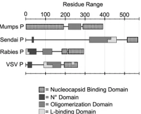
Another important feature of P is that its functional form is a self-associated oligomer. Self-association is required for support-ing viral RNA synthesis (17) but is not necessary for the associa-tion of P with other viral proteins such as L or N. The domain responsible for P oligomerization (POD) is located in the central region of P. PN°D and PNBDare linked to PODthrough flexible loops, which appears to be a common modular structure for all NSV P proteins. The length of P differs greatly among NSVs, and there is no homology for POD. Furthermore, the structure and the mode of self-association also vary considerably from one P to another. In the case of SeV P (568 amino acids), PODcorresponds to residues 320 to 433. Each SeV PODsubunit donates a single long helix to form a tetrameric coiled coil, with all helices being parallel to one another. Short helices at the N-terminal end of SeV POD contribute to additional tetrameric interactions. For VSV P (265 amino acids), PODcomprises residues 107 to 177 (18,19). VSV PODdimerizes by domain swapping of a-hairpin from each
sub-Received6 March 2013Accepted20 April 2013
Published ahead of print1 May 2013
Address correspondence to Ming Luo, [email protected].
Copyright © 2013, American Society for Microbiology. All Rights Reserved.
doi:10.1128/JVI.00653-13
on November 7, 2019 by guest
http://jvi.asm.org/
unit that participates in collating a-sheet of four antiparallel strands on each side. Two parallel␣-helices held together by a number of hydrophobic interactions form the core of the VSV P dimer. By comparison, the oligomerization mode of RABV PODis quite different from that of VSV POD, despite the two viruses being closely related members of the rhabdovirus family. RABV POD spans residues 92 to 131 in P (297 amino acids) and forms a dimer (20). However, each polypeptide contains two antiparallel helices linked by a loop. The dimer of RABV PODinvolves a four-helix bundle between two parallel subunits. This particular orientation places PN°Dand PNBDat the same end of RABV P, in contrast to other P proteins in which the two domains are at distant opposite ends of the oligomer (19–21). In VSV, it has been shown that a chimeric P dimer with one P mutant lacking PN°Dand another mutant lacking PNBD can fully support VSV RNA synthesis (17). In the case of the RABV P dimer, it is not clear what the functional implications are when all PN°Dand PNBDdomains are at the same end.
For this report, we carried out systematic studies of the do-mains of MuV P and their possible interactions with the nucleo-capsid. Previously, MuV P expressed as a recombinant glutathione S-transferase (GST) fusion protein was shown to bind a truncated N (N398) that was described as capable of assembling into NLP (4). In addition, deletion of the last 49 residues in MuV P abolished NLP binding, and the C-terminal domain (C343-391) alone was shown to be capable of binding NLP (4). In contrast, nucleocapsid binding by the P protein from other paramyxoviruses reportedly requires interactions with the C-terminal tail region of N (22–25). The previous report appears to suggest that MuV P contains a PNBDthat is similar to other paramyxoviruses, but its binding site on the nucleocapsid may be located in a different region of N. The crystal structure of the C-terminal domain of MuV P showed that it contains three packed␣-helices that may be induced to fold when P binds the nucleocapsid (7). We expanded the study of nucleocapsid binding to include all domains in MuV P. Our struc-tural and functional characterization of MuV P is discussed here with comparison to other NSV Ps.
MATERIALS AND METHODS
Materials.Restriction enzymes and T4 DNA ligase were purchased from New England BioLabs. pET28b and BL21(DE3) were purchased from Novagen. Primers for PCR were purchased from Invitrogen and Inte-grated DNA Technologies.
Plasmid construction.The P genes were amplified from a cDNA clone of MuV strain 88-1961 (GenBank accession no.AF467767.2) by PCR. Primers for amplification on the 5=end of the gene fragments con-tained NheI restriction sites, while the 3=primers contained XhoI sites. Sequences of the primers can be sent upon request. The P gene or gene fragments and the pET-28b plasmid were each digested with restriction enzymes NheI and XhoI and gel purified. The purified digested P genes were individually ligated in frame with the N-terminal polyhistidine tag, resulting in the vector MuV P-pET28b or MuV (P clone ID)-pET28b.
Protein expression and purification.Expression vectors were trans-formed intoEscherichia colistrain BL21(DE3). Bacteria were cultured in Luria-Bertani broth at 37°C until the optical density at 600 nm reached 0.6. Protein expression was induced with 1 mM IPTG (isopropyl--D -thiogalactopyranoside) for 18 h at 18°C. The cells were harvested by cen-trifugation and resuspended in binding buffer A containing 20 mM Tris (pH 7.9), 500 mM NaCl, and 5 mM imidazole. The cells were disrupted by sonication and then centrifuged for 1 h at 18,000 rpm. Soluble fractions were collected and passed through an Ni-affinity column (Chelating Sep-harose Fast Flow; GE Healthcare). The loaded Ni-affinity column was
washed with 5 column volumes (CV) of binding buffer A. The loaded column was washed with 5 CV of wash buffer A containing 20 mM Tris (pH 7.9), 500 mM NaCl, and 50 mM imidazole. Samples were eluted with elution buffer A containing 20 mM Tris (pH 7.9), 500 mM imidazole, and 500 mM NaCl. Proteins were further purified by size-exclusion chroma-tography (Sephacryl S-75 or S-200; GE Healthcare) in size exclusion buf-fer containing 20 mM Tris (pH 7.5) and 500 mM NaCl.
Purification of trypsinized MuV NLP.The MuV N protein was ex-pressed, and NLP was purified as previously reported (26). NLP has a ring structure composed of 13 N subunits and a single strand of random RNA. The purified NLP was allowed to incubate overnight at room temperature with trypsin at a 1-mg/100-mg ratio (trypsin:N). Trypsin was then inac-tivated by the addition of phenylmethylsulfonyl fluoride (1 mM). The digested NLP (NLP379) was concentrated to 8 mg/ml and purified by size-exclusion chromatography using a S-400 column (HiPrep Sephacryl S-400; GE Healthcare). The identity of the digested N was confirmed by N-terminal amino acid sequencing and mass spectrometry analysis. The size of the NLP379was consistent with that of a ring structure similar to the full-length NLP.
Identification of protease-resistant fragments of the MuV P protein. The full-length P protein was digested by using three different proteases: trypsin (TPCK [tolylsulfonyl phenylalanyl chloromethyl ketone] treated; Worthington),␣-chymotrypsin (Sigma), and proteinase K (Fisher). The amounts of enzymes used per mg of the P protein were 0.2 U, 0.1 U, and 0.2g for trypsin, chymotrypsin, and proteinase K, respectively. The P protein was digested with each protease for a maximum of 2 h at 37°C. During this time course, aliquots of each digestion mixture were taken at 5-min intervals and denatured for analysis in sodium dodecyl sulfate-polyacrylamide gel electrophoresis (SDS-PAGE) gels. SDS-PAGE gels of digestion reactions were electroblotted onto polyvinylidene difluoride membranes and stained with Coomassie blue. Bands corresponding to stable P fragments were cut out and sent to the Protein Analysis Core at the University of Texas Medical Branch (UTMB; Galveston, TX) for N-terminal amino acid sequencing. Products at optimal time points from the limited digestions of P were purified using the Gelfree 8100 fraction-ation system (Protein Discovery) in accordance with the manufacturer’s protocol. Fractions containing pure digestion products were subjected to mass spectrometry analysis at the Protein Analysis Core at UTMB.
Bioinformatics analysis.The secondary structure of MuV P protein was predicted using Jpred3 (27). PIV5 V protein (accession no.
AAA47882) was used in the Jpred3 homologue search.
Crystallization and data collection of MuV POD.P161-277 was ex-pressed and purified as described above and concentrated to 5 to 7 mg/ml. Crystallization conditions at 20°C were screened using Crystal Screen 1&2 (Hampton). An initial crystal hit was found in the handing drop setup with a reservoir solution containing 0.1 M sodium acetate (pH 4.6) and 2 M NaCl. Typical crystals appeared in hanging drops after 10 to 14 days at 20°C. Crystals were cryoprotected in a cryosolution containing the reser-voir solution supplemented with 25% glycerol. For the preparation of uranyl acetate derivatives, crystals were soaked stepwise in the reservoir solution containing 0.5, 1.0, 1.5, and 2.5 mM uranyl acetate at⬃10-min intervals. Uranyl acetate soaked crystals were cryoprotected by the same protocol as that used for the native crystals, except for the addition of 2.5 mM uranyl acetate to the cryosolution. Native crystals diffracted X-rays to a resolution of 2.2 Å when exposed to South East Regional Collaborative Access Team (SER-CAT) synchrotron beamline BL-22 BM X-rays at the Advanced Photon Source (APS).
Structure determination and refinement.The phases of X-ray dif-fraction data were determined by SIRAS using the program CNS (28). The uranium atom in the uranyl acetate derivative was used as an anomalous scatterer. An initial model was manually built into solvent flattened maps. The model was then subjected to several rounds of the Autobuild routine in PHENIX (29). The resulting model was subjected to several cycles of manual rebuilding using COOT (30) and refinement with PHENIX (29). TLS refinement was performed using the PHENIX program (31,32). The
on November 7, 2019 by guest
http://jvi.asm.org/
structure has been deposited in the Protein Data Bank under the code 4EIJ. Solvent accessible surfaces were calculated using CNS (28). Structure figures were created using PyMOL (33). The data collection and refine-ment statistics are shown inTable 1.
Contact area between the helices.In order to estimate how tight the interactions are between the different helices, calculations of the buried surface areas were performed in CNS (28) (Table 2). The total buried surface area is similar for each of the interchain interactions. Among the
three possible pairs of contacts, chain A-B proved to have the highest percentage of buried surface. However, the negligible differences among the three pairs strongly suggest that the oligomeric state of MuV PODis a tetramer.
Glutaraldehyde cross-linking.Protein cross-linking studies were car-ried out with the addition of 0.1% glutaraldehyde stock solution diluted in phosphate-buffered saline. Increasing concentrations of glutaraldehyde were added to 50l of P161-277 (3 mg/ml) and allowed to incubate for 4 h or overnight on ice. The reactions were quenched by addition of 18l of SDS-PAGE sample buffer. The samples were denatured and analyzed by SDS-PAGE.
Sedimentation velocity analysis.Sedimentation velocity experiments were performed in an XL-A analytical ultracentrifuge (Beckman Coulter) in two-channel Epon centerpieces. The data were collected at 280 nm and a speed of 40,000 rpm in an An60Ti four-hole rotor at 20°C. The concen-tration of protein loaded was⬃1 mg/ml. The data (Table 3) were analyzed with the program SEDFIT using both the c(s) and c(s,ff0) models (http: //www.analyticalultracentrifugation.com/default.htm). The c(s,ff0) model was used for molecular weight estimation of the sedimenting species. The buffer density, buffer viscosity, and protein partial specific volume used in these calculations were estimated with the program SEDNTERP (http: //bitcwiki.sr.unh.edu/index.php/Main_Page).
Pulldown assay.In order to map N-P interactions, a pulldown assay was performed. A small column containing 50l of charged Chelating Sepharose Fast Flow beads was first saturated with the P protein or a P fragment. After allowing the sample to flow through the column, the beads were washed with 10 CV of binding Buffer B, containing 20 mM Tris (pH 7.9), 50 mM NaCl, and 5 mM Imidazole. NLP, or NLP379, in binding Buffer B was added to the loaded column at a 1:1 molar ratio (N:P) and incubated at room temperature for 15 min. The NLP or NLP379 solution was allowed to flow through. Next, the loaded column was washed with 10 CV of binding Buffer B, followed by wash with 5 CV of wash Buffer B containing 20 mM Tris (pH 7.9), 50 mM NaCl, and 50 mM Imidazole. The protein was eluted with 5 CV of elution Buffer B contain-ing 20 mM Tris (pH 7.9), 50 mM NaCl, and 500 mM Imidazole. The eluate was denatured in SDS-PAGE sample buffer and electrophoresed in an SDS–12% PAGE gel. Gels were stained with 1% Coomassie brilliant blue dye. The full-length MuV P protein was used as a positive control for binding NLP. The purified NLP or NLP379in the absence of any P frag-ments was used as a negative control.
[image:3.585.301.545.77.169.2]BIAcore 2000 analysis.Real-time binding analysis of the MuV P pro-tein to the MuV NLP and its truncated form NLP379were analyzed using surface plasmon resonance on a BIAcore 2000 instrument (GE Health-care). Purified NLP or N379ligands were immobilized on the carboxylated surface of a CM5 sensor chip using an amine-coupling kit. Analyte full-length MuV P and P fragments P1-194, P161-277, and P286-391 were TABLE 1Crystal X-ray data, phasing, and refinement statistics
Parametera
BL-22 BMb
Native crystal Uranyl derivative
Data collection statistics
X-ray source SER-CAT, APS, IL SER-CAT, APS, IL
Wavelength (Å) 1.0 1.0
Space group R32 R32
Cell dimensions
a,b,c(Å) 80.348, 80.348, 165.17
78.919, 78.919, 165.447
␣,,␥(°) 90, 90, 120 90, 90, 120
Resolution (Å) 50–2.2 (2.24–2.2) 50–2.9 (2.96–2.9)
Rsym 0.062 (0.38) 0.081 (0.47)
AvgI/(I) 26.695 (4.145) 23.11 (3.0) Completeness (%) 99.9 (100) 99.6 (97.6)
Redundancy 4.6 (4.7) 3.5 (3.7)
No. of reflections 49,262 16,324
No. of unique reflections
10,717 (528) 4,601 (233)
Phasing statistics
Heavy atom sites 3⫻U(O)2
Resolution range (Å) 50–2.56 (2.76–2.56)
Figure of merit, acentric/centric 0.6566/0.3542 (0.6584/0.3531) Phasing power, iso/ano 1.3471/1.3104 (1.2658/1.4415) Rcullis, iso/ano 0.599/0.7606
(0.6059/0.9150)
Refinement statistics
Molecules (ASU) 2
Resolution (Å) 26.6–2.2 (2.31–2.2) Avg B-factor (Å2),
protein/water 43.05/46.52 Coordinate error (maximum likelihood based) 0.26
No. of reflections used 10,308 Rwork/Rfree 0.1814/0.2302 Completeness (%) 96.15 No. of atoms
Protein 942
Water 58
Glycerol 1
RMSD
Bond length (Å) 0.006 Bond angle (°) 0.887 Ramachandran plot
(%), favored regions
98.3
aR
sym⫽ ⌺|(I⫺ ⬍I⬎)|/⌺ ⬍I⬎, where⬍I⬎is the observed intensity;Rwork⫽ ⌺(||Fobs|⫺
k|Fmodel||)/⌺(|Fobs|).Rfreewas obtained for a test set of reflections (9.98%).
bValues for the highest-resolution shell are indicated in parentheses.
TABLE 2Surface area calculations
Chain
Surface (Å2)
% Buried
Buried Total
Chain A 6,382
Chain B 5,824
A-B 2,771 9,436 29.3
A-A= 2,570 10,193 25.2
B-B= 2,601 9,050 28.7
[image:3.585.301.546.685.723.2]Tetramer 5,065 13,809 36.7
TABLE 3Sedimentation velocity statistics
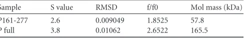
Sample S value RMSD f/f0 Mol mass (kDa)
P161-277 2.6 0.009049 1.8525 57.8
P full 3.8 0.01062 2.6522 165.5
on November 7, 2019 by guest
http://jvi.asm.org/
flowed over the immobilized NLP or NLP379CM5 chip at a flow rate of 20 l/min at 25°C. Regeneration of the chip surface after each cycle was performed using (0.015 M HEPES in 0.5 M NaCl). The change in the surface plasmon resonance angle is displayed as response units, where 1,000 response units (RU) is equal to 1 ng of analyte bound per nm2on the sensor surface. The experiments were carried out in triplicates and kinetic association (KA) and dissociation (KD) rate constants were deduced using the 1:1 Langmuir kinetic model with BIA evaluation software.
RESULTS
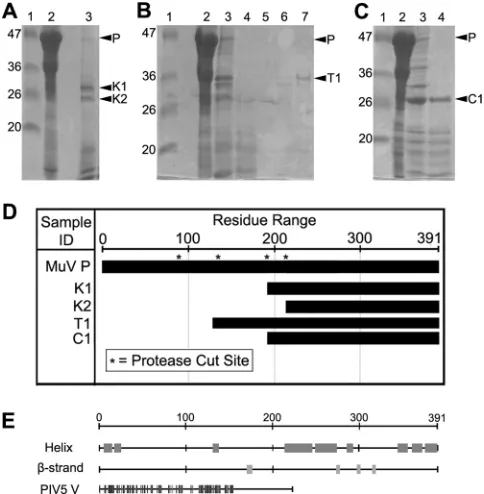
Digestion studies of MuV P.In order to study the structure and function of the MuV P protein, a single vector was constructed to express the MuV P protein inE. coli. After expression, His-tagged recombinant P was purified from the soluble fraction of the lysate. The P protein was further purified by size-exclusion chromatog-raphy and analyzed by SDS-PAGE. Purified P migrates to a posi-tion consistent with that observed in previous publicaposi-tions,⬃5 kDa higher than the calculated molecular mass of 41.5 kDa (26). Slow migration has also been observed for VSV P (34), suggesting that NSV P proteins appear to migrate more slowly in SDS-PAGE. The full-length P protein was shown to be susceptible to diges-tion by residual impurities over a brief time period following pu-rification. In order to define domains within MuV P, limited pro-teolytic digestion studies were performed. Purified full-length P protein was digested by using three different proteases: trypsin, chymotrypsin, and proteinase K. Digestion products were exam-ined by SDS-PAGE, which showed that each protease had a dif-ferent digestion pattern (Fig. 1AtoC). In order to identify the N and C termini of the stable fragments, individual bands corre-sponding to digested products were isolated and analyzed by N-terminal amino acid sequencing and mass spectrometry. The re-sults for the limited digestion studies are summarized inFig. 1D. Trypsin digestion resulted in a major stable fragment correspond-ing to residues 135 to 391 with an N-terminal sequence of MINRF and a molecular mass of 27.7 kDa. Chymotrypsin digestion re-sulted in a major stable fragment corresponding to residues 194 to 391 with an N-terminal sequence of AHPSP and a molecular mass of 21.7 kDa. Although bands corresponding to other partially di-gested products were seen on SDS-PAGE gels for both trypsin and chymotrypsin digestions, the molecular masses of these fragments could not be determined by mass spectrometry. Proteinase K di-gestion resulted in two stable fragments. Identification of mass by mass spectrometry was not successful. The residue numbers for the two K fragments (K1 and K2), therefore, were designated based on their N-terminal sequence, and the molecular masses were approximated from their position on SDS-PAGE. The N-ter-minal sequence for K2 was SVISA and likely corresponds to resi-dues 214 to 391. The N-terminal sequence for K1 was somewhat ambiguous, but the sequence in cycles 3 through 5 was PSP. This sequence with the size observed on SDS-PAGE of⬃22 kDa is consistent with a fragment encompassing residues 194 to 391. The chymotrypsin and K1 fragments are thus the same and consistent with the cleavage pattern of each enzyme, where each cleaves ad-jacent to the carboxyl group of bulky or aromatic amino acids. The sequence at this cleavage site of P is Y/AHPSP.
Bioinformatics analysis.Secondary structure prediction by Jpred3 identified nine different regions as probable helical regions (Fig. 1E). The prediction also identified four different regions that were likely to have-strand conformation. In addition, the Jpred3 search identified PIV5 V as a homologue of MuV P. PIV5 V has a 37% identity to MuV P, located within the first 155 residues and
164 residues of MuV P and PIV5 V, respectively. These data were used, along with the results of protease digestion, to design a clone library of MuV P fragments. These protein fragments were ex-pressed and purified by the same methods used for the full-length P. The yield of purified protein per liter varied for each fragment. A list of cloned P fragments and a summary of protein expression results are shown inFig. 2. P fragments that were consistent with the results of the protease digestion have a better expression level in the soluble fraction.
Structure of the P oligomerization domain.Several fragments were subjected to crystal screens. Crystals were grown with frag-ment P161-277, and the crystal structure was determined to 2.2-Å resolution. Only residues 213 to 277 could be fit correctly into the electron density maps. The traced polypeptide represented an ap-propriate protein volume for the unit cell with a solvent content of 60.04%, assuming a partial specific volume of 0.74 ml/g. Compos-ite omit maps were created to confirm that modeling was carried out correctly. The lack of residues 161 to 212 is likely due to diges-tionin situduring crystallization since crystal formation only oc-curred after 2 weeks. The asymmetric unit contains two chains (A FIG 1Limited digestion of MuV P by different proteases. In panels A to C, lanes 1, 2, and 3 correspond to the molecular mass ladder, undigested P pro-tein, and digested P propro-tein, respectively. (A) Proteinase K digestion resulted in two stable fragments (K1, residues 194 to 391; K2, residues 214 to 391) (lane 3). (B) Trypsin digestion resulted in a major stable fragment (T1, residues 135 to 391) (lane 3). Lanes 4 to 7 correspond to the different fractions separated using the Gelfree 8100 fractionation system. (C) Chymotrypsin digestion re-sulted in a major stable fragment (C1, residues 194 to 391) (lane 4). The identity of the major stable fragments was revealed by N-terminal amino acid sequencing and molecular weight determination, and is summarized in panel D. The molecular masses for fragments T1 and C1 were determined through mass spectrometry. The molecular masses of fragments K1 and K2 were ap-proximated from their position on SDS-PAGE gels. Protease cut sites (*) rep-resent the sequences identified using N-terminal sequencing. (D) Summary of bioinformatics analysis using Jpred3. Predicted helical regions are shown in the schematic to the right of “Helix”. Predicted beta-strand regions are shown in the schematic to the right of “-strand”. Homology to PIV5 V protein is shown in the schematic to the right of “PIV5 V”.
on November 7, 2019 by guest
http://jvi.asm.org/
[image:4.585.300.542.67.314.2]and B) that cover residues 213 to 273 and residues 215 to 277, respectively (Fig. 3A). Both chains are composed of a single long ␣-helix. However, chain A, unlike chain B, contains a kink at Gly 246. Chain A also contains a stretch of amino acids with extended conformation at the C-terminal end (residues 272 to 277) of its long helix.
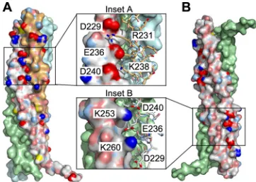
The crystallized MuV P fragment forms a tightly packed te-tramer through noncrystallographic and crystallographic symme-try contacts (Fig. 3B). We therefore define this tetramer as MuV POD. This tetrameric coiled-coil structure is reminiscent of SeV POD(Fig. 3C). However, the tetramer formed by SeV PODis com-posed of four parallel helices, whereas the tetramer formed by MuV PODis composed of two sets of parallel helices that are in opposite orientation (Fig. 3B). In the MuV PODtetramer that is primarily formed with hydrophobic interactions (Table 3), there are two zippers of charged side chain interactions at each end in addition to hydrophobic surface contacts among the helices. Be-tween the parallel pair of helices, the zipper is formed by Asp229, Glu236, and Asp240 (chain A) and Arg231 and Lys238 (chain B) as mentioned above (Fig. 4A, inset A). Between the antiparallel pair of helices, the zipper is formed by Lys253 and Lys260 (chain B) and Asp229, Glu236, and Asp240 (chain B=) (Fig. 4B, inset B). The helices wrap around each other and seem to clamp one an-other through these charge zippers at both ends of the tetrad.
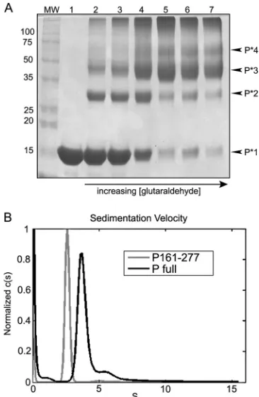
Glutaraldehyde cross-linking.In order to verify the
tetra-meric state of the MuV P protein, cross-linking reactions were performed using glutaraldehyde. Initially, full-length P was used in cross-linking experiments. However, because MuV P is highly susceptible to nonspecific digestion (35), the resulting cross-FIG 2Summary of MuV P fragments. A schematic representation of MuV P
modules is shown at the top. For expression levels, “⫹” indicates⬍15 mg/liter, “⫹⫹” indicates 15 to 25 mg/liter, and “⫹⫹⫹” indicates⬎25 mg/liter of purified recombinant protein. Fragments were derived from a combination of protease digestion and bioinformatics analysis.
FIG 3Structure of MuV POD. (A) Crystal structure of two parallel extended
␣-helices (chain A in green and chain B in cyan). Chain A contains residues 215 to 277, and a kink at Gly246 is noted. Chain B contains residues 213 to 273. (B) PODtetramer as found in the crystal. The crystal structures of PODs from SeV (C), VSV (D), and RABV (E) are shown for comparison. The range of residues for each PODis labeled. In panels C to E, each independent polypeptide chain is a different color.
FIG 4Tetrameric interactions. (A) Chain A-B interactions. In addition to extensive surface contacts, the main interactions between chains A and B also include a zipper of charged residues: Asp229, Glu236, and Asp240 (chain A) and Arg231 and Lys238 (chain B). In panel A, chain A is on the left, and chain B is on the right. The other two chains in the tetramer are monocolored. (B) Chain B-B=interactions. Residues Lys253 and Lys260 from chain B (left) also form a zipper of charged residues—Asp229, Glu236, and Asp 240 —from the antiparallel chain B=(right). This clamping by the zippers occurs on both ends of the tetramer. The tetramer in panel B is rotated left-handed by 90° com-pared to the tetramer in panel A.
on November 7, 2019 by guest
http://jvi.asm.org/
[image:5.585.329.513.62.341.2] [image:5.585.60.268.66.374.2] [image:5.585.330.511.500.629.2]linked products were composed of a mixture of different P frag-ments, and no clear data could be discerned (data not shown). Since the majority of the known protease sensitive sites are con-tained within the terminal regions, P161-277 was used in place of the full-length P protein, and the resulting cross-linked products were much easier to identify. After analysis in SDS-PAGE gels, bands representing monomers, dimers, trimers, and tetramers could be clearly discerned (Fig. 5A).
Sedimentation velocity analysis.To further confirm the oli-gomeric state determined by the PODcrystal structure, we carried out ultracentrifugation analyses to measure the sedimentation co-efficient of the purified P protein and fragment P161-277. For both P161-277 and full-length P, the data revealed a single domi-nant species (Fig. 5). The molecular mass derived from the sedi-mentation coefficient is 57.8 kDa for P161-277, which corre-sponds to the theoretical mass of a P161-277 tetramer (57.6 kDa). Furthermore, the molecular mass derived from sedimentation co-efficient is 165.5 kDa for full-length P, which is very close to the
theoretical molecular mass for a MuV P tetramer (168 kDa). We therefore concluded that the purified MuV P protein or P161-277 is a tetramer, in accordance with the results from the crystal struc-ture.
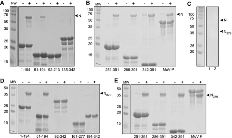
Nucleocapsid binding.A number of recombinant P fragments were expressed and purified (Fig. 1and2). In order to determine which parts of the MuV P protein interact with the nucleocapsid, a pulldown assay was developed. Purified P fragments were first added to a small Ni-affinity column in saturating amounts. After allowing the supernatant to flow through, the beads were washed once to remove any unbound protein. NLP of MuV prepared as previously reported (26) was added to the loaded column at a 1:1 molar ratio (N:P) and allowed to incubate at room temperature for 15 min. The NLP solution was then allowed to flow through. The loaded column was washed to remove any unbound protein. The proteins were then eluted from the column, and the products were denatured and electrophoresed in SDS-PAGE gels. A sum-mary of the results from the pulldown assays is shown inFig. 6. These assays confirmed that the extreme C-terminal domain, P342-391, is a nucleocapsid binding domain as previously re-ported (4). Other C-terminal fragments including residues 251 to 391, 286 to 391, and 342 to 391 each bound NLP (Fig. 6). Unex-pectedly, the results showed that the N-terminal region of MuV P (P1-194) was also capable of binding NLP. This has not been re-ported previously for a NSV P protein and suggests that the func-tional domains of MuV P may be different from those of other NSVs.
In addition to using NLP formed by the full-length NLP (540 amino acids), binding assays were also conducted using a trypsin digested form of the MuV NLP (NLP379). The digested NLP379was used to determine whether P binding could be abolished in NLP lacking the C-terminal region of N. Similar to the full-length NLP, NLP379was also bound by both the N-terminal (1 to 194 and 51 to 194) and C-terminal (251 to 391, 286 to 391, and 342 to 391) regions of MuV P (Fig. 6DandE).
Affinity of P fragments binding NLP.Based upon pulldown experiments, we concluded that MuV P contains two separate nucleocapsid binding domains: the N-terminal domain (P1-194) and the C-terminal domain (P286-391). In order to further char-acterize the interactions between the two separate nucleocapsid binding domains and NLP, surface plasmon resonance analysis was conducted. The recombinant full-length P, P1-194, P161-277, and P286-391 were investigated for their ability to interact with immobilized NLPs or NLP379. All analytes, except for P161-277 as a negative control, exhibited measurable binding (Fig. 7andTable 4). Kinetics experiments were conducted at multiple concentra-tions (Fig. 7). Modeling the association and dissociation of the P analytes with NLP using the 1:1 Langmuir binding model led to estimatedKAvalues of 2.02⫻105M⫺1for N1-194, 2.35⫻106 M⫺1 for P286-391, and 2.23 ⫻106 M⫺1 for the full-length P (Table 4). UsingKDcalculations, P286-391 also exhibited a rela-tively higher affinity for NLP with an estimated dissociation con-stant of 4.26⫻10⫺7M compared to N1-194 (4.95⫻10⫺6M). Next, we tested the ability of P to bind immobilized NLP379 (Fig. 7EtoH). Modeling the association and dissociation of the P analytes with NLP379using the 1:1 Langmuir binding model led to estimatedKAvalues of 2.17⫻106M⫺1for P1-194, 1.01⫻105M⫺1 for P286-391, and 1.61⫻106M⫺1for the full-length P (Table 4). Of the estimatedKAandKDvalues, P1-194 exhibited a relatively higher affinity for NLP379with an estimated dissociation constant FIG 5Oligomerization state of MuV P.(A) Cross-linking with
glutaralde-hyde. MuV P161-277 was cross-linked using glutaraldehyde in order to assess the oligomerization state. Cross-linking products were analyzed in SDS-PAGE gels. Lanes 1 to 5 correspond to 4 h of incubation with glutaraldehyde at the following concentrations: 0, 0.05, 0.06, 0.1, and 0.2%. Lanes 6 and 7 corre-spond to overnight incubation with 0.2 and 0.3% glutaraldehyde, respectively. Bands representing P161-277 monomers (P*1), dimers (P*2), trimers (P*3), and tetramers (P*4) can be seen at⬃15,⬃30,⬃45, and⬃60 kDa, respectively, and are noted to the right of the gel. (B) Sedimentation velocity analysis. Sedimentation coefficients were determined, and molecular masses were de-rived for P161-277 and full-length P (Table 3). The distribution plots showed a single species for each sample. The derived molecular mass for P161-277 (57.8 kDa) corresponded to the theoretical mass of a P161-277 tetramer (57.6 kDa). Furthermore, the molecular mass derived for full-length P (165.5 kDa) was very close to the theoretical molecular mass for a MuV P tetramer (168 kDa).
on November 7, 2019 by guest
http://jvi.asm.org/
[image:6.585.71.254.66.346.2](KD) of 4.6⫻10⫺7M compared to P286-391, which had aKD value of 9.88⫻10⫺6M. Control surface plasmon resonance ex-periments with P161-277, which contains the oligomerization do-main (OD), did not show measurable interactions with full-length NLP or NLP379. The surface plasmon resonance data indicate a 1.3-fold-higher affinity in P binding the full-length NLP versus NLP379but shows a slower dissociation rate (Fig. 7AandE). The curves ofFig. 7FandGshow that there are very fast on and off rates for both P1-194 and P286-391 to NLP379compared to the full-length NLP.
DISCUSSION
It has been anticipated that MuV P has a modular structure com-posed of separated functional domains. Here, we present an ex-perimental approach to more accurately map the functional do-mains of MuV P. In our proteolytic studies, the N-terminal region of P preceding residue 194 appeared to be less stable than the region that follows since more cleavage sites were found here. This is consistent with observations of other NSV Ps in which the N-terminal region is more disordered compared to the middle and C-terminal regions of P (11,23,36,37). A previous study deter-mined the structure of an extreme C-terminal segment (residues 343 to 391) of MuV P (7). Our proteolytic digestion study did not identify this smaller region of P as a stable fragment. A possible explanation for not being able to map P343-391 in the present study is that this C-terminal region is part of a larger C-terminal domain. The P343-391 region was previously proposed to be MuV PNBDby analogy to other P proteins. P343-391 may be a molten globule since its crystallization required the addition of stabilizing agents (7). On the other hand, the protein expression data did point to a potential linker between PODand the C-terminal do-main to be from residues 277 to 286. This could suggest that the
C-terminal domain of MuV P is larger than previously thought. P proteins of other NSVs were reported to contain flexible regions between PN°D, POD, and PNBD(1,21,36,38–43).
Crystallization studies clearly defined that MuV PODcomprises residues 213 to 277 when P161-277 was used in crystal growth. The structure of MuV POD was determined and shows a stable tetramer containing one pair of long parallel␣-helices (64 resi-dues) and a second pair of parallel helices in opposite orientation. The presence of P tetramers in solution was confirmed by analyt-ical ultracentrifugation and cross-linking with glutaraldehyde (Fig. 5). The unique organization of this tetrameric coiled-coil where parallel helices are joined with antiparallel helices has not been reported for a native protein before. In this distinctive assem-bly, interactions between the parallel helices are different from those between the antiparallel helices. In contrast, interactions between any neighboring pair of helices are identical in an all parallel or all antiparallel coiled-coil. The helices in the MuV POD tetramer are held together by a large hydrophobic contact area between each monomer (Table 2) and two zippers of charged side chain interactions (Fig. 4). This interesting orientation of antipa-rallel helices places the N-terminal and the C-terminal regions at both ends of MuV POD. This particular structure may well suit the functional requirements of MuV P as discussed below.
We have shown that P binds the full-length MuV NLP and truncated NLP379. These observations confirm that the P binding site in the MuV nucleocapsid does not fully require the N-tail, the same as that previously reported (4). In lieu of the apparent dis-pensability of this N-tail, nucleocapsid binding by MuV P may involve several regions of both N and P. Given this and the novel structure of MuV Pod, P may well be different from other NSV Ps in binding the nucleocapsid. With this open-minded approach, His-tagged fragments covering the full range of MuV P were tested FIG 6NLP pulldown assays. In order to determine which P fragments could interact with NLP, pulldown assays were performed. The headers of the gels in panels A, B, D, and E indicate whether the P protein (or P fragments) saturated column was incubated with NLP (⫹) or NLP379or not (⫺). Amino acid numbers corresponding to individual P fragments are labeled on the footer of the gels. The positions for N protein (labeled “N” in panels A and B) and trypsinized N (labeled “NLP379” in panels D and E) are noted (A and B), and trypsinized NLP (N379) are noted by arrows on the right sides of each gel (D and E). (C) Negative controls. Lanes 1 and 2 show the results for negative controls for NLP and trypsinized NLP379, respectively, with no P fragments added to the column.
on November 7, 2019 by guest
http://jvi.asm.org/
[image:7.585.100.485.67.297.2]FIG 7Evaluation of N-P interactions using surface plasmon resonance. (A to D) Full-length NLP was immobilized, and P protein analytes (full-length P, P1-194, P286-391, and P161-277, respectively) were injected over the CM5 chip surface. All analytes displayed a measurable binding response (1 RU⫽1 pg/mm2). (E to H) N379was immobilized, and P protein analytes (full-length P, P1-194, P286-391, and P161-277, respectively) were injected over the CM5 chip surface. All analytes displayed a measurable binding response. All experiments were carried out in triplicate.KAandKDvalues are listed inTable 4.
on November 7, 2019 by guest
http://jvi.asm.org/
for binding a well-prepared NLP composed of 13 N subunits and a piece of random RNA as previously described (26). In the His-tag pulldown assays, full-length MuV P and the previously iden-tified C-terminal domain, P343-391, were shown to bind NLP as expected. What was unexpected is that the N-terminal region of MuV P from residues 1 to 194 could also bind NLP. This indicates that specific nucleocapsid binding may require both the N-termi-nal and the C-termiN-termi-nal regions of MuV P, a binding mode that is different from all other known NSV Ps. The NLP binding by the N-terminal and C-terminal regions was confirmed by surface plas-mon resonance experiments. This analysis showed that the C-termi-nal region of P has a higher affinity for NLP. When the N-tail was removed by trypsin digestion (NLP379), the C-terminal region of P still binds NLP379with a lower affinity. The affinity of binding NLP379by the C-terminal region of P is similar as that of the N-terminal region of P binding to the full-length NLP. These ob-servations suggest that the C-terminal region of P interacts with the N-tail and additional regions of N, and the N-terminal region of P interacts with regions of N not including the N-tail. However, there is a 10.7-fold increase in binding NLP379by the N-terminal region of P versus the full-length NLP. This increase could point to the accessibility of the N0binding site for P upon removal of the N-tail. Evidence shows that this site is located in the RNA cavity for other N proteins (11,36,37). The N0binding motif of P could potentially bind in this site if encapsidated RNA was displaced from NLP379. The overall affinity of the full-length P for NLP is lower than that of the individual domains. The affinity values for the full-length tetrameric P may be underestimated because the NLP is representative of a single turn of the helical nucleocapsid and thus does not have the multiple binding sites available in the helical nucleocapsid (described below). In addition, the full-length P was not very stable in solution.
Comparisons of known structures of P uncovered three modes of P oligomerization (Fig. 3BtoE). In the SeV P tetramer, oli-gomerization results in a coiled-coil structure of four long parallel ␣-helices (21). Similarly, the subunits in the VSV P dimer are also parallel to each other even though the-sheet on each side is formed by domain swapping (19). The parallel orientation of P subunits in the oligomers places the N-terminal regions on one end and the C-terminal regions on the other. Since specific nu-cleocapsid binding in these viruses requires only the C-terminal region of P, the N-terminal region may be placed far away from the C-terminal region. In the RABV P dimer, however, the N-ter-minal and C-terN-ter-minal regions are each positioned at the same end due to the U-shaped structure that links two antiparallel helices in each PODsubunit (20). There is no indication that the N-terminal region of RABV P is involved in binding the nucleocapsid. It is not clear what functional implications this particular orientation of RABV P has. MuV P represents a third mode of oligomerization in
which parallel and antiparallel P subunits are associated together to form a tetramer. In the MuV P tetramer, the dimensions of the helix bundle are similar to those of the coiled-coil structure of the SeV P tetramer, but the orientations of the helices is different. The antiparallel orientation of the MuV P subunits places two N-terminal regions and two C-terminal regions on each end of the tetramer. This organization may be functionally required because both the N-terminal and the C-terminal regions of P are involved in binding the nucleocapsid, as shown in this report. The two terminal regions should be in close proximity to each other in order to effectively bind the nucleocapsid. The full P binding sites for each terminal regions in the nucleocapsid remain to be iden-tified for MuV. The results shown inFig. 6and7suggest that there are P binding sites within residues 1 to 379 in addition to the C-terminal tail of N. This mode of binding requires that P bind at least two or more N subunits either in the same turn or in succes-sive turns in the helical nucleocapsid. Since the conserved se-quences for initiation of replication (PrE I and II) are located in two successive turns of the nucleocapsid (44), it is possible that P plays a role in recognition of the two sites by the viral RdRp. Our results again demonstrated that NSV P proteins all have a modular structure with separated functional domains and are present as oligomers (Fig. 8). Although NSV P proteins harbor analogous functions, each may have a very different domain organization and can interact with other viral proteins, especially the nucleo-capsid, in a very different manner.
ACKNOWLEDGMENTS
We thank the staff of the SER-CAT at the APS, Argonne National Labo-ratory, for their assistance in data collection. Use of the APS was sup-ported by the U.S. Department of Energy, Office of Science, Office of Basic Energy Sciences, under contract W-31-109-Eng-38. SER-CAT-support-ing institutions may be found atwww.ser-cat.org/members.html.
We appreciate the generosity of the staff at the Stanford Synchrotron Radiation Lightsource (SSRL). Portions of this research were carried out at the SSRL, a national user facility operated by Stanford University on behalf of the U.S. Department of Energy, Office of Basic Energy Sciences. The SSRL Structural Molecular Biology Program is supported by the De-partment of Energy, Office of Biological and Environmental Research, and by the National Institutes of Health (NIH), National Center for Re-search Resources, Biomedical Technology Program, and the National In-stitute of General Medical Sciences. This study was supported in part by a NIH grant to M.L. (AI050066).
[image:9.585.350.495.66.183.2]R.C., T.J.G., and M.L. conceived and designed the experiments. R.C., T.J.G., S.P., and G.J.B. performed the experiments. R.C., T.J.G., S.P., C.D., TABLE 4Binding kinetics
Sample KA(M⫺1) KD(M)
P1-194⫹N full 2.02⫻105 4.95⫻10⫺6
P286-391⫹N full 2.35⫻106 4.26⫻10⫺7
P full⫹N full 2.23⫻106 4.48⫻10⫺7
P1-194⫹N379 2.17⫻10
6 4.60⫻10⫺7
P286-391⫹N379 1.01⫻10
5 9.88⫻10⫺6
P full⫹N379 1.61⫻10
6 8.65⫻10⫺7
FIG 8Diagram of the domain organization for four different NSV Ps. PODis typically located in the central region of P. PN°Dand PNBDare linked to POD through flexible loops. This appears to be a common modular structure for all NSV P proteins.
on November 7, 2019 by guest
http://jvi.asm.org/
G.J.B., P.E.P., and M.L. analyzed the data. R.C., T.J.G., S.P., C.D., G.J.B., P.E.P., and M.L. wrote the paper.
REFERENCES
1.Takacs AM, Das T, Banerjee AK.1993. Mapping of interacting domains between the nucleocapsid protein and the phosphoprotein of vesicular stomatitis virus by using a two-hybrid system. Proc. Natl. Acad. Sci. U. S. A.90:10375–10379.
2.Toriumi H, Honda Y, Morimoto K, Tochikura TS, Kawai A. 2002. Structural relationship between nucleocapsid-binding activity of the ra-bies virus phosphoprotein (P) and exposure of epitope 402-13 located at the C terminus. J. Gen. Virol.83:3035–3043.
3.Ryan KW, Portner A.1990. Separate domains of Sendai virus P protein are required for binding to viral nucleocapsids. Virology174:515–521. 4.Kingston RL, Baase WA, Gay LS.2004. Characterization of nucleocapsid
binding by the measles virus and mumps virus phosphoproteins. J. Virol. 78:8630 – 8640.
5.Assenberg R, Delmas O, Ren J, Vidalain PO, Verma A, Larrous F, Graham SC, Tangy F, Grimes JM, Bourhy H.2010. Structure of the nucleoprotein binding domain of Mokola virus phosphoprotein. J. Virol. 84:1089 –1096.
6.Gely S, Lowry DF, Bernard C, Jensen MR, Blackledge M, Costanzo S, Bourhis JM, Darbon H, Daughdrill G, Longhi S.2010. Solution struc-ture of the C-terminal X domain of the measles virus phosphoprotein and interaction with the intrinsically disordered C-terminal domain of the nucleoprotein. J. Mol. Recognit.23:435– 447.
7.Kingston RL, Gay LS, Baase WS, Matthews BW.2008. Structure of the nucleocapsid-binding domain from the mumps virus polymerase; an ex-ample of protein folding induced by crystallization. J. Mol. Biol.379:719 – 731.
8.Ribeiro EA, Jr, Favier A, Gerard FC, Leyrat C, Brutscher B, Blondel D, Ruigrok RW, Blackledge M, Jamin M.2008. Solution structure of the C-terminal nucleoprotein-RNA binding domain of the vesicular stomati-tis virus phosphoprotein. J. Mol. Biol.382:525–538.
9.Blanchard L, Tarbouriech N, Blackledge M, Timmins P, Burmeister WP, Ruigrok RW, Marion D. 2004. Structure and dynamics of the nucleocapsid-binding domain of the Sendai virus phosphoprotein in so-lution. Virology319:201–211.
10. Green TJ, Luo M.2009. Structure of the vesicular stomatitis virus nucleo-capsid in complex with the nucleonucleo-capsid-binding domain of the small polymerase cofactor, P. Proc. Natl. Acad. Sci. U. S. A.106:11713–11718. 11. Chen M, Ogino T, Banerjee AK.2007. Interaction of vesicular stomatitis
virus P and N proteins: identification of two overlapping domains at the N terminus of P that are involved in N0-P complex formation and encapsi-dation of viral genome RNA. J. Virol.81:13478 –13485.
12. Qanungo KR, Shaji D, Mathur M, Banerjee AK.2004. Two RNA poly-merase complexes from vesicular stomatitis virus-infected cells that carry out transcription and replication of genome RNA. Proc. Natl. Acad. Sci. U. S. A.101:5952–5957.
13. Mavrakis M, Mehouas S, Real E, Iseni F, Blondel D, Tordo N, Ruigrok RW.2006. Rabies virus chaperone: identification of the phosphoprotein peptide that keeps nucleoprotein soluble and free from nonspecific RNA. Virology349:422– 429.
14. Majumdar A, Bhattacharya R, Basak S, Shaila MS, Chattopadhyay D, Roy S.2004. P-protein of Chandipura virus is an N-protein-specific chap-erone that acts at the nucleation stage. Biochemistry43:2863–2870. 15. Mir MA, Panganiban AT.2006. Characterization of the RNA chaperone
activity of hantavirus nucleocapsid protein. J. Virol.80:6276 – 6285. 16. Leyrat C, Yabukarski F, Tarbouriech N, Ribeiro EA, Jr, Jensen MR,
Blackledge M, Ruigrok RW, Jamin M.2011. Structure of the vesicular stomatitis virus N-P complex. PLoS Pathog.7:e1002248. doi:10.1371 /journal.ppat.1002248.
17. Chen M, Ogino T, Banerjee AK.2006. Mapping and functional role of the self-association domain of vesicular stomatitis virus phosphoprotein. J. Virol.80:9511–9518.
18. Ding H, Green TJ, Luo M.2004. Crystallization and preliminary X-ray analysis of a proteinase-K-resistant domain within the phosphoprotein of vesicular stomatitis virus (Indiana). Acta Crystallogr. D Biol. Crystallogr. 60:2087–2090.
19. Ding H, Green TJ, Lu S, Luo M.2006. Crystal structure of the oligomer-ization domain of the phosphoprotein of vesicular stomatitis virus. J. Vi-rol.80:2808 –2814.
20. Ivanov I, Crepin T, Jamin M, Ruigrok RW. 2010. Structure of the dimerization domain of the rabies virus phosphoprotein. J. Virol.84: 3707–3710.
21. Tarbouriech N, Curran J, Ruigrok RW, Burmeister WP.2000. Tetra-meric coiled coil domain of Sendai virus phosphoprotein. Nat. Struct. Biol.7:777–781.
22. Curran J, Homann H, Buchholz C, Rochat S, Neubert W, Kolakofsky D.1993. The hypervariable C-terminal tail of the Sendai paramyxovirus nucleocapsid protein is required for template function but not for RNA encapsidation. J. Virol.67:4358 – 4364.
23. Longhi S, Receveur-Brechot V, Karlin D, Johansson K, Darbon H, Bhella D, Yeo R, Finet S, Canard B.2003. The C-terminal domain of the measles virus nucleoprotein is intrinsically disordered and folds upon binding to the C-terminal moiety of the phosphoprotein. J. Biol. Chem. 278:18638 –18648.
24. Houben K, Marion D, Tarbouriech N, Ruigrok RW, Blanchard L.2007. Interaction of the C-terminal domains of Sendai virus N and P proteins: comparison of polymerase-nucleocapsid interactions within the paramyxovirus family. J. Virol.81:6807– 6816.
25. Mavrakis M, McCarthy AA, Roche S, Blondel D, Ruigrok RW.2004. Structure and function of the C-terminal domain of the polymerase co-factor of rabies virus. J. Mol. Biol.343:819 – 831.
26. Cox R, Green TJ, Qiu S, Kang J, Tsao J, Prevelige PE, He B, Luo M. 2009. Characterization of a mumps virus nucleocapsid-like particle. J. Virol.83:11402–11406.
27. Cole C, Barber JD, Barton GJ.2008. The Jpred 3 secondary structure prediction server. Nucleic Acids Res.36:W197–W201.
28. Brunger AT, Adams PD, Clore GM, DeLano WL, Gros P, Grosse-Kunstleve RW, Jiang JS, Kuszewski J, Nilges M, Pannu NS, Read RJ, Rice LM, Simonson T, Warren GL.1998. Crystallography and NMR system: a new software suite for macromolecular structure determination. Acta Crystallogr. D Biol. Crystallogr.54:905–921.
29. Adams PD, Afonine PV, Bunkoczi G, Chen VB, Davis IW, Echols N, Headd JJ, Hung LW, Kapral GJ, Grosse-Kunstleve RW, McCoy AJ, Moriarty NW, Oeffner R, Read RJ, Richardson DC, Richardson JS, Terwilliger TC, Zwart PH. 2010. PHENIX: a comprehensive Python-based system for macromolecular structure solution. Acta Crystallogr. D Biol. Crystallogr.66:213–221.
30. Emsley P, Lohkamp B, Scott WG, Cowtan K.2010. Features and devel-opment of Coot. Acta Crystallogr. D Biol. Crystallogr.66:486 –501. 31. Painter J, Merritt EA.2005. A molecular viewer for the analysis of TLS
rigid-body motion in macromolecules. Acta Crystallogr. D Biol. Crystal-logr.61:465– 471.
32. Painter J, Merritt EA.2006. Optimal description of a protein structure in terms of multiple groups undergoing TLS motion. Acta Crystallogr. D Biol. Crystallogr.62:439 – 450.
33. Schrodinger LLC.2010. The PyMOL molecular graphics system, version 1.3r1. Schrodinger LLC, New York, NY.
34. Green TJ, Macpherson S, Qiu S, Lebowitz J, Wertz GW, Luo M.2000. Study of the assembly of vesicular stomatitis virus N protein: role of the P protein. J. Virol.74:9515–9524.
35. Karlin D, Ferron F, Canard B, Longhi S.2003. Structural disorder and modular organization inParamyxovirinaeN and P. J. Gen. Virol.84: 3239 –3252.
36. Karlin D, Longhi S, Receveur V, Canard B. 2002. The N-terminal domain of the phosphoprotein of morbilliviruses belongs to the natively unfolded class of proteins. Virology296:251–262.
37. Leyrat C, Jensen MR, Ribeiro EA, Jr, Gerard FC, Ruigrok RW, Black-ledge M, Jamin M.2011. The N0-binding region of the vesicular stoma-titis virus phosphoprotein is globally disordered but contains transient ␣-helices. Protein Sci.20:542–556.
38. Tarbouriech N, Curran J, Ebel C, Ruigrok RW, Burmeister WP.2000. On the domain structure and the polymerization state of the Sendai virus P protein. Virology266:99 –109.
39. Choudhary SK, Malur AG, Huo Y, De BP, Banerjee AK.2002. Charac-terization of the oligomerization domain of the phosphoprotein of human parainfluenza virus type 3. Virology302:373–382.
40. Rahaman A, Srinivasan N, Shamala N, Shaila MS.2004. Phosphopro-tein of the rinderpest virus forms a tetramer through a coiled coil region important for biological function: a structural insight. J. Biol. Chem.279: 23606 –23614.
41. Llorente MT, Garcia-Barreno B, Calero M, Camafeita E, Lopez JA, Longhi S, Ferron F, Varela PF, Melero JA.2006. Structural analysis of the
on November 7, 2019 by guest
http://jvi.asm.org/
human respiratory syncytial virus phosphoprotein: characterization of an alpha-helical domain involved in oligomerization. J. Gen. Virol.87:159 – 169.
42. Gerard FC, Ribeiro Ede A, Jr, Albertini AA, Gutsche I, Zaccai G, Ruigrok RW, Jamin M.2007. UnphosphorylatedRhabdoviridaephosphoproteins form elongated dimers in solution. Biochemistry46:10328 –10338.
43. Gerard FC, Ribeiro Ede A, Jr, Leyrat C, Ivanov I, Blondel D, Longhi S, Ruigrok RW, Jamin M.2009. Modular organization of rabies virus phos-phoprotein. J. Mol. Biol.388:978 –996.
44. Kolakofsky D, Roux L, Garcin D, Ruigrok RW.2005. Paramyxovirus mRNA editing, the “rule of six” and error catastrophe: a hypothesis. J. Gen. Virol.86:1869 –1877.