Int. J. Electrochem. Sci., 5 (2010) 1082 - 1089
International Journal of
ELECTROCHEMICAL
SCIENCE
www.electrochemsci.org
CoulArray Detector as a Tool for Estimation of Acute Toxicity
of Silver(I) Ions
Ondrej Zitka1, Dalibor Huska1, Vojtech Adam1, Ales Horna2, Miroslava Beklova3, Zdenka Svobodova4 and Rene Kizek1, *
1
Department of Chemistry and Biochemistry, Faculty of Agronomy, Mendel University in Brno, Zemedelska 1, CZ-613 00 Brno, Czech Republic
2
Tomas Bata University, T.G. Masaryka 275, CZ-762 72 Zlin, Czech Republic
3
Department of Veterinary Ecology and Environmental Protection, and 4 Institute of Toxicology, Faculty of Veterinary Hygiene and Ecology, University of Veterinary and Pharmaceutical Sciences, Palackeho 1-3, CZ-612 42 Brno, Czech Republic
*
E-mail: : [email protected]
Received: 5 July 2010 / Accepted: 15 July 2010 / Published: 10 August 2010
Biochemical markers suitable for monitoring of environmental pollution as well as for protecting of human health are searching. In this study, the effect of silver(I) ions on guppy fishes (Poecilia reticulata) was investigated. For this purpose, we employed hyphenated technique of high performance liquid chromatography coupled with multichannel CoulArray electrochemical detector. Content of cysteine in fishes treated with 0.5 and/or 1µg/l of silver(I) ions was 290and/or 240ng/g at the end of seven day long treatment. Reduced and oxidized glutathione were also determined. The enhance in GSSG content accompanied by decrease in GSH content confirms our hypothesis on increasing risk of oxidative stress in fishes due to treatment with silver(I) ions. In addition, we attempted to gain more information about health state of animals exposed to heavy metals from chromatograms measured by CoulArray detector. We counted areas of all signals detected up to 30 minutes in chromatograms of tissues from fishes treated with all concentrations of silver(I) ions. The content of thiols and thiols related compounds as a sum of signals areas enhanced with increasing concentration of silver(I) ions and time of exposition.
Keywords: heavy metal, silver, bioaccumulation, aquatic organisms, electrochemical detector
1. INTRODUCTION
osmosis,electrolysis, ion exchange or adsorption are using for removal of heavy metals from waters. Recently, bioremediation technologies were also employed to remove heavy metals from environment [4-11]. These technologies are based on extraction of heavy metals from soil by an organism and their subsequent deposition to cell or whole tissues, where the metals do not menace yet [12-15]. Organisms exposed to heavy metals ions protect themselves by biosynthesis of molecules containing –SH groups [2,8,16-18]. Low molecular mass protein called metallothionein and the most abundant peptide reduced glutathione belong to such molecules. Electrochemical detection is an attractive alternative method for thiols detection, because of its inherent advantages of simplicity, ease of miniaturization, high sensitivity and relatively low cost [7,16,19-24]. The main aim of this work is to detect thiolsin tissues of guppy fishes treated with silver(I) ions. We use high performance liquid chromatography coupled with multichannel electrochemical detector (HPLC-ED) for this purpose.
2. MATERIAL AND METHODS
2.1. Chemicals and material
Silver nitrate and all other reagents used were purchased from Sigma Aldrich (Sigma-Aldrich, USA) in ACS purity unless noted otherwise. Stock standard solutions were prepared with ACS water and stored in the dark -20 °C. Working standard solutions were prepared daily by dilution of the stock solutions. All solutions were filtered through a 0.45 µm Nylon filter discs (MetaChem, Torrance, CA, USA) prior to HPLC analysis.
2.2. Chromatographic techniques
HPLC-ED system consisted of two solvent delivery pumps (Model 582 ESA Inc., Chelmsford, MA), Metachem Polaris C18A reverse-phase column (150.0 × 2.1 mm, 5 µm particle size; Varian Inc., CA, USA) and a CoulArray electrochemical detector (Model 5600A, ESA, USA). The electrochemical detector includes three flow cells (Model 6210, ESA, USA). Each cell consists of four analytical cells. One analytic cell contains working carbon porous electrode, two auxiliary and two reference electrodes. Both the detector and the reaction coil/column were thermostated. The sample (5 µl) was injected using autosampler (Model 540 Microtiter HPLC, ESA, USA). Standardization and step-by-step optimization of various experimental conditions for detection of low molecular mass thiols can be found in the following papers [19,21,25,26]. The recoveries, intra and inter-day interceptions have been tested [19,26].
2.3. Biological experiment
solution where the fishes were kept constant, oxygen concentration and temperature were monitored during the experiment. The oxygen concentration varied within the range from 1.8 to 4.2 mg/l, the pH level from 6.3 to 7.0, and the temperature from 20.2 to 21.5 °C during the 7 days long experiment. Variations in the experimental conditions, mainly, in the oxygen level have not influenced viability and behaviour of the fishes. The sampled fish was killed by CO2, washed one time with distilled water
and one time with 0.5 M EDTA prior to the following processing. All experiments were authorised by ethic commission of Veterinary and Pharmaceutical University in Brno, Czech Republic.
2.4. Preparation of biological samples for electrochemical analysis
Weighed fish (approximately 0.2 g) were transferred to a test-tube, and liquid nitrogen was added. The samples were frozen to disrupt the cells. The frozen sample was transferred to mortar and grinding for 1 min. Then, 1 000 µl of 0.2 M phosphate buffer (pH 7.2) was added to the mortar, and the sample was grinded for 5 min. The homogenate was transferred to a new test-tube. The mixture was homogenised by shaking on a Vortex–2 Genie (Scientific Industries, New York, USA) at 4 °C for 30 min. The homogenate was centrifuged (14 000 g) for 30 min at 4 °C using a Universal 32 R centrifuge (Hettich-Zentrifugen GmbH, Tuttlingen, Germany). Before the analysis the supernatant was filtered through a membrane filter (0.45 µm Nylon filter disk, Millipore, Billerica, Mass., USA).
2.5. Descriptive statistics
Data were processed using MICROSOFT EXCEL® (USA). Results are expressed as mean ± standard deviation (S.D.) unless noted otherwise. Statistical significance of the differences between low molecular mass thiols quantified in control and silver(I) ions treated fishes was determined. Differences with p < 0.05 were considered significant and were determined by using of one way ANOVA test (particularly Scheffe test), which was applied for means comparison.
3. RESULTS AND DISCUSSION
3.1. Thiols as a biochemical marker
with multichannel CoulArray electrochemical detector. Based on the coulometric detection, the development of the coulometric electrode array detector constituted a major step towards improved selectivity and versatility. The array detector simply consists of a series of coulometric electrode pairs placed in series performing a multichannel (or array) detection. The system is based on analytical cells containing a platinum reference electrode that sets the electrochemical zero and four working electrodes that measure the redox reaction of interest. Each analytical cell therefore provides four channels [35]. We employed instrument consisted from three analytical cell, which means twelve channels detection. Typical chromatogram of low molecular mass thiols measured by HPLC coupled with twelve channel electrochemical detector is shown in Fig. 1.
Figure 1.Typical HPLC-ED chromatogram of low molecular mass thiols.
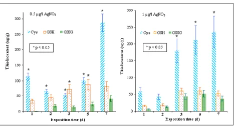
the end of the treatment. Similarly, the level of reduced glutathione(GSH) was increased. In the fishes treated with the lower silver(I) ions concentration, GSHsynthesis was about 40 % higher compared to GSH level determined in fishes exposed to 1 µg/l of silver(I) ions. The content of other marker of stress induced by heavy metals, oxidized glutathione (GSSG), was lower in tissues of fishes treated with 0.5 µg/l of silver(I) ions compared to the content of this compound in tissues of fishes exposed to 1µ g/l silver(I) ions. The enhance in GSSG content accompanied bydecrease in GSH content confirms our hypothesis on increasing risk of oxidative stress in fishes due to treatment with silver(I) ions
[image:5.612.74.543.201.455.2].
Figure 2.Content of thiols (cysteine, reduced and oxidized glutathione) determined in tissues of guppy fishes treated with silver(I) ions 0.5 and/or 1 µ g/l for seven days. Control was subtracted.
3.2. CoulArray detector in evaluation of fish acute toxicity
measured by CoulArray detector should be considered a new marker of acute toxicity after heavy metal ions treatment.
Figure 3. Sum of areas of all signals detected up to 30 minutes in chromatograms of tissues from fishes treated with all concentrations of silver(I) ions (0.5 and/or 1 µ g/l). Control was subtracted.
4. CONCLUSIONS
This paper describes a novel potential method to quantify thiols as a marker of metal exposure in fish, because heavy metal ions still represent a threat not only to aquatic ecosystems but also to the other ones. HPLC-ED used for the assessment of the metal exposure of guppy fishes seems to be versatile, robust and rapid technique. Moreover, electrochemical analyzers have high potential to be miniature, which opens new possibilities of the applications of the method and also to be used for detection not only thiols but also silver(I) ions [36-38].
ACKNOWLEDGEMENTS
[image:6.612.106.511.110.391.2]
References
1. J. Zukowska and M. Biziuk, J. Food Sci., 73 (2008) R21.
2. D. Huska, S. Krizkova, M. Beklova, L. Havel, J. Zehnalek, V. Diopan, V. Adam, L. Zeman, P. Babula and R. Kizek, Sensors, 8 (2008) 1039.
3. D. Huska, O. Zitka, V. Adam, M. Beklova, S. Krizkova, L. Zeman, A. Horna, L. Havel, J. Zehnalek and R. Kizek, Czech J. Anim. Sci., 52 (2007) 37.
4. P. K. Rai, Int. J. Phytoremediat., 10 (2008) 133.
5. E. Meers, F. M. G. Tack, S. Van Slycken, A. Ruttens, J. Vangronsveld and M. G. Verloo, Int. J. Phytoremediat., 10 (2008) 390.
6. W. S. W. Ngah and M. Hanafiah, Bioresour. Technol., 99 (2008) 3935.
7. V. Supalkova, D. Huska, V. Diopan, P. Hanustiak, O. Zitka, K. Stejskal, J. Baloun, J. Pikula, L. Havel, J. Zehnalek, V. Adam, L. Trnkova, M. Beklova and R. Kizek, Sensors, 7 (2007) 932. 8. S. Krizkova, P. Ryant, O. Krystofova, V. Adam, M. Galiova, M. Beklova, P. Babula, J. Kaiser,
K. Novotny, J. Novotny, M. Liska, R. Malina, J. Zehnalek, J. Hubalek, L. Havel and R. Kizek, Sensors, 8 (2008) 445.
9. A. Stafiej and K. Pyrzynska, Sep. Purif. Technol., 58 (2007) 49. 10. A. Stafiej and K. Pyrzynska, Microchem J., 89 (2008) 29.
11. S. Krizkova, V. Adam and R. Kizek, Chem. Listy, 103 (2009) 559. 12. C. Cobbett, New Phytol., 159 (2003) 289.
13. C. Cobbett and P. Goldsbrough, Annu. Rev. Plant Biol., 53 (2002) 159. 14. J. Zehnalek, J. Vacek and R. Kizek, Lis. Cukrov. Reparske, 120 (2004) 220. 15. J. Zehnalek, V. Adam and R. Kizek, Lis. Cukrov. Reparske, 120 (2004) 222.
16. J. Petrlova, S. Krizkova, O. Zitka, J. Hubalek, R. Prusa, V. Adam, J. Wang, M. Beklova, B. Sures and R. Kizek, Sens. Actuator B-Chem., 127 (2007) 112.
17. V. Adam, S. Krizkova, O. Zitka, L. Trnkova, J. Petrlova, M. Beklova and R. Kizek, Electroanalysis, 19 (2007) 339.
18. K. Stejskal, S. Krizkova, V. Adam, B. Sures, L. Trnkova, J. Zehnalek, J. Hubalek, M. Beklova, P. Hanustiak, Z. Svobodova, A. Horna and R. Kizek, IEEE Sens. J., 8 (2008) 1578.
19. J. Petrlova, R. Mikelova, K. Stejskal, A. Kleckerova, O. Zitka, J. Petrek, L. Havel, J. Zehnalek, V. Adam, L. Trnkova and R. Kizek, J. Sep. Sci., 29 (2006) 1166.
20. D. Potesil, R. Mikelova, V. Adam, R. Kizek and R. Prusa, Protein J., 25 (2006) 23.
21. D. Potesil, J. Petrlova, V. Adam, J. Vacek, B. Klejdus, J. Zehnalek, L. Trnkova, L. Havel and R. Kizek, J. Chromatogr. A, 1084 (2005) 134.
22. V. Adam, J. Petrlova, D. Potesil, J. Zehnalek, B. Sures, L. Trnkova, F. Jelen and R. Kizek, Electroanalysis, 17 (2005) 1649.
23. V. Adam, P. Hanustiak, S. Krizkova, M. Beklova, J. Zehnalek, L. Trnkova, A. Horna, B. Sures and R. Kizek, Electroanalysis, 19 (2007) 1909.
24. V. Adam, I. Fabrik, V. Kohoutkova, P. Babula, J. Hubalek, R. Vrba, L. Trnkova and R. Kizek, Int. J. Electrochem. Sci., 5 (2010) 429.
25. K. Stejskal, Z. Svobodova, I. Fabrik, V. Adam, M. Beklova, M. Rodina and R. Kizek, J. Appl. Ichthyol., 24 (2008) 519.
26. V. Diopan, K. Stejskal, M. Galiova, V. Adam, J. Kaiser, A. Horna, K. Novotny, M. Liska, L. Havel, J. Zehnalek and R. Kizek, Electroanalysis, 22 (2010) 1248.
27. M. Havelkova, J. Blahova, H. Kroupova, T. Randak, I. Slatinska, D. Leontovycova, R. Grabic, R. Pospisil and Z. Svobodova, Sensors, 8 (2008) 2589.
28. M. Havelkova, T. Randak, V. Zlabek, J. Krijt, H. Kroupova, J. Pulkrabova and Z. Svobodova, Sensors, 7 (2007) 2599.
30. M. Stiborova, M. Miksanova, M. Sulc, H. Rydlova, H. H. Schmeiser and E. Frei, Int. J. Cancer, 116 (2005) 667.
31. M. Stiborova, V. Martinek, H. Rydlova, T. Koblas and P. Hodek, Cancer Lett., 220 (2005) 145. 32. I. Sondi and B. Salopek-Sondi, J. Colloid Interface Sci., 275 (2004) 177.
33. D. M. Di Toro, H. E. Allen, H. L. Bergman, J. S. Meyer, P. R. Paquin and R. C. Santore, Environ. Toxicol. Chem., 20 (2001) 2383.
34. I. J. Morgan, R. P. Henry and C. M. Wood, Aquat. Toxicol., 38 (1997) 145. 35. J. L. Penalvo and T. Nurmi, J. Pharm. Biomed. Anal., 41 (2006) 1497.
36. S. Krizkova, D. Huska, M. Beklova, J. Hubalek, V. Adam, L. Trnkova and R. Kizek, Environ. Toxicol. Chem., 29 (2010) 492.
37. S. Krizkova, O. Krystofova, L. Trnkova, J. Hubalek, V. Adam, M. Beklova, A. Horna, L. Havel and R. Kizek, Sensors, 9 (2009) 6934.
38. H. H. Hassan, M. A. M. Ibrahim, S. S. Abd El Rehim and M. A. Amin, Int. J. Electrochem. Sci., 5 278.