Replication Stress and Genomic Instability
Graphical Abstract
Highlights
d
RECQL5 is upregulated in JAK2V617F mutant erythroblasts
in MPN patients
d
RECQL5 is a target of activated JAK2-PI3K signaling
d
RECQL5 stabilizes stalled forks during
JAK2V617F-associated replication stress
d
RECQL5 depletion sensitizes JAK2V617F-expressing cells to
hydroxyurea
Authors
Edwin Chen, Jong Sook Ahn,
David B. Sykes, ..., Daniel J. DeAngelo,
Anthony R. Green, Ann Mullally
Correspondence
[email protected]
In Brief
Oncogenic JAK2 signaling in MPN
patients leads to DNA damage, yet MPNs
are characterized by genomic stability.
Chen et al. show that the DNA helicase
RECQL5 maintains genomic integrity in
response to JAK2V617F-associated
replication stress. Moreover, RECQL5
depletion sensitizes JAK2V617F-mutant
cells to hydroxyurea, the most common
treatment for MPN.
Cell Reports
Report
RECQL5 Suppresses Oncogenic JAK2-Induced
Replication Stress and Genomic Instability
Edwin Chen,1,2,7Jong Sook Ahn,3,4David B. Sykes,5Lawrence J. Breyfogle,1Anna L. Godfrey,3,4Jyoti Nangalia,3,4 Amy Ko,1Daniel J. DeAngelo,6Anthony R. Green,3,4and Ann Mullally1,2,6,*
1Division of Hematology, Department of Medicine, Brigham and Women’s Hospital, Harvard Medical School, Boston, MA 02115 2Broad Institute of the Massachusetts Institute of Technology and Harvard, 415 Main Street, Cambridge, MA 02142
3Cambridge Institute for Medical Research, Medical Research Council/Wellcome Trust Stem Cell Institute, and Department of Haematology,
University of Cambridge, Hills Road, Cambridge CB2 0XY, UK
4Department of Haematology, Addenbrooke’s Hospital, Hills Road, Cambridge CB2 0XY, UK
5Center for Regenerative Medicine, Massachusetts General Hospital, 185 Cambridge Street, Boston, MA 02114 6Department of Medical Oncology, Dana Farber Cancer Institute, 44 Binney Street, Boston, MA 02115
7Present address: School of Molecular and Cellular Biology, Faculty of Biological Sciences, University of Leeds, Woodhouse Lane,
Leeds LS2 9JT, UK
*Correspondence:[email protected] http://dx.doi.org/10.1016/j.celrep.2015.11.037
This is an open access article under the CC BY license (http://creativecommons.org/licenses/by/4.0/).
SUMMARY
JAK2V617F is the most common oncogenic lesion in
patients with myeloproliferative neoplasms (MPNs).
Despite the ability of JAK2V617F to instigate DNA
damage in vitro, MPNs are nevertheless
character-ized by genomic stability. In this study, we address
this paradox by identifying the DNA helicase
RECQL5 as a suppressor of genomic instability in
MPNs. We report increased RECQL5 expression in
JAK2V617F-expressing cells and demonstrate that
RECQL5 is required to counteract
JAK2V617F-induced
replication
stress.
Moreover,
RECQL5
depletion sensitizes JAK2V617F mutant cells to
hy-droxyurea (HU), a pharmacological inducer of
repli-cation stress and the most common treatment for
MPNs. Using single-fiber chromosome combing,
we show that RECQL5 depletion in JAK2V617F
mutant cells impairs replication dynamics following
HU
treatment,
resulting
in
increased
double-stranded breaks and apoptosis. Cumulatively, these
findings identify RECQL5 as a critical regulator of
genome stability in MPNs and demonstrate that
replication stress-associated cytotoxicity can be
amplified specifically in JAK2V617F mutant cells
through RECQL5-targeted synthetic lethality.
INTRODUCTION
The propensity of cancer cells to undergo clonal evolution is enabled by a heightened state of genomic instability wherein cancer cells are continuously accumulating and repairing DNA damage. This increase in genomic flux allows cancer cells to accumulate somatic mutations that can drive disease
progres-sion. However, heightened genomic instability can also activate DNA damage-associated checkpoints, which can lead to apoptosis or cellular senescence. Cancer cells continuously tread a fine balance between cell death and survival in response to DNA damage (Negrini et al., 2010).
Chronic myeloproliferative neoplasms (MPNs) encompass a spectrum of clonal hematological disorders with an inherent ten-dency to transform into a more aggressive disease in the form of acute myeloid leukemia (AML). MPNs provide a window into cancer early during its ontogeny and give insights into the pro-cesses that regulate genome stability during malignant clonal evolution. The most common recurrent lesion in MPN patients is an activating V617F mutation in the JAK2 non-receptor tyro-sine kinase (JAK2V617F) that causes hyperactive JAK-STAT signaling and confers a capacity for cytokine-independent growth (Baxter et al., 2005; James et al., 2005; Kralovics et al., 2005; Levine et al., 2005). Recently, a growing body of work has suggested that JAK2V617F is associated with increased DNA damage: (1) increased numbers of gH2Ax-marked dou-ble-strand breaks (DSBs) have been detected in Ba/F3 pro-B cells overexpressing JAK2V617F (Marty et al., 2013) and in line-age-negative, Sca1-positive, c-Kit-positive (LSK) cells (enriched for hematopoietic stem cell [HSC] activity) from 6-month-old JAK2V617F-heterozygous knockin mice (Li et al., 2010); (2) JAK2V617F expression is associated with increased levels of DNA-damaging reactive oxygen species (Marty et al., 2013); (3) RAD51-positive foci indicative of increased DSB repair have been observed in CD34+ hematopoietic cells obtained from JAK2V617F-positive MPN patients (Plo et al., 2008); and (4) JAK2V617F expression in both human diploid fibroblasts and primary erythroblasts from MPN patients leads to higher rates of stalled replication forks, with improper processing of stalled replication intermediates representing a potential source of DSBs (Chen et al., 2014).
Given the genome-destabilizing functionalities of JAK2V617F and the inherent tendency for leukemic transformation in pa-tients with MPN, a reasonable supposition is that oncogenic
Cell Reports13, 1–8, December 22, 2015ª2015 The Authors 1
JAK2 signaling imposes a mutator phenotype on MPN cells, accelerating the accumulation of mutations and promoting clonal evolution and disease progression. However, longitudinal studies of MPN patients indicate that JAK2V617F-positive poly-cythemia vera (PV) and essential thrombopoly-cythemia (ET) patients (i.e., chronic-phase MPN) typically remain clinically and cytoge-netically stable over decades (Tefferi et al., 2014). A recent copy-number analysis of the genome of chronic-phase MPN patients has shown that cytogenetic abnormalities are rare (Klampfl et al., 2011), and an analysis of the mutational landscape of PV and ET patients revealed that each MPN patient harbors a modest num-ber of mutations per exome (approximately 6.5) (Nangalia et al., 2013).
[image:3.603.60.377.96.502.2]To reconcile the apparent paradox of JAK2V617F-induced DNA damage with the clinical and cytogenetic stability character-istic of chronic-phase MPN, we hypothesized that JAK2V617F, in addition to instigating a state of increased DNA damage, could also, in parallel, activate protective pathways that counteract and prevent DNA damage-induced apoptosis. In this report, we
Figure 1. JAK2V617F Increases the Expres-sion of RECQL5
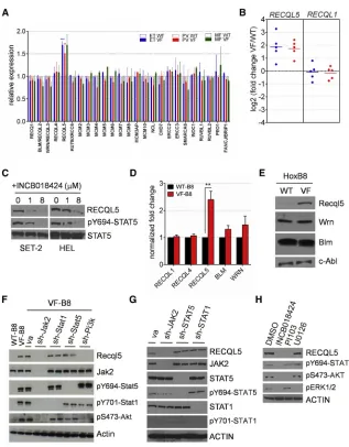
(A) Gene expression profiles depicting the expres-sion of 25 DNA helicases in 40 MPN patients (20 ET patients, 16 PV patients, and 4 MF patients). (B) qPCR validation of RECQL5 and RECQL1 expression in ET (blue) and PV (red) patients. (C) RECQL5 expression in SET-2 and HEL cells following treatment with 0–8 mM of the JAK2 in-hibitor INCB18424.
(D and E) Expression of Recq family members in WT-B8 and VF-B8 cells by qPCR (D) and western immunoblot (E). *p < 0.01.
(F) Recql5 expression in WT-B8 and VF-B8 cells following knockdown of Jak2 (sh-Jak2), the p85 subunit of PI3K (sh-Pi3k), Stat1 (sh-Stat1), or Stat5 (sh-Stat5).
(G) RECQL5 expression in HEL cells following knockdown of JAK2 (sh-JAK2), STAT5 (sh-STAT5), or STAT1 (sh-STAT1).
(H) RECQL5 expression in HEL cells following treatment for 16 hr with INCB018424 (1mM), the PI3K inhibitor PI103 (1mM), or the ERK1/2 inhibitor U0126 (5mM).
See alsoFigure S1.
identify increased expression of the DNA repair helicase RECQL5 in JAK2V617F-expressing cells and characterize its role in constraining JAK2V617F-induced repli-cation stress and maintaining genomic integrity in MPNs.
RESULTS
Activated JAK2 Signaling Regulates Expression of the RECQL5 Helicase in MPN Cells
concomitant with decreased STAT5 phosphorylation (Figure 1C). In aggregate, these data indicate that RECQL5 expression is regulated by activated JAK2 signaling in human disease-relevant contexts.
Physiological Levels of Jak2V617F Lead to Increased Recql5 Expression
To investigate the role of Recql5 under conditions that more closely recapitulate chronic-phase MPN in patients, we gener-ated disease-relevant cell lines from Jak2V617F knockin mice that we developed previously and characterized extensively ( Mul-lally et al., 2010, 2013). This mouse model closely recapitulates the features of human MPN, and Jak2V617F expression is phys-iological in the model, being driven from the endogenous Jak2 promoter. To generate cell lines, we engineered bone marrow progenitors from littermate wild-type or Jak2V617F knockin mice to express ab-estradiol-regulated Hoxb8 homeodomain-containing protein. Following 3 weeks of serial passaging and antibiotic selection, immortalized myeloid progenitor cells were generated from both wild-type mice (WT-B8) and Jak2V617F knockin mice (VF-B8) (Wang et al., 2006). Both the WT-B8 and VF-B8 lines resemble cells at a granulocyte-macrophage progen-itor (GMP) stage of myeloid maturation by cell surface immuno-phenotype analysis (Figure S1A). Immunoblotting showed equivalent levels of Jak2 expression in both cell lines but increased levels of phosphorylated Stat5 in VF-B8 cells relative to WT-B8 cells (Figure S1B). Additionally, VF-B8 cells exhibited an increased proliferative capacity and survival under reduced serum conditions (Figures S1C–S1D), indicating that VF-B8 cells recapitulate this key pathognomonic feature of MPN biology.
We next determined whether the expression of Recql5 was modulated by mutant Jak2 in these Hoxb8-immortalized cell lines. In accordance with the primary human data, VF-B8 cells ex-hibited elevated expression of Recql5 relative to WT-B8 cells by qPCR analysis (Figure 1D) and western immunoblotting ( Fig-ure 1E). No significant differential expression of other Recq family members (Recql1, Recql4, Blm, and Wrn) was observed. These data demonstrate that mutant JAK2 increases expression of the DNA repair helicase RECQL5 in both human and murine cells.
RECQL5 Is a Target of JAK2-PI3K Signaling
To determine which signaling pathways were necessary for the regulation of Recql5 by Jak2V617F, we used small hairpin RNAs (shRNAs) to knock down Jak2 and downstream effector molecules of Jak2 in VF-B8 cells. Expression of shRNAs target-ing Jak2 led to diminution of Recql5 levels relative to cells transduced with an empty vector control (Figure 1F), which is consistent with the finding that Recql5 is a downstream target of Jak2 signaling. Next, we assessed Recql5 levels in VF-B8 cells following transduction of shRNAs targeting Stat1, Stat5, or the p85 subunit of phosphatidylinositol 3-kinase (PI3K). The levels of Recql5 in VF-B8 cells were reduced following knockdown of PI3K but not Stat1 or Stat5 (Figure 1F). Similarly, RECQL5 levels were also attenuated following knockdown of JAK2 in HEL cells but were not attenuated following knockdown of STAT5 or STAT1 (Figure 1G), and treatment of HEL cells with the PI3K in-hibitor PI103 (but not the ERK1/2 inin-hibitor U0126) led to attenu-ated levels of RECQL5, accompanied by decreased AKT
phosphorylation (Figure 1H). Taken together, these data indicate that RECQL5 expression is increased upon JAK2 activation in a PI3K-dependent manner.
Recql5 Depletion Sensitizes Jak2V617F-Expressing Cells to Replication Stress
We next sought to clarify the function of RECQL5 upregulation in JAK2V617F-expressing cells. Previously, we had demonstrated that overexpression of JAK2V617F in human diploid fibroblasts resulted in increased replication fork stalling and replication stress (Chen et al., 2014). Because RECQL5 has been linked functionally to regulating stalled replication forks in normal cells, we hypothesized that the upregulation of RECQL5 may function to mitigate the deleterious consequences of replication stress in JAK2 mutant cells.
To test this, we knocked down Recql5 in VF-B8 cells using three independent shRNAs (Figure S2A). Densitometric analysis revealed a knockdown efficiency of 61%–95% for the three hair-pins. Under normal growth conditions, Recql5 knockdown in VF-B8 cells did not affect the proliferation rate (Figure S2B) or apoptosis (Figure S2C) relative to either equivalently modified WT-B8 cells or VF-B8 cells expressing empty vector alone (VA) controls. To simulate replication stress, we subjected WT-B8 and VF-B8 cells to low-serum conditions to deprive cells of de-oxynucleotide triphosphate (dNTP) (Bester et al., 2011). We observed that VA-expressing VF-B8 cells exhibited greater viability relative to WT-B8 cells under low-serum conditions ( Fig-ure 2A, left), which is in accordance with our previous data on non-genetically perturbed cells. In contrast, Recql5-depleted VF-B8 cells exhibited decreased viability relative to equivalently modified WT-B8 cells (Figure 2A, right). Hypersensitivity of Recql5-depleted VF-B8 cells to low-serum conditions was abro-gated completely upon repletion of deoxynucleotide (dNTPs) into the culture medium (Figure 2B). The results shown represent the average of three independent Recql5-targeting shRNAs and are consistent with data obtained for each individual hairpin ( Fig-ure S2D). Collectively, these data demonstrate that Recql5 pro-tects VF-B8 cells from endogenous replication stress instigated by low dNTP levels.
We next tested whether Recql5 depletion would also sensitize VF-B8 cells to exogenous instigators of replication stress, such as pharmacological agents. We tested hydroxyurea (HU) and camptothecin (CPT), which impair DNA replication by limiting production of dNTPs and inhibiting topoisomerase I activity, respectively. Strikingly, contemporaneous knockdown of Recql5 and exposure to either HU or CPT led to significantly decreased viability of VF-B8 cells compared with like-treated WT-B8 cells after 24 hr of drug treatment (Figures 2C, 2D, and 2G; Fig-ure S2D). Decreased cell viability was noted as early as 12 hr post-drug treatment (Figures S2E and S2F). In contrast, Recql5-depleted VF-B8 cells were not hypersensitive to the dou-ble-stranded breaking agents doxorubicin (DOX) and etoposide (ETP) (Figures 2E and 2F;Figure S2D). Concordant with these data, shRNA depletion of human RECQL5 also enhanced cyto-toxicity in HEL and SET2 cells, but only to pharmacological insti-gators of replication stress (HU, CPT, aphidicolin, and irinotecan) and not to pro-oxidants or DSB-generating drugs (Figures S2G and S2H).
Cell Reports13, 1–8, December 22, 2015ª2015 The Authors 3
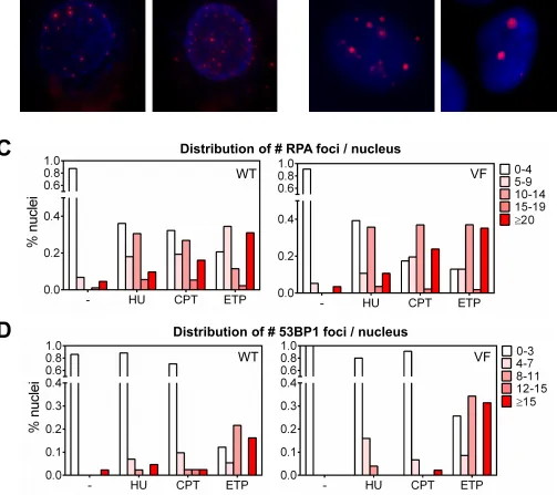
Replication stressors such as HU and CPT can also induce DSBs at sufficiently high doses. We therefore validated whether the HU and CPT doses associated with preferential cytotoxicity of Recql5-depleted VF-B8 cells were causing increased replica-tion stress or excessive formareplica-tion of DSBs. To differentiate be-tween these two phenomena, we treated parental WT-B8 and VF-B8 cells with HU (6mM) and CPT (4 nM) and performed immu-nocytochemical staining for foci containing the RPA protein (which marks stalled replication forks) or 53BP1 (which localizes to DSBs) (Figures S3A and S3B). At these HU and CPT dosages, we observed a marked increase in foci containing RPA (Figure S3C), with only a marginal increase in numbers of 53BP1-positive foci (Figure S3D). In contrast, etoposide (4 nM) generated both RPA-positive and 53BP1-RPA-positive foci (Figures S3C and S3D). In aggre-gate, these findings verify that the dosage of HU and CPT used to enhance the cytotoxicity of Recql5-depleted VF-B8 cells causes enhanced replication stress with only a slight elevation of DSBs.
Figure 2. Recql5 Depletion Sensitizes Jak2V617F-Expressing Cells to Replication Stress
(A–F) Viability of WT-B8 (black) and VF-B8 (red) cells transduced with VA or Recql5-targeting shRNAs following serum deprivation (SD) in 0.5% fetal calf serum (FCS) (A), SD with 100mM dNTPs (SD + dNTPs) (B), HU (0–250 mM) (C), CPT (0–10 nM) (D), DOX (0–25mM) (E), or ETP (0–10 nM) (F). For VA-transduced cultures, each point represents the mean of three in-dependent cultures. For Recql5-knockdown cultures, each point represents the mean of three separate Recql5-targeting shRNAs.
(G) Quantitation of annexin V positivity in cells treated with 100mM HU or 4 nM CPT.
(H–J) VF-B8 cells were transduced with Recql5-tar-geting shRNA simultaneously with either a wild-type Recql5 cDNA (RQ5wt), shRNA-resistant Recql5 cDNA (RQ5res), or RQ5res cDNA harboring a K58R mutation (RQ5res-K58R). Immunoblotting for Recql5 protein levels was performed (H), and cell viability was as-sessed (I and J).
Testing for statistical significance was performed us-ing Student’s t test (*p < 0.05, **p < 0.01, ***p < 0.001). See alsoFigure S2.
Given the potential off-target effects of shRNAs that may potentially influence the observed phenotypes, we next confirmed the specificity of the Recql5 shRNAs. We de-signed a Recql5 cDNA), RQ5(res), that was mutated at every third nucleotide to disrupt each of the shRNA-binding sites while retain-ing the correct amino acid encoded at each triplet codon. Expression of a wild-type Recql5 (RQ5(WT)) in VF-B8 cells together with a Recql5-targeting shRNA did not rescue Recql5 expression (Figure 2H) and failed to abrogate the increased sensitivity of Recql5-depleted VF-B8 cells to HU ( Fig-ure 2I). In contrast, expression of RQ5(res) resulted in high levels of Recql5 expression that were maintained despite expression of a Recql5 shRNA (Figure 2H) and successfully abrogated the hypersensitivity to HU of the VF-B8 cells co-expressing the Recql5 shRNA ( Fig-ure 2I). Moreover, a RQ5(res) cDNA harboring a K58R mutation within the helicase domain is incapable of abrogating HU hyper-sensitivity, revealing the essentiality of Recql5 helicase activity in protecting against replication stress (Figure 2J). Together, these data demonstrate that the effects of the shRNAs were on target and that Recql5 depletion was the critical factor for conferring increased sensitivity of VF-B8 cells to replication stress.
Recql5 Depletion Increases the Severity of Replication Fork Stalling in Jak2V617F-Expressing Cells Exposed to Exogenous Replication Stress
chromosome combing, which allows the direct visualization and analysis of replication tracts on individual, bromodeoxyuridine (BrdU)-labeled DNA fibers. We subjected WT-B8 or VF-B8 cells transduced with Recql5-targeting shRNAs or VA controls to this procedure. All cultures underwent a first labeling step with the BrdU analog iododeoxyuridine (IdU) under normal growth condi-tions, followed by a second labeling step with another BrdU analog, chlorodeoyuridine (CldU), in culture medium supple-mented with HU (Figure 3A). In this way, the extent of fork pro-gression can be measured both in the absence and presence of HU to determine any differential effects of HU on the cultures. We focused our initial analysis on fibers generated solely from the first labeling step to ascertain whether Recql5 depletion in the absence of HU altered DNA replication kinetics. For this anal-ysis, we measured the length of these fibers and calculated the average fork rate because decreased fork processivity is a robust indicator of fork stalling. In control cells, we observed a significant decrease in the mean replication rate in VF-B8 cells compared with WT-B8 cells (1.47 ± 0.18 kb/min [n = 88] in VF-B8 cells versus 1.97±0.21 kb/min [n = 94] in WT-B8 cells [p < 0.01]) (Figure 3B), consistent with reports published previously indicating a replication processivity impairment in JAK2V617F-expressing cells (Chen et al., 2014). However, the mean replication rate was not altered significantly by Recql5 knockdown in either WT-B8 or VF-B8 cells (WT-B8+sh1, 1.88±0.44 kb/min [n = 80]; WT-B8+sh2, 1.91 ±0.28 kb/min [n = 55]; VF-B8+sh1, 1.33±0.31 kb/min [n = 101]; VF-B8+sh2, 1.32 ± 0.39 kb/min [n = 50]). Critically, no difference was observed in fork rate between VF-B8 cells depleted for Recql5 relative to those transduced with an empty vector (Figure 3B). These data indicate that physiological levels of Jak2V617F expression in myeloid cells gives rise to a replication stress
phenotype, as evidenced by decreased fork processivity, but that Recql5 depletion alone has no additional affect.
We next explored the possibility that depletion of Recql5 in WT-B8 and VF-B8 cells could lead to altered replication dy-namics in the presence of HU. Because replication fork progress in VF-B8 cells is impaired globally relative to WT-B8 cells, to facilitate comparison, the extent of fork progress during the first (HU-free) labeling step was normalized and arbitrarily designated at 1, and the second (HU-treated) labeling step was depicted relative to the normalized first label. Using this analysis, we observed that the presence of HU impaired fork progression in all cultures tested (Figure 3C). However, the impairment in fork progression caused by exposure to HU was significantly greater in Recql5-depleted VF-B8 cells relative to control VF-B8 cells and Recql5-depleted WT-B8 cells (Figure 3C). This indicates that Recql5 depletion differentially impairs replication fork pro-gression in VF-B8 cells following HU exposure compared with WT-B8 cells.
The exacerbation in impairment of fork progression may be due to an increased frequency of fork collapse. To test for this, we measured the restart efficiency of a stalled replication fork, a process that is highly impaired following fork collapse. VF-B8 cells transduced with a control shRNA or shRNAs targeting Recql5 were initially exposed to HU for 60 min, followed by removal of HU and addition of BrdU (Figure 3D). Flow cytometric detection of BrdU-positive cells reflected cells that had effi-ciently restarted stalled forks. We observed significantly fewer BrdU-positive cells in HU-treated Recql5-depleted VF-B8 cells compared with cells expressing control hairpins (Figures 3E and 3F). Consistent with a role for Recql5 in protecting VF-B8 cells against HU-induced fork collapse, we noted that, following HU exposure, Recql5-depleted VF-B8 cells exhibited
Figure 3. Recql5 Protects against the Collapse of Stalled Replication Forks in Jak2V617F-Expressing Cells Exposed to Replication Stress
(A) Schematic of the time course for chromosome combing experiments (top). Shown are represen-tative replication structures of a single combed DNA molecule labeled with IdU (red) and CldU (green) (bottom).
(B) Fork rate in WT-B8 and VF-B8 cells transduced with empty vector alone (VA) or with two different Recql5-targeting shRNAs (sh1 and sh2). The re-sults represent the mean±SD for at least 50 fibers. (C) Quantification of HU-induced effects on fork dynamics. Relative fork progress is depicted as the normalized ratio of the second (HU-treated) label-ing step relative to the first (HU-free) labellabel-ing step. (D–F) Schematic of the timeline for fork restart ex-periments (D). Also shown are quantification of BrdU-positive cells (E) and representative flow cy-tometric plots of BrdU staining (F).
(G and H) Quantification ofgH2Ax-marked double-stranded breaks as assessed by immunofluores-cence detection (G) and western immunoblot (H). (D–H) Testing for statistical significance was per-formed using Student’s t test (*p < 0.05; **p < 0.01; n.s., not significant).
See alsoFigure S2.
Cell Reports13, 1–8, December 22, 2015ª2015 The Authors 5
an increase in DSBs, as indicated by higher numbers ofg H2Ax-positive foci (Figure 3G) and higher levels ofgH2Ax by immuno-blot analysis (Figure 3H). Together, these data demonstrate that Recql5 is essential to maintain fork stability in mutant JAK2-ex-pressing cells following exposure to HU.
RECQL5 Depletion Increases the Sensitivity of JAK2V617F-Positive Cells from MPN Patients to HU
Finally, we tested whether modulation of RECQL5 could also in-crease the sensitivity of JAK2V617F-positive cells from primary MPN patients to HU. Following depletion with RECQL5, CD34-positive peripheral blood mononuclear cells from JAK2V617F-positive myelofibrosis patients were grown in semi-solid medium supplemented with HU for 14 days (Figure 4A). Strikingly, following RECQL5 depletion, there was a preferential eradication of total BFU-E colonies relative to a control shRNA (Figure 4B). Moreover, genotyping of the colonies for JAK2V617F positivity revealed a preferential elimination of JAK2V617F-positive BFU-E colonies compared with autologous wild-type colonies following exposure to HU (Figure 4C;Figure S4). Collectively, this indicates that RECQL5 knockdown preferentially sensitizes JAK2V617F-positive BFU-E colonies from MPN patients to phar-macological induction of replication stress with HU.
DISCUSSION
Chronic MPNs represent a model of the early stages of leukemo-genesis and can provide insights into the balance between oncogene-associated DNA damage and the mechanisms that act to constrain it. JAK2V617F is the most common molecular driver of MPNs, and, in PV and ET, it is frequently the sole ge-netic driver identified. Although there is experimental evidence demonstrating that JAK2V617F induces DNA damage, clinical evidence indicates that chronic-phase MPNs follow a relatively indolent course over decades and that MPN genomes remain generally stable over time. In this study, we help resolve this apparent conundrum by demonstrating a role for the DNA repair helicase RECQL5 in regulating the balance between
JAK2V617F-induced DNA replication stress and genomic integ-rity in patients with MPNs.
RECQ helicases are a family of highly conserved genome sur-veillance enzymes. Humans possess five RECQ helicases— RECQL1, BLM, WRN, RECQL4, and RECQL5—that have both unique and overlapping roles in the regulation of DNA replication, repair, and transcription (Larsen and Hickson, 2013). Strikingly, we observed that the increased expression of RECQL5 in JAK2V617F mutant cells was specific and not seen with other RECQ helicases. This finding precludes the possibility that increased RECQL5 levels are due to an excess of S phase cells in cultures of JAK2V617F-expressing cells or that they represent an epi-phenomenon of increased DNA damage load. Rather, us-ing shRNA knockdown and various pharmacological agents to inhibit individual signaling pathways, we demonstrate that JAK2V617F increases RECQL5 expression through the PI3K-AKT signaling axis. In terms of the effectors downstream of PI3K-AKT signaling responsible for mediating RECQL5 activa-tion, it is possible that a direct transcriptional mechanism, such as modulating the activity of the FOXO family of transcription fac-tors, is involved. Indeed, a similar pathway has been shown pre-viously to be active in JAK2 mutant cells to modulate expression of the antioxidant protein catalase (Marty et al., 2013).
The mechanisms by which RECQL5 maintains genomic integ-rity in normal and cancer cells are ongoing areas of investigation. Germline loss-of-function mutations in RECQ family members have been associated with a predisposition to developing can-cer, and a key role has been described recently for RECQL4 in hematopoiesis (Smeets et al., 2014). However, RECQL5 dysre-gulation has not been implicated previously in human disease. Our data identify increased expression of RECQL5 as a means by which oncogene-induced replication stress is counteracted in JAK2-mutated MPNs. Concomitant induction of replication stress and RECQL5 expression downstream of JAK2-PI3K signaling potentially forms a feedback loop that regulates DNA damage accumulation. Depleting RECQL5 in JAK2 mutant cells disrupts this homeostasis and exposes a synthetic lethal vulner-ability of JAK2 mutant cells with pharmacological inducers of
Figure 4. RECQL5 Depletion Sensitizes JAK2V617F-Positive Cells from MPN Pa-tients to Replication Stressors
(A) Workflow for determining the effect of RECQL5 knockdown on the HU sensitivity of primary MPN samples.
(B and C) CD34+
peripheral blood mononuclear cells from three myelofibrosis patients were transduced with a non-targeting shRNA (scr) or RECQL5-targeting shRNA (sh1 and sh2) and cultured on methylcellulose in the presence and absence of 2mM HU. After 14 days, total BFU-Es were counted (B) and genotyped for JAK2V617F (C). *p < 0.05, **p < 0.01.
(D) Model depicting interactions between JAK2V617F, RECQL5 depletion, and exogenous pharmacological replication stress.
replication stress. Following depletion of RECQL5, JAK2V617F-expressing cells exposed to replication stressors (such as HU or CPT) exhibit more severe fork stalling and impaired fork restart-ing in comparison with isogenic wild-type cells. Moreover, RECQL5-depleted JAK2 mutant cells exhibited higher numbers of DSBs and underwent apoptosis at lower doses of HU compared with RECQL5-depleted wild-type cells (Figure 4D).
We envision two mutually non-exclusive scenarios to explain why JAK2 mutant cells are more sensitive than isogenic wild-type cells to contemporaneous RECQL5 depletion and exoge-nous replication stress. First, JAK2V617F-expressing cells are known to have more replication fork stalling and replication stress (Chen et al., 2014). Increased RECQL5 expression in these cells may be required to mitigate replication-associated genomic instability and maintain an appropriate balance of DNA damage/repair to ensure cell viability. RECQL5 depletion in JAK2V617F-expressing cells may therefore be more delete-rious than to equivalently modified wild-type cells because JAK2 mutant cells may have less leeway to cope with the additional replication stress in the form of exogenous administra-tion of HU. Second, JAK2 mutant cells may exhibit specific replication fork structures whose resolution is especially depen-dent on RECQL5 and, therefore, would be particularly sensitive to RECQL5 depletion. The mechanism of JAK2V617F-induced replication stress remains unresolved, but is likely to involve unscheduled euchromatinization during S phase by JAK2 directly (Dawson et al., 2009) or following activation of downstream STATs (Shi et al., 2006), leading to physical collisions between the replication machinery and transcriptional apparatus on the same competing DNA template. The resulting DNA:RNA hybrid structures (called R loops) are potentially mutagenic and genome-destabilizing. RECQL5 may play an important role in resolving R loops in JAK2 mutant cells. Future studies to distinguish between these possibilities will be interesting.
Finally, our data may have broader pharmacological implica-tions for cancer therapy. Although HU remains the frontline treat-ment for patients with ET and PV (Harrison et al., 2005), most studies have demonstrated that HU does not preferentially target the V617F-positive cell fraction in ET and PV patients (Antonioli et al., 2010), and, as a result, HU does not alter the natural history of MPNs. Our findings highlight a potential approach to amplify replication stress-associated cytotoxicity through helicase-tar-geted synthetic lethality. Indeed, there is precedent for this approach. Inhibition of the Werner helicase using small mole-cules has been shown to sensitize HeLa and U2OS cells to the replication stressor topotecan (Aggarwal et al., 2011). Nonethe-less, the development of small-molecule inhibitors of RECQL5 in MPNs would need to be approached with caution. In particular, the safety of RECQL5 inhibition with respect to its potential to provoke more genomic insults would need to be examined care-fully. However, exploiting the differential dependencies of cancer cells on DNA repair pathways has already demonstrated clinical efficacy through the use of PARP1 inhibitors in BRCA-mutant cancers (Bryant et al., 2005). Identifying and leveraging synthetic lethal relationships with DNA helicases may represent another similar approach for improved anti-cancer treatment strategies more broadly.
EXPERIMENTAL PROCEDURES
Generation of Myeloid Progenitor Cell Lines from Jak2V617F Knockin Mice
Myeloid progenitor cell lines were generated from a C57Bl/6 Jak2V617F-ex-pressing mouse (Mullally et al., 2010) and from a Jak2 wild-type littermate control by retroviral overexpression of an estrogen-dependent Hoxb8 tran-scription factor, as described previously (Wang et al., 2006). Conditionally immortalized myeloid progenitor cell lines from wild-type mice (WT-B8) and Jak2V617F knockin mice (VF-B8) were maintained in myeloid medium (RPMI medium + 10% fetal bovine serum (FBS) supplemented with 50 ng/ml murine stem cell factor (mSCF) and 1mMb-estradiol (Sigma).
Cell Viability Assays
WT-B8 and VF-B8 cells were counted, and 53104
cells were seeded in triplicate in each well of a 96-well plate in 100ml of myeloid medium. For drug treatments, agents were added freshly. For serum deprivation studies, cells were rinsed with 13PBS twice, and 13105
cells/well were seeded in a 96-well plate in RPMI medium + 0.5% FBS supplemented with 1mMb -estra-diol. Cell growth was measured using a standard Alamar blue assay (Life Technologies).
Chromosome Combing
Chromosome combing was performed as described previously (Chen et al., 2014). Briefly, 13105WT-B8 or VF-B8 cells were seeded in a well of a 96-well plate in a volume of 100ml. Replicating DNA was first labeled with 25mM IdU (Sigma), followed by 250mM CldU) (Sigma) for 20 min each. Cells were then harvested, genomic DNA was extracted, and individual DNA mole-cules were stretched on glass slides. The slides were immunostained with flu-orescently labeled anti-IdU (1:300) and anti-CldU (1:150) and analyzed under oil immersion on a Zeiss Axioscope 2 fluorescence microscope.
SUPPLEMENTAL INFORMATION
Supplemental Information includes Supplemental Experimental Procedures and four figures and can be found with this article online athttp://dx.doi.org/ 10.1016/j.celrep.2015.11.037.
ACKNOWLEDGMENTS
A.M. is supported by the NIH (K08 HL109734), the MPN Research Foundation, and the Jeanne D. Housman Fund for Research on Myeloproliferative Disor-ders and is a recipient of a Damon Runyon clinical investigator award. E.C. is a recipient of a Lady Tata Memorial Trust Award. A.R.G. is supported by Bloodwise (13003), the Wellcome Trust (104710/Z/14/Z), the Medical Research Council, the Kay Kendall Leukaemia Fund, the Cambridge NIHR Biomedical Research Center, the Cambridge Experimental Cancer Medicine Centre, the Leukemia and Lymphoma Society of America (07037), and a core support grant from the Wellcome Trust and MRC to the Wellcome Trust-Medical Research Council Cambridge Stem Cell Institute. We thank Drs. Benjamin Ebert and Steven Lane for critically reviewing the manuscript and Dr. Ross Levine for providing helpful insights during manuscript revision.
Received: June 9, 2015 Revised: September 16, 2015 Accepted: November 11, 2015 Published: December 10, 2015
REFERENCES
Aggarwal, M., Sommers, J.A., Shoemaker, R.H., and Brosh, R.M., Jr. (2011). Inhibition of helicase activity by a small molecule impairs Werner syndrome helicase (WRN) function in the cellular response to DNA damage or replication stress. Proc. Natl. Acad. Sci. USA108, 1525–1530.
Antonioli, E., Carobbio, A., Pieri, L., Pancrazzi, A., Guglielmelli, P., Delaini, F., Ponziani, V., Bartalucci, N., Tozzi, L., Bosi, A., et al. (2010). Hydroxyurea does
Cell Reports13, 1–8, December 22, 2015ª2015 The Authors 7
not appreciably reduce JAK2 V617F allele burden in patients with polycy-themia vera or essential thrombocypolycy-themia. Haematologica95, 1435–1438.
Baxter, E.J., Scott, L.M., Campbell, P.J., East, C., Fourouclas, N., Swanton, S., Vassiliou, G.S., Bench, A.J., Boyd, E.M., Curtin, N., et al.; Cancer Genome Project (2005). Acquired mutation of the tyrosine kinase JAK2 in human myelo-proliferative disorders. Lancet365, 1054–1061.
Bester, A.C., Roniger, M., Oren, Y.S., Im, M.M., Sarni, D., Chaoat, M., Bensi-mon, A., Zamir, G., Shewach, D.S., and Kerem, B. (2011). Nucleotide defi-ciency promotes genomic instability in early stages of cancer development. Cell145, 435–446.
Bryant, H.E., Schultz, N., Thomas, H.D., Parker, K.M., Flower, D., Lopez, E., Kyle, S., Meuth, M., Curtin, N.J., and Helleday, T. (2005). Specific killing of BRCA2-deficient tumours with inhibitors of poly(ADP-ribose) polymerase. Nature434, 913–917.
Chen, E., Beer, P.A., Godfrey, A.L., Ortmann, C.A., Li, J., Costa-Pereira, A.P., Ingle, C.E., Dermitzakis, E.T., Campbell, P.J., and Green, A.R. (2010). Distinct clinical phenotypes associated with JAK2V617F reflect differential STAT1 signaling. Cancer Cell18, 524–535.
Chen, E., Ahn, J.S., Massie, C.E., Clynes, D., Godfrey, A.L., Li, J., Park, H.J., Nangalia, J., Silber, Y., Mullally, A., et al. (2014). JAK2V617F promotes replica-tion fork stalling with disease-restricted impairment of the intra-S checkpoint response. Proc. Natl. Acad. Sci. USA111, 15190–15195.
Dawson, M.A., Bannister, A.J., Go¨ttgens, B., Foster, S.D., Bartke, T., Green, A.R., and Kouzarides, T. (2009). JAK2 phosphorylates histone H3Y41 and ex-cludes HP1alpha from chromatin. Nature461, 819–822.
Harrison, C.N., Campbell, P.J., Buck, G., Wheatley, K., East, C.L., Bareford, D., Wilkins, B.S., van der Walt, J.D., Reilly, J.T., Grigg, A.P., et al.; United Kingdom Medical Research Council Primary Thrombocythemia 1 Study (2005). Hydroxyurea compared with anagrelide in high-risk essential thrombo-cythemia. N. Engl. J. Med.353, 33–45.
James, C., Ugo, V., Le Coue´dic, J.P., Staerk, J., Delhommeau, F., Lacout, C., Garc¸on, L., Raslova, H., Berger, R., Bennaceur-Griscelli, A., et al. (2005). A unique clonal JAK2 mutation leading to constitutive signalling causes polycy-thaemia vera. Nature434, 1144–1148.
Klampfl, T., Harutyunyan, A., Berg, T., Gisslinger, B., Schalling, M., Bagienski, K., Olcaydu, D., Passamonti, F., Rumi, E., Pietra, D., et al. (2011). Genome integrity of myeloproliferative neoplasms in chronic phase and during disease progression. Blood118, 167–176.
Kralovics, R., Passamonti, F., Buser, A.S., Teo, S.S., Tiedt, R., Passweg, J.R., Tichelli, A., Cazzola, M., and Skoda, R.C. (2005). A gain-of-function mutation of JAK2 in myeloproliferative disorders. N. Engl. J. Med.352, 1779–1790.
Larsen, N.B., and Hickson, I.D. (2013). RecQ Helicases: Conserved Guardians of Genomic Integrity. Adv. Exp. Med. Biol.767, 161–184.
Levine, R.L., Wadleigh, M., Cools, J., Ebert, B.L., Wernig, G., Huntly, B.J., Bog-gon, T.J., Wlodarska, I., Clark, J.J., Moore, S., et al. (2005). Activating mutation
in the tyrosine kinase JAK2 in polycythemia vera, essential thrombocythemia, and myeloid metaplasia with myelofibrosis. Cancer Cell7, 387–397.
Li, J., Spensberger, D., Ahn, J.S., Anand, S., Beer, P.A., Ghevaert, C., Chen, E., Forrai, A., Scott, L.M., Ferreira, R., et al. (2010). JAK2 V617F impairs hemato-poietic stem cell function in a conditional knock-in mouse model of JAK2 V617F-positive essential thrombocythemia. Blood116, 1528–1538.
Marty, C., Lacout, C., Droin, N., Le Coue´dic, J.P., Ribrag, V., Solary, E., Vain-chenker, W., Villeval, J.L., and Plo, I. (2013). A role for reactive oxygen species in JAK2 V617F myeloproliferative neoplasm progression. Leukemia27, 2187– 2195.
Mullally, A., Lane, S.W., Ball, B., Megerdichian, C., Okabe, R., Al-Shahrour, F., Paktinat, M., Haydu, J.E., Housman, E., Lord, A.M., et al. (2010). Physiological Jak2V617F expression causes a lethal myeloproliferative neoplasm with differ-ential effects on hematopoietic stem and progenitor cells. Cancer Cell17, 584–596.
Mullally, A., Bruedigam, C., Poveromo, L., Heidel, F.H., Purdon, A., Vu, T., Aus-tin, R., Heckl, D., Breyfogle, L.J., Kuhn, C.P., et al. (2013). Depletion of Jak2V617F myeloproliferative neoplasm-propagating stem cells by inter-feron-ain a murine model of polycythemia vera. Blood121, 3692–3702.
Nangalia, J., Massie, C.E., Baxter, E.J., Nice, F.L., Gundem, G., Wedge, D.C., Avezov, E., Li, J., Kollmann, K., Kent, D.G., et al. (2013). Somatic CALR muta-tions in myeloproliferative neoplasms with nonmutated JAK2. N. Engl. J. Med.
369, 2391–2405.
Negrini, S., Gorgoulis, V.G., and Halazonetis, T.D. (2010). Genomic instability– an evolving hallmark of cancer. Nat. Rev. Mol. Cell Biol.11, 220–228.
Plo, I., Nakatake, M., Malivert, L., de Villartay, J.P., Giraudier, S., Villeval, J.L., Wiesmuller, L., and Vainchenker, W. (2008). JAK2 stimulates homologous recombination and genetic instability: potential implication in the heterogene-ity of myeloproliferative disorders. Blood112, 1402–1412.
Shi, S., Calhoun, H.C., Xia, F., Li, J., Le, L., and Li, W.X. (2006). JAK signaling globally counteracts heterochromatic gene silencing. Nat. Genet.38, 1071– 1076.
Smeets, M.F., DeLuca, E., Wall, M., Quach, J.M., Chalk, A.M., Deans, A.J., Heierhorst, J., Purton, L.E., Izon, D.J., and Walkley, C.R. (2014). The Roth-mund-Thomson syndrome helicase RECQL4 is essential for hematopoiesis. J. Clin. Invest.124, 3551–3565.
Tefferi, A., Guglielmelli, P., Larson, D.R., Finke, C., Wassie, E.A., Pieri, L., Gangat, N., Fjerza, R., Belachew, A.A., Lasho, T.L., et al. (2014). Long-term survival and blast transformation in molecularly annotated essential thrombo-cythemia, polycythemia vera, and myelofibrosis. Blood124, 2507–2513, quiz 2615.
Cell Reports
Supplemental Information
RECQL5 Suppresses Oncogenic JAK2-Induced
Replication Stress and Genomic Instability
Edwin Chen, Jong Sook Ahn, David B. Sykes, Lawrence J. Breyfogle, Anna L. Godfrey,
Jyoti Nangalia
3,4, Amy Ko
1, Daniel J. DeAngelo
6, Anthony R. Green
3,4, Ann Mullally
1,2,81Division of Hematology, Department of Medicine, Brigham and Women’s Hospital, Harvard Medical School, Boston, MA 02115
2Broad Institute of MIT and Harvard, 415 Main Street, Cambridge, MA 02142
3Cambridge Institute for Medical Research and MRC/Wellcome Trust Stem Cell Institute and Department of Haematology,
University of Cambridge, Hills Road, CB2 0XY, United Kingdom
4Department of Haematology, Addenbrooke’s Hospital, Hills Road, Cambridge, CB2 0XY, United Kingdom
5Center for Regenerative Medicine, Massachusetts General Hospital, 185 Cambridge Street, Boston, MA 02114
6Department of Medical Oncology, Dana Farber Cancer Institute, 44 Binney Street, Boston, MA 02115
7Present address: School of Molecular and Cellular Biology, Faculty of Biological Sciences, University of Leeds, Woodhouse
Lane, Leeds, LS2 9JT, United Kingdom
8Correspondences should be addressed to Dr. Ann Mullally, Brigham and Women’s Hospital, Karp Building,1 Blackfan Circle,
Boston, MA 02115, E-mail:[email protected];Phone: (617) 355-9002
E.C., J.S.A., D.B.S., J.B. and A.K. performed experiments. A.L.G., J.N., D.J.D., A.R.G. and A.M. provided patient samples. E.C., J.S.A., A.R.G. and A.M. designed experiments. E.C. and A.M. wrote the paper.
Supplemental Data
1. Figure S1
(Related to Figure 1). Characterization of wild-type and Jak2V617F-expressing
Hoxb8-immortalized myeloid progenitor cells.
2. Figure S2
(Related to Figure 2). Recql5 depletion sensitizes Jak2V617F-expressing cells to
replication stress.
3. Figure S3
(Related to Figure 2). RECQL5 depletion sensitizes human
JAK2V617F-expressing cell lines to replication stress.
4. Figure S4
(Related to Figure 4). RECQL5 depletion sensitizes JAK2V617F-positive cells
from MPN patients to replication stressors.
Supplemental Experimental Procedures
1.
Detailed description of myeloid progenitor cell line generation from Jak2V617F-knockin
mice
2.
Cells and reagents
3.
Alamar blue assay for measurement of cell viability
4.
Chromosome combing calculations and statistical analyses
5.
Vectors
6.
Generation of lentiviral particles and lentiviral infections
CD34
g
MEP
GMP
CMP
MEP
GMP
CMP
Jak2WT-Hoxb8ER
(WT-B8)
Jak2VF-Hoxb8ER
(VF-B8)
pY694-Stat5
Figure S1 (Related to Figure 1). Characterization
of wild-type and Jak2V617F-expressing
Hoxb8-immortalized myeloid progenitor cells. (A)
Immunophenotyping of wild-type and
Jak2V617F-expressing Hoxb8-immortalized myeloid progenitor
cells (WT-B8 and VF-B8, respectively). FACS plots
shown are gated on a lineage-negative,
Sca-1-positive, kit-positive fraction.
(B)
Western blot of
WT-B8 and VF-B8 cells show increased Stat5
activation in association with Jak2V617F mutation.
[image:12.595.51.553.108.728.2]Actin
ev
sh1
sh2
sh3
91
empty
sh-Recql5
(avg of 3)
sh1
sh2
sh3
sh1
sh2
sh3
H2O2 PL HU CPT
DOX MMC ETOP
IRT
APH
HU
H
2O
2PL
APH
CPT
IRT
DOX
MMC
ETOP
HEL
-2 -1 0 1 2