ACKNOWLEGEMENT
The author is extremely grateful to the Almighty for the successful completion of this work.
The author very much thankful to the vice chancellor of Tamilnadu Dr. M.G.R. medical university, and the special commissioner and former joint director Dr. I. Sornomariyammal MD(S) Directorate of Indian medicine and Homeopathy, Chennai for granting me to do this dissertation work.
The author would like to express her sincere gratitude to our principal Dr. M. Thinakaran MD(S) and vice principal Dr. R. Devarajan MD(S) Government siddha medical college for permitting and providing the necessary facilities to do this work.
Words hardly help us in expressing the gratitude. The author owe to our
professor and head of the department of Noi Naadal (PG) Dr. R Devarajan MD(S) for his valuable guidance and suggestions in
bringing out this dissertation work successfully.
The author feels pride to put my thanks to Dr. T. Rajasekar MD(S) Assistant Lecturer, Dr. S. Sundarrajan MD(S) Assistant lecturer for their valuable guidance and in all aspects from time to time.
The author expresses her whole hearted thanks to Dr. S.K. Sasi MD(S) Assistant Lecturer P.G. Noi Naadal Department for her encouragement and most valuable guidance to undertake this dissertation work.
The author would like to express her profound gratitude to Dr.A.Vasuki MD(S) Assistant Lecturer P.G. Noi Naadal Department who has been helping in dissertation work.
The author expresses her sincere thanks to Dr.V. Paramasivam MD (Pathology) H.O.D. Department of Pathology, Thirunelveli Medical College Hospital for their guidance in the modern approach.
The author expresses her special thanks to Dr. S. Mohan MD. Professor Department of General Medicine, G.S.M.C. Palayamkottai for his information’s regarding my dissertation work.
The author is thankful to Dr.V.S. Padma MBBS, DMRD Radiologist, G.S.M.C. Palayamkottai for her guidance in dissertation topic.
I express my deep thanks to Mr.P.Arumugam for his biostatical guidance.
The author also grateful to the librarian Mrs.T.Poongodi MA.M.Phil., of G.S.M.C. Palayamkottai for Co-operating in referring the books.
The author thanks for all her friends who helping this dissertation work successfully.
INTRODUCTION
India is a land of treasures. The nation is a paradise for its unique art, culture, civilization and indigenous medicine. Even in India, the southern peninsula is believed to be the Cradle of the human race where Tamil culture popularly called Dravidian culture flourished.
Siddha System of medicine is a traditional one, with prestigious background of Tamil culture. It’s perhaps the earliest medical science that laid stress on positive health, a harmonious blending of physical, mental, social, moral, and spiritual welfare of an individual.
Birth without any deformity and life without disease is a boon to human kind. It reminds the proverb “sound mind in a sound body”. There are two classes of disease, physical and mental. Siddha system guides the way to mukthi through physical and mental well being.
The word siddha is derived from siddhi, which means perfection of great supernatural power. The siddhars were the saints who have controlled the inner aspect of mind.
The siddha system of medicine is one of the ancient system of medicine. The siddha system has been purely associated with spiritual science, philosophy and astrology etc.
Siddha system of medicine is based upon the Panchapoothas [Five elements] and mukkuttram [Thridosha] theory.
“Tholkappiar” explained this as follows;
‘epyk; ePh; jP tsp tpRk;Nghile;Jk; fye;j kaf;fk; cyfkhjypd;”
The elements Vayu, Thee and Neer are primarily responsible for the formation of three humors, that is Muthatha. [Vali, Azhal and Iyam.] These are the three fundamental functional constituents of the human body and they have 1:1/2:1/4 in ratio
‘nka;asT thjnkhd;W” Nky;gpj;j Nkhiuahk; Iak; fhyd;Nw mwp”
But, when this equilibrium is upset or deranged these are known as “Mukkuttram” which there after leads to disease.
For proper understanding of these humours in equilibrium, a reasonable knowledge about the physiology of the body is imperative. Siddha physiology involves the ‘96’ basic thathuvas of the body apart from Seven udalkattukkal, Fourteen vegams [reflexal functions], Six suvaigkal [tastes], Three malangal [excretory products], Four udal thees (Fire of the body) and some features.
The great siddhar “ Theraiyar” in his famous venba described the prerequisites of the best physician as follows:-
The meaning of the above poem is that identification of the disease, and the primary causes along with knowing the factors that help the spreading of the disease and the ways of protecting the patient, without undergoing much difficulty are essential to become a great physician.
In Yugi vaidhya Chinthamani, yugi described 42 types of pitha diseases, “ kirumi pitham” is one of the entity of the pitha diseases.
SIDDHA PHYSIOLOGY
The science of the functions of living organisms and its components and the physical and chemical factors and processes involved is known as physiology.
The siddha physiology involves the:
Thathuvos - 96 basic elements Udal kattukkal - 7 somatic compounds Vegams - 14 reflexial functions Suvaigal - 6 tastes Udarthee - 4 body fire Udal vanmai - 3 immunities
I. 96 BASIC FACTORS
1. Five basic elements - Boothams 2. Five sense organs - Gnanaentrium 3. Functions of five sense organs - Pulankal
4. Five motor organs - Kanmaentrium 5. Five motor organs of action - Kanmavidayam 6. Four intellectual faculties - Anthakaranam 7. One wisdom - Arivu
14. Three regions - Mandalam 15. Three physical bindings - Edanai 16. Two deeds - Vinai 17. Three cosmic qualities - Gunam 18. Eight predominant passions - Raagam 19. Five status of the soul - Avathaigal
PANCHA POOTHM – FIVE BASIC ELEMENTS
The fundamental principles of siddha science involve the five basic elements namely.
1. Mann - Earth 2. Neer - Water 3. Thee - Fire 4. Vayu - Air 5. Aagayam - Ether
Characters of pancha pootham
1.Mann – Earth
All the organic living bodies and non organic substances are created by earth. Bones, muscles and tissues represent earth in body.
2. Neer – Water
It combines all the things serum, lymph, saliva etc., represent water in the body.
3. Thee – Fire
It gives colour and brightness to the thing. Digestion and circulation represent fire in the body.
4. Vayu – Air
All the spaces are filled by this pootham. Respiration and nervous system represent air in the body.
5.Aagayam – Ether
It lodges the other four poothams.
GNANENTHIRIYAM / PORI – FIVE SENSE ORGANS
PULAN - FUNCTIONS OF THE FIVE SENSE ORGANS;
1. Hearing 2. Touch 3. Vision 4. Taste 5. Smell
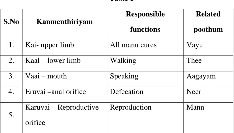
[image:10.612.105.508.268.498.2]KANMENTHIRIYAM – FIVER MOTOR ORGANS OF ACTION
Table 1
S.No Kanmenthiriyam
Responsible
functions
Related
poothum
1. Kai- upper limb All manu cures Vayu 2. Kaal – lower limb Walking Thee
3. Vaai – mouth Speaking Aagayam 4. Eruvai –anal orifice Defecation Neer
5.
Karuvai – Reproductive orifice
Reproduction Mann
ANTHAKARANAM – 4 INTELLECTUAL FACULTIES
1. Manam - The mind (or) the thinking faculty 2. puththi - Knowledge, the power of discrimination 3. Siddham - The deciding faculty
THASA NAADIKAL – 10 NERVES
These are subdivided into 10 kinds. These are.
1. Idakalai - From right big toe runs opposite side to the left nostril.. 2. Pinkalai - From left big toe runs opposite side to the right nostril. 3. Sulumunai - It is situated between the idakalai and pinkalai.
4. Purudan - It acts on the nerve of the right eye. 5. Kanthari - It acts on the nerve of lift eye. 6. Aththi - It controls the nerve of right ear. 7. Alambudai - It controls the nerve of left ear. 8. Sikuvai - It acts on the nerve of tongue
9. Sanguni - It controls the nerve of Reproductive organs 10.Gugu – It controls the nerve of rectum.
UYIR THAATHUKKAL – THREE HUMOURS
The physiological functions of the body are mediated by these three humours, which are made up of the five elements. These three functional factors maintain the integrity of the human body. According to different functions and sits each element is divided as follows.
VALI
Location:
Abanan, faeces. Idakalai, pelvic bone, Spermatic cord, skin, nerves, joints, hairs and muscles.
Functions:
Pain in the whole body, twitching, pricking pain, inflammation, reddish complexion, roughness of skin, hardness of limbs, astringent sense of taste in the mouth, constipation, oliguria, blackish discolouration of skin, stool, urine and muddy conjunctive.
Types of Vali:
Based on functions and locations it is classified into 10 types.
1. Uyirkkal – Piraanan:
Piranan means the forward (or) primary air force. It is mainly responsible for respiration and it is necessary for proper digestion and utilization of the food material.
2. Keelnokkukaal – Abaanan:
It expels faecal matter and urine. It constricts the anal sphincter. It also helps to spread the nutrients of digested food all over the body. Expulsion of sperm and menstrual flow is also under the control of abanan. Its derangement leads to disease of the bladder, rectum and reproductive system.
3. Paravukkaal – Viyaanan:
4. Melnokkukaal – Udhaanan:
Responsible for all kinds of upward motion such as nausea, vomiting and eructation.
5. Nadukkaal – Samaanan:
Samaanan means the equalizing air. It is considered essential for proper digestion, assimilation and carries digested nutrients to each and every organ. It is derangement will cause gastro intestinal, respiratory and neurological problems.
6. Vanthikaal – Naagan:
Responsible for higher intellectual functions. Causes opening and closing of eyes.
7. Vizhikkal – Koorman:
Responsible for vision, lacrimation and yawning.
8. Thummikkaal – Kirugaran:
It is situated in the tongue, salivary and nasal secretions. Induce appetite, salivation, sneezing, concentration of mind and responsible for taste sense.
9. Kottavikkal – Devathathan:
It causes laziness, ocular movements and anger.
10. Veengukkaal – Dhananjeyan:
AZHAL
Location:
Pirana vayu, bladder, moolagni, Heart, umbilical region, abdomen, sweating, saliva, blood, eyes and skin.
Characters:
It governs digestion, heat, visual perception, hunger, thirst, luster, complexion, under standing, intelligence, courage, softness of the body.
Types of Azhal
Azhal is the thermal life force of the body. It is subdivide into five types. They are.
1. Aakkanal - Anala pitham:
It is responsible to the digestion of food.
2. Vannayeri - Ranjaka Pitham:
It is responsible for the colour and contents of the blood. It is also responsible for the formation of tissues.
3. Nokku Azhal - Aalosaka Pitham:
It responsible for the perception of vision.
4. Aatralangi - Saathaga Pitham:
It controls the whole body and is responsible for fulfilling a purpose.
5. Olloliththe - Praasaka pitham:
IYAM
Location:
Samanan, suzhumunai, vinthu, head, fat, bone marrow, blood, Nose, Colon, Joints, Chest and tongue.
Characters:
In normal conditions it governs stability, lubrication, holding the joints in position, ability to cope with hunger, thirst, worry, heat etc.,
Types of Iyam:
1. Ali Iyam - Avalambagam:
It is situated in the lungs. It controls the heart and other four forms of Iyam.
2. Neeppi Iyam - Kiledhagam:
Present in the stomach. It makes the food wet and helps in digestion.
3. Suvaikaan Iyam - Pothogam
It is situated in the tongue. It helps in perception of taste.
4. Niraivu Iyam - Tharpagam :
It lies in head and is responsible for the cooling ness of the eye.
5. Ondri Iyam - Santhigam:
AASAYAM – FIVE VISCERAL CAVITIS
It is subdivided into five types. They are
1. Amarvasayam – stomach:
It lodges the ingested food.
2. Pahirvasayam – liver and small intestine
Separation and absorption of saaram from the digested food are done by this aasayam
3. salavaasayam – bladder
Responsible for the formation and excretion of urine.
4. Malavaasayam – large intestine and rectum
Responsible for the expulsion of undigested food parts and flatus.
5. sukkilavaasayam – Testes (or) ovary
Place for the formation and growth of the sperm and ovum.
KOSAM – FIVE MAJOR SYSTEMS
1. Annamayakosam – digestive system
Responsible for the digestion, separation of saaram (digestive juice) and sakkai (waste products). It nourishes all the tissues of the body.
2. Piranamayakosam – Respiratory system
Combination of piranan and kanmendhiryam.
4.Vingnaanamayakosam – Nervous system
Combination of puthi and Gnanendhiriyam
5.Anandhamaya kosam:
Combination of piranan and suluthi.
AATHARAM – SIX VITAL CENTRES:
1. Moolatharam - perineal region 2. Swathittanum - umbilical region 3. manipooragam - epigastric region 4. Anaagatham - cardiac region 5. Visuthi - neck region 6. Aognai - glabellar region
MALAM -3 PRINCIPLE OF MORAL EVIL:
1. Aanavam - Stage of selfishness 2. kanman - Fruits of deed 3. maayai - Stage of illusion
MANDALAM -3 REGIONS:
1. Gnayirumandalam
It is located in the cardiac region and 4 inches above the Stomach.
2. Thingalmandalam:
EDANAI – THREE PHYSICAL BINDINGS
Porul patru - Material bindings Puthalvar patru - Off spring bindings Ulaga patru - Worldly bindings.
VINAI – TWO DEEDS:
1. Nalvinai - Good deed 2. Thevinai - Bad deed
GUNAM – THREE COSMIC QUALITIES
1. Sathuva gunam
Godliness in all things
2. Raso gunam
Manifestation of passion, pride, courage, zeal, jealousy, knowledge etc.,
3. Thamo gunam
Badness in all aspect i.e. opposite to suthuva gunam
RAAGAM - EIGHT PREDOMINANT PASSIONS:
1. Kaamam - Desire 2. Krotham - Hatred 3. Lopam - Stingy 4. Moham - Lust 5. Matham - Pride
AVASTHAI - FIVE STATUS OF THE SOUL
1. Nanavu - Wakefulness 2. Kanavu - Dream
3. Urakkam - Sleep
4. Paerurakkam - Stage of stupor 5. Uyirpadakkam - Stage of Samadhi
UDAL KATTUGAL – 7 CONSTITUENT ELEMENTS OF THE BODY
It maintains the functions of different organs, systems and vital parts of the body. They play very important role in the development and nourishment of the body. They are,
1. Saram - Chyle
It contains nutrients from digested food and nourishes all the tissues, organs and systems. It enriches the blood.
2. Chenneer - Blood
It governs oxygenation in all tissues in vital organs. It is responsible for the nourishment, strength, vigor and colour of the body.
3.Oon – Muscle
5. Enbu – Bone
Support and protect the organs and is a fundamental requirement for posture, movement of the body.
6. Moolai – Bone marrow, Brain
Bone marrow nourishes the boney tissues. Brain is the central nerves system of the body.
7. Sukkilam (or) Suronitham – Sperm (or) Ovum
Responsible for reproduction.
14 VEGAMS – REFLEXIAL FUNCTIONS:
Reflexes are essential for the normal functions of human body, they are.
1. Abana Vayu - Downward force 2. Thummal - Sneezing 3. Siruneer - Micturation 4. Malam - Defaecation 5. Kottavi - Yawning 6. Pasi - Hunger 7. Neervetkai - Thirst 8. Erumal - Coughing
9. Elaippu - Exhaustiveness 10. Thookkam - Sleep
SUVAIKAL – SIX TASTES
Suvai is the peculiar sensation caused by the contact of soluble substances with the tongue. Combination of two poothas constitutes a suvai.
1. Sweet - Mann + Neer 2. Sour - Mann + Thee 3. Salt - Neer + Thee
4. Bitter - Vayu + Aagayam 5. Pungent - Vayu + Thee 6. Astringent - Mann + Vayu
UDAL AGANI – 4 BODY FIRES
The Agni –Azhal which is responsible for digestion, mediated through the samanavayu called as Udal Agni. It is classified into 4 types.
1. Samaagni 2. Vishamaagni 3. Deesagni 4. Mandhakini
UDAL VANMAI – THREE TYPES OF IMMUNITY
1. Iyarkaivanmai – Innate immunity
SIDDHA PATHOLOGY:
Pathology is a scientific study of changes in structure and functions of the body in a diseased condition.
Basis of siddha pathology:
According to siddha pathology, the human body is made of panchaboothams. This five basic elements exists in human body as uyir thathukkal. It is of 3 types namely Vali, Azhal and Iyam. These 3 essential humours are formed by the combination of
Idakalai + Abanan – Vali Pinkalai + Piranam – Azhal Suzhumunai + samanan –Iyam This uyirthathukkal is functioning as
thjkha; gilj;J -Creation
gpj;jtd;dpaha; fhj;J -Protection
Nrl;grPjkha; Jilj;J -Destruction
Uyirthathukkal are responsible for udalthathukkal. These basic structures of the body system are interlinked with one another. Any alterations in this basic form results in disease
Noi – disease
Synonyms
According to siddh aspect Noi (disease) is defined as
clYld; gpize;j caph; mZgtpf;Fk; ,d;g czh;r;rpf;F khwhd czh;r;rpNa gpzp vdg;gLfpwJ.
Neha; vd;gj ];J}y #f;Fk rhpuq;fshfpa rg;j jhJf;fSk;> tsp> jP - Ia khfpa Kf;Fw;wq;fSk; jk; jk; ,aw;ifj; jd;ikapdpd;W NtWgLk; NghJ Neha; vdg;gLk;.
- Neha; ehly;
Various factors are responsible for occurrence of disease such as changes in dietic factors, physical activities, and environmental factors.
This is quoted in the following schematic form.
Diet (Suvaigal) - czthjp nray;fs; Immoral activities - clyhjp nray;fs; Environmental factors - Rw;W#oy; NtWghLfs;
Changes in Five basic elements - gQ;r G+jk;
Changes in Three humours - caph; jhJf;fs;
Changes in Seven physical constituents - cly; jhJf;fs;
Man is thus linked with external world and any change in the elementary condition of the external world its corresponding changes in the human beings.
I. Variations in the intake of diet:
Any material that provides the nutritive requirements of an organisam to maintain growth and physical well – being is called as food.
Food comprises six suvaikal in appropriate proportion. Suvaikkal are formed by the combination of panchapootham, which are responsible for the uyirthathu and seven udalkattukkal.
In ‘THIRUKKURAL” the following quotations are given regarding food and food habits.
“khWgh by;yhj Tz;b kWj;Jz;zp Z}Wgh by;iy TapHf;F
Nutrients should be
Balanced with respect to
Any particular prevailing condition
Geographical location
Individual basic constitution Quantity, Quality and
Combination
An alteration in the normal, regular diet will produce changes in the proportion of the suvaikkal resulting in diseases.
Arusuvai – Uyirthathu – Udulthathu – Noi
Excessive intake of a particular suvai may produce hyper actives and develops some clinical manifestations. They are given below.
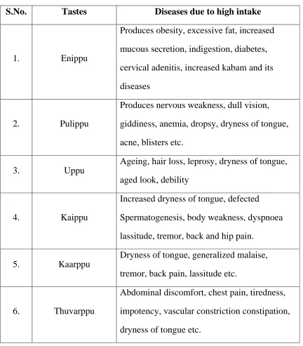
Table 2:
S.No. Tastes Diseases due to high intake
1. Enippu
Produces obesity, excessive fat, increased mucous secretion, indigestion, diabetes, cervical adenitis, increased kabam and its diseases
2. Pulippu
Produces nervous weakness, dull vision, giddiness, anemia, dropsy, dryness of tongue, acne, blisters etc.
3. Uppu
Ageing, hair loss, leprosy, dryness of tongue, aged look, debility
4. Kaippu
Increased dryness of tongue, defected Spermatogenesis, body weakness, dyspnoea lassitude, tremor, back and hip pain.
5. Kaarppu
[image:25.612.105.537.219.712.2]De-Arrangement of 3 humour
1. Vali – Thodam:
Darkness of motion
Body pain
Exaggerated Pricking pain Constipation
Paralysed limbs
Mental distress
Difficulty in work
Decreased Impairment of intelligence Giddiness
Increased iyam symptoms
2. Azhal thodam
Yellowish discolouration of skin, urine Increased appetite
Exaggearted Increased thirst
Burning sensation
Decreased sleep
3. Iyam thodam
Chills with rigor Pallor
Tightness Cough
Fullness of stomach Excessive sleep
Dyspnoea
Destruction of joint Giddiness
Decrease iyam in all Body fluids Increased sweating
Palpitation Exaggerated
II1. Alterations in udalthathukkal
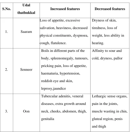
Table 3:
S.No.
Udal
thathukkal
Increased features Decreased features
1. Saaram
Loss of appetite, excessive salivation, heaviness, decreased physical constituents, dyspnoea, cough, flatulence.
Dryness of skin, tiredness, loss of weight, less ability in hearing.
2. Senneer
Boils in different parts of the body, spleenomegaly, tumours, pricking pain, loss of appetite, haematuria, hypertension, reddish eye and skin, leprosy,jaundice
Affinity to sour and cold, dryness, pallor
3. Oon
Tubercular adenitis, veneral diseases, extra growth around neck, cheeks, abdomen, thigh, genitalia
[image:28.612.105.560.138.601.2]4. Kozuppu (fat)
Identical features of increased oon, dyspnoea on exertion, extra musculature in gluteal region, external genitalia, chest, abdomen, and thigh
Loin pin,
spleenomegaly, emaciation
5. Enbu (Bone)
Excessive ossification and dentition
Joint pain, falling of teeth, falling and splitting of hairs and nails.
6.
Moolai (bone marrow)
Heaviness of body and eye, swollen interphalangeal joints, oliguria, non – healing ulcers.
Osteoporosis, blurred vision.
7.
Sukkilam (or) suronitham
Increased sexual activity, urinary calculi
IV.Environmental changes:
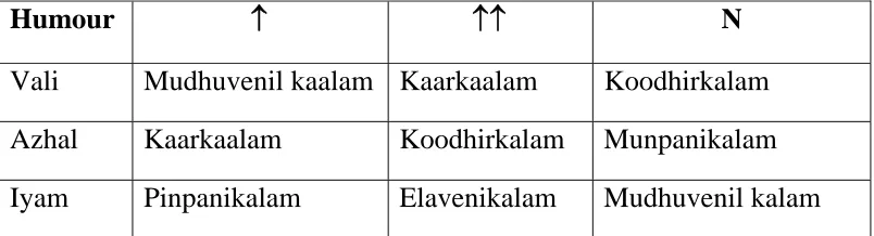
Table 4:Seasonal changes of humours
Humour ↑ ↑↑ N
Vali Mudhuvenil kaalam Kaarkaalam Koodhirkalam Azhal Kaarkaalam Koodhirkalam Munpanikalam Iyam Pinpanikalam Elavenikalam Mudhuvenil kalam
↑Thannilai valarchi. ↑↑piranilai valarchi. N - thannilai adaithal.
b.Regional changes of humours:
Kurinji - kabha diseases Mullai - pitha diseases Neythal - vadha diseases Marutham - no disease will occur Paalai - mukkuttra disease
V. Effects on self – suppression of 14 vegams
Reflexes are essential for the normal physiology when there is any self suppression to those reflexes, that will lead to the pathological state.
Vegankal Diseases
1.Vadham - Heart diseases, gastritis, umbilical hernia, body pain, liver disorder, constipation, oliguria, loss of appetite.
2.Thummal - Head ache, defect of special sensory organs and it is activities, pain over the face, hip joint pain.
3. Siruneer - Anuria, urethral ulcer, gas formation in the abdomen.
4. Malam - Diarrhoea, flatulence, knee pain.
5. Kottavi - urinary disorders, leucorrhoea, associated with schizophrenia, abdominal diseases.
6. Pasi - pricking pain all over the body, emaciation, apathetic face, painful joints
7. Neer - Same as that of pasi
8. Erumal - Increased cough, bad breath, heart disease 9. Elaippu - urinary disorder, syncope, rigor, peptic ulcer. 10. Thookkam - Heaviness of head, pain in the eyes, deafness 11. Vaanthi - Rashes, anemia, itching, eye diseases, asthma,
fever, cough
12. Kanneer - Heart diseases, eye diseases, wounds in the scalp, upper respiratory disorders.
DIAGNOSTIC METHODS:
Diagnosis is the mandatory process in the treatment of a patient. Envagai thervugal which is the unique and special method having a broad and important role in diagnosing a particular diseases. It is based upon the principles of poriyaal arithal, pulanaal arithal and vinaathal.
Poriyaal arithal means understanding by the five organs of perception, nose, tongue, eyes, skin and the ears.
Pulanaal arithal means understanding by the sense objects smell, taste, vision, somatic sense and sound.
Vinaathal means interrogating the patient, learning the history and symptoms of the disease by asking questions to the patient.
Envagai thervugal:
‘nka;f;Fwp epwe;njhdp tpopeh tpUkyk; iff;Fwp
-Njiuah; thf;F
1. Examination of tongue (eh)
By interrogation feeling, seeing the symptoms and signs are heard and examined. After examining, it must be compared, excluded and at last the final diagnosis is to be arrived.
Naa
It reflects the disease and so it gains importance in examining. The tongue is seen for the colour, shape, size, coating, fissures, growth, surfaces, sensations of taste and also salivary secretion
Niram
The normal colour of each humural body is explained. It there is any change from normal (ie) colour of eyes, tongue, mucous membrane, any erythema, hypo(or) hyper pigmentation in the skin, they are dealt under this.
Thoni
This not only explains the tone of speech but also the changes in modulations, pitch, sound, fluency, stammering, difficulty in articulation, repetition, listening, answering speech, associated with breathing difficulties etc.
Vizhi
Malam
The metabolic end product of our food after completing its work of supplying energy is expelled from the body as faeces. And thus any change in the colour, consistency, frequency, amount; components of motion exhibit the disease.
Moothiram
Urine plays an important role in revealing the diseased state in the form of changes in colour, specificgravity, odour, frequency, froth and deposits.
“te;j ePh;f;fwpvil kzk; Eiu vQ;rnyd; iue;jpa Ysit aiwFJ KiwNa”
Neikuri
“mUe;JkhwpujKk; mtpNuh jkjha;
m/fy; myh;jy; mfhyt+d; jtpHe;jow; Fw;wstUe;jp cwq;fp itfiw Mbf;fyrj; jhtpNa fhJ nga; njhUK$h;j;jf; fiyf;Fl;gL ePhpd; epwf;Fwp nea;fFwp epUkpj;jy; flNd:
Vali diseases – Rays of snake (ghk;ig Nghy; ePz;ly;) Azhal diseases – As a ring (Nkhjpuk; Nghy;)
Iya Disease – Stands as a peral (guthky; Kj;JNghy;)
Naadi
It is diagnostic entity and felt in the radial artery with the three fingers, fore finger (Vali), middle finger (Azhal) and ring finger tips (Iyam). Ratio is 1:1/2:1/4. It serves as a good indicator of all ill health. It has been considered for assessing the prognosis and diagnosis.
Mei
It deals all about the changes in the skin (i.e.) tactile sensation, the warmth, the chillness, sweat, numbness, fissures, plaques, papules, ulcers, inflammation etc.
Basically, siddha aims to maintain the equilibrium between the five elements despite out constant interaction with the outer world. The five elements which work as 3 vital forces in body and perform all physical and mental functions are constantly affected by time, space and nutrition.
AIM AND OBJECTIVES
The author has selected “ Kirumi pitham” for the dissertation subject because it is one of the disorders, which affect the individuals in higher incidence. Its occurrence is increased in recent times due to abnormal dietary habits and poor sanitation.
So this study mainly aims to define etiology, pathology, symptomo logy, and diagnostic methods of ‘Kirumi pitham’ by synchronizing the evidence, found in different siddha literature and formulating them after a detailed thorough study into an acceptable and adaptable form.
The following specific objectives have been drawn to achieve the above aim.
1. To collect the ancient siddha literature about pitha disease in general and kirumi pitham in particular
2. To evaluate the siddha basic physiology.
3. To Study the Clinical course of the disease ‘kirumi pitham’ with keen observation on the etiology, clinical Features and diagnosis.
4. The diagnosis of the disease by using siddha parameters like poriyal arithal, pulanal therdhal, vinadhal, Envagai thervugal, udal kattugal and mukkutra verupadugal.
5. To make a thorough physical examination of the patient.
ELUCIDATION ABOUT KIRUMI PITHAM
In Yugi Vaidhya Chinthamani kirumi pitham is mentioned under pitha
roga nithanam as,
fpUkp gpj;jk;
‘rPjkh abtapw;wpw; wpkpUz; lhfpr;
nrOikah Alk;ngq;Fe; jpdT khfp thjkh Alk;ngq;Fq; fLg;Gz; lhfp
tw;wpNa kyryKk; twz;L NghFk; fhjkha;f; fhy; iffs; fjypj; jz;L
fdj;jJ Nghy; ntr;nrd;W fLg;G thFk; ehjtha; kye;jd;dpw; fpUkp tPOk;
ehWNk Aly; fpUkpg; gpj;j khNk”
- A+fp itj;jpa rpe;jhkzp
The meaning of the words in this poem,
GO (fpUkp) - Worm
mbtapW - Lower abdomen
jpkph; - Colicky Pain
nrOik - Beauty
jpdT - Itching
fLg;G - Pain
rPjkha; abtapw;wpw; jpkpUz; lhfpr;
Lower abdominal pain
nrOikah Alk;ngq;Fe; jpdT khfp
Itching all over the body
thjkh Alk; ngq;Fq; fLg;Gz; lhfp
Generalised body pain
tw;wpNa kyryKk; twz;L NghFk;
Constipation Reduced urine out
fhjkha;f; fhy;iffs; fjypj;jz;L
fdj;jJ Nghy; ntr;nrd;W fLg;G thFk;.
Pain and swelling of the upper and lower limbs.
ehjtha; kye;jd;dpw; fpUkp tPOk;. ehWNk Aly; fpUkp gpj;jkhNk
Expulsion of worms in stool, and offensive odour of stool.
The Yugi’s lines are summarized as follows, 1. Lower abdominal pain
2. Itching all over the body. 3. Generalised body pain. 4. Constipation, and Reduced urine output.
PATHOLOGICAL VIEW OF DISSERTATION TOPIC IN
SIDDHA ASPECT
“gpzpapDw; gj;jpiag; NgRthd; gpzpKjy; thjgpj; jq;fg kd;ke;jphp je;jphp
tPjkh Alyuz; nka;k;Gu tuR nra; Kiw nrAkhjyhd; ………….”
- Njiuah; fhg;gpak;. thjkha; gilj;J
gpj;j td;dpaha; fhj;J Nrj;k rPjkha; Jilj;J
- NjiuaH kUj;Jt ghujk;
According to siddha aspect, Azhal is said to be the protective agent of all activities of our body. So that author mentioned ‘Azhal’ as ‘Manthiri’ in the above lines.
According to Yugi muni
‘Nghnkd;w gpj;jj;Jf; fpUg;gplNk Nfsha; Nguhd fz;lj;jpd; fPojhFk;”
It means place of the Azhal in body is below the neck.
responsible for maintaining good health. When some of the environmental factors like diet and immoral activities disturb the Azhal, it loses its control which may be diminished or exaggerated. This may leads to Azhal noigal
ALTERD THRIDOSHA IN KIRUMI PITHAM
In this disease the modified Azhal may affect the functions of vali humour.
Azhal
The deranged Azhal humour results in,
• Aakkanal (Anarpitham)
Derangement of anarpitham results in increased appetite in early stage.
• Vannayeri (Ranjakapitham)
Derangement of (Ranjakapitham) results in tiredness
• Aatralangi (Sathagapitham)
Sathaga pitham is affected in Kirumi Pitham the patients is not active because of generalized body pain.
• Olloliththe (Pirasagapitham)
Pirasagam pitham is affected in this disease produces dryness and itching all over the body.
• Nokkuazhal(Aalosagapitham)
Vali
When Azhal humour is increased, Vali humour also get increased.
• Uyirkkaal(Piraanan)
In kirumi pitham ,piraanan is affected which produces impaired utilization of nutrients of the body.
• Keelnokkukaal (Abaanan)
In kirumi pitham, derangement of abaanan leads to constipation and decreased urine output will occur.
• Paravakkaal (Viyaanan)
In “Kirumipitham” Viyaanan is affected produces generalized body pain.
• Nadukkaal (Samaanan)
In ‘Kirumi Pitham’ Samaanan is affected digestion and absorption are affected producing indigestion.
• Vizhikkaal(Koorman)
Derangement of koorman leads to conjunctivitis and photophobia.
• Thummikkaal(kirugaran)
In kirumi pitham, derangement of kirugaran leads to increased appetite in early stage.
• Kottavikkaal (Devathathan)
Iyam
In kirumi pitham azhal and vali humour is increased and iyam humour is decreased.
• Alliyam(Avalambagam)
In kirumi pitham derangement of avalambagam leads balancing is disturbed.
• Neeppi iyam(Kilethagam)
kilethagam is affected which produces increased appetite in early stage.
ALTERD UDAL KATTUKAL IN KIRUMI PITHAM
Udal Kattukal Changes when decreases
Saaram Fatigue
Senneer Itching all over the body Oon Weakness of sense organs Kozuppu loss of energy Enbu Hair falling Moolai Reduced urine output
Siddha Pathology deals with the diseased condition of the human, which is due to food alteration, environmental variation and immoral activities.
Detailed pathologic view of dissertation Topic
Modern Aspect
The small intestine is widest at its duodenal end and narrowest at the ileal end. For this reason foreign bodies may be impacted here.
“rPjkha; abtapw;wpw; jpkpUz; lhfp”
The usual Helminthic infestations are responsible for abdominal pain. The adult ascaris worms live in the upper part of the small intestine. Ascariasis causes prolonged irritation of the muscular coat of loop of intestine may cause muscular spasm. They may stimulate reflex peristalsis. For instance each time peristaltic wave travels along an overly excitable spastic gut, causing recurrent and often severe colicky pain in the abdomen.
“nrOikah Alk; ngq;Fk; jpdTkhfp” Pathology of Itching,
The eosinophils normally constitute about two percent of all the body leucocytes .Eosinophils are phagocytes, and they exhibit chemotaxis.
Eosinophils are often produced in large numbers in people with parasitic infection, and they migrate into tissues diseased by parasites. Although most of parasites are too large to be phagocytized cells, nevertheless the eosinophils attach themselves by way of special surface molecules to the parasite and release a substance that kills many of them.
“thjkh Alk;ngq;Fk; fLg;Gz;lhfp”
Ascariasis disturbed the small intestinal absorption. Requirement of nutrient to the body is decreased.
Difficiency of nutrients occur. It will cause generalized body discomfort (lassitude).
“tw;wpNa kyryKk; twz;L NghFk;”
Constipation may be defined as decreased in frequency of bowel movements and difficult (or) painful passage of hard stool.
Prolonged irritation of the muscular coat of loop of intestine may cause muscular spasm due to the spasm the frequencies of bowel movements are reduced leading to constipation.
“fhjkha; fhy; iffs; fjypj;jz;L fdj;jJNghy; ntr;nrd;W fLg;G thFk;”
Ascaris lumbricoides are robbing the host nutrition. The nutritional effects are seen when the worm burden is heavy. The worms may be present in enormous numbers, sometimes exceeding 500, occupying a large part of intestinal tract. This interferes with proper digestion and absorption of food. Ascariasis may contribute to protein energy malnutrition.
Maintenance of osmotic pressure of plasma is important for the proper distribution of water between blood and tissues.
‘ehjtha; kye;jd;dpw; fpUkp tpOk; ehWNk Aly; fpUkp gpj;jkhNk”
The Ascaris worms are restless wonderers, apparently showing great inquisitiveness, in that they tend to probe insinuate themselves into any aperture they find on the way. The wondering is enhanced when the host is ill.
REVIEW OF LITERATURE
Kirumi Pitham also explained in various literatures such as, Thanvanthiri vaitheyam and Segarasa sekaram
fpUkpNjh~f; Fwpfs;
‘(typj;jpLk;) kyk; tplhNj ehlNlhWe;jq;fp ahq;Nf fypj;jpLQ; rpWGOj;jhd; fhaj;ij kpff;nfLf;Fk; eypj;jpL kghdkl;Lk; erenrd; whpj;JNehth kyj;jpdpw; fpUkpj;Njh~ kpZnad tFf;fyhNk”
- jd;te;jphp itj;jpak; - 103 The Clinical Features of KirumiDhosa are abdominal pain, motion does not passes daily and it accumulates in the intestine, accumulation of the worms in the body causes generalized body pain, the worms causes itching and pain around the anus.
fpUkpgpj;jk;
clk;Giffhy; fLg;Gz;lhk; Xahj;jpdTe; jpkpUz;lhk; tplq;nfhs; kyj;jpw;fpUkptpOk; ntr;nrd;Wlk;G tw;wptUk; klq;fpr;ryKk; kykJTk; tusk;kjpf ehw;wKkhk;
jplq;nfhs; fpUkpg;gpj;jkJ nra;Aq;Fzj;ijj; NjHe;jwpNa”
- NrfuhrNrfu itj;jpak; -39
SMALL INTESTINE
The small intestine is continuous with the stomach at the pyloric sphincter and leads into the large intestine at the ileocaecal value. It is a little over 5 metres long and lies in the abdominal cavity surrounded by the large intestine. In the small intestine the chemical digestion of food is completed and most of the absorption of nutrient materials takes place.
The small intestine is described in three parts which are continuous with each other.
The duodenum is about 25 cm long and curves around the head of the pancreas. At its midpoint there is an opening, common to the pancreatic duct and the common bile duct, guarded by the hepatopancreatic sphincter.
THE JEJUNUM AND ILEUM
The mobile part of small intestine extends from the duodeno – jejunal flexure to the ileo –caecal junction, and is arranged in a series of coils which are suspended from the posterior abdominal wall by the mesentery. The coils are contained within the three and half-sided framework of the large gut. Proximal 2/5th of the small gut forms the jejunum
And distal 3/5th is known as the ileum.
External features of jejunam:
STRUCTURE OF THE SMALL INTESTINE (MOBILE PART)
It consists of four coats from without inwards 1. Serous Coat
2. Muscular Coat 3. Sub mucous Coat 4. Mucous Coat
SEROUS COAT:
It is derived from the peritoneum and invests the entire tube except the attachment of the mesentery.
MUSCULAR COAT
It consists of outer longitudinal and inner circular layers of smooth muscles, separated by the myenteric plexus of nerves (Auerbach’s plexus).
Contraction and relaxation of these muscle layers occurs in waves which push the contents of the tract onwards. This type of contraction of smooth muscle is called peristalsis. Muscle contraction also mixes food with the digestive juices. Onward movement of the contents of the tract is controlled at various points by sphincters consisting of an increased number of circular muscle fibres. They also act as valves preventing backflow in the tract. The control allows time for digestion and absorption of the food.
SUBMUCOUS LAYER
Meissner’s plexus, consisting of sympathetic and parasympathetic nerves which supply the mucous membrane lining.
MUCOSA
This consists of three layers of tissue:
a. Mucous membrane formed by columnar epithelium is the innermost layer and has three main functions: protection, secretion and absorption.
b. Muscularis mucosa, a thin outer layer of smooth muscle that provides involutions of the mucosa layer, e.g. gastric glands, villi.
Features in the mucous membrane of small gut:
The mucous membrane presents the following features a. Circular folds
b. Villi
c. Crypts of Lieberkuhn d. Solitary follicles e. Peyer’s patches
f. Entero – chromaffin cells.
a. Cirular folds are permanent mucous folds about 800 in number, more large and thickly set in jejunum, and absent in proximal one inch of duodenum and distal six inches of ileum.
b. Villi are fleshy tongue like in duodenum, leaf like in jejunum, and finger like in ileum. The villi act as little absorptive organs, present in the entire small gut except over the solitary follicles and Peyer’s patches. The villi cover the mucosal surface in a dense mat numbering 10 to 40 per mm2, each villus is about 0.5- 1mm long. The absorptive surface of the small gut is enormously increased by circular folds, villi and microvilli. The increased by circular folds is 3-fold, villi 10-fold, and microvilli 20-fold. Thus the total increase is 600-fold, exposing an area of 200 sq. metre.
c. Crypts of lieberkuhn (or) intestinal glands
d. Entero chromaffin cells.
Interposed between columnor cells of the glands are Argentaffin cells (or) enterochromaffin cells and goblet cells. Argentaffin cells secrete the intrinsic factor, which is essential for the absorption of vitamin B12. The goblet cells secrete mucus. There is another type of cell called paneth cells, which also secrete the enzymes.
e. Peyer’s patches
These are scanty, present in the lower part of jejunum and mostly circular in outline. The epithelial cells covering solitary follicles or peyer’s patches are known as M-Cells which help transport of antigens.
Functions of small intestine:
1. Mechanical function:
The mixing movements of small intestine help in the through mixing of chyme with the digestive juices like succus entericus , pancreatic juice and bile.
2. Secretary function:
Small intestine secrete succus entericus, enterokinase and the gastro intestinal hormones.
3. Hormonal functions:
4. Digestive function:
Though the digestion of various food substances commence in mouth and stomach it is completed only in small intestine. The digestive functions of small intestine are carried out by the enzymes of succus entericus secreted in small intestine.
5. Activation function:
The enterokinase secreted by small intestine activates typsinogen with trypsin. Trysin, in turn activates other enzymes.
6. Hemopoietic function:
The intrinsic factor present in the small intestine is necessary for absorption of vitamin B12 from gastro intestinal tract into the blood.
7. Hydrolytic function:
Succus entericus of small intestine provides water, which helps in all the hydrolytic processes of enzymatic reaction involved in digestion of various food stuffs.
Absorptive functions:
The presence of villi and microvilli in small intestinal mucosa increases the surface area of the mucosa. This facilitates the absorptive functions of intestine.
Absorption of carbohydrates:
Glucose is absorbed into blood and drained into portal vein. Absorption of glucose occurs by sodium co-transport.
Absorption of proteins:
The dextro-amino acids are absorbed by simple diffusion and most of levo-amino acids are transported actively by sodium- co –transport system.
Absorption of fats:
Most of the fats are absorbed in upper port of small intestine. Presence of bile is essential for fat absorption.
Absorption of water and minerals:
¾ In small intestine, sodium is absorbed actively.
¾ Calcium is actively absorbed mostly in upper part of small intestine.
¾ Water moves in (or) out of the intestinal lumen until the osmotic pressure of intestinal contents become equal to that of plasma.
Absorption of vitamins:
ASCARIS LUMBRICOIDES
“Infectious diseases will last as long as huminity excists”
Definition: Infection of A. Lumbricoides in man is known as ascariasis.
Causative Organism:
Kindom-Animalia Phylam-Nnematoda Class-Secemenda Family-Ascarididea Genus-Ascaris
Species-Ascaris lumbricoides
It is the common and longest intestinal nematode commonly known as round worm.
Geographical Distribution:
It is cosmopolitan, having a world-wide distribution, being especially prevalent in the tropics, such as China, India and South-East Asia. It occurs in person with unhygienic habits.
Incidence:
Habitat:
The adult worm lives in the lumen of the small intestine (jejunum) of man where it moves freely and maintains its position by its muscle tone.
Morphology:
Adult worm:
It resembles an ordinary earthworm and is the largest intestinal nematode parasitising man. When fresh from the intestine, it is light brown or pink in colour but is gradually changes to white. In shape it is rounded and tapers at both ends, the anterior end being thinner than the posterior. The mouth opens at the anterior end and possesses three finely toothed lips, one dorsal and two ventral. The digestive and reproductive organs float inside the body cavity containing an irritating fluid. The irritant action is due to the presence of a substance, ascaron or ascarase which is probably of the nature of primary albumoses (proteose). Allergic manifestations seen in infected individuals and amongst laboratory workers dissecting the worms are due to this ascaron.
Male :
Female :
It is longer and stouter than the male and measures 25 to 40 cm in length with a maximum diameter of 5mm. The posterior extremit, is neither curved nor pointed but is conical and straight. The anus is sub terminal and opens directly on the ventral aspect in the form of a transverse slit. The vulva opens at the junction of the anterior and the middle thirds of the body on the midventral aspect; this section of the worm is narrower and is called the vulvar waist. The egg-laying capacity of a mature female ascaris has been found to be enormous, liberating about 2, 00,000 eggs daily.
Eggs:
The eggs liberated by a fertilized female pass out of the human host with the faeces. The characteristics of a fertilised female pass out of the human host with the faces. Are as follows.
• Round or oval in shapes (60 to 75 µm in length by 40 to 50 µm in breadth.
• Always bile-stained and brownish (golden brown) in colour.
• Surrounded by a thick smooth translucent shell with an outer albuminous coat which is thrown into rugosities or mammillations: this outer coat is sometimes lost (decorticated egg).
• Contains a very large conspicuous unsegmented ovum (the nucleus is concealed by a large amount of coarse yolk granules). There is a clear crescentic area at each pole.
The female, even if not fertilised, is capable of liberating eggs. The
characteristics of thisunfertilised egg are as follow.
• Narrower, longer (80 μm in length by 55 μm in breadth) and more elliptical.
• Brownish in colour (bile – stained).
• Has a thinner shell with an irregular coating of albumin.
• Contains a small atrophied ovum with a mass of disorganised, highly refractile granules of various sizes.
• Does not float in salt solution (heaviest of all helminthic eggs.) Fertilised and unfertilised eggs may be found in a sample of stool but if a specimen shows only the unfertilised eggs, it signifies that the host is harbouring the female Ascaris.
Resistance of eggs
Round worm eggs are adversely affected by excessive heat and drying as caused by directs exposure to sun. However, they are remarkably resistant to most other environmental conditions.
Laboratory studies have revealed that the egg can survive and continue maturation even when immersed in 2% formalin, potassium, dichromate and 50% solutions of Acidic, Nitric, Hydrocholoric and Sulphuric acids. This factor adds to the longevity of the eggs in the environment.
Incubation period
Life – span
The worm has a life span of 10-12 months; the eggs have to longevity of 3-7 years
Life Cycle:
The worm passes its life cycle in one host and no intermediate host is required. Continuance of the species is maintained by transference from one individual to another. Man is the only known definitive host of A.lumbricoides. The various stages in the life cycle are described below.
Stage: 1
Eggs in Faeces; Fertilised eggs containing the unsegmented ovum are passed with the faeces. They are not infective to man when freshly passed.
Stage:2
Development in Soil. A rhabditiform larva is developed from the unsegmented ovum within the egg shell in 10 to 40 days time, depending on the atmospheric temperature and humidity. This takes place in the soil (that is outside the human host). The ripe egg containing the coiled-up embryo is infective to man before hatching, the larva undergoes moulting.
Stage :3
Stage: 4
Migration through the Lungs. The larvae liberated in the small intestine do not directly develop into mature worms. The newly hatched larvae burrow their way through the mucous membrane of the small intestine and are carried by the portal circulation to the liver: here they live for a period of 3 to 4 days. Finally they pass out of the liver and via right heart enter the pulmonary circulation. While in the lungs they grow much bigger and increase in length from 0.2 mm to 2mm and moult twice (first- on the fifth or sixth day and the second- after the tenth day). Breaking through the capillary wall they reach the lung alveoli. The time taken for such migration is on an average 10 to 15 days.
Stage: 5
Re-entry into the stomach and the small Intestine. From the lung alveoli the larvae crawl up the respiratory tract, they are propelled into the larynx and pharynx and are once more swallowed. The larvae pass down the esophagus to the stomach and localize in the upper part of the small intestine, their normal abode. Another moulting occurs between the twenty-fifth and the twenty-ninth day of infection.
Stage6:
RESERVOIR OF INFECTION
Man is the only reservoir
MODE OF INFECTION.
Infection is affected by swallowing ripe Ascaris eggs (embryonated eggs) with raw vegetables cultivated on a soil fertilised by infected human excreta. Water-supplies may be contaminated and infection may occur by drinking water. Where soil-pollution is common, the eggs may directly be conveyed to the mouth by dirty fingers.
Infection may also occur by inhalation of desiccated eggs in the dust reaching the pharynx and swallowed.
Factors favoring the spread of the transmission:
1. Simple life cycle.
2. Enormous egg production (2,00,000 eggs/ day)
3. These eggs are highly resistant to ordinary disinfectants (due to the ascroside.)
4. The eggs may remain viable for several years. 5. Social customs and living habits.
6. Disposal of faeces is unsuitable.
Immunology.
Specific antibodies (complement-fixing and precipitating) can be demonstrated in Ascaris infection. Hypersensitivity to ascaris is determined by skin test.
Pathogenesis:
Majority of the infections (about 86%) are symptomless. But the presence of even a few worms can be potentially dangerous. The worm inhibits the upper part of small intestine. The severity of the symptoms depends both on the number of eggs ingestible and on the previous infection history. The adult worm may produce its pathogenic effects in the following ways.
1. Migrating larvae 2. Adult worm
3. Toxins released from the worms
Symptoms produced by migrating larvae:
Loeffler’s syndrome
In heavy pulmonary infection typical symptoms such as Fever,
cough, dyspnoea, cyanosis, urticaria,
pain over the chest,
mucoid and bloody sputum are present.
he sputum may contain charcot–leydon crystals. The larvae may occasionally be found in sputum. But are seen more often in gastric washings. The clinical features generally clear in one (or) two weeks. Though it may sometimes be severe, it is rarely fatal. Loffler’s syndrome can also be caused by hypersensitivity to other agents, both living and non living.
In general circulation
Larvae pass beyond the pulmonary capillaries and reach the general circulation and they may reach other organs of the body such as liver, spinal cord, and kidneys, very rarely the larvae may occlude a small vessel in the heart and brain.
Symptoms due to adult worms:
The pathological effects present are caused by 1. Spoliative action
2. Toxic action 3. Mechanical effects
Spoliative action:
Ascaris lumbricoides robbing the host of its nutrition. It is otherwise called as nutritional effects, are usually seen when the worm burden is heavy. The worms may be present in enormous numbers, sometimes exceeding 500, occupying a large part of intestinal tract. This interferes with proper digestion and absorption of food. Ascariasis may contribute to protein energy malnutrition and vitamin A deficiency.
Patients have loss of appetite and are often restless abnormalities of jejunal mucosa are often present, including broadening and shortening of villi, elongation of crypts and round cell infiltration of laminapropria.
Toxic action:
These are due to the hypersensitivity to the worm antigens and may manifested as
Mechanical effects:
These are the most important manifestations of Ascaris lumbricoides. Mechanical effects can be due to the masses of worm causing luminal occlusion (or) even a single worm infiltrating into a vital area.
The adult worms live in the upper part of the small intestine, where they maintain their position due to their body muscle tone. They may stimulate reflex peristalsis, causing recurrent and often severe colicky pain in the abdomen. The worms may be clumped together into a mass, filling the lumen of intestine leading to intussusceptions. i.e., intestinal obstruction.
The worms are restless wonderers apparently showing great inquisitiveness, in that they tend to probe and insinuate themselves into any aperture they find on the way. The wandering is enhanced when the host is ill, particularly when febrile with the temperature above 390 F.
The male worm is more responsive to illness of the host, than the female. The worm may wonder up (or) down along the gut, going up it may enter the opening of the biliary (or) pancreatic duct causing acute biliary obstruction (or) pancreatitis. It may enter the liver parenchyma where it may leads to abscesses. The worm may go up to the esophagus and come out through the mouth (or) nose.
Ectopic ascariasis
The worms frequently migrate and enter the stomach and may pass up through the esophagus at night and coming out through the body openings such as nose, mouth, anus and vagina etc.. When migration occurs through respiratory system, may produce suffocation and asphyxia.
Symptoms due to sensitation
The metabolizes of ascaris, both during the period of biological incubation and after the worm mature in small bowels may produce sensitation phenomenon of allergic manifestation such as.
Skin -Urticaria Nose -Rhinitis
Lungs - Bronchial Asthma Intestinal tract - constipation
Eye - conjunctivitis and photophobia Excretory system - haematuria
Diagnosis
In this condition, diagnosis can be done by two ways (or) methods. 1. Direct evidence
Direct evidence:
This may be possible in the following situations.
1. When worms passed in the faeces particularly after the treatment when the parasite gets paralyzed and get propelled by peristalsis.
2. When the worm is vomited out.
3. On radiological examination this may be particularly obvious when to worms are lying parallel like “Trolley car lines”.
Demonstration of eggs:
Each female worm produces and lays massive number of eggs per day. One are two direct wet mounts are usually sufficient to diagnose and infection with even a small number of worms. There are three techniques, to demonstrate the eggs.
These are
1. Concentration technique. 2. Sedimentation technique. 3. Floatation technique.
All type of eggs as described above can be seen in the stool sample. The eggs can also be demonstrated on duodenal aspiration of the bile.
Sputum Examination:
In pulmonary involvement of sputum may reveal eosinophils, charcoat laydon crystals and occasionally larvae of the parasite.
In Direct evidence:
Prevention:
Ascariasis can be eliminated only if faecal contamination of soil can be prevented. The ascaris egg is highly resistant. Therefore the use of night soil as manure will lead to spread of the infection unless destruction of eggs in ensured by proper composting. Soak the vegetables and other garden crops containing iodine 200 ppm for 15 minutes which kills the eggs and larvae of ascaris and other helminthes.
Personal protection:
1. Water:
Drinking water should be boiled up to 1000 F and filtered to prevent infection through ingestion of infected cyclopse. Especially in area with high endemicity of these infections.
2. Food:
Under ground (or) fresh vegetables used for making salads should be thoroughly cleaned and preferably peeled before use. Pork, beef and fish should be cooked well to destroy the infective form of the parasite embedded in the flesh.
3. Skin:
The children should be encouraged to wear shoes while playing in the field which are likely to be contaminated with infected faeces.
Health education:
Personal hygiene:
1. The habit of washing hands after defaecation as well as before and taking food with antiseptic soaps should be encouraged.
2. Nails should be cut and cleaned daily.
Publicity:
The information on the mode of infection with the helminthes, methods of prevention and treatment should be widely disseminated.
1. Proper disposal of human stools. 2. Treatment of the affected individuals.
EVALUATION OF DISSERTATION TOPIC
Materials and methods
The clinical study on the disease kirumi pitham was carried out at the post graduate department of Noi Naadal in government siddha medical college, palayamkottai.
Case selection and supervision
The author have selected 10 cases of similar symptoms of Kirumi pitham under the supervision and monitoring by faculties and head of the department of post graduate Noi Naadal. All the cases were thoroughly examined and routine investigations performed.
Out of this 7 cases were selected for the study on Noi Naadal aspect of kirumi pitham. The clinical signs and symptoms of Kirumi pitham were taken from yugi Vaithiya Chindamani – 800).
1. Evaluation of clinical parameters:
The detail history and clinical features of the patients were taken carefully.
a. The clinical history contains.
1. Diet habits
6. Past history 7. Socio economic status 8. History of infectious disease 9. History of previous illness were collected from the patient.
b. Clinical features of kirumi pitham are
1. Lower abdominal pain 2. Itching all over the body
3.Generalised body pain(Discomfort) 4. Constipation
5. Pain and swelling in upper and lower limbs. 6. Worms in stools
7. Stools offensive in nature
were taken as criteria for selection of patients.
2. Diagnosis:
The diagnosis is made on the basis of interpretation of the following siddha principles.
1. Envagai Thervugal
3. Investigation:
Clinical investigation such as
Routine haematological examination a. Total Count
b. Differential Count c. Haemoglobin
d. Erythrocyte Sedimentation rate. e. Blood sugar
f. Blood Urea
Routine urine analysis
a. Albumin b. Sugar c. Deposits
Examination of stool:
Stool samples of affected patients by kirumi pitham were tested macroscopically for colour, quantity, nature (solid, semisolid, watery) odour and irugal.
Microscopical examination of stool for the presence of ova and cyst of ascaris lumbricoids were carried out.
OBSERVATION AND RESULTS
1. Results are observed with respect to the following aspects:
i. Age and Sex reference. ii. Mukkutranilai
iii. Udal Thathukkal iv. Envagai Thervugal
v. Clinical Features vi. Laboratory Findings.
[image:72.612.121.489.120.502.2]i. Age and Sex reference:
Table: 5
Age
Sex Total No. of
Cases Male Female
Up to 10 yrs - - -
10 – 20 yrs 5 1 6
ii. Mukkutranilai
Table 6: Derangement of Vali
S. No Types of Vali
No of cases
affected
Changes
1. Pranaan - -
2. Abanaan 4 Constipation 3 Viyanaan 7 Generalised body pain
4 Uthanaan - -
5 Samanaan 7 Reduced appetite
6 Nagaan - -
7 Koorman - -
8 Kirukaran 6 Reduced appetite 9 Devathathan 7 Tiredness
10 Denanjeyan - -
Table 7:Derangement of Azhal
S. No
Types of
Azhal
No of cases
affected
Changes
[image:73.612.117.512.524.710.2]Table 8:Derangement of Iyam
S. No Types of
Iyam
No of cases
affected
Changes
1 Avalampagam 6 Blanching function disturbed 2 Kilethagam 6 Reduced appetite
3 Bothagam - -
4 Thorpagam - - 5 Santhigam - -
iii.Udal Thathukkal:
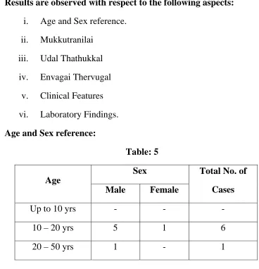
Table 9:
S. No Udal
Thathukkal
No of cases
affected
Changes
1 Saaram 7 Fatigue
2 Senneer 7 Itching all over the body
3 Oon 7 Tiredness
4 Kozhuppu - -
5 Enbu - -
6 Moolai - -
[image:74.612.118.510.322.602.2]iv.The Picture of Envagai Thervugal:
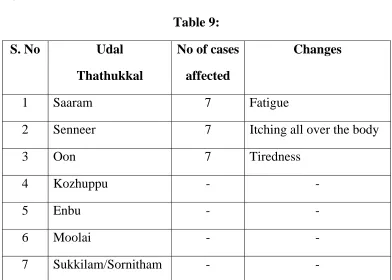
Table 10:
Case
No
Naa Niram Mozhi Vizhi Malam
Moothiram
Neerkuri
Naadi Sparisam
1 A A NA NA A NA PV A
2 A A NA NA A NA PV A
3 NA NA NA NA A NA PV A
4 NA NA NA NA A NA PV A
5 NA NA NA NA A NA VP A
6 A NA NA NA A NA VP A
7 A A NA A A NA VP A
A – Affected PV – Pitha Vatham NA – Not Affected VP – Vatha Pitham
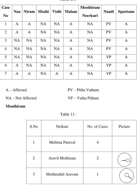
Moothiram
Table 11:
S.No Neikuri No. of Cases Picture
1 Mellena Paraval 4
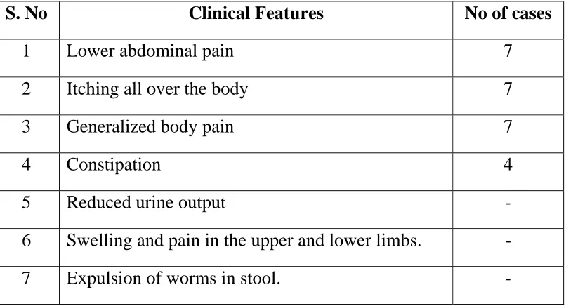
[image:75.612.92.544.105.734.2] [image:75.612.89.539.114.369.2]v. Clinical Features:
Table 12:
S. No Clinical Features No of cases
1 Lower abdominal pain 7
2 Itching all over the body 7 3 Generalized body pain 7
4 Constipation 4
5 Reduced urine output -
[image:76.612.116.519.120.338.2]vi. Laboratory Findings
Table 13
Cases
No
Blood Bio-Chemical Urine Motion
TC
cells /
cumm
DC cells ESR
Hb%
Sugar
msg%
Urea
msg% Alb Sug Dep Ova Cyst P% L% E%
1/2 hr
mm
1 hr
mm
1 10200 70 22 6 10 16 66 - - Nil Nil Nil AOP -
2 8100 55 40 5 8 16 65 - - Nil Nil Nil AOP -
3 9000 49 38 13 - - 80 - - Nil Nil Nil AOP -
4 9200 60 30 10 5 10 75 - - Nil Nil Nil AOP -
5 7200 54 36 10 10 20 80 - - Nil Nil Nil AOP -
STATISTICAL ANALYSIS OF KIRUMI PITHAM
Study subjects were analysed by the statistics mean median and percentages. The inference about the etiology was obtained by use the test of significance ‘Z’ proportion of single sample.
Observation and results:
The variations are related to kirumi pitham diseases where observed and assigned, under the rule of heading of the respective variations and phenomena.
Age
[image:78.612.147.484.547.711.2]Age is one of the crucial factor of incidences of kirumipitham. Since the disease are occurring among the youngest than the aged. The incidence of the disease was analysed in the basis of age is tabulated as follows.
Table 14:
S.No
Age
group
No of
cases
Significations
% Mean Median SD
The above table clearly shows the study subjects characteristics in terms age. The mean age is 16.4+ 8.6 years. The median age 13 years. Fifty percent of the study subjects are below the age of 13 years. The incidence of the kirumi pitham the age group of 10-14 is 57.27 where as the incidence in the other age group namely 15-19 and 35-39 are 28.5% and 14.3% respectively. But the above proportions are not statistically significant.
Etiology:
[image:79.612.83.550.310.551.2]The main and fore most etiology of the diseases are posted below and the incidences are analysed and interpreted.
Table 15:
S.No Name of etiology n
No of cases
affected
Affected
Percentage
Significant
1. Intake of unboiled water 7 7 100 Significant 2. Contaminated food 7 3 42.9 Not Significant 3. Poor sanitation 7 2 28.6 Not Significant 3. Contact in Soil 7 2 28.6 Not Significant 4. Bar foot 7 3 42.9 Not Significant 5. Improper Personal hygiene 7 5 71.4 Significant