NATIONAL INSTITUTE OF SIDDHA
Tambaram Sanatorium, Chennai - 47Affiliated to
THE TAMIL NADU DR. M.G.R. MEDICAL UNIVERSITY, CHENNAI - 600 032
A STUDY ON
PENN MALADU
(DISSERTATION SUBJECT)
For the partial fulfillment of the
requirements to the Degree of
DOCTOR OF MEDICINE (SIDDHA)
BRANCH I–MARUTHUVAM DEPARTMENT
INTRODUCTION
“Foypdp jpahopdp njd;g jk;kf;fs;
koiyr; nrhy; Nfshjth;“.
- jpUf;Fws;
The pipe is sweet, the bite is sweet,” say those who have not heard the
prattle of their own children.
“ngWktw;Ws; ahkwpt jpy;iy awptwpe;j
kf;fl;Ng wy;y gpw”
- jpUf;Fws;
Among all the benefits that may be acquired, we know no greater
benefit than the acquisition of intelligent children.
The basic biological function of a living organism is its capacity to
reproduce. Among humans for every 80 married couples who produce
children, there are 20 who are unable to have an offspring. Approximately
one-half of all cases can be traced to either partner.
In the fast pace of modern life, dietary habits, increasing prevalence of
obesity, stress, smoking, contraceptives have all contributed to the decline in
fertility.
In many southeast Asian Countries, infertility is considered a curse and
the inability to conceive is often a stigma attached to the female partner
though she may not always be the cause of it.
Approximately 15% of couples attempting their first pregnancy meet
this time should be therefore be regarded as possibly infertile and should be
evaluated.
AIM AND OBJECTIVES
1. To evaluate the therapeutic efficacy of Siddha Drugs, Maladu
Neenga Thailam and Saaravalli Mathirai in Penn Maladu (female
infertlity) patients.
2. To increase the fertility rate among the female infertility patients.
3. To regularise the menstrual abnormalities
4. To evaluate the toxicity & pharmacological actions in experimental
animals
5. To find out the adverse-effects of the drug in clinical trial, if any
6. To have a complete study of the disease, Penn Maladu (Female
Infertility) under the headings of
a) Enn Vagai Thervugal
b) Udal Kattugal
ngz; kyL
Foe;ijfisg; ngwhj jd;ikf;F ‘kyL’ vd;W ngah;.
kyil
1. Rj;j
kyL
2. fjyp
kyL
3.
fhf kyL vd;W %tifahfg; gFj;Js;sdh;
Rj;j kyL (Primary Infertility)
xU KiwahfpYk; fUj;jhpj;jNj fpilahj epiyikf;Fr; ‘Rj;j kyL’ vd;W ngah;.
fjyp kyL (Secondary Infertility)
thio kuk; xU Kiw Fiy js;spaJk; kPz;Lk; Fiy js;shky; epd;WtpLk;.
mJNghy; rpyh; xU kfit <d;W NkYk; fUj;jhpg;gNjapy;iy. ,jw;Ff; ‘fjyp kyL’
vd;W ngah;.
fhf kyL
fhfkhdJ ,uz;L ,dj;ijg; ngUf;fptpl;ljd; Nghpy; kPz;Lk; jd; ,dj;ijg;
ngUf;Ftjpy;iy. mJNghy; kfspu; ,uz;Nl kfit <d;W NkYk; fUj;jhpg;gNjapy;iy.
fd;k kyL
#y; nfhs;;th;. Mdhy; fUr;rpijT mbf;fb Vw;gl;L #y; fhyj;ij
ePbf;fnthl;lhky; jLf;Fk; epiyf;F ‘fd;k kyL’ vd;W ngah;.
fhuzq;fs;
kfspUf;F g+g;G epfo;T elg;gjhy; fUj;jhpf;fg;gLths; vd;W epue;jukhf
nrhy;tjw;fpy;iy.
(1) rpyUf;F
fhkf;fpsh;r;rpapy;yhkNy g+g;G epfo;r;rp eilngWfpwJ.
rpidg;igapy; ghprpid (Graffian Fallicle) Kjph;e;J rpid ntspg;gl;L
tUtjpy;iy.
(2)
rpy Ntisfspy; fUg;igr; rspr;rt;T rpidiag; gjpaitf;fj; jbg;GWtjpy;iy.
(3) kfsph;f;Ff;
fUj;jhpg;gjpy;
rpidg;gij kpfTk; gq;F nfhs;fpwJ.
rpidg;igapd;W Rod;W ntspg;gl;l rpid (Ovum) FQ;rhe;jj;jhy;
(Fimbriae) cwpQ;rg;gl;L> rpidg;ghijia milfpwJ. rpidg;ghijf; Foy;>
rpidia fUg;ig miwia Nehf;fp ce;jr; nra;fpwJ. ce;jpf; nfhz;Nl
nry;Yk; rpid tpe;J mZf;fisr; re;jpj;Jf; fUf;nfhs;fpwJ. MfNt>
rpidg;ghij rpw;rpy Neha;fspy; jhgpjkile;J> Foypy; jpR tsh;r;rp
Vw;gl;L> rpidAk;> tpe;JTk; re;jpf;f tha;g;gpy;yh fhuzj;jhy; kfsph;
(4) rpy
rkak;
Gzh;r;rpapy;
Vw;gl;l Nahdpf; frpT> fUg;ig Kff;frpT
Mfpaitfs; tpe;J mZ caph; thoj;jf;fjhf ,y;yhtpl;lhy; fUg;ig .miwia
miltjw;F Kd;Ng td;ikapoe;J kbe;J tpLfpd;wd.
(5)
kfspUf;F gpwg;GWg;Gfspy; jhgpj Neha;fs; Vw;gl;bUe;jhYk;
(6)
kfsph; gpwg;GWg;Gfs; rhptu Rfu;zhPjpahf ,y;yhikahYk;
(7)
fUg;ig Raepiyapy; ,y;yhky; khWgl;l epiyapy; ,Ue;jhYk;>
(8)
fUg;igapy; foiyf; fl;bfs; ,Ue;jhYk; fUj;jhpf;f Kbahky; kyl;Lj;jd;ik
ngz; kyl;bd; Fzk;
jhNkjhd;
khjhe;j
UJ
fhyj;jpy;
jf;fhd uj;jkJ rpte;J fhZk;
NtNkjhd; kQ;rs;epwq; fUikahFk;
tpidahd
ePyepwQ; rPo;Ngh yhFk;
NghNkjhd; uj;jkJ TUz;il ahFk;
nghy;yhj ky %j;jpu epwNk ahFk;
ehNkjhd;
nrhd;dgb
rpfpr;rh
rhuk;
ehl;LNshh;f; fwpantrd;W etpd;wpl; lhNu.
- a+fpKdp
kfspu; kUj;Jtk;
nghUs;
khj khjk; epfo;TWfpd;w g+g;Gf; fhyq;fspy; Vw;gLk; g+g;Gf;frpT
ed;F rpte;jpUj;jy;> kQ;rl;fUik epwkhfTk;> ePyepwr; rPo; NghyTk;
rpw;rpW cUz;ilfshf ntspg;Nghe;jy;> NehAw;w fhyq;fspy; ,opAk; kyk;>
rpWePh;fisnahj;jy; Nghd;w ,f;Fwp Fzq;fisg; ngw;wpUg;gjw;Fg; ngz; kyL
vd;W ngah;.
Meaning of the above:
(i) Menstrual blood is bright red, yellowish black, and bluish red in colour.
(ii) Menstrual blood is colloidal in consistency or like clots.
NOI VARUM VAZHIKAL (AETIOLOGY)
I.
ACCORDING TO MANMURUGIYAM
fUg;ig jd;dpy; tspepiwe; jpLjy;
fUf;Fopthapy; jirtsh;e; jilj;jy;
GOg;gy Njhd;wpf; fUTz; bLjy;
fUg;ig afj;Jg; ghrp gw;wy;
mjpy;nrq; FUjp fl;b epw;wy;
fUf;Fop kjh;j;jy; vDkpt;; thWk;
fUg;ig Aw;wpL Nehnad nkhopg.
1. Amenorrhoea
2. Any benign or malignant growths in the uterus like leiomyoma
3. Infections of the genital tract like Pelvic Inflammatory Disease,
genital tuberculosis
4. Peritubal adhesions and adhesions within the uterus
5. Blood stagnation within the uterus like adenomyosis
6. Due to obesity in conditions like Polycystic Ovarian Disease,
hypothyroidism.
kDTw kylh nkhd;W khjtdUr; nra;jhd;.
1. Congenital malformations of the genital tract or congenital sex
chromosomal abnormalities.
2. Due to alterations in three humours.
3. Increased Kapha humour.
III. ACCORDING TO ATHMARAKSHAMIRDHAM VAIDHIYA
SAARA SANGIRAHAM
ghug;gh ngz;kylhq; fUg;gf; Nfhspd;
gf;Ftj;ijr; nrhy;YfpNwd; gz;gha;f; NfS
Mug;gh Mz;kyNl ahU ky;yhy;
mg;gNd ngz;kyL ahU kpy;iy
Neug;gh jtkpUe;Jk; nghUis aPe;Jk;
Neuhf tpy;iyaJ #iy ahNy
Ntug;gh NjtijNah Nlohq; fUg;gk;
tphpj;J ed;wha; GfYfpNwd; tpUk;gpf;NfNs.
tsp epiwe; jpUg;gpd; GzUq; fhiyg;
ngz;zp
Dlk;G
typjU
nkd;g
jir tsh;e; jpUg;gpd; neQ;R Fj;Jk;
GOf;fs;
epuk;gpd;
KJF
NehFk;
ghrpg;gw;wpd; jiytyp Njhd;Wk;
fUf;Fop kjh;g;gpd; Vg;ge; Njhd;Wk;
From the above verse, it is well known that the etiology of Maladu is
same as mentioned in Manmurugium.
IV. ACCORDING
TO
AGATHIYAR VAIDHYA KAVIYAM - 1500
ghug;gh nfw;g fhyk; ghpTld; tha;T - Nja;T
Nrug;gh Te;jpNahL nrd;wpLq; fUj;jhd; khSk;
thug;ghGOj; jhZz;zpy; kynldr; nrhy;thug;gh
Mug;gh tpjidg; Nghf;fpyhz; gps;is nrwpf;Fe;jhNd.
• Increase in Vayu and Theyu elements.
• Infections of the genital organs.
CLASSIFICATION OF PENN MALADU
I. According to YUGIMUNI CHIKITCHA SAARAM there are 3 types
(i) Sudhtha Maladu
(ii) Kathali Maladu
(iii) Kaka Maladu
II. According to JEEVA RAKSHAMIRTHAM ANUBAVA DEVA
RAGASIUM & SARABENTHIRAR KARPA PINI there are 4 types.
III. According to MANMURUGIUM, there are 11 types
(i) Vali Maladu (vii) Puzhu Maladu
(ii) Anal Maladu (viii) Pettra Maladu
(iii) Iya Maladu (ix) Mutru Maladu
(iv) Alagai Maladu (x) Noi Maladu
(v) Varatchi Maladu (xi) Seyarkai Maladu
(vi) Karupai Maladu
IV. According to ATHMARAKSHAMIRTHAM VAIDHYA SAARA
SANGIRAHAM.
There are 7 types.
(i) Vatha Maladu
(ii) Pitha Maladu
(iii) Alagai Maladu
(iv) Petra Maladu
(v) Varatchi Maladu
(vi) Vayu Maladu
CLINICAL FEATURES
A. ACCORDING TO YUGIMUNI CHIKITCHA SAARAM
Athi Maladu - epue;ju kyl;bd; Fzk;
etpd;wplNt apLg;Gtapw; ngUj;Jf; fhZk;
eykhd NkdpaJ t+jpf; fhZk;
Ftpd;wplNt Kk;kbg;G tapw;wpy; Njhd;Wk;
Fztjpahe; Njtjh ngz;zh dhYk;
etpd;wplNt rd;kj;jd; kyNl ahFk;
rjhfhyq; fUg;gkJ jhpah njd;W
Gtpd;wplNt a+fpKdp rpfpr;rh rhuk;
Gfd;wpl;lhh; Nyhfj;J khe;jw; fhNk.
nghUs;: clypd; kw;w ghfq;fis tpl ,Lg;Gk;> tapWk; ngUj;jpUj;jy;> cly; mijj;Jf;
fhzy;> ce;jpapy; %d;W kbg;Gfs; fhZjy; Mfpa FwpFzq;fisf; nfhz;L vd;WNk
fUj;jhpf;fhkypUf;Fk; jd;ikf;F epue;ju kyL vd;W ngah;.
MEANING:
ehl;lNt thiokuj; jhW Nghy
ftpyNt ike;jdhq; fUg;G nkhd;W
fhjypah sPd;wgpd; fUg;g kpy;iy
jtpyNt fUg;gFop kiwe;jJ Nkjhd;
jq;fpNa KOFehs; jilAz; lhFk;
GtpyNt a+fpKdp rpfpr;rh rhuk;
g+jyj;jpy; khdplw;Fg; g+l;b dhNu
nghUs;: Fiy <d;w thio kWKiw Fiy <dhj;jd;ik Nghy kq;ifah;fs; xU Foe;ijia
<d;w gpd;dh; fUg;igf; Fop nrayw;W g+g;Gj; jil Vw;gl;L> Gzh;r;rpAw;w NghJk;
kWKiw fUjhpf;fhj jd;ikf;Ff; fjyp kyL vd;W ngah;.
Meaning: After delivering a baby, woman attains menopause at or below
the age of 40, (pre-mature menopause). This is called Kathali Maladu.
Kaka Maladu – fhf kyl;bd; Fzk;
MNkjhd; fhfj;jpd; kyNl ahFk;
mg;gNd fUg;igapy; Nrhhp nfl;L
NghNkjhd; fUg;igAe; Je;J Nkjhd;
Nghf;fhd fhAlNd fjL Nghyhk;
NtNkjh dpuz;L Ngh; ike;j Dz;lhk;
NtWtif fUg;gJ kyNl ahFk;
ehNkjhd; nrhd;dgb rpfpr;rh rhuk;
ehl;LNshh;f; fwpantd;W etpd;wpl;NlhNk.
nghUs;: ,uz;L kfit <d;W fUg;igahdJ Nrhhp NfLw;w VJtpdhy; kWKiw
B.
ACCORDING TO JEEVARAKSHAMIRTHAM ANUBAVA
DEVA RAGASIYAM, SARABENDRAR KARPAPINI
The features of Athi, Kathali and Kaka Maladu are more or less same
as described in Yugi Chikicha Saarum. And In addition,
Karpa Maladu
Most of the women deliver a dead foetus.
C.
According to MANMURUGIYAM
1. Vali Maladu:
‘tsp kylhapd; nre;ePh;’
In this type, the colour of the urine is red.
2. Anal Maladu
‘mdy; ky lhapd; kQ;rs; fiuj;njdr;
rPWePh; hpwq;Fnkdr; nrg;Gth; Gyth;’
In this type of infertility, the urine becomes bright yellow in colour.
3. Iya Maladu
‘Ik;ky lhapd; rpWePh; kpfTk;
ntSj;J Njhd;W nkd;gh; Gyth;’
NehT Njhd;wy; rpWePh; fLj;Jk;
ntSj;Jq; fWj;JQ; rpte;J kpwq;fy;
vDkpit ayif kyl;Lf; FwpNa’
- There is pain in the lower abdomen during menstruation.
- Burning micturition
- The colour of the urine is pale yellow, red or black.
5. Varatchi Maladu
''cz;zpD Kly Kyu;e;J Nghjy;
kpf nfhl;lhtp - Njhd;wy; gw;gy
mr;rf; fdt}fs; Njhd;wy; vDkpit
twl;rp kyl;bd; Fwpnad nkhopg""
• Weight loss
• Intractable yawning
• Experiences dreadful dreams often.
6. Karupai Maladu
‘tapW nghUky; czt whik
Vg;ge; Njhd;wy; fUf;Fop Rw;wp
tspepd; wpLjy; vDkpit fUg;ig
tspkyl;bd; Fwp ahnkd nkhopg’
Signs and Symptoms
¾ Flatulence
¾ Belching
¾ Menstrual irregularities like oligomenorrhoea, hypomenorrhoea,
amenorrhoea
7. Puzhu Maladu
‘kWg;G Njhd;wy; jpq;fs; tUKOf;
,uz;nlhU jpq;f spilap YWjy;
clyk; ntSj;jy; Fwpjpd TWjy;
,itGO kyl;Lf; Fwpnad nkhopg
fUg;ig apw;GO kpf;fpL khapd;
tapw;wb Fj;Jk; jpq;fl; g+g;Gj;
jirfOePh; Nghy; Njhd;Wk; Gzh;r;rpapy;
mopjU ePUw; whil fiwg;gLk;’
Signs and Symptoms
Dysfunctional uterine bleeding
Pallor
Pruritis vulva
Intense pain in the lower abdomen
The colour of the meustrual blood is like that of the water washed out from the meat
fOePh; Nghyr; rpWeP hpwq;fy;
iffh nyhpjy; vDkpit nay;yhk;
ngw;w kyl;bd; Fwpnad nkhopg’
Signs and Symptoms
Obesity
Burning Micturition
Burning sensation in the extremities
The colour of the urine is like the water washed out from the rice.
9. Mutru Maladu
‘kUe;jp Dk;gpw newpapD ePq;fh
kyL Kw;W kynld nkhopg’
In this type, there is no improvement even on treatment.
10. Noi Maladu
‘Nehapd; tUtJ Neha;ky lhFk;
tspKjw; gygpzp te;Jw khapDk;
fUf;fha; fUf;fs; jhuh thapDk;
ngz;ghy; Neha;ky nlhd;wpLk; nkd;g’
Chronic illnesses like endocrinal disorders (Hypothroidism), renal and
cardiac diseases lead to Infertility in females.
11. Seyarkai Maladu
mWitapd; njhopYwr; nra;j yhYk;
kUe;jpw; fUTwr; nra;j yhYk;
vd;Wk; Gzuh jpUj;j yhYk;
fUf;Nfh spy;tif Gzh;j yhYk;
nraw;if kyL kUtpl apay;Ng’
Surgical sterilization in the female or previous surgery in the abdomen leads to adhesions
Taking contraceptives to avoid pregnancy
Avoiding sexual intercourse
Sexual intercourse during first trimester.
D. ACCORDING
TO
ATHMARAKSHAMIRTHAM
The clinical features described in Athmarakshamirtham are same as in
Manmurugium.
E.
ACCORDING TO SARABENDRAR KARPA PINI
a) Karpa Sayu Puralal
‘Xjpa khkf tpy;yJ ePjpAiue;jpL Ntd;wpUNt
jhJFky; FypNy kyUz;lJ jhz;ilNa Guspy;
ePjpajha; tpOkh tKjy; Gfepd;wplkq; nfhUTk;
NtjKkpg;gbNa AiuahbL nkd;nfhb ePawpNa’
NtypL Gz;nzdNt typAz;nldpd; nkd;gfkh kyUq;
Nfhy kpyhJGuz;lJ ntd;wwp $wtdTljhNk’
From the above verse, it is known that women with Karpasayu Puralal
experiences pain along the ribs during coitus on the fourth day of mensturation.
b) Vayu Seruthal
“ePawpkhlyh; jhdjpy; tha;ThpNdak ehkKje;
jhafkhfpa khlydw;wpjpd; rhunkyh nkhopfpr;
rPnad ePnudNtaJNgha; tsh; Njfnky;yhk; typah
NahAk; tpjq;fspjhF kwpe;J nfhsnshz;nlhbapd;GwNt”
Since there is excessive vayu within the uterus, it reduces the virulence
of the sperm and make it dead.
Signs:
cz;lhNk jpuz;BhpU ehspdpnyhz; fztd; Gzuj;
jz;lhh; Njfnkyhk; typnad;dpw; wq;fpa gfkyh;e;j
gz;lhh; thA ciwe;J ntd;wwp...
In this type, the woman experiences pain all over the body during
coitus on fourth day of menstruation.
c) Thasai Seruthal
,d;GW khkyh; jd;dpYNk jir Nafkjha; tsu
md;GWkg; nghONj tpOkhtKjQ; rsp NghYUfp
td;gbah JiwahJ fyq;fp tope;jpLNk ntspapy;
Benign or malignant growth in the uterus interrupts the passage of the
semen.
Signs
... khjpil jhd; typfhZnkdpw;
fhj;jpu ty;FypNy jirnad;w wpfspub gpd;dKe;...
There is pain in the groin or hip region during coitus.
d) Ushnathikam
nky;ypjo; g+tpdpy; ty;typAl;bz nkd; NkYz;lhfpy;
ey;ypjkha; tpOnkd;dKje; jz;dhba ntz;nzajhk;
nfhy;yjp dpd;W kpFe;J ntJk;gpf; NfhKfkhk; topNa
nry;y Khpj;Jiw Ngha;tpL khkiwr; r+j;jpukpg;gbNa
Any inflammatory condition of the vagina, uterus or fallopian tubes
destroy the sperm.
Signs
Njd;nkhopiag; Gzh; NghjpdpNy Jil Nrh;e;J typf;Fnkdp;d;
khd;kyh; jhdjpYl;bz nkd;w khl;L ntd;Gq;
In this type, there is pain in both thighs during coitus.
e) Seethala Serkai
,g;gbg; g+tpd;w rPjsNk Awpdd; gkjhk; Ntisr;
In kapha diseases like pyogenic infections and genital tuberculosis, it
destroys the sperms.
Signs:
... ePq;fpL khh;gjpNy
fz;z typf;Fnkdpd; kyh;jhdjpw; fhz;gJ rPjsnkd;
nwz;zp kUe;Jfs; nra;jpL nrg;GtNdh FoNye;jpioNa
In this type, there is pain in the chest region during coitus.
f) Puzhu Undathal
thddp NahdpapNy kyh;kPjpdpd; khGOtq; FwpNyh
Czpa khtKje; jidNanaLj; Jz;L fspj;Jkpf
Mztkhf epiyj;J tsh;e;jpL khifapdhd; kfitf;
fhzpia nad;W fiue;jpL thhpJ fz;lwp fhhpifNa.
nghUs;: Nahdp kw;Wk; ngz; ,dTWg;Gfspy; fpUkpfspd; jhf;fj;jhy;
kfT mope;J tply;.
Signs:
... khjiu kUtpa NghjpdpNa
rpj;j kfpo;r;rp nfhshkY Nkikay; jPh;f;fpiy nad;Wnrhypy;
tpj;jf ty;FypNy GOntd;wwp
In this type, there is no orgasm.
g) Sanka Thosam
tPhpaNk tpOk; NghjpNyaJ tPo;tjpd; Kd;ghfr;
rhunkyhq; nfhLNgha; tpLNkapJ rj;jpaNk Aiuj;Njhk;
thuzp nfhq;if ey;yhapij ePawp khkiw Ehz; KiwNa
nghUs;: fhw;W vd;Dk; rq;if Njh\j;jhy; ngz; ,dTWg;Gfs;
ghjpf;fg;gLtjhy; Vw;gLtJ.
Signs:
ehafpNahL wthifapw; Fjpfhduk; gpOj;jpL nkd;whw;
wPPaJw; rq;iffshkJ ntd;wwp Nrahp tpopkhNj
In this type, there is pain in calf muscles during coitus.
h) Avaya Ettra Thazhvugal
Ehypilahs; FwpahokjhfT Ehy; nfhO ed; Fwpjhd;
rPyKld; rhpePs kpyhtpow;Nwa; tjpdhy Kjk;
nghydNt AiwahkYld;gpw; gue;J fye;JtpLk;
NtnyDik tpopahapJ fz;lwpNtjd; tpjpg;gbNa
Congenital elongation of the cervix, acute retroverted uterus, septate
vagina, transverse vaginal septum are the anatomic defects that result
in infertility.
i) Poorva Vinai
In this type, due to kanma vinai or excessive usage of medications,
their sperm or ovum becomes inactive.
COMPLICATIONS:
1. Malattu Vali
kyl;L Neha;fs; kUtp kpFjypd;
fUg;igapy; typgy Njhd;wp Kjph;e;J
tpyf;fpd; fhiy tFj;j kpUf;Fk;
On the days of menstruation, very severe dysmenorrhoea is present.
2. Malattu Janni
tpyf;fpd; fhiyg; Gspj;jiy Az;zy;
kpfT Kz;zy; cz;l jwhik
kpfFsp Nuw;wy; Kjypa gpwTk;
kyb jdf;Fk; fyitg; gpzp jUk;.
On the days of menstruation, if rotten foods or excessive foods are
MODERN ASPECT
I
NFERTILITY
Infertility is defined as a failure to conceive within one or more years of regular unprotected coitus.
Primary infertility denotes those patients who have never conceived.
Secondary infertility indicates previous pregnancy but failure to conceive
subsequently.
The relative prevalence of the etiologies of infertility is
Male factor - 30 – 40%
Female Factor - 40 – 50%
Both - 10%
An association between the age of woman and reduced fecundability
has been well documented. This decline begins in the early thirties and
accelerates in the late thirties and early forties.
FEMALE INFERTILITY
The female factor contribute to 40 – 50% of infertility.
The important causes of female infertility as given by FIGO manual (1990)
are,
- Tubal and peritoneal factors - 25 – 35% - Ovulatory factor - 15 – 25% - Endometriosis - 1 – 10%
ETIOLOGY
OVARIAN FACTORS
The ovulatory dysfunctions encompass:
Anovulation or oligo – ovulation
Luteinised unruptured follicle.
TUBAL FACTORS
The impaired tubal function includes,
Defective ovum pick up
Impaired tubal motility
Loss of cilia
Partial to complete obstruction of the tubal lumen
The impaired function of above anyone is related to ,
Tubal infection (or)
Peritubal adhesions following pelvic surgery or infection or
endometriosis.
PERITONEAL FACTORS:
In addition to peritubal adhesions, even minimal endometriosis may
produced infertility.
UTERINE FACTORS
Uterine Hypoplasia
Inadequate secretry enclometrium
CERVICAL FACTORS
Anatomic : Anatomic defects preventing sperm ascent
Congenital elongation of the cervix,
Second degree uterine prolapse
Acute retroverted uterus
Pin hole os
Physiologic
The fault lies in the composition of the cervical mucus, so much that
the spermatozoa fail to penetrate the mucus. The mucus may be,
Scanty – following amputation ionisation or deep cauterisation of the cervix.
Excessive, purulent – chronic cervicitis.
Presence of Antisperm antibodies or sperm immobilizing antibodies.
VAGINAL FACTORS
Atresia Vagina (Partial or Complete)
Transverse vaginal septum
Septate vagina
Narrow introitus.
IMMUNOLOGICAL FACTOR
and
GENERAL FACTORS
Advanced age of the woman beyond 35 is related to infertility
Use of lubricants during intercourse which may be spermicidal
Anxiety and apprehension
Infrequent intercourse, lact of knowledge of coital technique and timing
of coitus to utilize the fertile period are very much common even
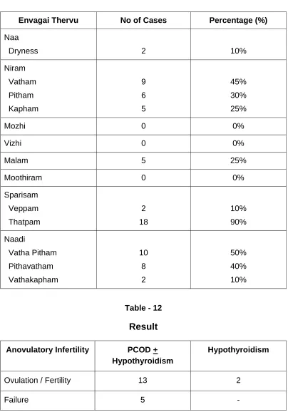
ANOVULATORY INFERTILITY
The ovarian activity is totally dependant on the gonadotrophins and the
normal secretion of gonadotrophins depends on the pulsatile release of GnRH
from hypothalamus.
As such, ovarian dysfunction is likely to be linked with disturbed
hypothalamo pituitary ovarian axis either primary or secondary from thyroid or
adrenal dysfunction.
Thus, the disturbance may result not only in anovulation but may also
produce oligomenorrhoea or even amenorrhoea. Anovulatory cycles usually
represent a lesser degree of disturbance with these normal pathways than
does amenorrhoea.
As there is no ovulation, there is no corpus luteum formation. In the
absence of progesterone, there is no secretary endometrium in the second
half of the cycle.
POSSIBLE CAUSES OF ANOVULATION
Any disturbance in hypothalamo – pituitary – ovarian action will result
in anovulation.
DISTURBANCE AT THE LEVEL OF HYPOTHALAMUS
1. Obesity or weight loss
2. Psychologic disturbances
3. Psychotrophic drugs
DISTURBANCE AT THE LEVEL OF PITUITARY
Primary Causes
1. Sheehan’s syndrome
2. Tumour – Prolactinoma
Secondary Causes
1. Hypo or Hyperthoroidism
2. Adrenal Hyperplasia
DISTURBANCES AT THE LEVEL OF OVARY
1. Polycystic ovarian disease (PCOD)
2. Premature ovarian failure
3. Luteinised unruptured follicles
CHRONIC ANVOULATION
At least 80% or more of gynecologic endocrine disorders result from
chronic anovulation. Women with chronic anovulation fail to ovulate
spontaneously but may ovulate with appropriate therapy. The ovaries of such
women do not secrete estrogen in a normally cyclic pattern. It is clinically
useful to differentiate those women who produce enough estrogen to have
withdrawal bleeding after progestogen therapy from those who do not; the
latter often have hypothalamic – pituitary dysfunction.
CHRONIC ANOVULATION WITH ESTROGEN PRESENT
This disorder is most commonly caused by polycytic ovarian syndrome.
Chronic anovulation with estrogen present may also occure with tumors of
the ovary. These include granulosa – theca cell tumors, Brenner tumors,
cystic teratomas, mucous cystadenomas, and Krukenberg tumors. Such
tumors can either secrete excess estrogen themselves or produce androgens
that are aromatized in extraglandular sites. Chronic anovulation and the
clinical features of PCOS result. Occasionally, areas of the ovary not involved
with tumors show the characteristic histologic changes of PCOS. Other
causes of chronic anovulation with estrogen present include adrenal
production of excess androgen, (usually adult-onset adrenal hyperplasia due
to partial 21-hydrozylase deficiency) and hypothyroidism.
CHRONIC ANOVULATION WITH ESTROGEN ABSENT
Women with chronic anovulation who have low or absent estrogen
production and do not experience withdrawal bleeding after progestogen
treatment usually have hypogonadotropic hypogonadism due to disease of
Isolated hypogonadotropic hypogonadism associated with defects of
smell (olfactory bulb defects) is known as the Kallmann syndrome, which is
due to a single gene defect in the X-linked KAL gene. Affected women are
sexually infantile and have a defect in the synthesis and/or release of GnRH.
Hypothalamic lesions that impair GnRH production and cause
hypogonadotropic hypogonadism include crainopharyngioma, germinoma
(pinealoma), glioma, Hand-Schuller-Christian disease, teratoma,
endodermal-sinus tumors, tuberculosis, sarcoidosis, and metastatic tumors that cause
suppression or destruction of the hypothalamus. Central nervous system
trauma and irradiation can also cause hypothalamic amenorrhea and
deficiencies in secretion of growth hormone, adrenocorticotropic hormone
(ACTH), vasopressin, and thyroid hormone. Rare, autosomal recessive
defects in the GnRH receptor have also been described.
More commonly, gonadotropin deficiency leading to chronic
anovulation is believed to arise from functional disorders of the hypothalamus
or higher centers. A history of a stressful event in a young woman is frequent.
Gonadotropin and estrogen levels are in the low to low-normal range as
compared with normal women in the early follicular phase of the cycle. In
addition, rigorous exercise, such as jogging or ballet, and diets that result in
excessive weight loss may lead to chronic anovulation, particularly in girls with
a history of prior menstrual irregularity. The amenorrhea in these women
does not appear to be a result of weight loss alone but a combination of a
decrease in body fat and chronic stress. An extreme form of weight loss with
In addition, chronic debilitating disease such as end-stage kidney
disease, malignancy, inflammatory bowel disease, and malabsorption can
lead to hypogonadotropic hypogonadism via hypothalamic mechanism.
Treatment of chronic anovulation due to hypothalamic disorders
includes ameliorating the stressful situation, decreasing exercise, and
correcting weight loss, as appropriate. These women are susceptible to the
development of osteoporosis; estrogen replacement therapy is recommended
to induce and maintain normal secondary sexual characteristics and prevent
bone loss in those who do not desire pregnancy, and gonadotropin or
gonadorelin therapy is indicated when pregnancy is desired. When
appropriate, therapy is directed at the primary disease of the hypothalamus.
Disorders of the pituitary can lead to the estrogen-deficient form of
chronic anovulation by at least two mechanisms – direct interference with
gonadotropin secretion by lesions that either obliterate or interfere with the
gonadotrope cells (chromophobe adenomas, Sheehan’s syndrome) or
inhibition of gonadotropin secretion in association with excess prolactin
(prolactinoma). Pituitary tumors may secrete no hormone, one hormone, or
more than one hormone. Prolactin levels are elevated in 50 to 70% of
patients with pituitary tumors, either because of prolactin secretion by the
tumor itself (in the case of prolactinomas) or because the tumor mass
interferes with the normal hypothalamic inhibition of prolactin secretion.
Prolactin excess associated with low levels of LH and FSH constitutes
a specific subtype of hypogonadotropic hypogonadism. One-tenth or more of
amenorrheic women have increased levles of prolactin, and more than half of
women with both galactorrhea and amenorrhea have elevated prolactin
estrogen production, but prolactin-secreting tumors on occasion are
associated with normal ovulatory menses or chronic anovulation with estrogen
present. In the latter half of pregnancy, prolactin-secreting pituitary tumors
may expand, leading to headache, compression of the optic chiasm,
bitemporal hemianopia, and blindness. Therefore, before inducing ovulation
for the purposes of achieving pregnancy, it is mandatory to exclude the
presence of a pituitary tumor.
Large pituitary tumors such as null cell adenomas—whether or not
hyper-prolactinemia is present—are likely to be associated with deficiency of
hormones in addition to gonadotropins.
Craniopharyngiomas, which are thought to arise from remnants of
Rathke’s pouch, occur most frequently in the second decade of life and often
extend into the suprasellar region. Many of these tumors calcify and can be
diagnosed by conventional skull film or CT. Patients often present with sexual
infantilism, delayed puberty, and amenorrhea due to gonadotropin deficiency;
secretion of TSH, ACTH, growth hormone, and vasopression may also be
impaired.
Panhypopituitarism can be caused by mutations in transcription
factors (Pit-1; Prop-1) involved in pituitary gland development, result
from surgical or radiation treatment of pituitary adenomas, or develop
THE OVARIAN CYCLE
Within the ovary, the menstrual cycle can be divided into three phases:
The follicular phase
Ovulation
The luteal phase
FOLLICULAR PHASE
The development of the oocyte is the key event in the follicular phase
of the menstrual cycle. The ovary contains thousands of primordial follicles
that are in a continuous state of development from birth, through periods of
anovulation, such as pregnancy, to the menopause. These initial stages of
follicular development are independent of hormonal stimulation. In the
absence of the correct hormonal stimulus however, follicular development
fails at the preantral stage, with ensuing follicular atresia. Development
beyond the preantral stage is stimulated by the pituitary hormones, (luteinizing
hormone (LH) and follicle-stimulating hormone (FSH)) which can be
considered as key regulators of oocyte development.
At the start of the menstrual cycle, FSH levels begin to rise as the
pituitary is released from the negative feedback effects of progesterone,
oestrogen and inhibin. Rising FSH levels rescue a cohort of follicles from
atresia, and initiate steroidogenesis.
Steroidogenesis
The basis of hormonal activity in preantral to pre-ovulatory follicles is
described as the ‘two cell, two gonadotrophin’ hypothesis. Steroidogenesis is
granulosa cells. The two cell, two gonadotrophin hypothesis states that these
cells are responsive to the gonadotrophins, LH and FSH respectively.
Within the theca cells, LH stimulates the production of androgens from
cholesterol. Within granulosa cells, FSH stimulates the conversion of thecally
derived androgens to oestrogens (aromatization). In addition to its effects on
aromatization, FSH is also responsible for proliferation of granulosa cells.
Although other mediators are now known to be important in follicular
development, this hypothesis is still the cornerstone to understanding events
in the ovarian follicle. The respective roles of FSH and LH in follicular
development are evidenced by studies on women undergoing ovulation
induction in whom endogenous gonadotrophin production has been
suppressed. If pure FSH alone is used for ovulation induction, an ovulatory
follicle can be produced but oestrogen production is markedly reduced. Both
FSH and LH are required to generate a normal cycle with adequate amounts
of oestrogen.
Androgen production within the follicle may also regulate development
of the preantral follicle. Low levels of androgens enhance aromatization and
therefore increase oestrogen production. In contract, high androgen levels
inhibit aromatization and produce follicular atresia. A delicate balance of FSH
and LH is required for early follicular development. The ideal situation for the
initial stages of follicular development is low LH levels and high FSH levels, as
seen in the early menstrual cycle. If LH levels are too high, theca cells
Selection of the Dominant Follicle
The developing follicle grows and produces steroid hormones under
the influence of the gonadotrophins LH and FSH. These gonadotrophins
rescue a cohort of preantral follicles from atresia. However, normally only one
of these follicles is destined to grow to a pre-ovulatory follicle and be released
at ovulation the dominant follicle.
The selection of the dominant follicle is the result of complex signalling
between the ovary and the pituitary. In simplistic terms, the dominant follicle
is the largest and most developed follicle in the ovary at the mid-follicular
phase. Such a follicle has the most efficient aromatase activity and the
highest concentration of FSH – induced LH receptors. The dominant follicle
therefore produces the greatest amount of oestradiol and inhibin. Inhibin
further amplifies LH-induced androgen synthesis, which is used as a substrate
for oestradiol synthesis. These features mean that the largest follicle
therefore requires the lowest levels of FSH (and LH) for continued
development. At the time of follicle selection, FSH levels are declining in
response to the negative feedback effects of oestrogen. The dominant follicle
is therefore the only follicle that is capable of continued development in the
face of falling FSH levels.
Ovarian-pituitary interaction is crucial to the selection of the dominant
follicle, and the forced atresia of the remaining follicles. When this interaction
is bypassed, as in ovulation induction with the administration of exogenous
gonadotrophins, many follicles continue to develop and are released at
ovulation with an ensuing multiple gestation rate of around 30 per cent.
During in vitro fertilization (IVF) the production of many ovulatory follicles is
number of embryos replaced can be carefully controlled. However, if such
multiple follicular development occurred unchecked in the normal cycle, it
would lead to the production of multiple gestations of high-order numbers,
with their associated problems.
Inhibin and Activin
Although folliculogenesis, ovulation and the production of progesterone
from the corpus luteum can be explained largely in terms of the interaction
between pituitary gonadotrophins and sex steroids, it is becoming clear that other
autocrine or paracrine mediators also pay a role. One of the most important of
these is inhibin.
Inhibin was originally described as a testicular product that inhibited
pituitary FSH production, hence its name. However, inhibin is also produced
by a variety of other cell types, including granulosa cells within the ovary.
Granulosa cell inhibin production is stimulated by FSH but in women, as in
men, inhibin attenuates FSH production. Within the ovary, inhibin enhances
LH – induced androgen synthesis. The production of inhibin is a further
mechanism by which FSH levels are reduced below a threshold at which only
the dominant follicle can respond, ensuring atresia of the remaining follicles.
Activin is a peptide that is structurally related to inhibin. It is produced
both by the granulosa cells of antral follicles, and also by the pituitary gland.
The action of activin is almost directly opposite to that of inhibin in that it
Insulin-like Growth Factors
Insulin-like growth factors (IGF-I and IGP-II) act as paracrine
regulators. Circulating levels do not change during the menstrucal cycle, but
follicular fluid levels increase towards ovulation, with the highest level found in
the dominant follicle. The actions of IGF-I and – II are modified by their
binding proteins; insulin-like growth factor binding proteins (IGFBPs).
In the follicular phase, IGF-I is produced by theca cells under the action
of LH. IGF-I receptors are present on both theca and granulosa cells. Within
the theca IGF-I augments LH induced steroidogenesis. In granulosa cells,
IGF-I augments the stimulatory effects of FSH on mitosis, aromatase activity
and inhibin production. In the preovulatory follicle, IGF-I enhances
LH-induced progesterone production form granuloss cells. Following ovulation,
IGF-II is produced from luteinized granulosa cells, and acts in an autocrine
manner to augment LH-induced proliferation of granulosa cells.
OVULATION
Late in the follicular phase, FSH induces LH receptors on granulosa
cells. Oestrogen is an obligatory co-factor in this effect. As the dominant
follicle develops further, follicular oestrogen production increases. The
production of oestrogen is eventually sufficient that the threshold required for
oestrogen to exert a positive feedback effect on pituitary LH secretion is
achieved. Once this occurr, LH levels increase, at first quite slowly (day 8 to
day 12 of the menstrual cycle) and then more rapidly (day 12 onwards.)
During this time, LH induces luteinization of granulosa cells in the dominant
follicle, so that progesterone is produced. Progesterone further amplifies the
surge of LH, Ovulation occurs 36 hours after the onset of the LH surge. The
LH surge is one of the best methods by which the time of ovulation can be
determined, and is the event detected by most over-the-counter ‘ovulation
predictor’ kits.
The periovulatory FSH surge is probably induced by the positive
feedback effects of progesterone. In addition to the rise in LH, FSH and
oestrogen that occur around ovulations, a rise in serum androgen levels also
occurs. These androgens are derived from the stimulatory effect of LH on
theca cells, particularly those of the non-dominant follicle. This rise in
androgens may have an important physiological effect in the stimulation of
libido, ensuring that sexual activity is likely to occur at the time of ovulation
when the woman is at her most fertile.
Prior to the release of the oocyte at the time of ovulation, the LH surge
stimulates the resumption of meiosis, a process which is completed after the
sperm enters the egg. In order for the ovary to release the oocyte at ovulation,
breakdown of the follicular wall is required. This event is coordinated by LH,
TSH and progesterone which stimulate the activity of proteolytic enzymes
such as plasminogen activators (which produce plasmin, which stimulates
collagenase activity) and prostaglandins. Prostaglandins not only stimuate the
activity of proteolytic enzymes, but also promote an inflammatory-type
response within the follicle wall, and by stimulation of smooth muscle activity
may help extrusion of the oocyte.
follicle syndrome, LUF). Although LUF appears to be an uncommon cause of
infertility, women wishing to become pregnant should be advised to avoid
taking prostaglandin synthetase inhibitors such as aspirin and ibuprofen which
may inhibit oocyte release.
LUTEAL PHASE
The luteal phase is characterized by the production of progesterone
from the corpus luteum within the ovary. The corpus luteum is derived both
from the granulosa cells that remain after ovulation, and from some of the
theca cells which differentiate to become theca lutein cells. The graunlosa
cells of the corpus luteum have a vacuolated appearance associated with the
accumulation of a yellow pigment, lutein from where the corpus luteum
derives its name. Extensive vascularization within the corpus luteum ensures
that the granulosa cells have a rich blood supply providing the precursors for
steroidogenesis.
The production of progesterone from the corpus luteum is dependent
on continued pituitary LH secretion. However, serum levels of progesterone
are such that LH and FSH production is relatively suppressed. This effect is
amplified by moderate levels of oestradiol and inhibin that are also produced
by the corpus luteum. The low levels of gonadotrophins mean that the
initiation of new follicular growth is inhibited for the duration of the luteal
phase.
Luetolysis
The duration of the luteal phase is fairly constant, being around 14
days in most women. In the absence of pregnancy and the production of
corpus luteum regresses at the end of the luteal phase, a process known as
luteolysis. The control of luteolysis in women remains obscure. As the
corpus luteum dies, oestrogen, progesterone and inhibin levels decline. The
pituitary is released from the negative feedback effects of these hormones
and gonadatrophins, particularly FSH, start to rise. A cohort of follicles which
happen to be at the preantral phase are rescued from atresia and a further
menstrual cycle is initiated.
SUMMARY OF OVARIAN EVENTS
FOLLICULAR PHASE
LH stimulates theca cells to produce androgens.
FSH stimulates granulosa cells to produce oestrogens.
The most advanced follicle at mid-follicular phase becomes the dominant follicle.
Rising oestrogen and inhibin produced by the dominant follicle inhibit pituitary FSH production.
Declining FSH levels cause atresia of all but the dominant follicle.
OVULATION
FSH induces LH receptors.
LH surge.
Proteolytic enzymes within the follicle cause follicular wall breakdown
THE LUTEAL PHASE
The corpus luteum is formed from granulosa and theca cells retained
after ovulation.
Progesterone produced by the corpus luteum is the dominant hormone
of the luteal phase.
In the absence of pregnancy, luteolysis occurs 14 days after ovulation.
PITUITARY GLAND
The process of follicular development, ovulation and the maintenance
of the corpus luteum has been described in terms of ovarian physiology. In
reality however, the ovary, pituitary and hypothalamus act in concert (the
hypothalamo-pituitary-ovarian asix) to ensure the growth and development of
(ideally) one ovarian follicle, and to maintain hormonal support of the
endometrium to allow implantation.
The pituitary hormones LH and FSH are, as we have seen, key
regulators of folliculogenesis. The output of LH and FSH from the pituitary
gland is stimulated by pulses of gonadotrophin-releasing hormone (GnRH)
produced by the hypothalamus and transported to the pituitary in the portal
circulation. The response of the pituitary is not constant but is modulated by
ovarian hormones, particularly oestrogen and progesterone. Thus low levels
of oestrogen have an inhibitory effect on LH (negative feeback) whereas high
levels of oestrogen actually stimulate pituitary LH production (positive
feedback). In the late follicular phase, serum levels of oestrogen are
sufficiently high so that a positive feedback effect is triggered thus generating
produces serum levels of oestrogen in the negative feedback range, so that
measured levels of gonadotrophins are low.
The mechanism of action of the positive feedback effect of oestrogen
involves an increase in GnRH receptor concentrations and an increase in
GnRH production, whilst the mechanism of the negative feedback effect of
oestrogen is uncertain.
In contrast to the effects of oestrogen, low levels of progesterone have
a positive feedback effect on pituitary LH and FSH secretion. Such levels are
generated immediately prior to ovulation, and contribute to the FSH surge.
High levels of progesterone such as those seen in the luteal phase inhibit
pituitary gonadotrophin production. Negative feedback effects of
progesterone are generated both via decreased GnRH production, and via
decreased sensitivity to GnRH at the pituitary level. Positive feed-back effects
of progesterone operate at the pituitary level only and involve increased
sensitivity to GnRH. Importantly, progesterone can only have these effects if
there has been prior priming by oestrogen.
As we have seen, oestrogen and progesterone are not the only
hormones to have an effect on pituitary gonadotrophin secretion. The peptide
hormones inhibin and activin have opposing effects on gonadotrophin
production: inhibin attenuates pituitary FSH secretion whereas activin
stimulates it.
Its role in the maintenance of corpus luteum in human is not well
documented, but the fact remains that there is high incidence of anovulation in
women with elevated plasma prolactin level.
Thyrotrophic Hormone (TSH)
TSH (thyroid stimulating hormone) is produced by the beta cells. It
acts on the thyroid gland and regulate the production of thyroxine. It has got
α and β subunits like those of FSH and LH, with functions of β subunits being
different. Abnormal TSH secretion is associated with menstrual and ovulatory
dysfunction.
THE HYPOTHALAMUS
The hypothalamus, via the pulsatile secretion of GnRH, stimulates
pituitary LH and FSH secretion. Production of GnRH not only has a
permissive effect on gonadotrophin production, but alterations in amplitude
and frequency of GnRH pulsation throughout the cycle are also responsible
for some fine tuning of gonadotrophin production.
The importance of GnRH secretion is seen in disorders such as anorexia
nervosa, and the amenorrhoea associated with excessive exercise. In these
disorders, GnRH production is suppressed leading to anovulation and amenorrhoea.
Ovulation can be restored in these women by the administration of GnRH in a
pulsatile manner (although this should be approached carefully since pregnancy is
relatively contraindicated in women whose body weight is significantly below average).
It is important to remember that GnRH is produced in a pulsatile
manner to exert its physiological effect. Drugs that are GnRH agonists (e.g.
endometriosis and other disorders. Although these drugs act as GnRH
agonists, they cause a decrease in pituitary LH and FSH secretion. The
reason for this is that these agonists are long acting and the continued
exposure of the pituitary to moderately high levels of GnRH causes down
regulation and desensitization of the pituitary. LH and FSH production is
therefore markedly decreased. Ovarian steroidogenesis is suppressed so
that serum oestrogen and progesterone fall to postmenopausal levels. Most
women become amenorrhoeic whilst taking GnRH agonists. A potential
disadvantage of the currently available GnRH agonists is that such
down-regulation and desensitization of the pituitary takes up to three weeks to exert
its effects. The initial effect of GnRH administration is to stimulate pituitary LH
and FSH production, leading to increased ovarian steroidogenesis. When a
patient commences GnRH therapy, this temporary increase in ovarian
steroidogenesis leads to a vaginal bleed within the first month of
HYPERPROLACTINEMIA
PROLACTIN
Functions
The primary function of prolactin is to enhance breast development in
pregnancy and to induce lactation after delivery. In addition, by binding to
specific receptors in the gonads, lymphoid cells and liver it affects fertility,
immunity and liver functions.
HYPERPROLACTINEMIA
A condition characterised by elevated serum prolactin levels, is the
most common endocrine disorder of the hypothalamo – pituitary axis. It could
result from a variety of conditions both physiological and pathological. The
prevalence varies from less than 1% of the general population, to almost 17
percent in women with reproductive disorders.
ETIOLOGY OF HYPERPROLACTINEMIA
Physiological Pathological
REM Sleep Tumors – Prolactinoma
Pregnancy Hypothalamic (pituitary lesions) Nipple Stimulation Idiopathic
Stress Polycystic ovarian disease
Coitus Hypothyroidism
Chest wall injury
Renal Failure
Liver Failure
Drugs – dopamine analogs
Phenothiazines Estrogen
Opiates Cimetidine
EFFECT ON FEMALE REPRODUCTIVE FUNCTION
Prolactin has a significant effect on the female reproductive function by
acting on the hypothalamo-pituitary ovarian axis. In addition, it has a direct
action on the ovaries, which is supposed to be responsible for the menstrual
irregularities associated with hyperprolactinemia ; probably regulating ovarian
steroidogenesis by its actions on the aromatase enzyme. It is interesting to
note that the action of prolactin on the ovaries varies in the different phases of
the menstrual cycle.
In the follicular phase, elevated prolactin level can disrupt normal
follicular development, cause atresia of the dominant follicle and inhibit
ovulation. On the other hand, the role of prolactin in the luteal phase is not
very clear, as it is supposed to both stimulate (by inducing LH receptor
formation) and inhibit corpus luteal functions (by inhibiting corpus luteal
steroidogenesis). Animal experiment have found that elevated prolactin level
can induce the development of adenomyosis – a condition characterized by
the implantation or extension of the endometrial glands into the myometrium,
apart from ovulatory dysfunction that could cause the infertility associated with
hyperprolactinemia.
Effect of Hyperprolactinemia on female reproductive function
1. Disrupts normal follicular development.
2. Atresia of the dominant follicle.
DIAGNOSIS
As prolactin is a dynamic hormone that responds readily to a variety of
stimuli, caution must be exercised during diagnosis.
Women typically present with a history of amenorrhea, oligomenorrhea
or infertility. Occasionally galactorrhea may be the only presenting symptoms.
Both sexes may present with visual field defects and headaches if associated
with a pituitary tumour.
Common Presenting symptoms in prolactin related disorders.
1. Amenorrhoea
2. Oligomenorrhoea
3. Galactorrhoea
4. Unexplained Infertility
5. Headache
6. Visual field defects
7. Symptoms of hypothyroidism
8. Drug intake
9. Decreased libido
Adjuvant investigation in a case of hyperprolactinemia
1. Serum TSH
2. BUN - Blood Urea Nitrogen
3. Serum Creatinine
4. Liver function tests
5. Visual field testing
6. CT - Brain
THYROID DISORDERS IN INFERTILITY
Thyroid disorders both hypo and hyperthyroidism are known to have a
profound effect on pregnancy and reproduction.
HYPERTHYROIDISM
The effect of severe hyperthyroidism on the fertility potential of an
individual is well documented, but the effects of the mild and moderate forms
of this disorders are not very clear.
ETIOLOGY OF HYPERTHYROIDISM
1. Grave's disease.
2. Solitary toxic nodule.
3. Toxic multinodular goitre.
4. Acute thyroiditis- viral
a. Autoimmune
b. Post-radiotherapy
5. Thyrotoxicosis.
6. Exogenous iodine administration.
7. Metastatic differentiated thyroid carcinoma.
CLINICAL FEATURES
Symptoms
¾ Hyperactivity,
¾ irritability, dysphoria
¾ Heat intolerance and sweating
¾ Palpitations
¾ Fatigue and weakness
¾ Weight loss with increased appetite
¾ Diarrhoea
¾ Polyuria
¾ Oligomenorrhoea, loss of libido.
Signs
Tachycardia: atrial fibrillation in the elderly
¾ Tremor
¾ Goiter
¾ Warm, Moist skin
¾ Muscle weakness, proximal myopathy
¾ Lid refraction or lag
HYPERTHYROIDISM AND FEMALE FERTILITY
Elevated thyroxine concentrations lead to increased levels of the sex
hormons binding globulin. This, in turn, accounts for the raised
concentrations of Estradiol and testosterone in the blood. Apart from this , the
fallicular phase baseline serum FSH and LH concentrations are also
increased with an attenuated mid-cycle LH surge. Consequently oligovulatory
or anovulatory cycles with a wide range of menstrual disorders (ranging from
amenorrhoea to menometrorrhagia) may be seen.
DIAGNOSIS
The diagnosis of hyperthyroidism is based upon clinical findings and
serological assays.
Assessment of the clinical manifestations would give a reasonable
indication to the presence of the disorders, which could then be confirmed by
serum hormonal assay.
The best screening tool, however, is the serum TSH assay. This is
based on the fact that as the level of the serum T3 and T4 rise, the
concentration of TSH will fall exponentially giving an accurate estimation of
the severity of the condition.
Once the diagnosis is established further tests to identify a possible
cause may be carried out- ultrasound, radioactive iodine uptake, anti thyroid
Radioactive Iodine uptake
Ultrasound
Antithyroid antibodies.
HYPOTHYROIDISM
Hypothyroidism is characterized by a spectrum of clinical
manifestations that are directly or indirectly related to the deficiency of the
thyroid hormones.
Moderate and severe degrees of hypothyroidism have a detrimental
effect on the reproductive potential of both men and women, but the same
cannot be said of the mild and sub-clinical forms.
Primary hypothyroidism is due to thyroid gland failure, while secondary
hypothyroidism occurs due to disorders of the hypothalamo - pituitary axis,
which results in the inadequate production of bio-active TSH.
ETIOLOGICAL FACTORS OF HYPOTHYROIDISM
Primary
1. Congenital - Agenesis
Ectopic thyroid remnants.
2. Defect in synthesis -
Iodine deficiency
Dyshormogenesis
Antithyroid drugs
3. Autoimmune
4. Atrophic.
5. Infective.
6. Post - surgery.
7. Post - radiotherapy.
8. Peripheral resistance to thyroid hormones.
Secondary
1. Hypopituitarism.
2. Isolated TSH deficiency.
CLINICAL FEATURES
Symptoms
¾ Tiredness, weakness
¾ Dry skin
¾ Feeling cold
¾ Hair loss
¾ Difficulty concentrating and poor memory
¾ Constipation
¾ Weight gain with poor appetite
¾ Dyspnea
¾ Hoarse voice
¾ Menorrhagia (later oligomenorrhoea or amenorrhoea)
Signs
¾ Dry coarse skin; cool peripheral extremities
¾ Puffy face, hands and feet (myxedema)
¾ Diffuse aloepecia
¾ Bradycardia
¾ Peripheral oedema
¾ Delayed tendor reflex relaxation
¾ Carpal tunnel syndrome
¾ Serous cavity effusions
HYPOTHYROIDISM AND FEMALE FERTILITY
The effect of hypothyroidism on the reproductive potential has been
well documented in women probably due to the fact this disorder is more often
seen in females.
Menstrual irregularities, spontaneous first trimester miscarriages,
permature deliveries, unexplained stillbirths and infertility are some of the
manifestations.
Almost 70 percent of infertility in hypothyroid females is due to
anovulation.
Hypothyroidism is also common in women with unexplained infertility,
and not often seen in women with tubal factor.
Menstrual irregularities are seen in approximately 23 to 25 percent.
Menorrhagia, sometimes seen in these women is due to a combination
of anoulation, poor uterine muscle tone and platelet dysfunction.
DIAGNOSIS
Confirmation of the diagnosis is by serological assay, which would
demonstrate a deficiency of the thyroid hormones.
Thyroid function Test
Test Use Misleading
Total T4 Hypothyroidism Pregnancy
Free T4 Thyrotoxicosis
Hypothalmo - pituitary disease
Estrogen therapy
NSAID therapy
Total T3 Thyrotoxicosis screening
TSH Neonatal hypothyroidism
Thyrotoxicosis
Hypothalamo pituitary disease
Once the diagnosis is established, the presence of autoimmune
disorders must be looked for. Assay of antimicrosomal antibodies and
antithyroglobulin antibodies are useful indicators of the risk of progression.
Screening for hypothyroidism is best done using a sensitive TSH
assay. However, this may not be very accurate in conditions that do not
produce enough bioactive TSH as seen in recent onset hypothyroidism and in
POLYCYSTIC OVARIAN SYNDROME (PCOS)
In 1935, Irving F. Stein and Michael L. Leventhal first described a
symptom complex associated with infertility. They described 7 patients, 4 of
whom were obese, with amenorrhoea, hirsutism and enlarged polycystic
ovaries. Based on their observation that, several amenorrhoeic patients
menstruated after ovarian biopsy, they subjected the 7 patients to bilateral
wedge resection, wherein, they removed half to three-fourth of each ovary.
They observed that all 7 women resumed regular menses and 2 of them even
conceived.
The National Institute of Health Conference (1990) gave a working
definition whereby it is enough if a patient has chronic ovulatory dysfunction
and any evidence of hyperandrogenism excluding other causes such as adult
onset congenital adrenal hyperplasia (CAH) Cushing’s syndrome, androgen
secreting neoplasms.
The incidence varies between 0.5-4 per cent, more common amongst
infertile women. It is prevalent in young reproductive period.
PATHOLOGY
Typically, the ovaries are enlarged two to five times the normal size.
Stroma is increased. The capsule is thickened and pearly white in colour. On
bisection, multiple follicular cysts measuring about 8-10mm in diameter are
crowded around the cortex.
Histologically, there is thickening of tunica albuginea. The cysts are
hypertrophy (stromal hyperthecosis). Patient may present with features of
diabetes mellitus (insulin resistance).
CLINICAL FEATURES
The patient complains of increasing obesity, menstrual abnormalities
in the form of oligomenorrhoea, amenorrhoea or DUB (Dysfunctional uterine
bleeding) and infertility.
Amenorrhoea was the presenting complaint for all the patients in the
series reported by stein and leventhal, however, Gold heizer in his class
review of 187 reports described 1079 cases, he identified amenorrhoea in an
average of 47% of patients only, while 16% had regular menses. Conway and
co-workers found 20-25% of patients with ultrasound findings of polycystic
ovarian syndrome, to have regular period. Grossly abnormal menstrual
cycles usually signal ovulatory dysfunction. There may be hirsutism. Virilism
is rare. The patient may not always be obese.
Acanthosis nigricans is characterized by specific skin changes due to
insulin resistance. The skin is thickened and pigmented. Commonly affected
sites are nape of the neck, inner thighs, and axilla.
HAIR-AN syndrome in patients with PCOS is characterized by
hyperandrogenism, insulin resistance and acanthosis nigricans.
Internal examination reveals bilateral enlarged cystic ovaries which
INVESTIGATIONS
Sonography
Transvaginal sonography is specially useful in obese patient. Ovaries
are enlarged in volume. Increased number of peripherally arranged cysts are
seen.
Serum values
• LH level is elevated and / or the ratio LH: FSH is > 3:1.
• Reversible oestradiol: oestrone ratio – The oestrone level is
markedly elevated.
• SHBG level is reduced.
• Androstenedione is elevated.
• Serum testosterone and DHEA –S may be marginally elevated.
• Raised serum insulin level (insulin resistance) / or the ratio of
fasting glucose : fasting insulin is <4.5
Laparoscopy
Bilateral polycystic ovaries are characteristic of PCOS.
PATHOPHYSIOLOGY
Exact pathophysiology of PCOS is not clearly understood. It may be
discussed under the following heads .
(a) Hypothalamic – Pituitary compartment abnormality.
(b) Androgen excess
(d) Obesity and insulin resistance.
(e) Long-term consequences
HYPOTHALAMIC – PITUITARY COMPARTMENT IN PCOS
¾ Increased pulse frequency of GnRH leads to increased pulse
frequency of L.H. (Leptin, a peptide, secreted by fat cells and by the
ovarian follicle, in presence of hyperinsulinaemia may be responsible
for this).
¾ GnRH is preferential to LH rather than FSH.
¾ Increased pulse frequency and amplitude of LH results in tonically
elevated level of LH.
¾ FSH level is not increased. This is mainly due to the negative feed
back effect of chronically elevated oestrogen and the follicular inhibin.
¾ Increased free oestradiol due to reduced sex hormone binding globulin
(SHBG) bears positive feedback relationship to LH.
¾ The LH: FSH ratio is increased.
ANDROGEN EXCESS
Abnormal regulation of the androgen forming enzyme (P450 C17) is
thought to be the main cause for excess production of androgens from the
A. Ovary produces excess androgens due to:
(i) Stimulation of theca cells by high LH
(ii) P450 C17 enzyme hyperfunction
(iii) Defective aromatisation of androgens to oestrogen
(iv) Stimulation of theca cells by IGF-1 (insulin growth factor-1).
B. Adrenals are stimulated to produce excess androgens by:
(i) Stress
(ii) P450 C17 enzyme hyperfunction
(iii) Associated high prolactin level (20%).
C. Systemic metabolic alteration
(i) Hyperinsulinaemia causes :
(a) Stimulation of theca cells to produce more androgens.
(b) Insulin results in more free IGF-1. By autocrine action,
IGF-1 stimulates theca cells to produce more androgens.
(c) Insulin inhibits hepatic synthesis of SHBG, resulting in
more free level of androgens.
Features Suggestive of insulin resistance are: BMI >27 kg / M2,
Acanthosis nigricans and waist to hip ratio > 0.85.
(ii) Hyperprolactinaemia
In about 20 per cent cases, there may be mild elevation of prolactin
level due to increased pulsitivity of GnRH or due to dopamine deficiency or to
ANOVULATION
Because of low FSH level, follicular growth is arrested at different
phases of maturation (5-10 mm diameter). The net effect is diminished
oestradiol and increased inhibin production. Due to elevated LH, there is
hypertorophy of theca cells and more androgens are produced either from
theca cells or stroma.
There is defective FSH induced aromatisation of androgens to
oestrogens.
Follicular microenvironment is therefore more androgenic rather than
oestrogenic.
Unless there is oestrogenic follicular micro-environment, follicular
growth, maturation and ovulation cannot occur. There is huge number if
atretic follicles that contribute to increased ovarian stroma (hyperthecosis).
LH level is tonically elevated without any surge. LH surge is essential for
ovulation to occur.
OBESITY AND INSULIN RESISTANCE
Obesity has been classically regarded as an important feature but its
presence it extremely visible being found in 35-60% of polycystic ovarian
syndrome. It has no diagnostic value but the greater the body mass index,
the higher the testosterone levels and hirsutism. They have a characteristic
distribution. Hyperinsulinemia and hyperandrogenism however, are not
confined to obese polycystic ovarian syndrome. However, insulin levels are
higher, luteinizing hormone, SHBG and IGFBP-1 levels are lower in obese,
compared to non-obese individuals. Obesity, defined as BMI greater than 27
is found in 30-35% of women with polycystic ovarian syndrome. Hirsutism is
found in 70-73% of obese polycystic ovarian syndrome compared to 56-58%
of normal weight polycystic ovarian syndrome and infertility is 40% more in
polycystic ovarian syndrome with BMI greater than 30 . Also menstrual
cycles are more irregular.
LONG TERM CONSEQUENCES IN A PATIENT SUFFERING FROM PCOS
INCLUDES
The excess androgens (mainly androstenedione) either from the
ovaries or adrenals are peripherally aromatised to oestrone (E1). There is
concomitant diminished SHBG. Cumulative excess unbound E2 and oestrone
results in a tonic hyperoestrogenic state. There is endometrial hyperplasia.
Risk of developing diabetes mellitus due to insulin resistance..
Risk of developing endometrial carcinoma due to persistently elevated
level of oestrogens. Oestrogen effects are not opposed by
progesterone because of chronic anovulatory state.
Risk of hypertension and cardiovascular disease due to abnormal lipid
DIAGNOSIS OF OVULATION
The various methods used in practice to detect ovulation are grouped
as follows:
Diagnosis of ovulation
Indirect
Menstrual history
Evaluation of peripheral or endorgan changes • BBT (Basal Body Temperature)
• Cervical mucus study • Vaginal cytology • Horomone estimation
- Serum progesterone - Serum LH
- Serum oestradiol • Endometrial biopsy
Sonography
Direct
Laparoscopy
Conclusive
Pregnancy
INDIRECT
The indirect or presumptive evidences of ovulation are commonly used