JOURNAL OF VIROLOGY, Jan. 1994, p. 338-345 Vol.68, No. 1 0022-538X/94/$04.00 +o
Copyright © 1994,American Society forMicrobiology
Capsid Assembly
and Involved Function
Analysis of Twelve
Core
Protein
Mutants
of Duck
Hepatitis B Virus
WENGANG YANG,* JUTAO GUO, ZHENG YING, SU HUA, WENPING DONG,ANDHONGSHAN CHEN Instituteof MedicinalBiotechnology, Chinese Academy ofMedicalSciences, Tiantan,
Beijing 100050, People's Republic ofChina Received 17 May 1993/Accepted 7October 1993
The roles of different regions of the duck hepatitis B virus (DHBV) coreproteinonviralcapsid assemblyand related functions were examined. Twelvedeletion andinsertion mutations which covered 80% of the DHBV C openreading frame were constructed and expressed inEscherichiacoli. The N-terminalregion (aminoacids 3 to66) of DHBV core protein was important for its tertiary structure andfunction in E. coli. Theexpressedcore mutants without this region apparently inhibited E. coli growth. The results of transmission electron microscopy of E. coli thin sections, capsid agarose gel, and sucrosegradient sedimentation demonstrated that afew DHBV core mutants with insertion in the N terminus and deletion in theC terminus retained theability to form core-like particles in E. coli. However, other mutations in most of N-terminal and central regions strongly inhibited the self-assembly ability of DHBV core protein in E. coli. In addition, the mutant with a C-terminal region deletion (amino acids 181 to 228) lost most of the nucleicacid-binding activityof the DHBV core protein.
Hepadnaviruses are small DNA viruses and replicate through a pregenomic RNA intermediate (22), which, before the process of reverse transcription, is encapsidated into an icosahedral nucleocapsid (core particle) together with a viral polymerase (2, 10-12; for a review, see reference 7). The core gene ofhepadnaviruses encodes distinctly different proteins, named thecore,the precore, and a membrane-associated core geneproduct exhibitingeantigenicity (21). Initiation of trans-lation at the 3' start codon of the core gene leads to synthesis of the coreprotein, which assembles intracytoplasmically into the viralcapsid.
Recent studiessuggest thatthe coreproteins of hepadnavi-ruses playboth structural and functional roles in the replica-tion processoftheviruses(1, 3-6,9, 16-18, 20, 21, 24, 27-29, 31, 32). Muchattention wasfocusedonthe C-terminal region of thisprotein, in which several domains have beenfound to mediate different functions in the viral life cycle; e.g., the C-terminal region is dispensable for human hepatitis B virus (HBV) capsid assembly in Escherichia coli (3, 6) but is importantfor the nucleic acid binding activity of the HBV core proteinand for the capsid stability (3,6, 9).Thearginine-rich sequence inthe C-terminal region of theHBVcoreprotein is involvedin the nuclearlocalizationof thecapsid (5,27), which is necessary for covalently closedcircularDNAformation and amplification of hepadnaviruses (23).AC-terminal domain of the core protein of the duck hepatitis B virus (DHBV) is required for viral DNA maturation and assembly of nucleo-capsid intothe viral envelope (29). The C-terminal region of HBV (28) and DHBV (20) core protein contains phosphory-lation sites which may be important for the intracellular transportation of the core gene products and for the viral replication process (20, 28).
Littleis known about the roles of the N-terminal and central regions ofhepadnavirus core protein in capsid assembly and
*Corresponding author. Mailing address: DepartmentofCell Biol-ogy, UNMCancer Center,University ofNewMexico,900Caminode Salud, Albuquerque, NM 87131-5226. Phone: (505) 277-7977. Fax:
(505)277-9494.
other functions necessary for the viral life cycle. To obtain somecluestothesequestions,wehave nowcarriedoutaseries ofmutationalanalysis ofDHBV coreprotein.The resultsshow thatthe N-terminal regionwasimportantfor maintainingthe tertiarystructureof DHBV coreprotein. For thecoreparticle assembly, mutations ofsmallinsertionsat the N terminus and deletions at the C terminus can be tolerated. Deletion or insertionin mostof theN-terminalandcentralregions largely changedtheself-assembly ability of DHBV coreproteinin E. coli. In addition, using the mutants above, we located a nonspecific DNA-binding activity inthe C-terminal region of DHBV coreprotein.
MATERIALSAND METHODS
Plasmids. Allexpression plasmids were constructed with a prokaryoticexpression vector, pBV220 (30), which contained thebacteriophagePLand PR promoters and thephageXc1857 gene,encodingatemperature-sensitive cI857repressor.
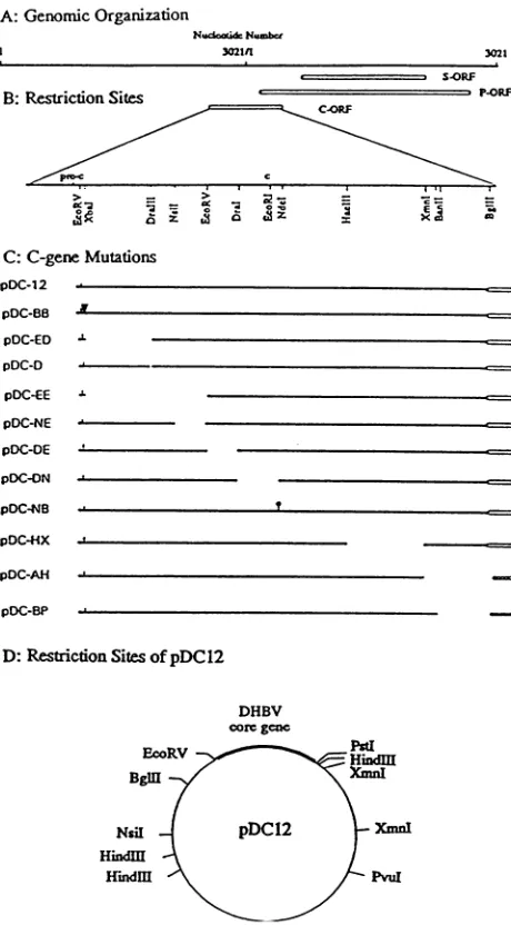
Construction of deletion and insertion mutations in the DHBV core gene. Plasmid pDC12containing DHBV 76 (25) coregene wasgenerated bycloningtheEcoRV-BglII fragment (nucleotides [nt] 2654 to 391 in the DHBV 76 genome) into EcoRV and BamHI sites ofpBV220. The DHBV coregene fragmentwaspurified from plasmid pDD81, which containeda head-to-tail tandem EcoRI dimer of the DHBV 76 genome. Accordingto the DNA sequences of DHBV 76 (25) and the expression vector pBV220 (30), this plasmidwas expected to express a256-amino-acid polypeptide identical in its primary sequence tothatpartof the naturalproductbecausetherewas a3-amino-acidinsertion(HKC) before the N-terminal aspartic acid residue (the second amino acid of the DHBV core protein) and a 5-amino-acid deletion (PSPRK) at the C terminus of the DHBV core protein, replaced by 11 amino acids(VDLQPSFCFGG) with the cloningstrategy(Fig. 1).All plasmidsexpressingdeleted and inserted DHBV coreprotein mutants weregenerated on the basis ofplasmid pDC12.
To construct aseries of deletion mutation which would cover most parts of the DHBV C open reading frame, we used different restriction sites scattering within this gene (Fig. 1) 338
on November 9, 2019 by guest
http://jvi.asm.org/
A: GenomicOrganization
1dcad&N.-6cr
I 30211
S.ORF B:RestrictionSites
C.ORF
w 4 - gc
> _ 2E
4 a a
C:C-geneMutations
_
,l
pDC-BB pDC-88
pOC-ED pDC-D
pOC-EE
pDC-NE pOC-DE
pOC-ON pDC-NB
PDC4HX
pDC-AH
pDC-BP
D:RestrictionSitesofpDC12
DHBV coregene
EcoRV
Bgl
Nsil
Hindl
HImdM
FIG. 1. Schematic view of the DHBV genomic organization and
the mutations of the core gene constructed in this study. (A) The
genomic organization of DHBV 76 (25) contained as a head-to-tail
dimer in plasmid pDD81 (see Materials and Methods). Nucleotide
numbers follow the numbering system of Mandart et al. (14). ORF,
openreading frame. (B)Restrictionsites in thecoregene relevantfor
the construction of the mutations. (C) Construction of various core
genemutations. Symbols:I, insertion of threeaminoacids(HKC); V, insertion offour aminoacids (SGSA);t,insertionoffour aminoacids
(GIPY). The 5aminoacidsattheC terminus(PSPRK)werereplaced
byanonspecific 11amino acids (VDLQPSFCFGG)IE1,by 6 amino
acids (SFCFGG) (011111), orbysevenaminoacids (PSFCFGG) (_).
Other positions of deleted amino acids in the deletion mutants and
sequences encoded by linkers are listed in Table 1. (D) Restriction
sitesofpDC12.
and three types of linkers (10-ntBamHI linker,
CGGGATC-CCG; 8-nt BamHl linker, CGGATCCG; 8-nt XhoI linker,
CCTCGAGG) in some mutations for maintaining in-frame
deletion or insertion. All mutations were confirmed by DNA
sequencing with Sequenase(United StatesBiochemical Corp.,
Cleveland,Ohio).
InpDC-ED, theEcoRV-DraIII fragment(nt 2654to 2805 in
DHBV 76) was deleted. This plasmid was constructed by
cutting
pDC12
with Dralll(nt 2805 in DHBV 76, the unique site in pDC12); removing protruding 3' termini withT4 DNA polymerase (19); cutting with PvuI (nt 969 in pBV220, the unique site inpDCl2
[Fig. ID]), purifying the DraIII-PvlI fragment containing the central and C-terminal regionsof the DHBV core gene, and subcloning into EcoRV (nt 2654 in DHBV) andPvuI
(nt 969 in pBV220) sites of pDC12.In pDC-D, one amino acid (phenylalanine, aminoacid 53 of the DHBV core protein) was deleted by cutting pDCl2 with
DraIll
(nt 2805 in DHBV 76, the unique site in pDC12), removing protruding 3' termini with T4 DNApolymerase, and recircularizating with T4 DNA ligase.In pDC-EE, the EcoRV-EcoRV fragment (nt 2654 to 2913 in DHBV 76) was deleted. It was constructed by cutting pDC12 with EcoRV, purifying the large fragment, and insert-ing a 10-nt
BamHI
linker sequence (CGGGATCCCG) in this site.In pDC-NE, the
NsiI-EcoRV
fragment (nt 2849 to 2913 in DHBV 76) was deleted. This plasmid was generated by partially cutting pDC12 withNsiI (two sites in pDC12: nt2894 in DHBV 76 and nt 3071 in pBV220), removing protruding 3' termini with T4 DNA polymerase, cutting withPvlI (nt969 inpBV220),purifying the large vector-containingNsiI-PvuI frag-ment (nt 2849 in DHBV 76 to nt 969 in pBV220) acted as subclone vector, cutting pDC12 with EcoRV (two sites in pDC12: nt 2654 and 2913 in the DHBV 76 core gene) and
PvuI,
purifying the EcoRV-PvuI fragment (nt 2913 in DHBV 76 to nt 969 in pBV220) as the insert fragment, andsubcloninginto the
NsiI-PvuI
vector fragment.In pDC-DE, theEcoRV-DraI fragment (nt 2913 to 2961 in DHBV 76) was deleted. It was generated by a method involv-ing bacteriophage M13 (26). Briefly, the pDC12 fragment of
XbaI-PstI
(nt 2662 in DHBV 76 and nt 26 in pBV220; both were unique sites in pDC12) was cloned into the unique sites ofXbaI andPstI
of M13mp18and M13mpl9,respectively.The single-stranded DNA of recombinant M13mp18 andM13mp19 was purified; it contained the complementary sequence be-tween the two ends of polylinker including insertedXbaI-PstI fragment. These two kinds of M13 single-stranded DNA were annealed in 0.2 M NaCI for 4 h at 62°C to form a partiallydouble-stranded 0-form structure in which both EcoRV and Dral were unique sites in this double-stranded DNA region. pDC-DE was constructed by digesting the 0-form structure with EcoRV and
Dral
to remove theEcoRV-DraI fragment(nt 2913 to 2961 in DHBV 76), inserting aXhoI linker (CCTCGAGG) to ensure that the right C open readingframefollowed,
recutting with
XbaI
and PstI, and recloning intoXbaI and PstI sites of pDC12.In pDC-DN, the DraI-NdeI fragment (nt 2961 to 18 in DHBV 76) was deleted. This plasmid wasgenerated bycutting pDC12 with
NdeI
(unique site in pDC12), filling in the overhangs with Klenow enzyme, recutting with BglII (nt 3402 in pBV220; unique site in pDC12), purifying the largerNdeI-BglII
fragment which acted as vector (see Fig. lB and D for positions of NdeI and BglII), purifying the small 673-bp fragment for recutting with Dral (nt 2961; unique site in this 673-bp fragment), and recloning the DraI-BglII fragment into theNdeI-BglII
large fragment, resulting in anin-frame deletion of 78-bp.In pDC-HX, the
HaeIII-XmrnI
fragment (nt 164 and 309 in DHBV 76) was deleted. It was generated as pDC-DE, that is, by first generating the M13 0-form structure containing the double-stranded XbaI-Pstl fragment of pDC12, digesting this form with HaeIII and XmnI (both were unique sites in this double-stranded DNA region), inserting a 10-ntBamnHI linker (CGGGATCCCG), and recloning the deletedXbaI-PstIfrag--m
.L
J. -4c==
--C===
I
on November 9, 2019 by guest
http://jvi.asm.org/
[image:2.612.65.295.71.487.2]340 YANG ET AL.
mentinto the corresponding position of pDC12, resulting in an in-frame deletion of 135 bp.
In pDC-AH, theXmnI-HindIII fragment (nt 309 in DHBV 76 to nt 30 in pBV220) in pDC12 was deleted. It was constructed by purifyingXmnI-PvuI (nt 309 in DHBV 76 to nt 969 inpBV220), the large fragment of pDC12 which acted as vector (see Fig. 1B and D for positions of XmnI and PvuI),
purifying theHindIII-PvuI fragment (nt 30 to 969 in pBV220; the overhangs of HindIll have been filled with Klenow
en-zyme), and recloning into the XmnI-PvuI large fragment of
pDC12, resulting inanin-frame deletion with 6(SFCFGG) of 11 nonspecific amino acids coming from the vector at the C terminus of thismutant.
InpDC-BP, the BanII-PstI fragment (nt 336 in DHBV 76 to nt26 inpBV220) was deleted. pDC12 was digested with BanlI
(nt336 and 358 in DHBV 76; no site in pBV220) and PstI (nt 26 in pBV220; unique site in pDC12), protruding 3' termini were removed withT4DNApolymerase, and the plasmid was recircularizated withT4 DNA ligase resulting in an in-frame deletion with seven nonspecific amino acids (PSFCFGG) deriving from thevector atthe C terminus of this mutant.
Twoinsertion mutations, pDC-BB and pDC-NB, of DHBV coregenewereconstructed bydigesting pDC12 withXbaI or
NdeI, respectively(both of which were unique sites in plasmid
pDC12), filling in the overhangs with Klenow enzyme, and
insertingan 8- or10-ntBamHI linker (CGGATCCG, CGGG
ATCCCG), respectively, resulting in insertion of four amino
acids,SGSAorGIPY in the coreprotein mutants encoded by
pDC-BB orpDC-NB, respectively.
Expression ofDHBVcore gene mutants andsucrose gradi-ent sedimentation analysis. Transformed E. coli DHScx was inoculated into LB medium in the presence of 50 ,ug of
ampicillin per ml, grown at 32°C overnight, diluted with
high-expression medium (3.2% tryptone, 2% yeast extract, 1x M9salt, 0.1 mMMgSO4, 0.001mMFeCI3, 50 ,ug of ampicillin perml),andgrownasabove untiltheoptical densityat600nm
(OD600)
was 0.6. The cultures were induced by shifting the temperatureto42°C for4to5 hfor expression. The cell pellets were lysed with lysis buffer (50 mM Tris hydrochloride [pH8.0], 1 mMdisodiumEDTA, 100mMNaCI,1% Triton X-100, 1 mg of lysozyme per ml) at 0°C for 30 min, and DNase I, RNaseA, andMgCI2wereadded to final concentrations of 50
U/ml, 10
jig/ml,
and 50 mM, respectively. The mixture was incubated at roomtemperature for 15 min. After the cellular debris had been removed by centrifugation, the supernatant wassubjectedto asedimentation in a linear 10 to50% sucrosegradient(SW27rotor at100,000 xgfor 3 h at4°C).Fractions were monitored for DHBV core protein by sodium dodecyl
sulfate-polyacrylamide gel electrophoresis (SDS-PAGE) and Westernimmunoblotting (see below).
PolyclonalrabbitantibodytoDHBVcoreparticles. DHBV coreparticleswerepurifiedfromDHBV-infected duck liver by the gradient centrifugation method described previously (8).
Briefly, theinfected duck liver washomogenized in buffer (10 mMTrishydrochloride[pH 7.6], 1 mM disodium EDTA, 0.3%
[vol/vol]
1-mercaptoethanol).
The celldebris were removed by twocentrifugationsteps at 12,000 x gfor 30 min at 4°C. The core particles were collected by ultracentrifugation (SW40 rotor;100,000 xg for4h at4°C), suspended in buffer (10 mM Tris hydrochloride [pH 7.6], 100 mM NaCI, 1 mM disodiumEDTA, 0.3%
[vol/vol]
3-mercaptoethanol,
1% Nonidet P-40), and thenappliedto a5to45% linear sucrose gradient. Afterbeing centrifuged for4 h at 50,000 x g in an SW 40 rotor, fractions were monitored by endogenous DNA polymerase assay. Peakfractions were pooled and precipitated by a third
centrifugation (SW40 rotor; 100,000 x g for 10 h at 4°C).
Purified DHBV core particles (200,ug)were emulsified with complete Freund's adjuvant and injected intradermally at several sites onthe back ofaNewZealand White rabbit. After being given two intramuscular boosters with emulsion of DHBV core particles (200 jLg each time) and incomplete
Freund's adjuvant, the animal was bled to obtain antiserum. Transmission electron microscopy. Theinduced E. coli cells expressing different DHBV core protein mutants were fixed overnight at 4°C in 2.5% glutaraldehyde-2%
paraformalde-hyde. The fixed bacteria were rinsed with 0.1 M phosphate(pH 7.3), postfixed with 1% OS04, rinsed again, dehydrated in a graded series of ethanol, cleared in propylene oxide, and embedded in Epon 812. Thin sections (silver grey) were cut, stained with uranyl acetate and lead citrate, andexamined with a H-800 transmission electron microscope.
SDS-PAGE and Western blotting. Bacterial suspensionsor partially purified recombinant proteins were solubilized with an equal volume of 2 x sample buffer (100 mM Tris hydro-chloride [pH 6.8], 5% SDS, 10% [vol/vol] 3-mercaptoethanol, 20% glycerol, 0.02%bromophenol blue). Samples were heated for 5 min at 100°C and centrifuged, and the supernatantwas applied to the gel. SDS-PAGE was performed as previously described (13) on a 15% resolving gel. For Western blotting, the proteins on the SDS-PAGE gel were transferred electro-phoretically to a nitrocellulose (NC) membrane and examined for DHBV core antigen by using a polyclonal rabbitantibody raised against the purified DHBV core particles (see above).
Agarose gel electrophoresis of DHBV core particles. The supernatant of the bacterial lysate containing DHBV core protein mutants was analyzed on a capsid agarose gel (3, 21a). The samples weremixed with 5 x loading buffer (50% glycerol, 0.02% bromophenol blue) and loaded onto a 1% agarose gel with ethidium bromide (0.5 ,ug/ml). The gel was run in lx TAE buffer (19) or in 10 mM sodium phosphate buffer (pH 7.5), and the gel was detected under UV light. For immuno-staining, the protein was capillary transferred to NC and detected with DHBV core protein antibody.
Nucleic acid binding assays. Nucleic acid binding activities of the expressed DHBV core protein mutants were assayed by the Southwestern (DNA-protein) blotting method (6) with minor modification. Bacterial suspensions were incubated in SDS sample buffer plus 3-mercaptoethanol for 10min at 60°C and then separated by SDS-PAGE (15% running gel). After incubation of the gel for 4 to 6 h at room temperature in refolding buffer (10 mM Tris hydrochloride [pH 7.5], 1 mM disodium EDTA, 50 mM NaCI, 1 mM dithiothreitol, 4 M urea), proteins were electrophoretically transferred to an NC membrane in 25 mM Tris hydrochloride [pH 8.3]-192 mM glycine buffer. NC membranes were presaturated in binding buffer (10 mM Trishydrochloride [pH 7.5], 0.1 mM disodium EDTA, 1 mM dithiothreitol, 125 mM NaCI, 2x Denhardt's solution) overnight at 4°C and then incubated for 1 h at 30°C in bindingbuffer containing 20 ng ofnick-translated plasmid pDD81, which contained a dimer DHBV 76 genome, or pUC18 (approximately108 cpm/,ug) per ml. After incubation, the NC membranes were extensively washed in washing solu-tion (10 mM Tris hydrochloride [pH 7.5], 0.1 mM disodium EDTA, 50 mM NaCI, 0.5x Denhardt's solution) at 37°C, dried, and exposed to X-ray film.
RESULTS
Expression of theDHBV core protein mutants in E.coli. In this study, 12 plasmids with various deletion and insertion mutations in the DHBV C open reading frame were con-structed(Fig. 1). The deletion mutations covered 80% of this J. VIROL.
on November 9, 2019 by guest
http://jvi.asm.org/
CAPSID MUTANT ASSEMBLY IN DUCK HEPATITIS B VIRUS 341
p, x o :Z w w m a
04 m z a a z w m w
b I I I I I I I I I I
0 a a a a00) a
x 00a a a a a00 00
i
a Ad Av vf A v4 v v4 v4 0
1 1 1 1 1 1
-97 -66
-55
-42
-40
-31
-21
0mpaDa z m of~~~p
ffi m wW z 0 ¢ g g m a CIA
I I IIII I I I I r4 el
a a a a 0aa a a a a a
I I I I I I I I I I I I
-66
-55
-42
-40
-31
A
B
[image:4.612.318.561.77.204.2]1 234 S67 9 101112
FIG. 3. The expressed DHBV core protein mutants encoded by pDC-ED and pDC-EE inhibited the growth of E. coli. The
trans-formedcellswere grownovernightat32°Cin LBcontaining ampicillin (50 ,ug/ml).Theculture mediumwasdiluted 1:100into20 ml offresh LB-ampicillin broth andgrown at 32°C.The
0D1(6
was determined every h. When theO1360
wasaround 0.2(A)or0.6(B),2 ml of the cultures was removed, added to one-third volume ofLB-ampicillinbroth at 65°C, and grown at 42°C for 5 h. Then the OD600 was
measured. Symbols: _,
OD600
beforeinduction; WA,OD60,)after5h of induction; -, beginning of induction. Bars: 1, pDC-ED; 2,
pDC-EE; 3, pDC-NE; 4, pDC-DE; 5, pDC-DN; 6, pDC-HX; 7, pDC-AH; 8, pDC-BP; 9, pDC-BB; 10, pDC-NB; 11,pDC-12; 12,E.
coli DH55a.
tteS:-^ies-i>e.;.
;^,syY,*-,,,;
ffw:: ;-.S
:
7' * _ w^
-!+:-
+tz:fifrti
StES --:o:
<;tF * Ty0%00500
-21
-14
FIG. 2. Expression of different DHBVcore protein mutantsin E. coli. Freshlyinduced E.colicellscontainingtheindividualplasmidwith the core genemutation were lysedwith SDSbuffer and analyzed by SDS-PAGE (15% polyacrylamide) followed by staining with Coo-massie blue (A) ortransferred ontoan NCfilter and reacted with a
polyclonalrabbit anti-DHBVcoreparticle antibody (seeMaterials and Methods)for Western blotanalysis (B).Sizesareshown in kilodaltons.
gene. Expression of these 12 DHBVcoreproteinmutantswas
induced by raising the temperature of the culture, which caused inactivation of a temperature-sensitive repressor
en-coded by the phage X c1857 gene.The predicted sizes of the
expressedDHBVcore proteinmutants,accordingtodeduced amino acid sequences, were detected in an SDS-PAGE gel stained with Coomassie blue (Fig. 2A) and confirmed by a Western blot (Fig. 2B) with polyclonal rabbit antibody raised against the core particles purified from DHBV-infected duck liver(see Materials and Methods).
It was interesting to find that there were no apparent expressed protein bands for plasmidspDC-EDandpDC-EEin Coomassie blue-stained SDS-PAGEgels (Fig. 2A). However, the results ofWestern blot (Fig. 2B) and DNA-binding (see Fig. 6A) studies demonstrated that the DHBV core protein
mutants were indeed expressed in the bacteria harboring pDC-ED and pDC-EE, respectively, but were in a state of degradation compared with the other DHBV core protein
mutants. On the otherhand,asshown inFig. 2A, theamounts
of totalproteinsinthe bacteriallysates containing pDC-EDor pDC-EE were smaller than those in the other strains. This
indicated that the mutatedDHBVcoregene in pDC-EDand pDC-EEorexpressed productswould be toxictothe host cells. By using an assayofbacterial growth rate, we demonstrated that it was the expressed products encoded by pDC-ED or
pDC-EE which were responsible for this effect, because the
growth rate of E. coli carrying these two plasmids began to
decreasejust after inductionat42°C (Fig. 3). This inhibition by expressed productswas moreapparent attheearlyexponential growth phase of the host cells (compare Fig. 3A and B). In pDC-ED and pDC-EE, most of the N-terminal sequence of the DHBVcoreproteinwasdeleted; that is, the regions coding for amino acids 3to 53 and 3to 89 of the core protein were deleted in pDC-ED and pDC-EE, respectively (Fig. 1;Table 1). However, deletion of the region coding for amino acids 67
to 89 inpDC-NE did not result in eitherdegradation of the
core products or inhibitory effects on bacterial growth as in pDC-ED andpDC-EE (Fig. 2 and 3; alsoseeFig. 6A). These datasuggested that the existence of the N-terminal sequence, probably within amino acids 3to66,wasnecessarytomaintain the basicstructureand function of the expressed DHBVcore protein in E. coli and presumably was important for the
self-assembly processof the DHBVcore particle.
Transmission electron micrographs of thin sections of E. coli expressing different DHBV core protein mutants. In a preliminary experiment, we noted that the DHBV core-like
TABLE 1. Deleted amino acidsequencesand insertionsatdeletion sitescausedbydeletion orinserted linker
Plasmid Positionofdeletion Insertionatdeletionsite
(amino acid)
pDC-ED 3-53 No insertion
pDC-EE 3-89 RDPD
pDC-D 53 No insertion
pDC-NE 67-89 N
pDC-DE 89-105 SSR
pDC-DN 106-131 Noinsertion
pDC-HX 181-228 GIP
pDC-AH 229-262 Nonspecific"
pDC-BP 237-262
Nonspecific'
"Sixnonspecificaminoacids(SFCFGG)attheC terminus.
"Sevennonspecificaminoacids(PSFCFGG)atthe C terminus.
A
B
1.4 1.0 0.8
0.6-0.4 fl
0.2-0.0
1 2 3 4 S6 789101112
VOL.68, 1994
on November 9, 2019 by guest
http://jvi.asm.org/
[image:4.612.319.561.595.701.2]342 YANG ET AL.
particles
can beseenin the transmission electronmicrograph
of E. coli thin sections
harboring
pDCI2.
These sphericalparticles
with uniformmorphology arranged
in acrystalline
model,
located almost at the ends of E.coli,
are easy torecognize.
Thisunique
feature would undoubtedly facilitate the studies of theself-assembly ability
of the DHBV coreprotein
mutants. For this purpose, the pellets of freshlyinduced bacteria expressing different DHBV core protein mutants were directly fixed, dehydrated, and embedded in
Epon
812. Afterbeing
stained with uranyl acetate and leadcitrate,
the thin sections of E. coli were examined under a transmission electronmicroscope.Among12DHBVcore
protein
mutants,3(pDC12,
pDC-BB,and
pDC-BP)
self-assembled toform the core-likeparticles
in E.coli.(Fig.
4, panels 2, 3,and13).Thecoreparticlesassembledby
the differentmutantsshowed similarsize, averaging28nmin diameter. Unlike pDC12and pDC-BB, pDC-BP lost the crys-tallinearrayability
inE. coli. The mutationsin theseplasmids
werelocatedatboth 5' and3' endsof the Copenreadingframe
(Fig. 1).
Itwasnotable forpDC-AH,in which thedeletion ofan additional 8 amino acids based onpDC-BP
(amino
acids237to 262 deleted inpDC-BP) (Fig.
1;Table1)
largely changed themorphology
of assembledparticles.AsshowninFig. 4, panel 12,manysmall
particles
with adiameter ofca. 20nmaccumulatedrandomly
attheends ofE. coli.In contrast,other DHBVcoreproteinmutantscouldnotall form the
typical
coreparticles,
whichwerereplaced by
inclu-sion bodies inE.coli(Fig.
4, panels4to11).
Thesecoreproteinmutants contained deletion mutations in the N-terminal and central
regions (pDC-ED, pDC-D, pDC-EE, pDC-NE,
pDC-DE, pDC-DN,
andpDC-HX)
or aninsertion mutation in the centralregion (pDC-NB) (Fig.
1; Table1).
Compared withpDC12,
whoseproducts
have theself-assembly
ability in E.coli,
there wasonly
one amino acid deleted(phenylalanine,
residue 53 of the DHBV core
protein)
or four amino acids inserted(GIPY
inserted between residues 132 and 133 of thisprotein)
in the mutants encodedby
pDC-D and pDC-NB,respectively. However,
these small mutations resulted in the failureof the formation ofthe core particlesin E. coli.To support the results of
electron-microscopic observation,
we tried to
partially purify
expressed DHBV core proteinmutants from E. coli for the purpose of
performing capsid
agarose
gel
and sucrosegradient
sedimentationanalysis.
We found thatlysozyme-detergent
treatment cannot releaseex-pressed products
into the soluble supernatant of bacteriallysate; however,
after thelysate
wasdigested
withDNase Iand RNaseA,
theexpressed products
were released from the insoluble materials. A Western blot of SDS-PAGE gelsshowed lower molecular
weights, compared
withpredicted
sizes,
for the core mutants which did not form core-likeparticles
underelectron-microscopic observation;
theproductsencoded
by N-terminally
deletedmutants,pDC-ED
andpDC-EE, nearly
disappeared (data
notshown).
Theresultsindicated thatproteolysis
had occurredduring
the nuclease treatment. On theotherhand,
alarge proportion
of releasedcore mutants encodedby pDC-12, pDC-BB,
pDC-BP,andpDC-AH,which allformed electronmicroscopicallyobservablecore-likeparti-cles in E.
coli,
still located at the positions with predictedmolecular
weights
after nuclease digestion (data not shown),suggesting
thatthe assembled state ofthese proteinsmay beresponsible
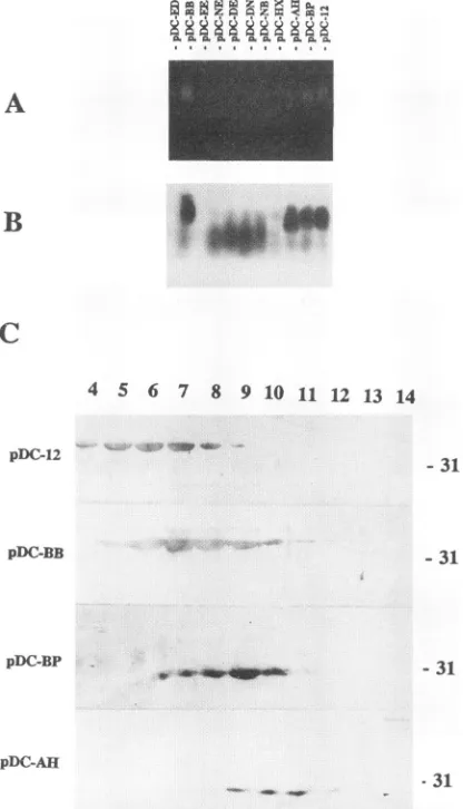
for this resistancetoproteolysis.For
capsid
agarose gel analysis, the lysate supernatant of different core mutants was used. Ethidium bromide-stained bands were seenonly
in the samples of bacteria harboringpDC12, pDC-BP, pDC-BB,
and pDC-AH(Fig.
5A).Accord-ing
tothedatareported previously (3),these bandswere mostprobably RNA encapsidated in core particles, which were resistant to treatment with RNase A. To confirm that these staining bands indeed represented core protein, the gel was transferredtoNC and the filter was detectedby immunostain-ing. The result showed thatcore proteinswere locatedat the same positions as that of ethidium bromide-stained bands, whereas othermutantsdisplayed thesmearbandsatthe lower position (Fig. 5B).
The lysate supernatants of the four strains capable of self-assemblingcore mutants weresubjectedto sucrose gradi-entsedimentation (see Materials and Methods). All exhibited sedimentation velocities indicative of theirparticulate nature (Fig. SC). The mutants encoded by pDC12 and pDC-BB sedimented faster than the mutants coded by pDC-BP and pDC-AH.
Taken together, these data revealed directly that both termini of DHBVcoreproteincan tolerate some insertion and deletion mutations which do not block the formation ofthe core-likeparticles inE. coli; however, deletion orinsertionin N-terminal and central regions of this protein strongly or entirely inhibits this assembly process.
Localization of a DNA-binding domain in the DHBV core protein.TheHBV coreprotein has nucleic acid-binding activity (3, 6, 9,15,17).The functions of thisactivity involve thestability ofcoreparticles (3) and details of viral genome replication(9). In DHBV coreprotein,manyof the positively charged arginine residues (19 of 29) are scattered in the C-terminal 84-amino-acid region. This Arg-rich region might have a nucleic acid-binding function. Todemonstrate this function and locate the possible binding domain in the DHBVcore protein,we used Southwesternblotting to detect DNA-binding abilities of the differentcoreproteinmutants.AsshowninFig.6A,allDHBV coreproteinmutantsboundeffectivelytothe denatured pDD81 DNAprobe (see Materials and Methods), except the mutant encoded by pDC-HX, in which amino acids 181 to 228 were deleted(Fig. 1; Table 1). The result suggested that amino acids 181 to228of DHBV coreprotein contained, atleastinpart, a DNA-binding domain.
Thebinding of the expressedHBVcore protein by cellular nucleic acid (3) indicates that this binding ability of hepadna-virus core proteinwas not specific toviral nucleic acid itself. The results shown in Fig. 6B, lane 1, demonstrate that the expressed DHBVcoreprotein (encoded by pDC12) canbind a nonspecific DNA probe, pUC18, as effectively as pDD81, which contained a head-to-tail double-copied DHBV DNA. The binding with pUC18can be blockedcompletely by dena-tured salmonspermDNA (Fig.6B, lane 2).
DISCUSSION
This article presents the consequences ofdifferent DHBV core protein mutants on the self-assembly and nucleic acid bindingofthis virus.
The fact that core-like particlescanbe assembledin E. coli (3, 4, 6) indicates that the core protein itself contains all information necessary for the self-assembly process. As the basis of the assembly which would involve intermolecular recognition and interaction, intramolecular folding of individ-ual molecules must happen first to form a suitable tertiary structure. The results reported here suggest that the N-terminalregion of theDHBV coreprotein is important for its
tertiary structure. The DHBV core mutants with a deleted N-terminalregion apparently inhibited thegrowth ofthe host cells more than did the other core mutants with central or C-terminal deletions.Thisfunctional difference caused by the expressed products indicates thatastructuralvariation proba-J. VIROL.
on November 9, 2019 by guest
http://jvi.asm.org/
CAPSID MUTANT ASSEMBLY IN DUCK HEPATITIS B VIRUS 343
iD910
a
%
W
i .E
_
.E P.
_~~1
2E^ wi}].
13
_FIG4 Tasisso elcto mirorah ofthi
setoso_.cl
xrsigtedfeetmuat
fDBoepoenrslnue
E.coicel wr fie
oengh
at4C i2.5%guaadhd-%prfradhd.
Th ie el eerne n hsht p.)
otiein1sicai t
omtmprtr1fr2h
r7e
nhshte(H73)aan
dhdaedi rae eie fehao,ceaenprpln
oxd,ademeddiiEo
1.Thnscins(ivrgry,eecu,sand ihuaylaeaean
edctrt_adexmndude_ -0Magifcaton
3000 ar 0 m __ ,VOL.68, 1994
on November 9, 2019 by guest
http://jvi.asm.org/
344 YANG ET AL.
A
B
A
B
C
4 5 6 7 8 9 10 11 12 13 14
PDC-12
pDC-BB
pDC-BP
--31
-31
-31
pDC-AH
[image:7.612.70.278.75.438.2]31 FIG. 5. Capsidagarosegel andsucrosegradient sedimentation
anal-ysesof bacterial lysates. The bacteriaexpressing different DHBVcore
proteinmutantswerelysed with lysozyme-detergent anddigested with
DNaseIand RNaseA(see Materials and Methods). After centrifuga-tiontopelletthecellulardebris, thesupernatantoflysateswasloadedon
1%agarosegelcontaining 0.5 pLgofethidium bromidepermlandrun
in TAEbuffer. Thegelwasdetected under UV light (A),orcapillary
transferred to NC for detection immunologically with DHBV core
protein antibody (B). The supernatant of bacteria harboring pDC12, pDC-BB, pDC-AH, and pDC-BPwassubjectedtoalinear 10to50%
sucrose gradientsedimentation (SW27 rotor; 100,000 x g for3 h at
4°C). The core protein content of each fraction (from the bottom fraction 1 tothe topfraction 16)was analyzed by SDS-PAGE (15%
running gel) followed by Western blot. Only the region of the gradient where DHBVcoreprotein mutantsweredetected is shown (C). The positionof the31-kDa marker isindicatedtotheright ofpanelC.
bly occurred in the N-terminal deleted DHBV core protein
mutants.
Thefinding by Birnbaum andNassal (3) that the C-terminal 39residues of the HBVcoreprotein is dispensable for capsid
formation in E. coli prompted us to address whether DHBV
coreproteincantolerateanN-terminalorcentralmutation for assembling the capsid. The results of electron microscopy, capsid gel, and sucrose gradient centrifugation demonstrate
thatasmall-fragmentinsertion atthe N terminusaswell as a
deletion in the C-terminal region can be tolerated in the
assembly process. Deletion or insertion in most of the
N-n mwmrwA inwwzzazmS A
n m 0 e
Pt A , P,N,AvAA A Q
g31 *
21-FIG. 6. DNA-binding activities of DHBV core protein mutants
expressedin E. coli. (A)Southwestern blot resultsfor differentcore
protein mutants.Thesuspensions of bacteria induced for expressing different mutants were lysed with an equal volume of 2x SDS reducing buffer (see Materials and Methods)at60°C for 10 minand analyzed by SDS-PAGE (15% running gel),and thegelwasincubated
atroomtemperaturefor4to6h inrefolding buffer.Proteinswerethen
electrophoreticallytransferredtoanNC membrane. After presatura-tioninbinding buffer,themembranewasincubatedat30°Cfor 1 h in the same buffercontaining 20ng of nick-translated plasmidpDD81 (denatured, approximately 108cpm/p.g)perml, extensivelywashed in
washing solution, dried, and exposed to X-ray film. (B) Inhibitory effectsofsalmonspermDNAon thebinding activityofcore protein (expressed by pDC12). The denatured plasmid pUC18 (ca. 108cpm/ ,ug, 20 ng/ml in the binding buffer) was used in lanes 1 and 2 to
determine a nonspecific nucleic acid-binding ability of DHBVcore
protein.Ahigh concentration of denatured salmonsperm DNA(500 xg/ml in binding buffer) was added during both presaturation and DNAbinding (lane 2).
terminal and central regionsofDHBV coreprotein reported in this study stronglyorcompletely inhibited its self-assembly in E. coli. This finding was supported by the data on the sensitivitytoproteolysis ofthe DHBV core mutantsduring the partialpurification;thatis,duringtreatmentwith DNaseIand RNase A, all of theDHBV core mutants,which didnotshow core-like particles under electron microscopy, degradated to
positions on the SDS-PAGE gel smaller than the predicted
sizes. This resultwasconsistent with the datareported by Zhou et al. (32)thatprotease treatmentcleaved unassembledHBV core proteintosmallerproducts.
Yuand Summers(29) reported the effects of four classes of DHBV core protein C-terminally truncatedmutants on viral DNAmaturation and virus assembly. Viral DNAsynthesis in class III and IV mutants was reduced 10-fold or severely defective, respectively (29). pDC-BP reported here fell near theboundary between classIIIandIV mutants;pDC-AHwas aclassIV mutant.Themorphology of the assembledparticles
in E. coli harboring pDC-BP orpDC-AH was different from that of particles encoded by pDC12. Consistently,
immuno-stainingand ethidiumbromidestaininginacapsidagarosegel
(Fig.5)showedabandofproductencodedbypDC-AH witha somewhat lower position than the others, indicating that structuralchangeshadoccurredfortheseparticles.Ourresults supporttheconclusion that the stericconstraint of themutant
capsidsisoneof the reasonsresponsible forthe inhibition of viral DNAsynthesis.
InHBV, identification of thepossible amino acidsequences responsible for the binding between the core protein and nucleic acid was controversial (6, 15). Most studies (3, 6, 9)
suggest that the C-terminal arginine-rich region is a nucleic acid-binding domain and that this bindingcontributes
impor-J. VIROL.
KUiik.ukxi
. . . 4 4 . I . 4 . .on November 9, 2019 by guest
http://jvi.asm.org/
[image:7.612.337.521.76.206.2]tantly to HBV capsid stability (3) and viral DNA replication (9). Here, we detected a nucleic acid-binding activity of DHBV coreprotein by the Southwestern blot method (Fig. 6). On the basis of the results that most of this activity was lost in the mutant encoded by pDC-HX, whereas no other mutant showed a detectable change of the nucleic acid-binding activ-ity, we suggest that at least part of the nucleic acid-binding domainof DHBV core protein is located in the deletion region of pDC-HX (amino acids 181 to 228). The high content of positively charged amino acids (10 arginine residues and 5 lysine residues in this region of 48 residues) indicates that it may be a nonspecific binding secondary to interactions with negatively charged nucleic acids. The results of the binding studieswith pUC18 were consistent with this prediction (Fig. 6B). It was interesting that the C terminus of DHBV core protein does not involve this nucleic acid-binding activity, although eight arginine residues, two lysine residues, and two histidine residues are located in this 34-amino-acid region. These data might beinterpretedto indicate that the position of afragment in the core protein molecule (for example, on the surface or in the inner part) or some unknown secondary structurein the fragment may determine its role on the nucleic acid binding ofDHBV core protein. Moreover, the results of nonspecific nucleic acid binding of core protein in this study cannot eliminatethe possibility that some different but highly specific binding between the core protein and viral genome is involved in the encapsidation of theviral RNA pregenome.
ACKNOWLEDGMENTS
We thank Samuel L.Stanley,Jr.,and Ellen Liforhelp in prepara-tion of the manuscript. We thank Chang Zhiqin for kindly providing expression vector pBV220.
This work wassupported by the National Natural Foundation of China (grants 39000005 and 39100007).
REFERENCES
1. Argos, P., and S. D.Fuller. 1988. A model for the hepatitisBvirus coreprotein: prediction of antigenic sites and relationship toRNA viruscapsid proteins. EMBO J. 7:819-824.
2. Bartenschlager, R., M. Junker-Niepmann, and H. Schaller. 1990. The Pgeneproduct of hepatitisBvirus isrequired as a structural component for genomic RNA encapsidation. J. Virol. 64:5324-5332.
3. Birnbaum, F., and M. Nassal. 1990. Hepatitis B virus nucleocapsid assembly: primary structure requirements in the core protein.J. Virol. 64:3319-3330.
4. Cohen, B. J., and J. E.Richmond. 1982. Electron microscopy of hepatitis Bcore antigen synthesized in E. coli. Nature (London) 296:677-678.
5. Eckhardt, S. G., D. R. Milich, and A. McLachlan.1991. Hepatitis Bvirus core antigen has two nuclear localization sequences in the arginine-rich carboxyl terminus.J. Virol. 65:575-582.
6. Gallina, A., F.Bonelli, L. Zentilin, G. Rindi, M. Muttini, and G. Milanesi. 1989.Arecombinant hepatitis B core antigen polypeptide with theprotamine-like domain deleted self-assembles into capsid particles but fails to bind nucleic acid. J. Virol. 63:4645-4652.
7. Ganem, D., andH. E.Varmus. 1987. The molecular biology of the hepatitis B viruses. Annu. Rev. Biochem. 56:651-693.
8. Guo, J. T.,and H. S. Chen. 1990.Endogenous DNA polymerase activity in duck hepatitis B virus replicative complexes and its inhibition in vitro. Chin. J. Virol. 6:260-265.
9. Hatton, T., S. L. Zhou, and D. N. Standring. 1992. RNA- and DNA-bindingactivities in hepatitis B virus capsid protein: a model fortheir roles inviral replication.J. Virol. 66:5232-5241.
10. Hirsch, R., J. Lavine, L. Chang, H. Varmus, and D. Ganem. 1990. Polymerase gene products of hepatitis Bviruses are required for genomic RNA packaging as well as for reverse transcription. Nature (London)344:552-555.
11. Hirsch, R., D. Loeb, J. Pollack, and D. Ganem. 1991. cis-acting
sequence required for encapsidation of duck hepatitis B virus pregenomic RNA. J. Virol. 65:3309-3316.
12. Junker-Niepmann, M., R. Bartenschlager, and H. Schaller. 1990. A short cis-acting sequence is required for hepatitis B virus pregenome encapsidation and sufficient for packaging of foreign RNA. EMBO J. 9:3389-3396.
13. Laemmli, U. K. 1970. Cleavage of structural proteins during the assembly of the head of bacteriophage T4. Nature (London) 227:680-685.
14. Mandart, E., A. Kay, and F. Galibert. 1984. Nucleotide sequence of a cloned duck hepatitis B virus genome: comparison with woodchuck and human hepatitis B virus sequences. J. Virol. 49:782-792.
15. Matsuda, K., S. Satoh, and H. Ohori. 1988. DNA-binding activity of hepatitis B e antigen polypeptide lacking the protamine-like sequence of nucleocapsid protein of human hepatitis B virus. J. Virol. 62:3517-3521.
16. Nassal, M. 1988. Total chemical synthesis of a gene for hepatitis B virus core protein and its functional characterization. Gene 66: 279-294.
17. Petit, M.-A., and J. Pillot. 1985. HBc and HBe antigenicity and DNA-binding activity of major core protein p22 in hepatitis B virus core particles isolated from the cytoplasm of human liver cells. J. Virol. 53:543-553.
18. Pugh, J. C., A. Zweidler, and J. Summers. 1989. Characterization ofthe major duck hepatitis B virus core particle protein. J. Virol. 63:1371-1376.
19. Sambrook, J., E. F. Fritsch, and T. Maniatis. 1989. Molecular cloning: a laboratory manual, 2nd ed. Cold Spring Harbor Labo-ratory, ColdSpring Harbor, N.Y.
20. Schlicht, H.-J., R. Bartenschlager, and H. Schaller. 1989. The duck hepatitis B virus core protein contains a highly phosphory-lated C terminus that is essential for replication but not for RNA packaging. J. Virol. 63:2995-3000.
21. Schlicht, H. J., and H. Schaller. 1989. The secretory core protein ofhuman hepatitis B virus is expressed on the cell surface. J. Virol. 63:5399-5404.
21a.Summers, J. Unpublished data.
22. Summers, J., and W. S. Mason. 1982. Replication of the genome of a hepatitis B like virus by reverse transcription of an RNA intermediate. Cell29:403-415.
23. Tuttleman, J., C. Pourcel, and J. Summers. 1986. Formation of the pool of covalently closed circular viral DNA in hepadnavirus infected cells. Cell 47:451-460.
24. Wasenauer, G., J. Kock, and H. J. Schlicht. 1992. A cysteine and a hydrophobic sequence in the noncleaved portion of the Pre-C leader peptide determine the biophysical properties of the secre-tory core protein (HBe protein) of human hepatitis B virus. J. Virol. 66:5338-5346.
25. Yang, W. G.,H. S. Chen, Q. Jin, G. Hu, J. S. Yuan, and Y. D.Hou. 1992. Sequence analysis of duck hepatitis B virus genome of variant isolated fromchinese ducks. Chin. Sci. Bull.37:1126-1129. 26. Yang, W. G.,Y.Zheng, and H. S. Chen. A new method to generate specific deletion mutation by using bacteriophage M13. Chin. J. Virol., in press.
27. Yeh, C.-T., Y.-F. Liaw, and J.-H. Ou. 1990. The arginine-rich domain of hepatitis B virus precore and core proteins contains a signal for nucleartransport. J. Virol. 64:6141-6147.
28. Yeh, C.-T., and J.-H. Ou. 1991. Phosphorylation of hepatitis B virusprecore and core proteins. J. Virol. 65:2327-2331.
29. Yu, M., and J. Summers. 1991. A domain of the hepadnavirus capsid protein is specifically required for DNA maturation and virusassembly.J. Virol. 65:2511-2517.
30. Zhang, Z. Q. 1990.Construction of a procaryotic expression vector pBV220.Chin. J. Virol. 6:111.
31. Zhou, S. L., and D. N. Standring. 1992. Cys residues of the hepatitis B virus capsid protein are not essential for the assembly of viral core particles but can influence their stability. J. Virol. 66:5393-5398.
32. Zhou, S. L., S. Q. Yang,and D. N. Standring. 1992. Characteriza-tion of hepatitis B virus capsid particle assembly in Xenopus oocytes. J. Virol.66:3086-3092.