R E S E A R C H A R T I C L E
Open Access
Drug repurposing screen identifies lestaurtinib
amplifies the ability of the poly (ADP-ribose)
polymerase 1 inhibitor AG14361 to kill breast
cancer associated gene-1 mutant and wild type
breast cancer cells
Guelaguetza Vazquez-Ortiz
1, Cristine Chisholm
1, Xiaoling Xu
1, Tyler J Lahusen
1, Cuiling Li
1, Srilatha Sakamuru
2,
Ruili Huang
2, Craig J Thomas
2, Menghang Xia
2and Chuxia Deng
1*Abstract
Introduction:Breast cancer is a devastating disease that results in approximately 40,000 deaths each year in the USA. Current drug screening and chemopreventatitive methods are suboptimal, due in part to the poor specificity of compounds for cancer cells. Poly (ADP-ribose) polymerase 1 (PARP1) inhibitor (PARPi)-mediated therapy is a promising approach for familial breast cancers caused by mutations of breast cancer-associated gene-1 and -2 (BRCA1/2), yet drug resistance frequently occurs during the treatment. Moreover, PARPis exhibit very little effect on cancers that are proficient for DNA repair and clinical efficacy for PARPis as single-agent therapies has yet to be illustrated.
Methods:Using a quantitative high-throughput screening approach, we screened a library containing 2,816 drugs, most of which are approved for human or animal use by the Food and Drug Administration (FDA) or other countries, to identify compounds that sensitize breast cancer cells to PARPi. After initial screening, we performed further cellular and molecular analysis on lestaurtinib, which is an orally bioavailable multikinase inhibitor and has been used in clinical trials for myeloproliferative disorders and acute myelogenous leukemia.
Results:Our study indicated that lestaurtinib is highly potent against breast cancers as a mono-treatment agent. It also strongly enhanced the activity of the potent PARPi AG14361 on breast cancer cell growth bothin vitroand in vivoconditions. The inhibition of cancer growth is measured by increased apoptosis and reduced cell proliferation. Consistent with this, the treatment results in activation of caspase 3/7, and accumulation of cells in the G2 phase of the cell cycle, irrespective of their BRCA1 status. Finally, we demonstrated that AG14361 inhibits NF-κB signaling, which is further enhanced by lestaurtinib treatment.
Conclusions:Lestaurtinib amplifies the ability of the PARP1 inhibitor AG14361 to kill BRCA1 mutant and wild-type breast cancer cells, at least in part, by inhibiting NF-κB signaling. Each of these drugs has been approved for clinical trials for several different cancers, thus, their combination treatment should be applicable for a breast cancer trial in the future.
* Correspondence:[email protected]
1Genetics of Development and Disease Branch, National Institute of Diabetes,
Digestive and Kidney Diseases, National Institutes of Health, 9000 Rockville Pike, Bethesda, MD 20892, USA
Full list of author information is available at the end of the article
Introduction
Breast cancer is one of the most prevalent cancers in women worldwide and it is estimated that a million women will develop this disorder each year. About 8% of breast cancer cases are inheritable, associated with mu-tations of highly penetrant breast cancer susceptibility genes, such as breast cancer-associated gene-1 and -2 (BRCA1/2) and other tumor suppressor genes [1-7]. In addition, it has been estimated that BRCA1 mutation carriers have a 50 to 80% risk of developing breast cancer before the age of 70 years [8-11].
Both BRCA1 and BRCA2 play essential roles in many biological processes [12-17]. A common feature of BRCA1/ 2-associated tumorigenesis is massive genetic instability, primarily due to the fact that cells lacking BRCA1 or BRCA2 have impaired ability to undergo homologous recombination (HR) [15,16,18,19], therefore these cells cannot effectively repair HR-mediated DNA damage, including DNA double-strand breaks (DSBs). The genetic instability often leads to altered expression of many genes and signaling pathways making it difficult to inhibit tu-morigenesis and progression by targeting a single mo-lecular target. Recently, significant work in the area of synthetic lethality has led to new approaches for the treat-ment of BRCA1/2-deficient cancers using high efficacy poly (ADP-ribose) polymerase 1 (PARP1) inhibitors (PARPi) with high efficiency [20-24].
The PARP family plays important roles in DNA damage repair. For example, PARP1 is involved in the repair of DNA single-stranded breaks (SSBs) [25-27]. Inhibition of PARP1 activity could also result in DSB formation when unrepaired SSBs meet the replication fork, causing its collapse. Because BRCA1/2 mutant cells are defective in repairing DSBs, PARPi inhibition may result in accumu-lation of DSBs in these cells and eventually lead to apop-tosis. This may account for the molecular basis of why BRCA1- and BRCA2-deficient cells are extremely sensitive to PARPi [20-22]. However, it was shown that several cell lines derived from mouse BRCA1 mutant mammary can-cers [28] and a human pancreatic BRCA2 mutant cancer cell line [29] exhibited resistance to PARPi. While the exact cause for the resistance was unclear, it was hypothe-sized that some specific alterations/mutations might block the sensitivity of these cancer cells to PARPi [30]. It was subsequently demonstrated that resistance to PARPi could occur through multiple mechanisms (reviewed in [31]), such as impaired expression of 53BP1 [32,33], restoration of BRCA function [34], and induction of P-glycoprotein expression [35,36]. To overcome the resistance, combin-ational therapies using multiple chemotherapeutic agents have been used to enhance the ability to kill BRCA1/2-related cancers [35,36].
In theory, all clinically used drugs have effects on biological systems other than those for which they were
designed; therefore, drug repurposing consists of develop-ing new applications for existdevelop-ing drugs. There has been an increase in the interest of drug repurposing due to the high cost of drug development and time involved in bring-ing new drugs to the market [16,37]. It has been estimated that it costs approximately more than USD 800 million to
develop a new drug de novo and the time estimated to
develop a new drug that complies with the regulatory requirements for safety, efficacy and quality goes in the order of 10 to 17 years [38].
In this study, a drug repurposing approach using the National Institutes of Health Chemical Genomics Center (NCGC) Pharmaceutical Collection (NPC) [39], a library containing drugs approved for clinical use or that have been in clinical trials, was used to identify drugs that amplify the ability of AG14361, a potent PARP1 inhibi-tor [21], to inhibit the growth of both human and mouse breast cancer cells, irrespective of their BRCA1 status.
Methods
Cell lines and viral vectors
Our initial study for human cell lines was performed in three isogenic models derived from the primary cell lines: 92 J, MDA-MB-231 (American Type Culture Collection, ATCC) and T47D (ATCC) and their BRCA1 mutant sublines 92 J-sh-BRCA1, MDA-MB-231-sh-BRCA1 and T47D-sh-BRCA1 respectively. The 92 J cell line, which is derived from a xenograft tumor of MDA-MB-231, forms mammary tumors much faster than the parent MDA-MB-231 cells when implanted into nude mice.
BRCA1 short hairpin RNA (shRNA) constructs in the pLKO.1-based vector were obtained from Open Biosys-tems (GE Healthcare, Little Chalfont, UK). A control lenti-viral shRNA vector, packaging vector pCMV-dR8.2, and envelope vector VSV-G was obtained from Addgene (Cambridge, MA, USA). The BRCA1 shRNA construct (TRCN0000039837) was used to produce lentiviral parti-cles for generation of stable BRCA1 knockdown cells. Lentivirus was produced in 293 T cells and the media col-lected for later transduction of target cells. Cells were transduced with lentiviral supernatant and then selected
with 2 μg/ml puromycin to generate cells with stable
knockdown of BRCA1. The viral supernatant was used to infect 92 J, MDA-MB-231 and T47D cells
Mouse BRCA1 mutant cell line 69 derived from
mam-mary tumor ofBrca1Co/Co; MMTV-Cre;p53+/− mice
con-taining a targeted deletion of full-length BRCA1 [40] and BRCA1 wild-type (wt) cell line Ras, derived from mammary tumor MMTV-Ras mice [41].
Growth assays
For the growth curve assay, 5 × 10⁴cells were plated per
well of a six-well plate and medium was changed every
every 12 hr cells were detached by trypsinization and counted with a Z1 Coulter counter (Beckman Coulter, Brea, CA, USA). Plating for each time point was done in triplicate for each 92 J isogenic pair. In order to elimin-ate artifacts that could be produced by the cell line, we validated the other two pairs of human isogenic cell lines following the same protocol.
NCGC Pharmaceutical Collection (NPC) and quantitative high throughput screening
The NPC drug library consists of 2,816 small molecule compounds at the time of this screening [39]. Fifty-two percent of the compounds are drugs approved for hu-man or animal use by the United States Food and Drug Administration (FDA), 22% are drugs approved in Europe, Canada or Japan, and the remaining 25% are drugs approved in other countries or compounds that have been tested in clinical trials.
For the initial screening, the library was prepared as 15 inter-plate titrations, which were serially diluted 1:2.236 in dimethyl sulfoxide (DMSO, (Thermo Fisher Scientific, Waltham, MA, USA) in 384-well plates. The stock con-centrations of the test compounds ranged from 10 mM to
0.13 μM. Transfer of the diluted compounds from
384-well plates to 1,536-384-well plates was performed using an Evolution P3 system (Perkin Elmer, Wellesley, MA, USA). Each treatment plate included concurrent DMSO and positive control wells and concentration-response titrations of controls, all occupying columns 1 to 4. Cell viability was measured using a luciferase-coupled ATP quantization assay of metabolically active cells (ATPliteTM 1step Luminescence Assay System, Perkin Elmer). Cells were
dispensed at 2,000 cells/5 μL/well in 1,536-well white,
solid-bottom assay plates using a flying reagent dispenser (FRD). The assay plates were incubated at 37°C for 5 hr to
allow for cell attachment, followed by addition of 5μl of
compounds via pin tool. After compound addition, plates were incubated for 48 hr at 37°C. At the end of the
incu-bation period, 5 μL of ATPlite reagent was added, plates
were incubated at room temperature for 20 to 30 min, and luminescence intensity was determined using a ViewLux plate reader (Perkin Elmer).
Data analysis
Analysis of compound concentration-response data was performed as previously described [36]. Briefly, raw plate reads for each titration point were first normalized relative to the positive control compound (-100%) and
DMSO-only wells (0%) as follows: % activity = ((Vcompound –
VDMSO)/(Vpos–VDMSO)) × 100, where Vcompounddenotes
the compound well value, Vposdenotes the median value
of the positive control wells, and VDMSO denotes the
median values of the DMSO-only wells, and then correc-ted by applying a NCGC in-house pattern correction
algorithm using compound-free control plates (DMSO-only plates) at the beginning and end of the compound plate stack. Concentration-response titration points for each compound were fitted to a four-parameter equa-tion yielding concentraequa-tions of half-maximal activity
(AC50) and maximal response (efficacy) values. Compounds
were designated as class 1 to 4 according to the type of concentration-response curve observed [42,43]. Curve clas-ses are heuristic measures of data confidence, classifying concentration-responses on the basis of efficacy, the num-ber of data points observed above background activity, and the quality of fit. Compounds with class 1.1, 2.1, 1.2 or 2.2 (>50% efficacy) curves are considered active. Compounds with class 4 curves are considered inactive and compounds with all other curves classes are considered inconclusive. Compounds that were selectively active (showed a potency difference of >3-fold) in one cell line or with or without the combination compound were selected for confirmation and follow-up studies.
Determination of synergistic effect and additive effect The theoretical addictive effect of compounds with AG14361 was based on the fractional inhibition of these compounds when used separately. If the 50% inhibition
concentration (IC50) of each drug is administered
to-gether, by the union of two events, the predictive addictive
killing is calculated as Etotal= E1+ E2–E1× E2(where E1
is IC50of drug 1 and E2is IC50of drug 2), which is 75%.
This classifies a drug synergistic if, when treated with the 50% inhibition dose of each drug, the synergistic killing effect should be significantly greater than 75%.
Cell proliferation assays for validation of synergistic effect In order to validate the synergistic effect of the selected
drugs in vitro we performed cell viability assay using a
luciferase-coupled ATP quantization assay of metabolic-ally active cells (ATPliteTM 1step Luminescence Assay System, Perkin Elmer) in a 96-well plate and 3-(4,5-dimethylthiazol-2-yl)-2,5-diphenyl tetrazolium bromide
(MTT). For MTT, 1 to 2 × 10⁴ cells were plated per
one well of a 24-well plate. Target drugs at various con-centrations were dissolved in DMSO and then added to
the cells in 10% fetal bovine serum-containing Dulbecco’s
modified Eagle’s medium (DMEM), IC50concentration of
AG14361 were also added to each well. The final DMSO concentration was kept at 0.1% after the addition to medium. After 48 hr medium was removed and 0.3 ml of 0.1% MTT in phosphate-buffered saline (PBS) was added
in each well. After incubation for 30 min in a 37°C CO2
Histological and immunohistochemical analysis of tumor samples
For immunohistochemistry procedures, the tumors were fixed in phosphate-buffered formalin, embedded
in paraffin, cut in 4-μm thickness, and stained.
Immuno-histochemical analysis of proliferating cell nuclear antigen (PCNA) was performed using a labeled streptavidin-biotin technique described previously. Anti-PCNA monoclonal antibody PC 10 (Dako, Carpenteria, CA, USA), which reacts exclusively with nuclei, was used at a dilution of 1:200. The number of PCNA-positive cells was counted in
five high-power fields (0.135 mm2fields at × 200
magnifica-tion) selected at random, and the PCNA labeling index for each field was calculated as the percent of PCNA-positive cells (relative to the total). Apoptosis in tumor cells was detected using the terminal deoxynucleotidyl transferase-mediated dUTP-biotin nick end-labeling (TUNEL) assay, as described previously. In the same manner as PCNA, five
fields (0.135 mm2fields at × 200 magnification) were
se-lected at random, and the apoptotic index of each field was calculated as the percentage of TUNEL-positive cells.
RT-PCR and real-time PCR
Total RNA from cells or tissues were extracted with
RNA STAT-60™ following the manufacturer’s protocol
(Tel-Test, Inc., Gainesville, FL, USA), and cDNA was
generated by Cells-to-cDNA™II (Ambion, Inc., Austin,
TX, USA). Quantitative RT-PCR was performed using a SYBR green PCR Master Mix (Applied Biosystems, Carlsbad, CA, USA) and the 7500 Real-Time PCR system (Applied Biosystems). Primers used are listed below:
BRCA1F 5′-ctgatgtgctttgttctgga-3′BRCA1R: 5′-ggctatcct
ctaagagtgaca-3′, β-actinF: 5′gatggagttgaagtgagtttcgtg-3′,
β-actinR 5-gcgggaaatcgtgcgtgcgtgacatt-3′, IL8F: 5′-aat
ctggcaaccctagtctgcta-3′, IL8R: 5′-aaaccaaggcacagtgga
aca-3′, p50F: 5′-cagctcttctcaaagcagca-3′, p50R: 5′tcca
ggtcatagagaggctca-3′, MMP9F: 5′-cctgtgtgttcccgttcatct-3′,
MMP9R: 5′-cgctggaatgatctaagccca-3′, COX2F: 5′-gaagtgg
ggtttaggatcatc-3′, COX2R: 5′-cctttcactttcggataacca-3′,
p65F: 5′-gcaggctcctgtgcgtgtct-3′, p65R: 5′-ggtgctcagggatg
acgtaaag-3′, IL6F: 5′-cactgggcacagaacttatgttg-3′, IL6R:
5′-aaaataattaaaatagtgtcctaacgctcat-3′.
Luciferase reporter assay
The reporter plasmid, pNF-κB-luc, containing the κ
B-enhancer consensus sequences ((TGGGGACTTTCC
GC) × 5) and nuclear factorκB (NF-κB)-dependent firefly
luciferase gene was purchased from Stratagene (La Jolla, CA, USA). 92 J, and MDA-MB-231 isogenic cells were
transiently transfected with two plasmids (pNF-κB-luc
plasmid and renilla) using the LipofectAMINE 2000 Plus regent. Cells were seeded in a 24-well plate the day prior to transfection to achieve 80 to 85% confluence on the following day. Twelve hours after transfection, cells were
incubated for an additional 24 hr in medium containing
IC50 of AG14361, and lestaurtinib as mono-treatment
and in combination and harvested for luciferase reporter
assays. NF-κB transcription activity assay was also
performed in a HeLa cell line, which carries a stably integrated luciferase reporter (Signosis, Inc., Santa
Clara, CA, USA) after treatment with IC50 of AG14361
and/or lestaurtinib for 24 hr. Luciferase activity was measured with a luminometer using the Luciferase Assay System (Signosis). Renilla activity was detected to normalize any variations.
Caspase 3/7 activity
The 92 J isogenic cell lines were dispensed in culture
medium at 2,000 cells/5μl/well in 1536-well
white/solid-bottom assay plates. The cells were incubated a minimum of 5 hr at 37°C. The compounds (23 nl/well) were added via the pin tool and then Resveratrol or AG14361 were added. The treated cells were incubated for 5 or 24 hr at 37°C, followed by the addition of the Caspase-Glo 3/7
(Promega, Madison, WI, USA) reagent at 5μl/well. After
30 min incubation at room temperature, the luminescence intensity of the assay plates was measured using a View-Lux Plate Reader (Perkin Elmer).
Cell cycle analysis
After the 12, 24, 36 and 48 hr of drug treatment as mono-treament and combination, 92 J cells were trypsinized and washed twice in PBS (pH 7.4). Cells
were fixed in 2 mL 70% ethanol (stored at −20°C),
vigorously vortexed and incubated at 4°C for 4 hr. The cells were then washed with ice-cold PBS and
resuspended in 200 μl PBS. Subsequently, cell
suspen-sion was incubated with 20 μl DNase-free RNase
(10 mg/mL) and 1 ml of DNA intercalating dye PI
(50μg/ml, Triton-X 100 1.0%) at 4°C for 30 min. Cell cycle
phase analysis was performed by flow cytometry using Epics-XL II FACS Caliber flow cytometer (Beckman Coulter, Brea, CA, USA), and data were analyzed by Multicycle AV software (Phoenix Flow Systems, San Diego, CA, USA).
Tumor formation assay in nude mice
Ninety-two J-shBRCA1 and 92 J-PLK cells after trypsini-zation were resuspended in PBS, the cells were injected into the fourth mammary fat pad on both sides of female
nude mice at 1 × 10⁶cells/100μl/spot. There were four
palpable (about 14 days after cell implantation). The mice were injected with AG14361 (30 mg/kg) intraperitoneally five times per week and lestaurtinib (10 mg/kg) three times per week, respectively. The recipient mice were housed in pathogen-specific facility, kept in a 12 hr light and dark cycle, and fed with a regular diet. Mice were monitored for tumor formation, and were euthanized when the tumors were 3.5 cm, which required harvesting, or there were tumor ulcerations. Tumor size was mea-sured every three days before day 15 and every four days thereafter with a caliper, and tumor volume was calculated
by using the formula V = 2/3πrxryrz(r is radius and x, y, z
refer to each axis, and π= 3.14). All animal experiments
were approved by the Animal Care and Use Committee of National Institute of Diabetes, Digestive and Kidney Dis-eases (ACUC, NIDDK).
Statistical analyses
All analyses were performed with the assistance of Graph-Pad Prism software version 4.0a (GraphGraph-Pad Software, San
Diego, CA, USA). APvalue of less than 0.05 was
consid-ered statistically significant.
Results
Isogenic cell lines carrying acute knockdown of BRCA1 expression
In order to screen the NCGC Pharmaceutical Collection (NPC) Library, we generated three pairs of human iso-genic cancer cell lines from T47D, MDA-MB-231, and 92 J, which were derived from an xenograft tumor in nude mice implanted with MDA-MB-231 cells infected them with lentivirus carrying an shBRCA1 or a control (PLK) vector. Downregulation of BRCA1 mRNA and protein were verified by qRT-PCR (Figure 1A), and Western blot (Figure 1B). We tested levels of prolifera-tion by MTT (Figure 1C), clonogenic assays (Figure 1D), and tumor growth (Figure 1E) of these isogenic cell lines, and found that the 92 J pair (92 J-shBRCA1 and 92 J- wtBRCA1) and MDA-MB-231 pair (MDA-MB-231-shBRCA1 and MDA-MB-231-wtBRCA1) exhibited similar growth, irrespective of their BRCA1 status (Figure 1E). The growth of T47D-shBRCA1 was slower than that of T47D-wtBRCA1 (Figure 1E), therefore, this pair of cells was not suitable for drug screening in our fur-ther experiment. We have also tested two Brca1-deficient mouse cell lines, 780 and 69, and two Brca1 wild-type cell lines, NK and Ras, which were derived from mammary tumors of MMTV-Neu and MMTV-Ras, respectively [40,41]. The cell lines 69 and Ras showed similar pro-liferation rate (Additional file 1A), colony formation (Additional file 1B), and tumor growth in nude mice (Additional file 1C), therefore they were used for fur-ther studies.
Screening for drugs that synergistically kill breast cancer cells in the presence of PARP1 inhibitor AG14361
To identify small molecule drugs that can synergize with PARPi for breast cancer cells, the 92 J isogenic pair of cell lines was used to screen the NPC library in the absence or presence of AG14361 at a concentration
that killed 50% (IC50) of cells (92 J-wt-BRCA1, 17μM,
92 J-sh-BRCA1, 25 μM, respectively). Compounds were
prepared in 15 concentrations with 2.236-fold dilution titrations in DMSO, with final concentrations ranging
from 0.11 pM to 92 μM. This identified a total of 32
compounds that showed both high potency as
mono-treatment (<30 μM) and had a synergistic effect with
AG14361 (Additional file 2: Table S1). Among these 32 compounds, 17 showed similar levels in killing both shBRCA1 and wtBRCA1 cells, six were more specific for killing shBRCA1 cells; and the remaining nine killed wtBRCA1 cells better.
Next, we focused on 17 compounds that showed simi-lar levels in killing both shBRCA1 and wtBRCA1 cells. Confirmation studies were performed with 92 J cells on a new aliquot of these compounds taken from the ori-ginal library. Strong synergistic activity was confirmed in 11 of the 17 compounds tested (The first 11 in the Table S1 in Additional file 2). The remaining six compounds, which showed relatively weak synergy with AG14361 in the ini-tial screen (Table S1 in Additional file 2), exhibited even weaker synergy (see Additional file 3).
Lestaurtinib and its combination with AG14361 on breast
cancer cell lines growthin vitro
Among 11 compounds that showed similar levels in killing both BRCA1 mutant and wild-type cells in the presence of AG13461, we decided to focus on lestaurtinib (CEP-701) first. Lestaurtinib is a tyrosine kinase inhibitor and has been used in several clinical trials for myeloproliferative disorders and acute myelogenous leukemia [44-46]. In order to independently verify the activity and synergistic effect of lestaurtinib with AG14361, we performed ATP release assay in a 96-well format with lestaurtinib that was obtained from a different vendor. Besides the 92 J pair of cell lines, an additional human isogenic pair of breast cancer cell lines (MB-231-wtBRCA1 and MDA-MB-231-shBRCA1), and two mouse mammary cell lines, Ras and 69, were also used in the assay. The data con-firmed the synergistic cytotoxic effect of lestaurtinib with AG14361 in all three pairs of cell lines (Figure 2). We also performed cell proliferation assays using MTT with a 24-well format, and obtained similar results (data not shown).
Lestaurtinib and its combination with AG14361 on mouse
breast cancer cell growthin vivo
vehicle, AG14361 and lestaurtinib, alone or in combin-ation. For tumors derived from both types of cells, mono-treatment of AG14361 slightly delayed tumor growth compared with the vehicle group, and lestaurtinib treat-ment significantly delayed tumor growth, that took tumors 30 more days, on average, to reach a similar size
compared with the tumors in the vehicle-treated group (Figure 3A,B). Of note, the combination of AG14361 and lestaurtinib resulted in complete tumor regression in two 92 J-shBRCA1 mice and two 92 J-wtBRCA1 mice (Figure 3A,B). In these cases, the tumors initiated at about
day 9 and reached about 300 mm3 at day 36, and then
A
B
D
T47D 231 92J
C
231
92J
T47D
sh
wt
BRCA1 BRCA1
* *
wt BRCA1
sh BRCA1
Tumor Volume (mm
3)
E
Number of Colonies
Tumor Volume (mm
3
)
231
Tumor Volume (mm
3)
92J
[image:6.595.57.539.87.523.2]T47D
they gradually reduced volume and eventually disappeared (Figure 3C,D). The growth of the remaining 10 tumors treated with lestaurtinib/AG14361 was delayed about 20 days compared with tumors with lestaurtinib mono-treatment. During the treatment, the recipient mice gradually increased their body weight, which was pro-portional to the progression of their tumors (Figure 3E,F), suggesting that the treatment did not cause an adverse ef-fect on animal health, as measured by weight loss. Thus, the combined lestaurtinib and AG14361 markedly delayed tumor growth in all treated mice and even caused complete regression in four out of fourteen tumors derived from 92 J-shBRCA1 and 92 J-wtBRCA1 cells, respectively. Because our data clearly indicate that these tumors initiated in the beginning and were gradually regressed later (Figure 3C,D) and such regression was not observed in the 42 tumors with vehicle, lestaurtinib or AG14361 mono-treatment derived from each cell line, we believe the regression is caused by the combined
treatment of lestaurtinib and AG14361. This suggests that the combination of lestaurtinib and AG14361 could be useful in a therapeutic setting.
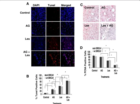
Next, we examined cell proliferation and apoptosis in tumors of control and drug-treated mice. There was an increase in TUNEL-positive cells in tumors with an in-creasing order from no treatment, AG14361, lestaurtinib, to AG14361/lestaurtinib (Figure 4A,B). The apoptotic index of the combination therapy reached approximately 90%, which was significantly higher than those from mice with lestaurtinib (55%), and AG14361 (30%) mono-treatment, whereas no significant difference between shBRCA1 and wtBRCA1 tumors was detected (Figure 4B). Immunochemical staining using an antibody for prolifer-ating cell nuclear antigen (PCNA) detected a reversed order of decreasing PCNA-positive cells from these tumors (Figure 4C,D). Thus, the inhibition of tumor growth of these treatments is correlated with increased apoptosis and decreased cell proliferation.
%
L
iving Cells
%
L
iving Cells
B
%
L
iving Cells
Lestaurnitib Concentrations (nM)
%
L
iving Cells
%
L
iving Cells
C
%
L
iving Cells
[image:7.595.59.538.88.447.2]A
Treatment results in caspase 3/7 activation and cell cycle abnormalities
To understand the cause of the enhanced apoptosis, cas-pase 3/7 activity was measured in the 92 J pair of cells after treatment with AG14361 and/or lestaurtinib at their
IC50 concentrations. Our data demonstrated that all the
treatments activated caspase 3/7 in a time-dependent manner. As a mono-treatment, lestaurtinib was a more potent stimulator of caspase 3/7 activity than AG14361, and the combination of both drugs significantly enhanced this activity during the period of treatment of both cell lines, especially before 36 hr (Figure 5A,B).
Next, we determined the cell cycle distribution of 92 J-shBRCA1 and 92 J-wtBRCA1 cells after different drug treatments at four time points using flow cytometry. The proportion of cells in the G1 phase was markedly increased upon treatment of AG14361 at 12 hr at the
[image:8.595.59.538.87.454.2]which is similar to the mono-treatment of AG14361. Both cell lines showed similar responses (Figure 5D). Thus the treatment of both drugs recapitulated features of mono-treatment of these two drugs, consequently leading to increased apoptosis and inhibition of cell proliferation.
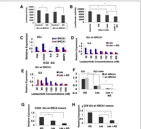
AG14361 inhibits NF-κB signaling that is enhanced by
lestaurtinib treatment
We previously identified some compounds in this library
that inhibit NF-κB signaling [47]. Among the eleven
com-pounds confirmed in our secondary screen that exhibit strong synergy with PARPi in killing breast cancers
irre-spective of their BRCA1 status, four can inhibit NF-κB
activity (lestaurtinib, bortezomib, ouabain, and digitoxin).
These data suggest that inhibition of NF-κB might be one
of the reasons for the synergistic killing of these cancer
cells with PARPi. To investigate this theory, we
ana-lyzed the effect of AG14361 on NF-κB transcriptional
activity using a luciferase reporter. The data showed that AG14361 treatment inhibited transcriptional activity of both shBRCA1 and wtBRCA1 cells (Figure 6A). Next,
we investigate the effect of AG14361 on NF-κB signaling
by investigating the expression levels of several down-stream genes. We found that treatment of AG14361 at its
IC50 significantly repressed expression of all the genes
tested in wtBRCA1 cells; whereas in shBRCA1 cells,
AG14361 could significantly repress expression of IL-6,
IL8, andMMP9, but did not significant change expression
of p65 and COX2 (Figure 6B). These data show that
AG14361 at its IC50concentration could affect expression
of some transcriptional downstream genes of NF-κB
al-though a stronger effect was observed in wtBRCA1 cells than in shBRCA1 cells.
A
Control
AG
Les
AG + Les
B
DAPI Tunel Merged
% T
u
nel
P
ositive
C
ells
*
* * * *
*
Control AG
Les Les + AG
C
D
%
P
CNA Positive
C
ells
* *
[image:9.595.59.538.90.447.2]* *
Next we investigated whether the inhibitory effect of
NF-κB by AG14361 was potentiated by the presence of
lestaurtinib by examining the expression of a NF-κB
lucif-erase reporter that is stably integrated into the genome of HeLa cells. The data indicated that while mono-treatment
of AG14361 and lestaurtinib at their IC50 significantly
inhibited the NF-κB transcriptional activity, the
combin-ation of both enhanced the effect (Figure 6C). We then examined the expression of several downstream genes,
IL-6, IL8, p50, and COX-2. Our data indicated that
lestaurtinib inhibited expression of these genes in a dose-dependent manner and this effect was significantly
enhanced in the presence of AG14361 at its IC50
concentration (Figure 6D,E, and Additional file 4). We have also examined the expression of these genes in the tumors treated with AG14361 and/or lestaurtinib that were harvested at their end point. The data indicated that AG14361 mono-treatment had no
obvious effect on IL-6 and COX2 expression, whereas
lestaurtinib significantly inhibited their expression. This effect was enhanced with the combined treatment with both drugs (Figure 6F-I).
Discussion
The majority of BRCA1/2-related breast cancers exhibit high grade and are insensitive to most available hormonal or targeted therapeutic agents [1-7,48,49]. Moreover many sporadic breast cancers also exhibit reduced or dimin-ished expression of BRCA1 [50,51]. Thus the finding that BRCA1/2-associated breast cancers are highly sensitive to
PARPi has been considered as a very promising approach for breast cancer [20-24,52]. However, like many other therapeutic agents, PARPi treatment is also associated with drug resistance after initial response to the treatment (reviewed in [31]). For example, in phase II trials, 400 mg twice daily exposure of PARPi olaparib only resulted in the delay of breast cancer and ovarian cancer progression (both with median progression-free survival of about six months), and all patients with BRCA1/2 mutations even-tually died of cancers [53,54].
In mouse models, prolonged treatment of the PARPi, AZD2281 also resulted in drug resistance presumably due to upregulation of Abcb1a/b genes encoding
P-glycoprotein efflux pumps [36]. Do Sotoet al. [28] found
that while a PARPi was able to kill naïve BRCA1 mutant
cells with high specificity both in vitro and in vivo, it
exhibited minimal specificity in inhibiting several cell lines derived from mouse Brca1 mutant mammary tumors. Altogether, these observations reinforced the need for screens for additional drugs that efficiently kill BRCA1/2-associated cancer cells when combined with PARPi. Of note, it has been demonstrated that PARPi, when combined with agents that impair DNA repair, are also effective in killing cancer cells containing wild-type BRCA1/2 [36,55,56].
In this study, we screened a library that contains 2,816 small molecules, most of which are approved for human or animal use by the FDA or other countries [39,47], in the presence of AG14361 at a constant sublethal dose in order to identify compounds that kill breast cancer cells
A
B
C
% C
aspase
A
ctivation
D
92J sh BRCA1
[image:10.595.59.540.90.312.2]Cells Cells
synergistically with PARPi. Our initial screen identified seventeen compounds that have similar levels in killing both shBRCA1 and wtBRCA1 cells, six compounds that are more specific for killing shBRCA1 cells, and nine com-pounds that kill wtBRCA1 cells better. After validation, lestaurtinib was selected for further investigation. Lestaur-tinib is an orally bioavailable multikinase inhibitor for a number of kinases including protein kinase C-related kinase 1 (PRK1) [57], FMS-like tyrosine kinase 3 (FLT3) [44,45], JAK2 [46,58], Trk-A/B/C [59,60]. Lestaurtinib has
been used in clinical trials for myeloproliferative disorders, and acute myelogenous leukemia [44-46], but there has been no report of its application for breast cancer treat-ment yet. Our data indicated that lestaurtinib is highly potent against tumor cells derived from both mouse and human breast cancers as a mono-treatment agent. In combination with AG14361, this effect is synergistically enhanced as reflected by further delay of tumor progres-sion. We also found that four out of fourteen tumors com-pletely regressed during combination treatment, while no
Relative Expression
92J
IC50 AG * * * *
* *
* *
A
* *B
D
C
F
E
Relative Expression
92J sh BRCA1
92J wt BRCA1
Relative Expression
H
Relative Expression
* *
Relative Expression
* *
G
HELA Cells *
* *
* *
[image:11.595.59.539.87.525.2]*
regression was observed in the other three groups of mice (control, mono-treatment of either AG14361 or lestaurti-nib) carrying a total of 42 tumors derived from each of cell line tested. Therefore, the synergy between AG14361 and lestaurtinib treatment is significant in these cancer cells. The complete tumor regression in these four groups of animals may reflect a differential threshold response of these mice to the treatment compared with the other recipients. In the clinic setting, complete tumor regression upon the therapeutic treatment is the most desirable outcome, however it does not always happen. In most cases, patients display partial response at different degrees, perhaps, due to individual difference in response to the treatment [53,54]. Nonetheless, the significant delay of tumor progression could prolong the life of patients and provide valuable time for further therapeutic therapies. Our data are reminiscent of this feature. We are in the process of screening further drug combinations in order to achieve the most desirable outcome in the near future.
The effect of lestaurtinib are primarily on G2 arrest, apoptosis and reduced proliferation of cancer cells irrespective of their BRCA1 status. Mono-treatment of AG14361 exhibited a similar, yet mild effect on apoptosis and proliferation; however, it affected all phases of the cell cycle. Of note, the combination of both drugs results in dramatic expansion of cells in the G2 phase at the expense of the S phase. This may account for the much more se-vere growth retardation and markedly enhanced cell death. Of note, we found that four out of eleven com-pounds, including lestaurtinib, which exhibits synergy
with PARPi, could inhibit NF-κB activity based on our
previous study [47]. NF-κB is a transcription factor that
plays important roles in cell cycle progression, cell survival and inflammation [1,52,59,60]. Therefore we tested the
effect of AG14361 on NF-κB and found it could also
in-hibit NF-κB activity, albeit to a less extent compared with
lestaurtinib. When combined together, AG14361 and les-taurtinib exhibited a much stronger inhibitory effect on the
expression of a number of genes in the NF-κB signaling
pathway, such asp50,p65,IL6,IL8,COX2andMMP9that
are involved in cancer cell proliferation, inflammation, invasion and/or cell death [45,50,54,58,59].
Conclusions
Our data indicated that lestaurtinib is a potent thera-peutic agent for killing breast cancer cells and it amp-lifies the ability of the PARP1 inhibitor AG14361 to kill breast cancer cells irrespective to their BRCA1 status. This effect is, at least in part, by inhibiting
NF-κB signaling. Because lestaurtinib and PARPi are
drugs approved for clinical trials for several different cancers [44-46,53,54], we believe this combination will be applicable for a breast cancer trial in the near future.
Additional files
Additional file 1:Murine cell lines Ras and 69 proliferationin vitro and tumor growth in allografts. (A)Fold change in the cell growth rate in respect to the initial inoculated Ras and 69 cells lines. Data shown represent mean standard deviation (SD) from triplicate experiments.(B) Example of clonogenic assay performed on Ras and 69 cell lines. A total of 1,000 cells were seeded in 10 ml plates and grown for 21 days and counted at the end point. Data represent the mean SD from triplicate experiments.(C)Cells were injected into the mammary fat pad of nude mice and the tumor volume was measured until they reached 3.5 mm3. Each group consisted of five mice and each mouse had two tumors, the measurements on the graph represent the average of ten tumors per group.
Additional file 2: Table S1.Identification of clinically used drugs that kill 92 J isogenic pair as single agents and show the synergistic toxic effect with AG14361.
Additional file 3:Cell viability assay of deserpiline that was eliminated for further analysis after the cherry pick. (A)Viability assay of deserpiline from the primary high throughput screen.(B)Viability assay of the same drug from the secondary screen. The synergy is less obvious in the secondary screen as some points of the curve touch, or even cross with the expected additive values curve.
Additional file 4:Synergistic effect of lestaurtinib in combination with AG14361 in Ras and 69 cell lines.Expression ofCOX2(A,B), IL8(C,D), andp50(E,F)in 92 J pair of isogenic cells in the presence of AG14361 and/or various concentrations of lestaurnitib for 24 hr revealed by real-time PCR. Data show the mean of three different readings and are marked by columns with ± standard deviation (SD) error bars.
Abbreviations
BRCA1/2:breast cancer type 1/2 susceptibility; DMEM: Dulbecco’s modified Eagle’s medium; DSBs: double-strand breaks; HR: homologous
recombination; IC50: 50% inhibition dose; NF-κB: nuclear factorκB; PARP1: poly(ADP-ribose)polymerase 1; PARPi: PARP inhibitor; PBS: phosphate-buffered saline; PCNA: proliferating cell nuclear antigen; qRT-PCR: quantitative reverse transcription-polymerase chain reaction; shBRCA1: sample with short hairpin RNA (shRNA) against BRCA1; SSBs: single-strand breaks; TUNEL: terminal deoxynucleotidyl transferase-mediated dUTP-biotin nick end-labeling; wtBRCA1: sample with wild-type BRCA1 status.
Competing interests
The authors declare that they have no competing interests.
Authors’contributions
GVO designed and performed the experiments, analyzed and interpreted the data and wrote the manuscript. CC, SS and TJL performed the experiments. XX developed the cell lines. CL performed the mice surgeries, CJT synthesized the AG14361. RH and MHX designed the high-throughput experiments and performed the data analysis. CXD designed the experiments, interpreted the data and wrote the manuscript. CJT, MHX and CXD revised the manuscript for important intellectual content. All authors read and approved the final manuscript.
Acknowledgements
We gratefully acknowledge the critical reading and helpful discussion of members of the Deng laboratory. This work was supported by the Intramural Research Program of the National Institute of Diabetes, Digestive and Kidney Diseases, National Institutes of Health, USA.
Author details 1
Genetics of Development and Disease Branch, National Institute of Diabetes, Digestive and Kidney Diseases, National Institutes of Health, 9000 Rockville Pike, Bethesda, MD 20892, USA.2NIH Chemical Genomics Center, National Center for Advancing Translational Sciences, National Institutes of Health, 9800 Medical Center Drive, Rockville, MD 20850, USA.
References
1. Alberg AJ, Helzlsouer KJ:Epidemiology, prevention, and early detection of breast cancer.Curr Opin Oncol1997,9:505–511.
2. Zhang J, Powell SN:The role of the BRCA1 tumor suppressor in DNA double-strand break repair.Mol Cancer Res2005,3:531–539. 3. Brody LC, Biesecker BB:Breast cancer susceptibility genes. BRCA1 and
BRCA2.Medicine (Baltimore)1998,77:208–226.
4. Eccles DM, Pichert G:Familial non-BRCA1/BRCA2-associated breast cancer.
Lancet Oncol2005,6:705–711.
5. Cong L, Ran FA, Cox D, Lin S, Barretto R, Habib N, Hsu PD, Wu X, Jiang W, Marraffini LA, Zhang F:Multiplex genome engineering using CRISPR/Cas systems.Science2013,339:819–823.
6. Nathanson KL, Weber BL:“Other”breast cancer susceptibility genes: searching for more holy grail.Hum Mol Genet2001,10:715–720. 7. Oldenburg RA, Meijers-Heijboer H, Cornelisse CJ, Devilee P:Genetic
susceptibility for breast cancer: how many more genes to be found?
Crit Rev Oncol Hematol2007,63:125–149.
8. Whittemore AS, Gong G, Itnyre J:Prevalence and contribution of BRCA1 mutations in breast cancer and ovarian cancer: results from three U.S. population-based case-control studies of ovarian cancer.Am J Hum Genet 1997,60:496–504.
9. Mavaddat N, Peock S, Frost D, Ellis S, Platte R, Fineberg E, Evans DG, Izatt L, Eeles RA, Adlard J, Davidson R, Eccles D, Cole T, Cook J, Brewer C, Tischkowitz M, Douglas F, Hodgson S, Walker L, Porteous ME, Morrison PJ, Side LE, Kennedy MJ, Houghton C, Donaldson A, Rogers MT, Dorkins H, Miedzybrodzka Z, Gregory H, Eason J,et al:Cancer Risks for BRCA1 and BRCA2 mutation carriers: results from prospective analysis of EMBRACE.
J Natl Cancer Inst2013,105:812–822.
10. Chen S, Iversen ES, Friebel T, Finkelstein D, Weber BL, Eisen A, Peterson LE, Schildkraut JM, Isaacs C, Peshkin BN, Corio C, Leondaridis L, Tomlinson G, Dutson D, Kerber R, Amos CI, Strong LC, Berry DA, Euhus DM, Parmigiani G:
Characterization of BRCA1 and BRCA2 mutations in a large United States sample.J Clin Oncol2006,24:863–871.
11. Easton D:Breast cancer genes–what are the real risks?Nat Genet1997,
16:210–211.
12. Yoo KH, Hennighausen L:EZH2 methyltransferase and H3K27 methylation in breast cancer.Int J Biol Sci2012,8:59–65.
13. Hu Y:BRCA1, hormone, and tissue-specific tumor suppression.Int J Biol Sci2009,5:20–27.
14. Deng CX, Brodie SG:Roles of BRCA1 and its interacting proteins.Bioessays 2000,22:728–737.
15. Deng CX:BRCA1: cell cycle checkpoint, genetic instability, DNA damage response, and cancer evolution.Nucleic Acids Res2006,34:1416–1426. 16. Boguski MS, Mandl KD, Sukhatme VP:Drug discovery, Repurposing with a
difference.Science2009,324:1394–1395.
17. Dine J, Deng CX:Mouse models of BRCA1 and their application to breast cancer research.Cancer Metastasis Rev2012,32:25–37.
18. Deng CX, Wang RH:Roles of BRCA1 in DNA damage repair: a link between development and cancer.Hum Mol Genet2003,12:R113–R123. 19. Venkitaraman AR:Linking the cellular functions of BRCA genes to cancer
pathogenesis and treatment.Annu Rev Pathol2009,4:461–487. 20. Farmer H, McCabe N, Lord CJ, Tutt AN, Johnson DA, Richardson TB,
Santarosa M, Dillon KJ, Hickson I, Knights C, Martin NM, Jackson SP, Smith GC, Ashworth A:Targeting the DNA repair defect in BRCA mutant cells as a therapeutic strategy.Nature2005,434:917–921.
21. Bryant HE, Schultz N, Thomas HD, Parker KM, Flower D, Lopez E, Kyle S, Meuth M, Curtin NJ, Helleday T:Specific killing of BRCA2-deficient tumours with inhibitors of poly(ADP-ribose) polymerase.Nature2005,
434:913–917.
22. Marchetti C, Imperiale L, Gasparri ML, Palaia I, Pignata S, Boni T, Bellati F, Benedetti Panici P:Olaparib, PARP1 inhibitor in ovarian cancer.Expert Opin Investig Drugs2012,21:1575–1584.
23. Glendenning J, Tutt A:PARP inhibitors–current status and the walk towards early breast cancer.Breast2011,20:S12–S19.
24. Helleday T:The underlying mechanism for the PARP and BRCA synthetic lethality: clearing up the misunderstandings.Mol Oncol 2011,5:387–393.
25. Strom CE, Johansson F, Uhlen M, Szigyarto CA, Erixon K, Helleday T:Poly (ADP-ribose) polymerase (PARP) is not involved in base excision repair but PARP inhibition traps a single-strand intermediate.Nucleic Acids Res 2011,39:3166–3175.
26. Bryant HE, Helleday T:Poly(ADP-ribose) polymerase inhibitors as potential chemotherapeutic agents.Biochem Soc Trans2004,32:959–961. 27. Dantzer F, Schreiber V, Niedergang C, Trucco C, Flatter E, De La Rubia G,
Oliver J, Rolli V, Menissier-de Murcia J, de Murcia G:Involvement of poly (ADP-ribose) polymerase in base excision repair.Biochimie1999,81:69–75. 28. De Soto JA, Wang X, Tominaga Y, Wang RH, Cao L, Qiao W, Li C, Xu X,
Skoumbourdis AP, Prindiville SA, Thomas CJ, Deng CX:The inhibition and treatment of breast cancer with poly (ADP-ribose) polymerase (PARP-1) inhibitors.Int J Biol Sci2006,2:179–185.
29. Gallmeier E, Kern SE:Absence of specific cell killing of the BRCA2-deficient human cancer cell line CAPAN1 by poly(ADP-ribose) polymerase inhibition.
Cancer Biol Ther2005,4:703–706.
30. De Soto JA, Deng C:PARP-1 inhibitors, are they the long-sought genetically specific drugs for BRCA1/2-associated breast cancers?
Int J Med Sci2006,3:117–123.
31. Chiarugi A:A snapshot of chemoresistance to PARP inhibitors.
Trends Pharmacol Sci2012,33:42–48.
32. Oplustilova L, Wolanin K, Mistrik M, Korinkova G, Simkova D, Bouchal J, Lenobel R, Bartkova J, Lau A, O’Connor MJ, Lukas J, Bartek J:Evaluation of candidate biomarkers to predict cancer cell sensitivity or resistance to PARP-1 inhibitor treatment.Cell Cycle2012,11:3837–3850.
33. Bunting SF, Callen E, Wong N, Chen HT, Polato F, Gunn A, Bothmer A, Feldhahn N, Fernandez-Capetillo O, Cao L, Xu X, Deng CX, Finkel T, Nussenzweig M, Stark JM, Nussenzweig A:53BP1 inhibits homologous recombination in Brca1-deficient cells by blocking resection of DNA breaks.Cell2010,141:243–254.
34. Edwards SL, Brough R, Lord CJ, Natrajan R, Vatcheva R, Levine DA, Boyd J, Reis-Filho JS, Ashworth A:Resistance to therapy caused by intragenic deletion in BRCA2.Nature2008,451:1111–1115.
35. Issaeva N, Thomas HD, Djureinovic T, Jaspers JE, Stoimenov I, Kyle S, Pedley N, Gottipati P, Zur R, Sleeth K, Chatzakos V, Mulligan EA, Lundin C, Gubanova E, Kersbergen A, Harris AL, Sharma RA, Rottenberg S, Curtin NJ, Helleday T:6-thioguanine selectively kills BRCA2-defective tumors and overcomes PARP inhibitor resistance.Cancer Res2010,70:6268–6276. 36. Rottenberg S, Jaspers JE, Kersbergen A, van der Burg E, Nygren AO,
Zander SA, Derksen PW, de Bruin M, Zevenhoven J, Lau A, Boulter R, Cranston A, O’Connor MJ, Martin NM, Borst P, Jonkers J:High sensitivity of BRCA1-deficient mammary tumors to the PARP inhibitor AZD2281 alone and in combination with platinum drugs.Proc Natl Acad Sci U S A2008,
105:17079–17084.
37. Nzila A, Ma Z, Chibale K:Drug repositioning in the treatment of malaria and TB.Future Med Chem2011,3:1413–1426.
38. Tobinick EL:The value of drug repositioning in the current pharmaceutical market.Drug News Perspect2009,22:119–125. 39. Huang R, Southall N, Wang Y, Yasgar A, Shinn P, Jadhav A, Nguyen DT,
Austin CP:The NCGC pharmaceutical collection: a comprehensive resource of clinically approved drugs enabling repurposing and chemical genomics.Sci Transl Med2011,3:80ps16.
40. Xu X, Wagner KU, Larson D, Weaver Z, Li C, Ried T, Hennighausen L, Wynshaw-Boris A, Deng CX:Conditional mutation of Brca1 in mammary epithelial cells results in blunted ductal morphogenesis and tumour formation [see comments].Nat Genet1999,22:37–43.
41. Brodie SG, Xu X, Qiao W, Li WM, Cao L, Deng CX:Multiple genetic changes are associated with mammary tumorigenesis in Brca1 conditional knockout mice.Oncogene2001,20:7514–7523.
42. Huang R, Xia M, Cho MH, Sakamuru S, Shinn P, Houck KA, Dix DJ, Judson RS, Witt KL, Kavlock RJ, Tice RR, Austin CP:Chemical genomics profiling of environmental chemical modulation of human nuclear receptors.
Environ Health Perspect2011,119:1142–1148.
43. Inglese J, Auld DS, Jadhav A, Johnson RL, Simeonov A, Yasgar A, Zheng W, Austin CP:Quantitative high-throughput screening: a titration-based approach that efficiently identifies biological activities in large chemical libraries.Proc Natl Acad Sci U S A2006,103:11473–11478.
44. Fathi AT, Chabner BA:FLT3 inhibition as therapy in acute myeloid leukemia: a record of trials and tribulations.Oncologist2011,
16:1162–1174.
45. Knapper S, Burnett AK, Littlewood T, Kell WJ, Agrawal S, Chopra R, Clark R, Levis MJ, Small D:A phase 2 trial of the FLT3 inhibitor lestaurtinib (CEP701) as first-line treatment for older patients with acute myeloid leukemia not considered fit for intensive chemotherapy.Blood2006,
46. Diaz T, Navarro A, Ferrer G, Gel B, Gaya A, Artells R, Bellosillo B, Garcia-Garcia M, Serrano S, Martinez A, Monzo M:Lestaurtinib inhibition of the Jak/STAT signaling pathway in hodgkin lymphoma inhibits proliferation and induces apoptosis.PLoS One2011,6:e18856.
47. Miller SC, Huang R, Sakamuru S, Shukla SJ, Attene-Ramos MS, Shinn P, Van Leer D, Leister W, Austin CP, Xia M:Identification of known drugs that act as inhibitors of NF-kappaB signaling and their mechanism of action.
Biochem Pharmacol2010,79:1272–1280.
48. Andre F, Zielinski CC:Optimal strategies for the treatment of metastatic triple-negative breast cancer with currently approved agents.Ann Oncol 2012,23:vi46-51.
49. Carey L, Winer E, Viale G, Cameron D, Gianni L:Triple-negative breast cancer: disease entity or title of convenience?Nat Rev Clin Oncol2010,
7:683–692.
50. Lee MH, Lahusen T, Wang RH, Xiao C, Xu X, Hwang YS, He WW, Shi Y, Deng CX:Yin Yang 1 positively regulates BRCA1 and inhibits mammary cancer formation.Oncogene2012,31:116–127.
51. Birgisdottir V, Stefansson OA, Bodvarsdottir SK, Hilmarsdottir H, Jonasson JG, Eyfjord JE:Epigenetic silencing and deletion of the BRCA1 gene in sporadic breast cancer.Breast Cancer Res2006,8:R38.
52. Fong PC, Boss DS, Yap TA, Tutt A, Wu P, Mergui-Roelvink M, Mortimer P, Swaisland H, Lau A, O’Connor MJ, Carmichael J, Kaye SB, Schellens JH, de Bono JS:Inhibition of poly(ADP-ribose) polymerase in tumors from BRCA mutation carriers.N Engl J Med2009,361:123–134.
53. Audeh MW, Carmichael J, Penson RT, Friedlander M, Powell B, Bell-McGuinn KM, Scott C, Weitzel JN, Oaknin A, Loman N, Lu K, Schmutzler RK, Matulonis U, Wickens M, Tutt A:Oral poly(ADP-ribose) polymerase inhibitor olaparib in patients with BRCA1 or BRCA2 mutations and recurrent ovarian cancer: a proof-of-concept trial.Lancet2010,376:245–251. 54. Tutt A, Robson M, Garber JE, Domchek SM, Audeh MW, Weitzel JN,
Friedlander M, Arun B, Loman N, Schmutzler RK, Wardley A, Mitchell G, Earl H, Wickens M, Carmichael J:Oral poly(ADP-ribose) polymerase inhibitor olaparib in patients with BRCA1 or BRCA2 mutations and advanced breast cancer: a proof-of-concept trial.Lancet2010,376:235–244.
55. Wesierska-Gadek J, Zulehner N, Ferk F, Skladanowski A, Komina O, Maurer M:PARP inhibition potentiates the cytotoxic activity of C-1305, a selective inhibitor of topoisomerase II, in human BRCA1-positive breast cancer cells.Biochem Pharmacol2012,84:1318–1331.
56. Moskwa P, Buffa FM, Pan Y, Panchakshari R, Gottipati P, Muschel RJ, Beech J, Kulshrestha R, Abdelmohsen K, Weinstock DM, Gorospe M, Harris AL, Helleday T, Chowdhury D:miR-182-mediated downregulation of BRCA1 impacts DNA repair and sensitivity to PARP inhibitors.Mol Cell2011,
41:210–220.
57. Kohler J, Erlenkamp G, Eberlin A, Rumpf T, Slynko I, Metzger E, Schule R, Sippl W, Jung M:Lestaurtinib inhibits histone phosphorylation and androgen-dependent gene expression in prostate cancer cells.PLoS One 2012,7:e34973.
58. Hexner EO, Serdikoff C, Jan M, Swider CR, Robinson C, Yang S, Angeles T, Emerson SG, Carroll M, Ruggeri B, Dobrzanski P:Lestaurtinib (CEP701) is a JAK2 inhibitor that suppresses JAK2/STAT5 signaling and the proliferation of primary erythroid cells from patients with myeloproliferative disorders.Blood2008,111:5663–5671.
59. Festuccia C, Gravina GL, Muzi P, Pomante R, Ventura L, Ricevuto E, Vicentini C, Bologna M:In vitro and in vivo effects of bicalutamide on the expression of TrkA and P75 neurotrophin receptors in prostate carcinoma.Prostate2007,67:1255–1264.
60. Festuccia C, Muzi P, Gravina GL, Millimaggi D, Speca S, Dolo V, Ricevuto E, Vicentini C, Bologna M:Tyrosine kinase inhibitor CEP-701 blocks the NTRK1/NGF receptor and limits the invasive capability of prostate cancer cells in vitro.Int J Oncol2007,30:193–200.
doi:10.1186/bcr3682
Cite this article as:Vazquez-Ortizet al.:Drug repurposing screen identifies lestaurtinib amplifies the ability of the poly (ADP-ribose) polymerase 1 inhibitor AG14361 to kill breast cancer associated gene-1 mutant and wild type breast cancer cells.Breast Cancer Research201416:R67.
Submit your next manuscript to BioMed Central and take full advantage of:
• Convenient online submission
• Thorough peer review
• No space constraints or color figure charges
• Immediate publication on acceptance
• Inclusion in PubMed, CAS, Scopus and Google Scholar
• Research which is freely available for redistribution


![Dimethanolbis[N′ (3 pyridylmethylene)benzohydrazide]sodium(I) iodide](data:image/gif;base64,R0lGODlhAQABAIAAAP///wAAACH5BAEAAAAALAAAAAABAAEAAAICRAEAOw==)