RESEARCH NOTE
Development and characterization of 17
polymorphic microsatellite markers for the reef
manta ray (
Mobula alfredi
)
Amelia J. Armstrong
1*, Christine L. Dudgeon
1, Carlos Bustamante
2, Michael B. Bennett
1and Jennifer R. Ovenden
1Abstract
Objective: Limited sample sizes are often a problem for species of conservation concern when using genetic tools to make population assessments. Lack of analytical power from small sample sizes can be compensated for by use of a large marker set. Here we report on development and characterization of 17 novel microsatellite markers for the reef manta ray (Mobula alfredi).
Results: Loci were screened on 60 reef manta rays (M. alfredi) sampled from the east coast of Australia. The number of alleles per locus varied from 2 to 13 with observed heterozygosities ranging between 0.300 and 0.917. The devel-opment of these 17 additional markers increases the total number of microsatellite markers available for this species to 27.
Keywords: Population genetics, Conservation genetics, Microsatellite loci, Short tandem repeats
© The Author(s) 2019. This article is distributed under the terms of the Creative Commons Attribution 4.0 International License (http://creat iveco mmons .org/licen ses/by/4.0/), which permits unrestricted use, distribution, and reproduction in any medium, provided you give appropriate credit to the original author(s) and the source, provide a link to the Creative Commons license, and indicate if changes were made. The Creative Commons Public Domain Dedication waiver (http://creat iveco mmons .org/ publi cdoma in/zero/1.0/) applies to the data made available in this article, unless otherwise stated.
Introduction
The reef manta ray (Mobula alfredi) occurs in tropical and subtropical waters of the Indian and Pacific oceans [1]. The species forms aggregations in coastal waters throughout its range, which provides for targeted eco-tourism in many coastal regions [2]. However, these aggregations may suffer depletion from competing by-catch and targeted fisheries, particularly for their highly-valued body parts that are used in ‘traditional medicines’ [3–6]. Like many large shark and ray species, M. alfredi
is long-lived with conservative life history parameters (i.e. slow growth, late maturity, low fecundity) that make populations highly-susceptible to over-exploitation [7]. As such, the global conservation concerns for this spe-cies have resulted in its classification as ‘Vulnerable to Extinction’ on the IUCN Red List [8], and has led to list-ing in Appendix II of the Convention on the International Trade in Endangered Species and Appendices I and II of
the Convention on Migratory Species. Future research priorities identified for reef manta rays include investi-gation of population structure (regional and global) and connectivity, based on genetic analyses [9].
To date, the limited genetic work conducted on DNA from reef manta rays has been primarily in the context of broader phylogenetic studies [10, 11]. One recent phylogenetic study resulted in the collapse of the genus
Manta into Mobula, and a reassignment of the
posi-tion of M. alfredi within this genus [12]. An earlier study used nuclear DNA obtained from M. alfredi sampled in Japanese waters to develop ten species specific micro-satellite markers [13]. However, preliminary studies on of M. alfredi from the east coast of Australia found that only eight of the previously developed markers were polymorphic, with the remaining markers either non-polymorphic or non-amplifiable. Collection of adequate sample sizes to overcome the limitation of few markers is often difficult, expensive and inappropriate for species of conservation concern. Therefore, we address this issue through the development of additional microsatellite markers for M. alfredi that may allow for robust estima-tion of populaestima-tion sizes without the need to sample large
Open Access
*Correspondence: [email protected]
1 School of Biomedical Science, The University of Queensland, Brisbane,
Australia
numbers of animals, and will facilitate future investiga-tions into fine-scale structure (e.g. relatedness).
For studies focused on non-model species of conser-vation concern, microsatellite markers can be amplified from relatively small quantities of DNA and provide a relatively low cost per sample for genotyping (espe-cially when working with pre-developed marker sets). While genotype-by-sequencing approaches (i.e. RAD-seq, DArTseq) are increasingly used for genomic appli-cations to non-model species, the strict requirements surrounding DNA quality, quantity and contamination may limit application to historical, degraded or small amounts of sampled-DNA. Furthermore, microsatellite markers provide the potential for a standardised marker set that can facilitate comparisons of genetically distinct populations across broad temporal and geographical scales. Here we report the development of 17 polymor-phic microsatellite markers for M. alfredi, to augment the 10 previously developed, and detail marker performance through the genotyping of 60 individual manta rays from the east coast of Australia. We anticipate that microsatel-lite markers developed here will inform ongoing regional studies on population connectivity, and may facilitate global population comparisons through the provision of an enlarged and published marker set.
Main text
Methods and results
Microsatellite primer sequences were devel-oped de novo from DNA obtained from a Mobula alfredi voucher specimen from Lady Elliot Island Reef, Australia. A genomic library was developed with the TruSeq Nano DNA sample prep kit (Illumina, San Diego, CA) and sequenced using an Illumina HiSeq 2000 platform (San Diego, CA) that supported the acquisition of 2 × 125 base pair (bp) paired-end reads, following the protocols supplied by the manufacturer. Sequences between 150 and 400 bp were explored for microsatellite motifs using the software QDD v.3.1 [14] following the microsatellite discovery pro-tocol described by Vargas-Caro et al. [15]. In brief, perfect microsatellite repeat motifs with > 10 repeat-ing units were identified and loci with product sizes estimated at less than 100 bp were removed. Addi-tional loci where flanking regions contained ‘runs’ (of a single base) or repeated sequence elements were excluded. Selected sequences consisting of repeat motifs and flanking sequences were blasted against NCBI GenBank using default parameters to exclude loci potentially located in coding regions. Primers with homology outside the target flanking region or that may amplify more than one locus were excluded by blasting back against the genomic library using
GENEIOUS v 9.1.8 (http://www.genei ous.com) [16]. Of the remaining candidate primer pairs, we selected 48 to take through to development, giving priority to loci with tri or tetra nucleotide repeat motifs. For the 48 candidate microsatellite loci developed, a ‘CAG-tail’ was attached to the 5′ end of the forward primers allowing downstream use of fluorescent PCR product labelling. Loci were combined in multiplexed PCRs to avoid overlapping product sizes. The 5′ end of the reverse primer had a ‘GTTT-tail’ attached to ensure complete adenylation, thereby protecting PCR prod-ucts from non-template nucleotide addition and facil-itating accurate genotyping [17].
Genotyping was conducted using genomic DNA extracted from ethanol preserved tissue biopsies col-lected non-lethally (2015–2018) from M. alfredi
in waters surrounding Lady Elliot Island (− 24.11°, 152.71°) and North Stradbroke Island (− 27.42°, 153.54°), Queensland, Australia. Possible sample dupli-cation was avoided, as each manta ray’s unique ventral skin pattern was photographed at the time of biopsy collection [18]. Loci were amplified in a 12 µl PCR reac-tion comprised of 1–2 µl of genomic DNA (10–15 ng), 0.5 µl primer stock, 6 µl of 2 × MyTaq mix (Bioline Aus-tralia), with remaining volume made up to 12 µl with Milli-Q H2O. PCR cycling conditions were a 95 °C for 60 s step, followed by 38 cycles of 95 °C for 60 s, 95 °C for 15 s, 55 °C for 15 s and 72 °C for 15 s, with a final extension step of 72 °C for 15 min. The diluted PCR products (1:10 dilution) were denatured with forma-mide and an internal size standard (Genescan 500-LIZ) was added. Fragment separation was performed with an ABI 3730 DNA Analyser. Allele scoring was under-taken using the GENEIOUS Microsatellite Plugin Ver-sion 1.4 (GENEIOUS v 9.1.8) [16]. Loci were initially amplified in singleplex reaction against 6–12 individu-als. Loci that successfully amplified were characterised against an additional 18–24 individuals to determine information content before selection for inclusion in a multiplex PCR. After removing loci that either did not amplify, produced an inconsistent product, or were mono-morphic, the 48 candidate loci were reduced down to a final set of 17 (Table 1).
heterozygosity (Ho= 0.88) with heterozygosity ranging widely for di-nucleotide loci (Ho= 0.35–0.80). Exact tests identified two loci (MaQDD23, p = 0.0212 & MaQDD39,
p = 0.036) not in Hardy–Weinberg equilibrium, as cal-culated by GENEPOP v 4.2 [21]. Linkage disequilib-rium tests (GENEPOP v4.2) revealed significant linkage between only one pair of loci (MaQDD04 & MaQDD11,
p = 0.00), after Bonferroni correction.
Conclusions
The need for high quality, standardized genetic mark-ers resulted in the development of 17 novel microsatellite for reef manta rays. This marker set supplements micros-atellite primers already available for the species [13], and
enables research questions to be addressed that require the higher statistical power provided by a larger marker set. It is hoped that these additional markers also facilitate col-laboration between studies of reef manta ray populations globally through the provision of named primers for which resultant genotypes and diversity metrics can be collabora-tively compared and contrasted for reef manta ray popu-lations globally. The combination of these novel markers with those already developed previously for reef manta rays [13] provides up to 27 markers for use in population structure and genetic effective population size investiga-tions. The delineation of population structure and genera-tion of populagenera-tion size estimates have been highlighted as research priorities to help guide conservation action for the reef manta ray [9].
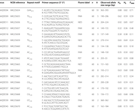
Table 1 Summary details for 17 microsatellite loci developed for Mobula alfredi
NCBI National Center for Biotechnology Information GenBank Reference; Primer Sequence F forward, R reverse (All forward primers had a GTT on the 3′ end & all reverse primers had CAG tag on the 5′ end (not included here), n is the number of individuals genotyped, A is the number of alleles observed, HO is the observed heterozygosity, HE is the expected heterozygosity, PHWE is the probability of the locus deviating from Hardy–Weinberg equilibrium
Locus NCBI reference Repeat motif Primer sequence (5′-3′) Fluoro label n A Observed allele
size range (bp) HO HE PHWE
MAQDD04 MK225654 GAA (27) F: ACA TCC TGC AGA GGC TGT AA
R: TCC TTA TCT TGT TCC GGC AG VIC 60 10 363–393 0.88 0.82 0.71
MAQDD09 MK225655 TG(15) F: CGT CGA CTG TTC ACA CTT CC
R: CTT CCT GGC TGG TAG GTA CG FAM 60 5 190–206 0.62 0.59 0.59
MAQDD11 MK225656 TC(19) F: TTT ACC TAT AGA TGG GTC AGG GAC
R: GCA GAT CGC TGT AGC TGT GA NED 60 8 204–224 0.92 0.85 0.07
MAQDD12 MK225657 AC(19) F: AAA GGA TCT GGG AAT ACG GG
R: GTG TTG GGA TCT CTG GTG CT FAM 60 4 149–155 0.32 0.36 0.48
MAQDD18 MK225658 TG(13) F: GGG AGA CAT TGA AAC GTG TG
R: GAA TGT GAT GGA GAG CAC TTG FAM 60 3 137–149 0.30 0.38 0.11
MAQDD21 MK225659 TG(13) F: GGC ATA CAA AGT GCT GGA GG
R: ATC TCA ACC CTG CGT GGA C NED 60 13 270–314 0.65 0.69 0.24
MAQDD22 MK225660 AC(17) F: CGG AAT AGC TGA CCC TCA GA
R: ATT GTG ATG GAG TGG TCC GT FAM 60 3 134–138 0.68 0.55 0.18
MAQDD23 MK225661 AC(10) F: CCC ATC ACT GAT AAT GAG CGT
R: GTG CAG TCA AAG GAA TGG GT FAM 60 2 148–150 0.35 0.50 0.02
MAQDD26 MK225662 AC(16) F: ATG TCA ACC GGA CTG GAA AT
R: TGT ATG CGT ATC TAG CAT CTTCA PET 60 6 203–33 0.62 0.59 0.08
MAQDD28 MK225663 AC(23) F: GCT GCA GGG AAG AAG CTA AA
R: TTT GTC CGA ATA TCT TGC CA NED 60 6 151–165 0.58 0.55 0.95
MAQDD29 MK225664 GT(21) F: GCT GAG AGG AAG CTA TTG GC
R: GAA GAT ACA GGA AGA TAA ATT GGC A FAM 60 8 193–213 0.63 0.60 0.90
MAQDD31 MK225665 AC(20) F: AAC TGC CCA TTC ACA TTT CC
R: CCC ATA GCT ACC ATC AGG GA PET 60 13 282–314 0.72 0.72 0.34
MAQDD33 MK225666 CA(21) F: GAC GAG ACA TCC CAA ACA CC
R: TTT CCT GGA TCC ACA ACC TC FAM 60 7 200–228 0.65 0.59 0.31
MAQDD37 MK225667 GT(14) F: CGT TGC ATC CAT CTG ACG TA
R: GAT GCA CCA GAG CAA CCA C PET 60 4 170–192 0.58 0.51 0.36
MAQDD38 MK225668 CA(14) F: CCA AAC ATA ACC CAG GCA AG
R: CAG CCT TGG TGA ACT GAG TG VIC 60 5 156–172 0.65 0.68 0.96
MAQDD39 MK225669 GT(14) F: AGC AGC TCT GTG GAA GCA GT
R: GCA CCC ATT TCC AAT CAG TT FAM 60 2 360–362 0.65 0.50 0.04
MAQDD43 MK225670 AC(14) F: TCC CTG GCT GTA TTG CCT AC
[image:3.595.62.537.100.490.2]Limitations
At the time of primer development and manuscript prepa-ration, the study did not have access to a sufficient number of samples from other locations for cross-population com-parisons. Recent genotyping suggests marker applicability for M. alfredi sampled on Australia’s west coast (approxi-mately 5600 km from east coast sampling locations) (author’s unpublished data). Due to sample availability and permit restrictions, this study did not test cross-species (within the family Mobulidae) primer amplification for the newly developed microsatellite loci.
Authors’ contributions
AJA carried out primer design filtering, genotyping, data analysis and manuscript writing; CB advised and assisted with primer design; CLD assisted with genotyping and was a major contributor in writing the manuscript; JRO assisted with primer design, genotyping, and was a major contributor in writing the manuscript; MBB provided funding, permits, ethical clearance, and feedback on the manuscript. All authors read and approved the final manuscript.
Author details
1 School of Biomedical Science, The University of Queensland, Brisbane,
Australia. 2 Facultad de Recursos Naturales Renovables, Universidad Arturo
Prat, Iquique, Chile.
Acknowledgements
The authors wish to acknowledge the services of the Australian Equine Genet-ics Research Centre Genotyping & Sequencing Facility (AEGRC).
Competing interests
The authors declare that they have no competing interests.
Availability of data and materials
The datasets analysed during the current study are not at present publicly available due ongoing publication, but are available from the corresponding author upon reasonable request. Sequences have been uploaded to GenBank and can be found at their accession numbers listed in Table 1.
Consent for publication Not applicable.
Ethics approval and consent to participate
All research was conducted in accordance with the approval of the University of Queensland’s Animal Ethics Committee (Permit SBS/319/14/ARC/EA/LEIER), the Great Barrier Reef Marine Park Authority (Permit G16/37856.1), the Depart-ment of National Parks, Sports and Racing (Permit QS2015/CVL1440) and the Department of Agriculture and Fisheries (Permit 199045).
Funding
This work was supported by an Australian Research Council Linkage Grant (LP150100669). AA is supported by a University of Queensland Research Scholarship. The funders were not involved in the design, collection, analysis and interpretation of the data, nor in writing the manuscript.
Publisher’s Note
Springer Nature remains neutral with regard to jurisdictional claims in pub-lished maps and institutional affiliations.
Received: 27 November 2018 Accepted: 15 April 2019
References
1. Marshall AD, Compagno LJV, Bennett MB. Redescription of the genus Manta with resurrection of Manta alfredi (Krefft, 1868) (Chondrichthyes; Myliobatoidei; Mobulidae). Zootaxa. 2009;2301:1–28.
2. O’Malley MP, Lee-Brooks K, Medd HB. The global economic impact of manta ray watching tourism. PLoS ONE. 2013. https ://doi.org/10.1371/ journ al.pone.00650 51.
3. Couturier LIE, Marshall AD, Jaine FRA, Kashiwagi T, Pierce SJ, Townsend KA, et al. Biology, ecology and conservation of the Mobulidae. J Fish Biol. 2012. https ://doi.org/10.1111/j.1095-8649.2012.03264 .x.
4. Croll DA, Dewar H, Dulvy NK, Fernando D, Francis MP, Galván-Magaña F, et al. Vulnerabilities and fisheries impacts: the uncertain future of manta and devil rays. Aquat Conserv. 2016. https ://doi.org/10.1002/aqc.2591. 5. O’Malley MP, Townsend KA, Hilton P, Heinrichs S, Stewart JD.
Charac-terization of the trade in manta and devil ray gill plates in China and South-east Asia through trader surveys. Aquat Conserv. 2017. https ://doi. org/10.1002/aqc.2670.
6. Steinke DI, Bernard AM, Horn RL, Hilton P, Hanner R, Shivji MS. DNA analysis of traded shark fins and mobulid gill plates reveals a high proportion of species of conservation concern. Sci Rep. 2017. https ://doi. org/10.1038/s4159 8-017-10123 -5.
7. Dulvy NK, Pardo SA, Simpfendorfer CA, Carlson JK. Diagnosing the dan-gerous demography of manta rays using life history theory. Peer J. 2014.
https ://doi.org/10.7717/peerj .400.
8. Marshall AD, Kashiwagi T, Bennett MB, Deakos M, Stevens G, McGregor F, et al. Mobula alfredi (amended version of the 2011 assessment). IUCN Red List; 2018. http://dx.doi.org/10.2305/IUCN.UK. Accessed 13 Nov 2018. 9. Stewart JD, Jaine FRA, Armstrong AJ, Armstrong AO, Bennett MB, Burgess
KB, et al. Research priorities to support effective manta and devil ray con-servation. Front Mar Sci. 2018. https ://doi.org/10.3389/fmars .2018.00314 . 10. Kashiwagi T, Marshall AD, Bennett MB, Ovenden JR. The genetic signature
of recent speciation in manta rays (Manta alfredi and M. birostris). Mol Phylogenet Evol. 2012. https ://doi.org/10.1016/j.ympev .2012.03.020. 11. Walter RP, Kessel ST, Alhasan N, Fisk AT, Heath DD, Chekchak T, et al. First
record of living Manta alfredi × Manta birostris hybrid. Mar Biodivers. 2014. https ://doi.org/10.1007/s1252 6-013-0183-2.
12. White WT, Corrigan S, Yang L, Henderson AC, Bazinet AL, Swofford DL, et al. Phylogeny of the manta and devilrays (Chondrichthyes: mobulidae), with an updated taxonomic arrangement for the family. Zool J Linn Soc. 2018. https ://doi.org/10.1093/zooli nnean /zlx01 8.
13. Kashiwagi T, Broderick D, Lance SL, Bennett MB, Ovenden JR. Develop-ment and characterization of ten microsatellite loci for the reef manta ray Manta alfredi. Conserv Genet Resour. 2012. https ://doi.org/10.1007/s1268 6-012-9705-7.
14. Meglécz E, Pech N, Gilles A, Dubut V, Hingamp P, Trilles A, et al. QDD version 3.1: a user-friendly computer program for microsatellite selec-tion and primer design revisited: experimental validaselec-tion of variables determining genotyping success rate. Mol Ecol Resour. 2014. https ://doi. org/10.1111/1755-0998.12271 .
15. Vargas-Caro C, Bustamante C, Bennett MB, Ovenden JR. Towards sustain-able fishery management for skates in South America: The genetic popu-lation structure of Zearaja chilensis and Dipturus trachyderma (Chondrich-thyes, Rajiformes) in the south-east Pacific Ocean. PLoS ONE. 2017. https ://doi.org/10.1371/journ al.pone.01722 55.
16. Kearse M, Moir R, Wilson A, Stones-Havas S, Cheung M, Sturrock S, et al. Geneious Basic: An integrated and extendable desktop software platform for the organization and analysis of sequence data. Bioinformatics. 2012.
https ://doi.org/10.1093/bioin forma tics/bts19 9.
17. Brownstein MJ, Carpten JD, Smith JR. Modulation of Non-Templated Nucleotide Addition by Taq DNA Polymerase: Primer Modifica-tions that Facilitate Genotyping. Biotechniques. 1996. https ://doi. org/10.2144/96206 st01.
18. Marshall AD, Dudgeon CL, Bennett MB. Size and structure of a photo-graphically identified population of manta rays Manta alfredi in southern Mozambique. Mar Biol. 2011. https ://doi.org/10.1007/s0022 7-011-1634-6. 19. Van Oosterhout C, Hutchinson WF, Wills DPM, Shipley P.
•fast, convenient online submission •
thorough peer review by experienced researchers in your field • rapid publication on acceptance
• support for research data, including large and complex data types •
gold Open Access which fosters wider collaboration and increased citations maximum visibility for your research: over 100M website views per year •
At BMC, research is always in progress.
Learn more biomedcentral.com/submissions
Ready to submit your research? Choose BMC and benefit from:
20. Peakall R, Smouse PE. GenAlEx 6.5: genetic analysis in Excel. Population genetic software for teaching and research—an update. Bioinformatics. 2012. https ://doi.org/10.1093/bioin forma tics/bts46 0.