R E S E A R C H
Open Access
Remarkable stability in patterns of blood-stage
gene expression during episodes of non-lethal
Plasmodium yoelii
malaria
Amy Cernetich-Ott, Thomas M Daly, Akhil B Vaidya, Lawrence W Bergman and James M Burns Jr
*Abstract
Background:Microarray studies using in vitro cultures of synchronized, blood-stagePlasmodium falciparummalaria parasites have revealed a‘just-in-time’cascade of gene expression with some indication that these transcriptional patterns remain stable even in the presence of external stressors. However, direct analysis of transcription in P. falciparumblood-stage parasites obtained from the blood of infected patients suggests that parasite gene expression may be modulated by factors present in the in vivo environment of the host. The aim of this study was to examine changes in gene expression of the rodent malaria parasite,Plasmodium yoelii17X, while varying the in vivo setting of replication.
Methods:UsingP. yoelii17X parasites replicating in vivo, differential gene expression in parasites isolated from individual mice, from independent infections, during ascending, peak and descending parasitaemia and in the presence and absence of host antibody responses was examined usingP. yoeliiDNA microarrays. A genome-wide analysis to identify coordinated changes in groups of genes associated with specific biological pathways was a primary focus, although an analysis of the expression patterns of two multi-gene families inP. yoelii, theyirand pyst-afamilies, was also completed.
Results:Across experimental conditions, transcription was surprisingly stable with little evidence for distinct transcriptional states or for consistent changes in specific pathways. Differential gene expression was greatest when comparing differences due to parasite load and/or host cell availability. However, the number of differentially expressed genes was generally low. Of genes that were differentially expressed, many involved biologically diverse pathways. There was little to no differential expression of members of theyirandpyst-amultigene families that encode polymorphic proteins associated with the membrane of infected erythrocytes. However, a relatively large number of these genes were expressed during blood-stage infection regardless of experimental condition. Conclusions:Taken together, these results indicate that 1)P. yoeliigene expression remains stable in the presence of a changing host environment, and 2) concurrent expression of a large number of the polymorphicyirandpyst-a genes, rather than differential expression in response to specific host factors, may in itself limit the effectiveness of host immune responses.
Keywords:Malaria,Plasmodium yoelii, Blood-stage parasites, DNA microarrays, In vivo gene expression profiles
* Correspondence:[email protected]
Centre for Molecular Parasitology, Department of Microbiology and Immunology, Drexel University College of Medicine, 2900 Queen Lane, Philadelphia, PA 19129 USA
Background
Of the four species of Plasmodiumthat infect humans,
Plasmodium falciparum is most commonly associated with severe disease and mortality, especially in young children. Immunity to this species does develop, but requires repeated exposure over many years [1].
Sequen-cing and annotation of the P. falciparum genome
revealed ~5,300 genes, many of which are predicted to encode proteins involved in host cell invasion and im-mune evasion, but the function of a large proportion remains unknown [2,3]. A detailed microarray study of gene expression using blood-stage P. falciparum para-sites cultured in vitro showed that a large percentage of the genome is expressed during the asexual stage, and that there is a surprising ‘continuous cascade’ of gene expression. Most genes are expressed only once during the asexual cycle and groups of genes involved in similar processes are active at the same time [4,5]. This highly coordinated expression profile implies tightly controlled regulation, although as relatively few transcription fac-tors have been identified in Plasmodium, precisely how gene regulation occurs remains incompletely under-stood. In these transcriptional studies, in vitro cultured
P. falciparum parasites were tightly synchronized to evaluate gene expression at distinct points during the life cycle, and growth conditions were controlled to avoid introducing unwanted stressors and other confounding factors.
Transcriptional evaluation using malaria parasites iso-lated from infected individuals is more technically chal-lenging and is influenced by the diversity of both host and parasite, their interaction and the ensuing immune response. Additional environmental factors such as body temperature, nutritional status and hormone levels [6] also vary considerably between individuals and may in-fluence parasite growth, gene expression and ultimately disease outcome. In one study, a transcriptional analysis of ring-stage parasites isolated directly from human sub-jects revealed distinct transcriptional profiles, thought by the authors to occur in response to the in vivo environ-ment and not necessarily detectible using parasites cul-tured in vitro [7]. There has been some debate as to the reasons for the observed differences [8]. More such studies are necessary to fully examine parasite gene expression patterns in vivo and to determine how they change in response to host factors and immune pressures.
The genomes ofPlasmodiumparasites contain a num-ber of multi-gene families, including the relatively well-studied var genes of P. falciparum which encode ~60 antigenic variants of P. falciparum erythrocyte mem-brane protein-1 (PfEMP-1).PfEMP-1 is expressed on the surface of the infected red blood cell (iRBC) membrane, and mediates binding to a variety of host endothelial cell
receptors in tissues such as brain, placenta, lung and kidney [9]. Interestingly, the var genes appear to be unique to P. falciparum and possibly the chimpanzee parasite Plasmodium reichenowi [10,11] and are not present in other plasmodial species [2,12-15]. However, the genomes of these species of malarial parasites also contain multi-gene families thought to encode iRBC sur-face proteins, although the function of these proteins remains largely unknown. It is thought that altering ex-pression of members of a multi-gene family functions as an immune evasion strategy while maintaining the es-sential function of the encoded protein. The Plasmo-dium Interspersed Repeats (PIR) multi-gene family is one such family that is well conserved in the human malaria parasite Plasmodium vivax (vir, n = 245), the monkey parasite Plasmodium knowlesi (kir, n = 68) and the rodent malaria parasites Plasmodium berghei (bir, n = 245), Plasmodium chabaudi (cir, n = 135) and Plas-modium yoelii(yir, n = 838) [12,14-17]. Unlike the clonal expression of P. falciparum var genes [18,19], many pir
genes are concurrently transcribed during blood-stage infection [20-22], and in the case of the yir family, this expression appears relatively stable during a primary in-fection. Sequencing of the P. yoeliigenome revealed an-other multi-gene family designated pyst-a (n = 140), which are homologous to P. chabaudi glutamate-rich proteins and to a single hypothetical protein in P. fal-ciparum (PF14_0604 [3]), P. vivax (Pvx_117290, [3]) and P. knowlesi (PKH_124210, [3]). The glutamate-rich proteins of P. chabaudi, such as Pc90 (also known as Pc(em)93, Pc(em)96 and Pch105/RESA) are thought to localize to the cytoplasmic face of the RBC membrane and have been shown to be immunogenic [23-29]. To date, the function of the PYST-A family of proteins during blood-stage infection in P. yoelii has not been studied.
Rodent models of malaria present a unique opportun-ity to examine patterns of gene expression in vivo, allow-ing for use of cloned parasite strains replicatallow-ing in inbred mice. These conditions may reduce some vari-ability seen when using clinical isolates ofP. falciparum
parasites and are technically much more manageable. These in vivo model systems also allow for examination of gene expression at various time points post-infection and make it possible to manipulate the host immune system under controlled conditions. In this study, theP.
focus but yir and pyst-a gene expression under these conditions was also examined. Results indicate that P. yoeliigene expression was remarkably stable supporting the notion that in vivo, external stressors and environ-mental factors have limited influence on gene transcrip-tion inPlasmodiumblood-stage parasites.
Methods
Ethics statement
All animal studies were reviewed, approved and con-ducted in compliance with the Institutional Animal Care and Use Committee (IACUC) of Drexel University Col-lege of Medicine (protocol approval ID #18874). This IACUC operates with Public Health Service approval (Animal Welfare Assurance Number: A-322-01).
Mice and parasites
Five to six week old male BALB/cByJ mice were pur-chased from The Jackson Laboratory (Bar Harbor, Maine, USA). Male BALB/cJ and B-cell-deficient JHD
mice [30] on a BALB/cJ background were purchased from Taconic Farms Inc. (Germantown, NY, USA). All animals were housed in the Animal Care Facility of Drexel University College of Medicine under specific pathogen-free conditions. Food and water were provided ad libitum and the room was maintained on a 12-hour light–dark cycle. The lethal and non-lethal 17XL and 17X strains ofP. yoelii were originally obtained from Dr William P Weidanz (University of Wisconsin, Madison, WI, USA) and maintained as cryopreserved stabilates. Blood from P. yoelii-infected mice contains a mixture of ring, trophozoite and schizont stage parasites as replica-tion is asynchronous. During infecreplica-tion with theseP. yoelii
lines, gametocytes are not observed and parasitized blood is not infective for mosquitoes.
To establish cloned lines of P. yoelii 17X, groups of three to four male BALB/cByJ mice were infected i.v. with 25, 10 or one P. yoelii17X iRBC(s). Blood parasit-aemia was monitored by the enumeration of parasitized erythrocytes in thin tail-blood smears stained with Giemsa (EMD Chemicals, Inc., Gibbstown, NJ, USA). Parasites from the lowest infective dose were selected as clones.
PyRMP preparation and generation of anti-PyRMP sera
Male BALB/cByJ mice were infected with 1x10⁶P. yoelii
17X iRBCs and blood was obtained when parasitaemia was ~30%. Infected RBCs were separated from unin-fected RBCs by density gradient centrifugation on a 70% Percoll gradient (GE Healthcare, Uppsala, Sweden). Recovered iRBCs were treated with PBS-0.05% saponin and erythrocyte membranes were separated from intact parasites by differential centrifugation. Membranes from uninfected RBCs were also isolated as a control. Protein
concentrations were determined using the bicinchoninic acid protein assay (Pierce Chemical Company, Rockford, IL, USA). The P. yoelii reticulocyte membrane protein (PyRMP) fraction, uninfected membrane proteins and parasite-associated antigen were separated by SDS-PAGE (15 μg/lane) on a 10% polyacrylamide gel and stained with Coomassie Blue. For the generation of poly-clonal anti-PyRMP sera, mice were immunized subcuta-neously three times at three-week intervals with 50μg/ dose of the PyRMP preparation formulated with 25 μg Quil A adjuvant (Accurate Chemical and Scientific Cor-poration, Westbury, NY, USA). Two weeks following the third immunization, sera was collected.
Immunoblot analysis
ThePyRMP fraction, uninfected membrane proteins and parasite-associated antigen were separated by SDS-PAGE as above and electroblotted onto nitrocellulose mem-branes. Membranes were then blocked with 5% (w/v) non-fat milk in TBS (25 mM Tris–HCl, pH 8.0, 150 mM NaCl) and probed with normal rabbit serum (1:20,000), polyclonal rabbit antiserum raised against recombinant
P. yoeliiMSP-8 (1:20,000), normal mouse serum (1:1000), or mouse anti-PyRMP (1:1000) diluted in TBS containing 0.1% (v/v) Tween 20 and 1% (w/v) BSA. Bound antibodies were detected by chemiluminescence using horseradish peroxidase conjugated protein A (Pierce Chemical Com-pany) or rabbit anti-mouse IgG (Invitrogen, Carlsbad, CA, USA) and the SuperSignal West Pico Substrate (Pierce Chemical Company).
Indirect-immunofluorescence assay
Blood was collected from P. yoelii 17X infected mice, washed and resuspended at ~25% haematocrit in PBS containing 1% (w/v) gelatin. Thin blood films were pre-pared, air dried and fixed in acetone:methanol (1:1) for 20 min at −20°C. Fixed smears were incubated for 30 min at 37°C in a humidified chamber with
anti-PyRMP sera or normal mouse sera diluted 1:200 in PBS. Bound antibody was detected using tetramethyl rhoda-mine (TRITC)-conjugated goat anti-mouse immuno-globulin G (IgG) (Invitrogen) diluted 1:250 in PBS. Slides were stained with Hoechst 33258 diluted 1:47,000 in 1X PBS, washed, and mounted with AntiFade reagent (Invitrogen Molecular Probes, Eugene OR, USA). Images (1,000X magnification) were obtained using an Olympus BX60 fluorescent microscope (Olympus America Inc., Melville, NY, USA) and a SPOT RT Slider Digital Cam-era System (Diagnostic Instruments, Sterling Heights, MI, USA).
Immunizations and infections
infected RBCs. Infected RBCs for RNA isolation were obtained early in infection when parasitaemia was ~15% (day 10–11), at peak parasitaemia (~35-40%, day 14) or during descending parasitaemia (~15%, day 18). Alterna-tively, mice were immunized subcutaneously three times at three week intervals with 50μg of thePyRMP fraction formulated with 25 μg of Quil A adjuvant. Control groups received Quil A alone. Two weeks following the final immunization, animals were challenged with 1x10⁵
P. yoelii 17X iRBCs. Infected RBCs for RNA isolation were obtained from PyRMP-immunized and adjuvant control mice on day 10–12 of infection.
Plasmodium yoeliiDNA microarrays
Plasmodium yoelii DNA microarrays were produced in the Molecular Genomics Core Facility, Drexel University College of Medicine, under the direction of LWB. Each array contained 65-base oligonucleotides, spotted in du-plicate, representing ~6,700 coding regions predicted from the P. yoelii genome sequence analysis [12] and present in the current PlasmoDB and NCBI databases. Each quadrant of an array contained a pool of P. yoelii
oligos (~2200), spotted in triplicate, which served as a positive control. Blood from P. yoelii-infected mice was obtained from individual animals or pooled from groups of animals (five to 10) as needed. Infected RBCs were isolated as above by Percoll density gradient centrifuga-tion. Infected RBCs were saponin lysed and pelleted P. yoelii parasites were resuspended directly in the TRIzol Reagent (Invitrogen). Total RNA was extracted, precipi-tated and purified using an RNeasy RNA isolation kit (Qiagen, Inc., Valencia, CA, USA).
For all microarrays, gene expression in P. yoelii 17X blood-stage parasites was evaluated relative to a standard comparator of purifiedP. yoelii17XL total RNA.P. yoelii
RNA (5 μg/sample) was amplified in the presence of aminoallyl-dUTP using the Amino Allyl MessageAmp II aRNA amplification kit (Ambion, Inc., Austin, TX, USA). aRNA was then fluorescently labelled by reaction with monofunctional, NHS-activated Cy3 or Cy5 dyes (Amersham Biosciences Inc., Piscataway, NJ, USA). Cy dye labelled aRNA was purified with yield and specific activity of each probe determined by absorption spec-troscopy. Pairs of Cy3 and Cy5 labelled aRNA probes (1 μg/probe) were pooled, fragmented (Ambion RNA Fragmentation Reagent) and hybridized to the P. yoelii
microarrays for 14–16 hours at 65°C. Following hybridization and washing, slides were scanned using a GenePix 4000A microarray laser scanner (Axon Instru-ments, Inc., Union City, CA, USA) and the fluorescence intensity of each DNA feature was determined at 532 nm (Cy3) and 635 nm (Cy5). Data for each gene was obtained from replicate features on each array. In addition, replicate arrays and standard dye flips were
also hybridized for each comparison. Fluorescence data were acquired and initially analysed using GenePixPro 5.1 Software (Axon Instruments, Inc.). Irregular and missing features flagged during image acquisition, fea-tures with a diameter of≤60μm and features with a sig-nal to noise ratio (SNR) of <2 on the 532 nm channel and SNR <2 on the 635 nm channel were removed from the analysis.
Data analysis
In order to evaluate differential gene expression in P. yoelii 17X parasites replicating in various in vivo envir-onments, Cy3 and Cy5 fluorescence intensity data were processed and analysed using limma version 2.18.2 [31,32] through limmaGUI version 1.20.0 [33] in the R programming environment [34] The normexp method with an offset value of 16 was utilized for background correction [35,36]. Data were normalized using the print-tip loess method [37]. After fit of a linear model to the expression data, genes differentially expressed by at least two-fold were identified (log2-fold change >1 or <−1). Such changes in gene expression were consid-ered significant based on a p-value≤0.01 obtained from the moderated t-statistic and adjusted for multiple test-ing ustest-ing the method of Benjamini and Hochberg [38]. The false discovery rate was 1%. An initial data analysis revealed relatively small numbers of differentially expressed genes across conditions. As such, the final statistical analysis was completed by multiple pairwise comparisons of groups, accepting the increased possi-bility of a type I error - erroneously detecting differ-ences in gene expression.
The number of yir and pyst-a genes expressed in a given population ofP. yoelii17X parasites was estimated based on normalized signal intensity relative to the en-tire P. yoelii gene set. Fluorescence data were captured and filtered as described above and analysed using the Acuity 3.1 Microarray Informatics Software package (Axon instruments, Inc). On each array, Cy3 and Cy5 signal intensities were normalized such that the ratio of the median fluorescence intensity of 192 positive control features equalled 1. A normalized fluorescence intensity for all genes was calculated by setting the fluorescence intensity of the positive control to 3,000. Mean fluores-cence intensity (MFI) was then calculated for each gene across an array set (i.e., replicate arrays, dye swaps) and percentiled. Based on mapping data available through PlasmoDB, 464/859 yir gene oligos and 74/140
pyst-a gene oligos on the arrays detect unique tran-scripts (Additional file 1). Non-discriminating yir and
Plasmodium yoeliigene/protein categories
In an attempt to determine if the expression of genes involved in similar processes changes in a coordinated manner in response to the in vivo environment,P. yoelii
genes were grouped into several categories. Bioinfor-matic data available from the sequencing and annotation of theP. yoeliigenome were considered. As this data set is incomplete, a substantial set of data available on orthologs ofP. yoeliigenes present in the genomes ofP. falciparum, P. vivax, P. knowlesi, P. berghei and P. cha-baudiwere consulted [3]. Likewise, published compara-tive genomic data and in depth transcriptional profiling data were also considered. These included the analysis of the P. falciparum transcriptome of in vitro cultured P. falciparum blood-stage parasites [4], the analysis of changes in P. falciparum gene expression in cultured blood-stage parasites upon exposure to various chemical compounds [39] and genome-wide comparisons of P. yoelii, P. berghei and P. chabaudi [40]. Using these resources, differentially expressed P. yoelii genes were grouped into three broad categories: i) genes conserved across malarial species; ii) genes specific to the rodent malarial parasites; and iii) genes that appear to be unique toP. yoelii. Within these broad categories, differ-entially expressed genes were assigned to 16 subcategor-ies (see Figure 1 and Additional file 2). The evaluation of genes encoding mitochondria-associated proteins (Additional file 3) included the set of P. yoeliiorthologs of putative P. falciparum mitochondrial proteins [7,41] (Mather and Vaidya, unpublished data) for which signal intensity across arrays was consistently above the 50th percentile.
Data deposition
The microarray data reported in this paper have been deposited in the Gene Expression Omnibus database (GEO) (accession no. GSE31274) [42]. Deposited data are MIAME compliant [43,44].
Results
Little variation in gene expression inPlasmodium yoelii 17X blood-stage parasites obtained from individual mice challenged with the same inoculum of iRBCs
In the P. yoelii 17X model, peak parasitaemia generally occurs on day 14–16, reaching parasitaemias of 40-45% in BALB/c mice. During this time, the primary immune re-sponse that ultimately leads to parasite clearance develops. The percentage of reticulocytes peaks at ~70% slightly later post-infection (days 16–18). Parasites are generally cleared from the circulation by day 22–25 post-infection (Figure 2). As a starting point in evaluating variability in gene expression in blood-stage parasites replicating in vivo in different hosts, three mice were simultaneously infected with 1x10⁵ P. yoelii 17X iRBCs from a single
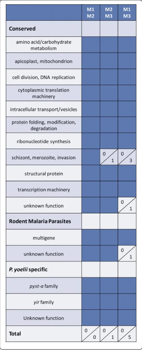
donor mouse. Parasite diversity was minimized by using a clonedP. yoelii17X line. Host diversity was minimized by using age-matched, male, inbred BALB/cByJ mice housed together. Parasite RNA was isolated early during infection when parasitaemia was ascending as follows: mouse 1 -day 11 at 13.0% parasitaemia; mouse 2 - -day 10 at 11.1% parasitaemia; mouse 3 - day 10 at 18.6% parasitaemia. UsingP. yoeliiDNA microarrays (~6,700 oligos), gene ex-pression profiles were then compared between individual mice with three pair-wise comparisons (M1/M2, M2/M3 and M1/M3). In all comparisons, a change in expression of at least two-fold (p ≤0.01) was considered significant. As shown in Figure 1 and Additional file 2, gene expres-sion profiles between individual mice were remarkably consistent. There were no differentially expressed genes between mouse 1 and mouse 2. Only one gene, mero-zoite surface protein-1 (MSP1), was down-regulated ~2.3-fold in mouse 3 relative to mouse 2. There were only five genes differentially expressed when compar-ing mouse 1 and mouse 3 which included MSP1, two rhoptry proteins and two proteins with unknown function. As such, there were minimal differences in gene expression profiles of P. yoelii 17X parasites isolated from three separate hosts after a 10–11day period of replication in vivo.
In the above analysis, no members of theyiror pyst-a
multigene families were differentially expressed between individual animals. To estimate the number of yir and
pyst-a genes expressed in each population of P. yoelii
17X parasites, signal intensity on each set of arrays was normalized and percentiled relative to the entireP. yoelii
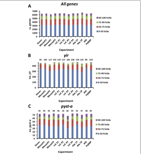
gene set (Figure 3A). Focusing the analysis on a set of 464yir gene oligos that could detect unique transcripts, expression (>50thpercentile) of a relatively large number of yirgenes (105, 117, 143) was measurable in parasites isolated from the three individual mice (Figure 3B). Combined, 20–40 of these were considered to be expressed at moderate (76-90th percentile) or high (>90th percentile) levels. Likewise, based on data obtained with a subset of 74 unique oligos, expres-sion (>50th percentile) of multiple pyst-a genes (34, 35, 38) was observed in the three populations of P. yoelii 17X parasites, with 16–18 detected at moderate to high levels (Figure 3C). Differences in the number of ‘expressed’ yir and pyst-a genes in each group are due to a small number of genes whose signal inten-sity was near the threshold set for detection (50th percentile) but where fold-change was less than two and/or not statistically significant.
Differential gene expression inPlasmodium yoelii17X blood-stage parasites marginally increases over time
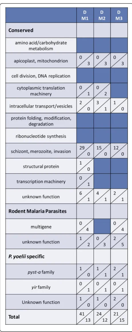
gene expression in the three infected mice described in Figure 1 above (day 10–11) were compared to that of the population of P. yoelii 17X parasites isolated from the donor animal used to initiate infection on day 0. The donor mouse was infected using cryopreserved P. yoelii
[image:6.595.57.290.90.672.2]17X iRBCs. On day 11 of infection in this donor mouse when parasitaemia was 18.7%, P. yoelii17X iRBCs were isolated for infection of M1, M2 and M3 and for RNA isolation. As shown in Figure 4 and Additional file 4, some variation in expression profiles was noted in three pair-wise comparisons between the donor (D) and each infected mouse (M1, M2, M3). Overall however, the number of differentially expressed genes remained
Figure 2Course ofPlasmodium yoelii17X blood stage infection in male BALB/cByJ mice.Animals (n = 5) were infected by intraperitoneal injection with 1x10⁵P. yoelii17X infected RBCs.
Parasitaemia and percent reticulocytes were monitored in thin tail-blood smears stained with Giemsa. Average percent parasitaemia (red) and reticulocytes (blue) at various days post-infection are shown.
Figure 1There is little differential gene expression between
Plasmodium yoelii17X parasites isolated from individual mice.
[image:6.595.305.538.476.654.2]relatively low as the expression of only 54, 36 and 36 genes was significantly altered in M1, M2 and M3 rela-tive to the donor mouse, respecrela-tively. The magnitude of the changes ranged from 2 to 5.8-fold. Genes associated with schizont rupture and/or merozoite invasion of RBCs were consistently up-regulated in mouse 1, 2 and 3 relative to the donor animal, and included P. yoelii
orthologs of several rhoptry proteins, MSP1, MyoA, MTIP, SERA and subtilisin-like protease 2. Similar to the comparisons between individual mice, expression of a fairly large number of the yir (95/464) and pyst-a
(34/74) genes was detected above background (>50th percentile) in the donor mouse (Figure 3). Changes in
yir and pyst-a gene expression from day 0 in the donor mouse to day 10–11 in the three infected mice were not remarkable.
Gene expression profiles inPlasmodium yoelii17X blood-stage parasites remain relatively stable in independently infected mice
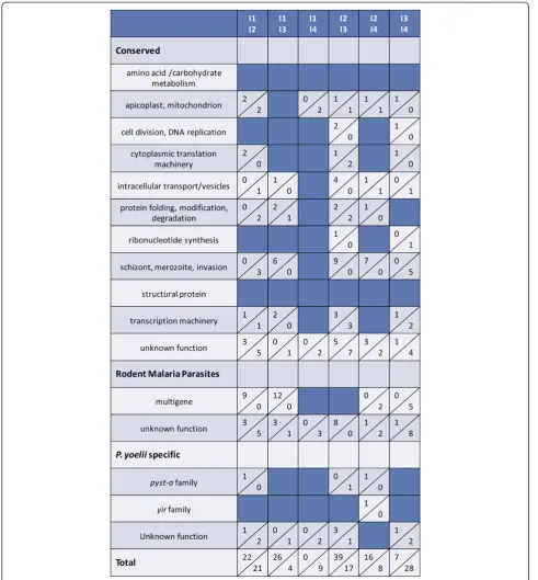
Given the relatively few differences noted between donor and individual infected mice, it was of interest to examine variability in P. yoelii 17X gene expression across independently initiated infections (I1, I2, I3, I4) in animals housed separately in time and space. On four separate occasions, blood from a donor mouse was used to infect groups of BALB/cByJ mice (n=10). For each infection, blood from P. yoelii 17X infected mice was obtained and pooled on days 10–11 of in-fection when parasitaemia was ascending and was ~15%. P. yoelii 17X RNA from these four independ-ent infections permitted six pair-wise comparisons.
Gene expression profiles varied to a greater
[image:8.595.57.288.84.665.2]degree between these independently initiated infec-tions (Figure 5 and Additional file 5) than between individual animals concurrently infected with the same inoculum of P. yoelii 17X iRBCs (Figure 1 and Additional file 2). Nevertheless, differences were again lower than expected with the number of genes differ-entially expressed between any two infections ranging from only 9 to 56 genes. In each case, at least 40% of differentially expressed genes are predicted to encode proteins of unknown function. The remaining genes in each set were associated with multiple and diverse
functions. Consistent with the comparisons thus far, each population of P. yoelii 17X blood-stage parasites expressed a relatively large number of yir genes (105 to 138) and pyst-a genes (32 to 39) over background (>50th percentile) (Figure 3).
Plasmodium yoeliigene expression is influenced by parasitaemia and/or reticulocyte levels
[image:9.595.55.547.88.618.2]Thus far,P. yoeliigene expression was measured early dur-ing acute infection and at consistent parasitaemia. How-ever, P. yoelii17X preferentially invades reticulocytes and
at this early point in infection, the number of reticulocytes in circulation is low (~15-20%). A sharp increase in erythropoiesis leads to a marked influx of new reticulocytes into peripheral circulation and a concurrent rise inP. yoelii
17X parasitaemia. It was of interest to determine if changes in parasite load and permissive host cell availability affected
P. yoeliigene expression in vivo.P. yoelii17X iRBCs from a single donor mouse were used to infect a set of BALB/ cByJ mice. RNA was isolated from P. yoelii iRBCs har-vested on day 10 (14% parasitaemia, 14.4% reticulocytes) and on day 14 (43.5% parasitaemia, 57.9% reticulocytes) of infection. Comparing P. yoelii 17X gene expression pro-files, there were 15 genes significantly up-regulated and 75 genes significantly down-regulated on day 14 relative to day 10 (Figure 6 and Additional file 6). Overall, the magnitude of the changes in gene ex-pression was again modest varying only 2 to 5.7-fold. Putative protein functions appeared to be diverse, with genes encoding proteins of unknown function being the most commonly represented group. These tended to be down-regulated on day 14 relative to day 10. In addition and despite an overall increase in the number of differentially expressed genes, the ex-pression of yir and pyst-a gene families remained relatively constant (Figures 3 and 6).
To further evaluate changes in parasite gene expression during the course of infection,P. yoelii17X RNA was iso-lated on day 10 (19% parasitaemia, 18.6% reticulocytes) and on day 18 (14% parasitaemia, 70.2% reticulocytes). Al-though parasite load is similar at these time points, parasit-aemia is ascending on day 10 and target host cells are relatively scarce. By day 18 post-infection, parasitaemia is declining due to immune-mediated mechanisms and reti-culocytes are abundant. Comparing day 10 and day 18, there were 192 differentially expressed P. yoelii genes, in-cluding 67 genes of unknown function (Figure 6 and Additional file 6). Of those differentially expressed, 94 were up-regulated and 98 were down-regulated on day 18 versus day 10. This set of differentially expressed genes represents the most variable expression seen across all ex-perimental conditions. Of interest, genes encoding pro-teins associated with the cytoplasmic translation machinery tended to be down-regulated on day 18 (n = 11) versus day 10, and included several ribosomal proteins. In addition, a small set of genes (n = 24) encod-ing proteins involved with schizont rupture and/or mero-zoite invasion of RBCs tended to be up-regulated on day 18 and these included glidosome- and rhoptry-associated genes, MSP1 and MSP7. Finally, there were 17 genes en-coding apicoplast/mitochondrion-associated proteins that were differentially expressed, with up and down-regulated genes equally represented. As before, members the yir
[image:10.595.57.289.86.589.2]and pyst-a multigene families were not differentially expressed while the number of genes in each family
Figure 6Parasitaemia and/or host cell availability influence differential gene expression ofPlasmodium yoelii17X parasites.
Groups of wild-type animals (n=5-10) were infected withP. yoelii17X iRBCs and parasite RNA was isolated on day 10, day 14 or day 18 post-infection. Immunologically intact (WT) and B-cell deficient JHD
expressed on day 18 remained stable (Figure 3, 155 yir
genes, 37pyst-agenes).
Changes in immune pressure do not significantly alter Plasmodium yoelii17X gene expression during a primary infection
To specifically evaluate whether the presence or absence of host immune pressure affects parasite gene expression
in vivo, two experimental approaches were taken. In the first approach, all antibody-dependent immune pressure was removed by use of B cell deficient mice. Naïve, im-munocompetent BALB/c mice and B cell deficient, JHD
mice on a BALB/c background were concurrently infected with P. yoelii 17X iRBCs obtained from the same donor animal.P. yoeliigene expression in the two hosts was evaluated when ascending parasitaemia reached ~15%. Surprisingly, changes in gene expression were negligible with only seven differentially expressed genes, five of which were of unknown function (Figure 6 and Additional file 6). Noyirorpyst-agenes were differ-entially expressed between the populations of P. yoelii
17X parasites.
In a second approach, antibody-mediated immune pressure was applied by immunizing animals with a preparation of membrane proteins isolated fromP. yoelii
17X infected reticulocytes (P. yoelii reticulocyte mem-brane proteins, PyRMPs). SDS-PAGE and Coomassie-blue staining of proteins associated with the membranes of uninfected versusP. yoelii17X infected RBCs revealed several protein bands unique to the PyRMP fraction
(Figure 7A, lanes 1 & 2). Immunoblot analysis to assess separation of reticulocyte membrane proteins from para-site membrane proteins showed that the GPI-anchored merozoite surface protein 8 was present in parasite but not reticulocyte membrane fractions (Figure 7B). Anti-bodies from mice immunized with the PyRMP prepar-ation recognized 8–10 distinct polypeptides present in thePyRMP fraction (Figure 8). In indirect immunofluor-escence assays, these antibodies specifically stained pro-teins localized to both the intracellular parasite and the iRBC membrane (Figure 9). To determine if the pres-ence of PyRMP-specific antibodies affected parasite growth and gene expression,PyRMP immunized and ad-juvant control (Quil A) mice were challenged with P. yoelii17X iRBCs from the same donor mouse. To some degree, PyRMP immunization limited parasite replica-tion early during infecreplica-tion but over time these animals developed relatively high and persistent parasitaemia and were sacrificed (Figure 10). To determine if the presence of anti-PyRMP antibodies influenced patterns of gene expression, RNA was isolated fromP. yoelii17X parasites on day 10 from Quil A control mice (15.6% parasitaemia) or day 12 from PyRMP immunized mice (13.3% parasitaemia) post-infection and subjected to microarray analysis (Figure 6 and Additional file 6). Similar to the results in JHD mice in the absence of
[image:11.595.57.293.88.218.2]anti-body responses, increasing immune pressure by PyRMP immunization led to negligible differences in gene ex-pression. Comparison of expression profiles of P. yoelii Figure 7Membrane preparations fromPlasmodium
yoelii-infected RBCs contain unique parasite proteins.Animals were infected withP. yoelii17X and infected RBCs were collected. (A). Proteins associated with the membranes of uninfected (lane 1) orP. yoelii17X infected erythrocytes (lane 2) or with intracellular P. yoelii17X parasites (lane 3) were separated by SDS-PAGE and stained with Coomassie Blue. (B). To evaluate the ability to separate thePyRMPs from parasite associated proteins, isolated protein fractions were separated as in A, blotted onto nitrocellulose and probed with rabbit anti-mousePyMSP8 polyclonal sera.PyMSP8 is a GPI-anchored membrane protein ofP. yoeliitrophozoites and merozoites. Normal rabbit sera (NRS) served as a negative control. Molecular weights in kiloDaltons (kDa) are indicated.
[image:11.595.305.539.450.641.2]17X parasites isolated from Quil A versusPyRMP immu-nized mice revealed only 10 differentially expressed genes including three with no known function. Unexpectedly, no members of theyirorpyst-amulti-gene families were differentially expressed. Consistent with the earlier data however, expression (>50th percentile) of 134 yir genes
and 36pyst-a genes in Quil A control mice and 143 yir
genes and 36 genes pyst-a in PyRMP immunized mice was detected (Figure 3, Inf. #1 &PyRMP). Combined, the data indicate that in vivo, P. yoelii 17X gene expression was only minimally altered by increasing or decreasing antibody-mediated immune pressure.
Discussion
Transcription in P. falciparum and P. vivax parasites appears to be tightly regulated, resulting in a‘continuous cascade’of gene expression during blood-stage develop-ment [4,5,45]. There is some evidence that this tran-scriptional programme continues unabated even in the presence of external stresses. In one study, anti-folate treatment of cultures of drug-sensitive P. falciparum
blood-stage parasites did not lead to increased expres-sion of the defined drug target, dihydrofolate reductase-thymidylate synthatase. Additionally, there were no genes differentially regulated greater than two-fold be-tween control and drug treated cultures [46]. Similarly, Gunasekeraet al.[47] found little variation in transcrip-tion patterns between untreated and chloroquine-treated
P. falciparum parasites growing asynchronously in cul-ture. These results were unexpected, as genome-wide studies in organisms such as Saccharomyces cerevisiae,
Mycobacterium tuberculosis and Candida albicans
revealed differential expression of hundreds of genes in response to drug treatment, and these changes often confirmed or revealed associated drug targets and affected pathways [48-51]. However, other studies in
Plasmodiumexamining the effects of drug treatment or manipulation of culture conditions have found more substantial transcriptional changes (~300-400 differen-tially expressed genes), especially in genes predicted to encode proteins exported to the host cell cytoplasm and the RBC membrane [52,53]. The work shown here extends these efforts significantly by focusing on malaria parasites replicating in vivo, in a more complex setting. WithP. yoelii 17X blood-stage parasites, global gene ex-pression profiles were remarkably consistent even in the presence of a changing host environment.
[image:12.595.58.291.89.292.2]Initially, three genetically identical, age-matched BALB/cByJ mice were infected with the same inoculum ofP. yoelii 17X iRBCs obtained from a donor mouse.P. yoelii RNA was isolated from each animal after a 10– 11 day period of replication in vivo. As the in vivo envir-onment is expected to be comparable in this setting, only a small set of differentially expressed genes was anticipated. Parasite gene expression patterns in each of the three animals were virtually identical with only five to six differentially expressed genes. Between the initi-ation of infection and harvest of iRBCs for RNA isola-tion, P. yoelii parasites in this study completed 10–12 replication cycles in vivo. Over this time, the number of
Figure 9Anti-PyRMP antibodies detectPlasmodium yoelii
proteins associated with the intracellular parasite and the reticulocyte membrane.Immunofluorescence ofP. yoelii17X infected reticulocytes using polyclonalPyRMP sera or normal mouse sera. Row A: Differential interference contrast images; Row B: Parasite DNA stained with DAPI (blue); Row C: Localization ofP. yoelii proteins recognized by anti-PyRMP sera (TRITC, red). Background staining with normal mouse sera (NMS) is shown.
Figure 10Course ofPlasmodium yoelii17X infection following
PyRMP immunization.Animals (n = 5) were immunized three times with thePyRMP preparation plus Quil A adjuvant. A separate group of animals received Quil A alone. Both groups were challenged with 1x10⁵P. yoelii17X iRBCs intraperitoneally. Parasitaemia was
[image:12.595.58.291.476.635.2]differentially expressed genes was expected to increase somewhat relative to the population of P. yoelii iRBCs used to initiate the infection. This was the case, but the increase was relatively small involving only 40–60 genes out of the set represented by ~ 6,700 oligonucleotides on the arrays. A small cluster of genes expressed in late-stage parasites and potentially involved in merozoite in-vasion of host cells were differentially expressed between the donor mouse and recipients. This may not be sur-prising as iRBCs were harvested early during ascending parasitaemia when P. yoelii 17X parasites can be found in varying degrees in both normocytes and reticulocytes. This change in gene expression may reflect utilization of multiple invasion pathways. Finally, a number of poten-tially influencing variables were further increased by com-paring gene expression profiles ofP. yoelii17X blood-stage parasites harvested from four independent infections initiated with iRBCs obtained from separate donor mice. Remarkably, the number of differentially expressed genes in pair-wise comparisons remained low (10–50 genes). These represent genes that are involved in diverse or un-known functions and at present, the associated changes do not appear to reflect a biologically significant response to the host environment. This, combined with a false discov-ery rate of 1% associated with this analysis suggest that de-tection of a low number of differentially expressed but largely unrelated genes could simply represent ‘noise’ in-herent to large microarray analysis.
Non-lethal P. yoelii 17X parasites replicate preferen-tially in reticulocytes but do invade and develop within mature RBCs when reticulocytes are limiting. Parasites harvested on day 10 of P. yoelii 17X infection will be present in both reticulocytes and normocytes. As a result of an influx of new reticulocytes into circulation,P. yoelii
17X parasites will be found almost exclusively in reticu-locytes by day 14 post-infection. As such, substantial dif-ferences in patterns of P. yoelii 17X gene expression in parasites harvested on day 10 versus day 14 were expected. In addition to the shift in host cell tropism, the ongoing infection increases parasite burden, host stress and immune pressure that could also influenceP. yoelii17X expression profiles. While the number of dif-ferentially expressed genes in this comparison increased to 95, these were not clustered based on related function or biological process and the majority were of unknown function. In an early study, utilizing lethalP. yoelii17XL parasites, a similar number of genes altered in associ-ation with a shift in host cell preference from normo-cytes to reticulonormo-cytes were identified [54]. The function of these genes will be revisited as the annotation of both
P. falciparum and P. yoelii genomes progresses. To fur-ther examine the role of host cell availability and immune pressure, gene expression in parasites isolated on day 10 and day 18 of infection was compared. Parasitaemia at
these two time points is comparable. However on day 18, 70-75% of RBCs in circulation are reticulocytes andP. yoe-lii iRBCs are rapidly being cleared from circulation by immune-mediated mechanisms. Nearly 200 genes were differentially expressed in P. yoeliiiRBCs on day 18 rela-tive to day 10 of infection; the largest set in this study. On day 18, several genes encoding components of the cyto-plasmic translation machinery were down-regulated while a second set associated with merozoite invasion were up-regulated. InP. falciparum, transcription of genes encod-ing the cytoplasmic translation machinery generally peaks in ring-stage parasites at ~12 hours post-invasion, fol-lowed by a marked down-regulation in trophozoite-and schizont-stage parasites. In contrast, transcription of genes involved in schizont rupture and merozoite in-vasion is relatively low through most of the asexual cycle, peaking at ~42 hours post-invasion [4,5]. Differ-ences noted in gene expression inP. yoeliiparasites on day 18 versus day 10 of infection may partially be explained by a modest shift noted in distribution of parasite stages in circulation toward increased schi-zonts and decreased trophozoites. It is also clear that infected blood isolated on day 18 contains damaged host cells and dying parasites, as well as free extracellu-lar parasites. These factors likely contributed to a mixed alteration in expression profiles at this late time point during infection.
To focus on the role of the host immune response on parasite gene expression, two approaches to actively alter host immune pressure were taken. In the first approach, all antibody mediated immune pressure was removed by infecting B cell deficient JHD mice. It was expected that
in the absence of antibodies, P. yoelii growth would be unchecked with an increase in the diversity of parasite populations and gene expression profiles. This did not occur as only seven genes were differentially expressed when P. yoelii parasites isolated from B cell deficient JHD mice were compared with those from
In addition to addressing global changes in gene expres-sion, the expression of members of two multi-gene families in P. yoelii, the yirand pyst-a were of particular interest. Previous studies have shown that at a population level, many yir genes are transcribed during blood-stage infec-tion, in a seemingly random order. Transcription at the level of individual parasites appears to be tightly controlled, with each parasite transcribing between one and three yir
genes [55]. In the present study, significant differences in the expression ofyir family members across experimental conditions were not observed, including the comparison of
P. yoelii17X day 10 and day 18 parasites. This is in agree-ment with the study by Cunninghamet al.examining the expression of a subset of theyirrepertoire on days 12 and 18 post-infection [20]. Of note, expression of a fairly large number of the yir genes (89–155) was consistently observed using signal intensity greater than the 50th per-centile on each array as a benchmark. These expressedyir
genes were distributed across five previously reported phylogenetic groups, with no obvious bias toward any one group [56].
The expression ofpyst-agenes followed a similar pattern to theyirfamily, with little to no differential expression be-tween conditions. It appears that a large proportion of pyst-afamily members are expressed in a mixed blood-stage in-fection. Whetherpyst-agene expression is more restricted in individual parasites has not been determined. Noyiror
pyst-a members were differentially expressed in wild-type versus JHD animals, in agreement with a study examining
expression of a subset of yirmembers in wild-type versus Rag2 knockout mice, which lack mature B and T cells [20]. A more extensive analysis ofyirandpyst-aexpression dur-ing primary and secondary infections usdur-ing a microarray approach would be of interest. It has been suggested that the polymorphic erythrocyte membrane antigens encoded by theyirand/orpyst-amultigene families may function as part of a parasite immune evasion strategy. For immune evasion, the current data suggest that changing the pattern of gene expression may be less important than showering the host immune system with a large repertoire of poly-morphic antigens at any given time. In fact, prior immunization of mice with the PyRMP preparation may have further impeded the development of protective im-mune responses following challenge infection as these ani-mals developed a persistent infection which was difficult to clear. In agreement with the current findings, members of the cir multi-gene family do not seem to be differentially expressed during the course of blood-stageP. chabaudi in-fection, but there is some indication thatcir gene expres-sion varies in parasites localized to different host tissues [21]. However, it is still possible that much like theP. falcip-arumEMP1 and thevargene family, theyirand/orpyst-a
encoded proteins may possess specific functions required for parasite growth and development in vivo.
Overall, in vivo, P. yoelii17X gene expression did not appear to be appreciably influenced by the host environ-ment. These data in the P. yoelii 17X model differ from that reported by Dailyet al.[7] who detected distinct pat-terns of gene expression inP. falciparumparasites isolated from malaria-infected patients. In light of these data, changes in the expression of genes encoding a set of mito-chondrial proteins (n = 148), across all experimental con-ditions was examined. Significant changes that would be consistent with distinct physiological states or a response to environmental stress were not observed (Additional file 3). Although parasite gene expression was assessed in vivo, theP. yoeliimodel is still less complex than with
P. falciparum infected human subjects. Here, a single cloned line ofP. yoelii17X that does not produce gameto-cytes was utilized to simultaneously infect genetically identical mice housed in environmentally controlled, spe-cific pathogen free conditions. In some regards, this lack of gametocyte-stage parasites is advantageous, as detec-tion of differential gene expression due to varying gameto-cyte levels across samples can be ruled out. On the other hand, the repeated passage of thisP. yoelii17X line in the vertebrate host and/or the lack of exposure to the mos-quito vector could have altered regulatory mechanisms that control expression of certain gene subsets (i.e.yiror
pyst-agenes). It is also possible that the greater diversity in human hosts (genetic, environmental) may have a greater influence on parasite gene expression than we observed in theP. yoeliimodel. Finally, each isolate ofP. falciparummay exhibit unique elements of a‘hard-wired’ programme of gene expression that can be detected when comparing isolates obtained from individual malaria patients. Co-infection of human subjects with such dis-tinct P. falciparum clones will increase diversity in the overallP. falciparumgene expression profile detected in a single host and may allow preferential growth ofP. falcip-arumclones in specific in vivo environments.
Conclusions
Additional files
Additional file 1:yirandpyst-aoligonucleotides onPlasmodium
yoeliiDNA microarrays that mapped to a single gene.In assessing the number ofyirandpyst-agenes expressed in a given sample, only data obtained withyirandpyst-aoligos that mapped to a single gene were considered. Mapping data available on PlasmoDB [3] was used to focus the analysis on 464/859yiroligos (top portion of Table) and 74/140 pyst-aoligos (bottom portion of Table) onP. yoeliiDNA microarrays.
Additional file 2:Differential gene expression betweenPlasmodium yoelii17X parasites isolated from individual mice.Three animals (M1, M2 and M3) were infected withP. yoelii17X iRBCs from a single donor mouse and gene expression analysed usingP. yoeliiDNA microarrays. Of three possible pair-wise comparisons, significant differential gene expression was only seen between M2 and M3 (black text) and M1 versus M3 (blue text). For each differentially expressed gene, oligo ID and gene name are listed [3]. Log2Fold Changes and adjustedp-values are
also included, as are the predicted number of amino acids (AA) and molecular weight in Daltons (MW). Where available,P. falciparum orthologs and associated Plasmodb.org accession numbers are included (Pf Ortholog # and Pf Name). Functional groupings (Category) are listed at the far right of the Table. For detailed information regarding data analysis and gene categorization, please see the Methods.
Additional file 3:Comparative expression ofPlasmodium yoelii
genes encoding mitochondria associated proteins.Data on the differential expression of genes encoding mitochondria associated proteins is shown for all comparisons. The gene set includedP. yoelii orthologs of putativeP. falciparummitochondrial proteins [7,41] Mather and Vaidya, unpublished data] for which signal intensity across arrays was consistently above the 50thpercentile. Shown are theP. falciparumGene
ID and Name,P. yoeliiOligo ID and Name and for each comparison, the corresponding the log2fold change and adjustedp-value. Log2fold
change values >1 are shaded in red, log2fold change values <−1 are
shaded in green and adjustedp-values <0.01 are shaded in yellow.
Additional file 4:Differential gene expression between parasites isolated from a single donor mouse and three individual animals.
Three animals (M1, M2 and M3) were infected withP. yoelii17X iRBCs from a single donor mouse (D) and gene expression analysed using P. yoeliiDNA microarrays. Three pair-wise comparisons were possible: D versus M1 (black text), D versus M2 (blue text) and D versus M3 (red text). For differentially expressed genes, oligo ID, gene name, log2fold change,
adjustedp-value, predicted number of amino acids, predicted MW, availableP. falciparumortholog information and functional categories are provided as described for Additional file 2.
Additional file 5:Differential gene expression betweenPlasmodium yoelii17X parasites isolated from independent infections.Groups of animals were infected on four separate occasions (I1, I2, I3 and I4) using four separate donor animals and gene expression analysed usingP. yoelii DNA microarrays. Six possible pair-wise comparisons were considered: I1 versus I2 (black text), I1 versus I3 (blue text), I1 versus I4 (red text), I2 versus I3 (green text), I2 versus I4 (purple text) and I3 versus I4 (blue text). For differentially expressed genes, oligo ID, gene name, log2fold change,
adjustedp-value, predicted number of amino acids, predicted MW, availableP. falciparumortholog information and functional categories are provided as described for Additional file 2.
Additional file 6:Differential gene expression on days 10/14, 10/18 and in the absence/presence of host antibody responses.Groups of wild-type animals were infected withP. yoelii17X iRBCs and parasite RNA was isolated on day 10 (D10), day 14 (D14) or day 18 (D18) post-infection. Immunologically intact (WT) and B-cell deficient JHD (JHD)
animals were similarly infected and parasite RNA was isolated on day 10. A third set of animals was immunized three times with a preparation of P. yoeliireticulocyte membrane proteins plus Quil A as adjuvant (RMP) or with Quil A alone (QA) prior toP. yoelii17X challenge. Parasite RNA was then isolated on day 10 (QA) or day 12 (RMP) post-infection. Gene expression was analysed usingP. yoeliiDNA microarrays and the following comparisons were made: D10 versus D14 (black text), D10 versus D18 (blue text), WT versus JHD (red text), and QA versus RMP
(green text). For differentially expressed genes, oligo ID, name, log2fold
change, adjustedp-value, predicted number of amino acids, predicted MW, availableP. falciparumortholog information and functional categories are provided as described for Additional file 2.
Abbreviations
CON-A/CM: Conserved, amino acid/carbohydrate metabolism;
CON-AP/M: Conserved, apicoplast, mitochondrion; CON-CD: Conserved, cell division, DNA replication; CON-CTM: Conserved, cytoplasmic translation machinery; CON-ITV: Conserved, intracellular transport/vesicles; CON-PM: Conserved, protein folding, modification, degradation;
CON-RS: Conserved, ribonucleotide synthesis; CON-SMI: Conserved, schizont, merozoite, invasion; CON-SP: Conserved, structural protein;
CON-TM: Conserved, transcription machinery; CON-UF: Conserved, unknown function; RMP-MG: Rodent malaria parasites, multigene family; RMP-UF: Rodent malaria parasites, unknown function; PY-pyst-a:P. yoelii specific,pyst-afamily; PY-yir:P. yoeliispecific,yirfamily; PY-UF:P. yoelii specific, unknown function.
Competing interests
The authors declare that they have no competing interests.
Authors’contributions
ACO conceived, designed and performed the experiments and wrote the paper. JMB conceived and designed experiments, analyzed the data, and wrote the paper. ABV analyzed the data. LWB analyzed the data and contributed reagents/materials. TMD contributed reagents/materials. All authors read and approved the final manuscript.
Acknowledgements
This work was supported by NIH-NIAID grant AI069147 (JMB). The funders had no role in study design, data collection and analysis, decision to publish or preparation of the manuscript.
Received: 24 January 2012 Accepted: 22 July 2012 Published: 6 August 2012
References
1. Baird JK:Host age as a determinant of naturally acquired immunity to Plasmodium falciparum.Parasitol Today1995,11:105–111.
2. Gardner MJ, Hall N, Fung E, White O, Berriman M, Hyman RW, Carlton JM, Pain A, Nelson KE, Bowman S, Paulsen IT, James K, Eisen JA, Rutherford K, Salzberg SL, Craig A, Kyes S, Chan MS, Nene V, Shallom SJ, Suh B, Peterson J, Angiuoli S, Pertea M, Allen J, Selengut J, Haft D, Mather MW, Vaidya AB, Martin DM, Fairlamb AH, Fraunholz MJ, Roos DS, Ralph SA, McFadden GI, Cummings LM, Subramanian GM, Mungall C, Venter JC, Carucci DJ, Hoffman SL, Newbold C, Davis RW, Fraser CM, Barrell B:Genome sequence of the human malaria parasitePlasmodium falciparum.Nature2002, 419:498–511.
3. Aurrecoechea C, Brestelli J, Brunk BP, Dommer J, Fischer S, Gajria B, Gao X, Gingle A, Grant G, Harb OS, Heiges M, Innamorato F, Iodice J, Kissinger JC, Kraemer E, Li W, Miller JA, Nayak V, Pennington C, Pinney DF, Roos DS, Ross C, Stoeckert CJ Jr, Treatman C, Wang H:PlasmoDB: a functional genomic database for malaria parasites.Nucleic Acids Res2009,37:D539–543. 4. Bozdech Z, Llinas M, Pulliam BL, Wong ED, Zhu J, DeRisi JL:The
transcriptome of the intraerythrocytic developmental cycle of Plasmodium falciparum.PLoS Biol2003,1:e5.
5. Llinas M, Bozdech Z, Wong ED, Adai AT, DeRisi JL:Comparative whole genome transcriptome analysis of threePlasmodium falciparumstrains. Nucleic Acids Res2006,34:1166–1173.
6. LeRoux M, Lakshmanan V, Daily JP:Plasmodium falciparumbiology: analysis of in vitro versus in vivo growth conditions.Trends Parasitol2009, 25:474–481.
8. Lemieux JE, Gomez-Escobar N, Feller A, Carret C, Amambua-Ngwa A, Pinches R, Day F, Kyes SA, Conway DJ, Holmes CC, Newbold CI:Statistical estimation of cell-cycle progression and lineage commitment in Plasmodium falciparumreveals a homogeneous pattern of transcription in ex vivo culture.Proc Natl Acad Sci U S A2009,106:7559–7564. 9. Kraemer SM, Smith JD:A family affair: var genes, PfEMP1 binding, and
malaria disease.Curr Opin Microbiol2006,9:374–380.
10. Trimnell AR, Kraemer SM, Mukherjee S, Phippard DJ, Janes JH, Flamoe E, Su XZ, Awadalla P, Smith JD:Global genetic diversity and evolution of var genes associated with placental and severe childhood malaria.Mol Biochem Parasitol2006,148:169–180.
11. Bull PC, Buckee CO, Kyes S, Kortok MM, Thathy V, Guyah B, Stoute JA, Newbold CI, Marsh K:Plasmodium falciparumantigenic variation. Mapping mosaic var gene sequences onto a network of shared, highly polymorphic sequence blocks.Mol Microbiol2008,68:1519–1534. 12. Carlton JM, Angiuoli SV, Suh BB, Kooij TW, Pertea M, Silva JC, Ermolaeva MD,
Allen JE, Selengut JD, Koo HL, Peterson JD, Pop M, Kosack DS, Shumway MF, Bidwell SL, Shallom SJ, van Aken SE, Riedmuller SB, Feldblyum TV, Cho JK, Quackenbush J, Sedegah M, Shoaibi A, Cummings LM, Florens L, Yates JR, Raine JD, Sinden RE, Harris MA, Cunningham DA, Preiser PR, Bergman LW, Vaidya AB, van Lin LH, Janse CJ, Waters AP, Smith HO, White OR, Salzberg SL, Venter JC, Fraser CM, Hoffman SL, Gardner MJ, Carucci DJ: Genome sequence and comparative analysis of the model rodent malaria parasitePlasmodium yoelii yoelii.Nature2002,419:512–519. 13. Hall N, Karras M, Raine JD, Carlton JM, Kooij TW, Berriman M, Florens L,
Janssen CS, Pain A, Christophides GK, James K, Rutherford K, Harris B, Harris D, Churcher C, Quail MA, Ormond D, Doggett J, Trueman HE, Mendoza J, Bidwell SL, Rajandream MA, Carucci DJ, Yates JR 3rd, Kafatos FC, Janse CJ, Barrell B, Turner CM, Waters AP, Sinden RE:A comprehensive survey of the Plasmodium life cycle by genomic, transcriptomic, and proteomic analyses.Science2005,307:82–86.
14. Pain A, Bohme U, Berry AE, Mungall K, Finn RD, Jackson AP, Mourier T, Mistry J, Pasini EM, Aslett MA, Balasubrammaniam S, Borgwardt K, Brooks K, Carret C, Carver TJ, Cherevach I, Chillingworth T, Clark TG, Galinski MR, Hall N, Harper D, Harris D, Hauser H, Ivens A, Janssen CS, Keane T, Larke N, Lapp S, Marti M, Moule S, Meyer IM, Ormond D, Peters N, Sanders M, Sanders S, Sargeant TJ, Simmonds M, Smith F, Squares R, Thurston S, Tivey AR, Walker D, White B, Zuiderwijk E, Churcher C, Quail MA, Cowman AF, Turner CM, Rajandream MA, Kocken CH, Thomas AW, Newbold CI, Barrell BG, Berriman M:The genome of the simian and human malaria parasitePlasmodium knowlesi.Nature2008,455:799–803.
15. Carlton JM, Adams JH, Silva JC, Bidwell SL, Lorenzi H, Caler E, Crabtree J, Angiuoli SV, Merino EF, Amedeo P, Cheng Q, Coulson RM, Crabb BS, Del Portillo HA, Essien K, Feldblyum TV, Fernandez-Becerra C, Gilson PR, Gueye AH, Guo X, Kang'a S, Kooij TW, Korsinczky M, Meyer EV, Nene V, Paulsen I, White O, SA R, Ren Q, Sargeant TJ, Salzberg SL, Stoeckert CJ, Sullivan SA, Yamamoto MM, Hoffman SL, Wortman JR, Gardner MJ, Galinski MR, Barnwell JW, Fraser-Liggett CM:Comparative genomics of the neglected human malaria parasitePlasmodium vivax.Nature2008,455:757–763. 16. Janssen CS, Barrett MP, Turner CM, Phillips RS:A large gene family for
putative variant antigens shared by human and rodent malaria parasites. Proc Biol Sci2002,269:431–436.
17. Janssen CS, Phillips RS, Turner CM, Barrett MP:Plasmodium interspersed repeats: the major multigene superfamily of malaria parasites.Nucleic Acids Res2004,32:5712–5720.
18. Chen Q, Fernandez V, Sundstrom A, Schlichtherle M, Datta S, Hagblom P, Wahlgren M:Developmental selection of var gene expression in Plasmodium falciparum.Nature1998,394:392–395.
19. Scherf A, Hernandez-Rivas R, Buffet P, Bottius E, Benatar C, Pouvelle B, Gysin J, Lanzer M:Antigenic variation in malaria: in situ switching, relaxed and mutually exclusive transcription of var genes during intra-erythrocytic development inPlasmodium falciparum.EMBO J1998,17:5418–5426. 20. Cunningham DA, Jarra W, Koernig S, Fonager J, Fernandez-Reyes D, Blythe
JE, Waller C, Preiser PR, Langhorne J:Host immunity modulates transcriptional changes in a multigene family (yir) of rodent malaria.Mol Microbiol2005,58:636–647.
21. Ebbinghaus P, Krucken J:Characterization and tissue-specific expression patterns of thePlasmodium chabaudicir multigene family.Malar J2011, 10:272.
22. Fernandez-Becerra C, Pein O, de Oliveira TR, Yamamoto MM, Cassola AC, Rocha C, Soares IS, de Braganca Pereira CA, del Portillo HA:Variant proteins
ofPlasmodium vivaxare not clonally expressed in natural infections.Mol Microbiol2005,58:648–658.
23. Gabriel JA, Holmquist G, Perlmann H, Berzins K, Wigzell H, Perlmann P: Identification of aPlasmodium chabaudiantigen present in the membrane of ring stage infected erythrocytes.Mol Biochem Parasitol 1986,20:67–75.
24. Wanidworanun C, Barnwell JW, Shear HL:Protective antigen in the membranes of mouse erythrocytes infected withPlasmodium chabaudi. Mol Biochem Parasitol1987,25:195–201.
25. Wunderlich F, Brenner HH, Helwig M:Plasmodium chabaudimalaria: protective immunization with surface membranes of infected erythrocytes.Infect Immun1988,56:3326–3328.
26. Wunderlich F, Helwig M, Schillinger G, Speth V, Wiser MF:Expression of the parasite protein Pc90 in plasma membranes of erythrocytes infected withPlasmodium chabaudi.Eur J Cell Biol1988,47:157–164.
27. Wiser MF, Leible MB, Plitt B:Acidic phosphoproteins associated with the host erythrocyte membrane of erythrocytes infected withPlasmodium bergheiandP. chabaudi.Mol Biochem Parasitol1988,27:11–21. 28. Wiser MF, Giraldo LE, Schmitt-Wrede HP, Wunderlich F:Plasmodium
chabaudi: immunogenicity of a highly antigenic glutamate-rich protein. Exp Parasitol1997,85:43–54.
29. Giraldo LE, Grab DJ, Wiser MF:Molecular characterization of aPlasmodium chabaudierythrocyte membrane-associated protein with glutamate-rich tandem repeats.J Eukaryot Microbiol1998,45:528–534.
30. Chen J, Trounstine M, Alt FW, Young F, Kurahara C, Loring JF, Huszar D: Immunoglobulin gene rearrangement in B cell deficient mice generated by targeted deletion of the JH locus.Int Immunol1993,5:647–656. 31. Smyth GK:Linear models and empirical bayes methods for assessing
differential expression in microarray experiments.Stat Appl Genet Mol Biol 2004,3:3.
32. Smyth GK, Michaud J, Scott HS:Use of within-array replicate spots for assessing differential expression in microarray experiments.Bioinformatics 2005,21:2067–2075.
33. Wettenhall JM, Smyth GK:limmaGUI: a graphical user interface for linear modeling of microarray data.Bioinformatics2004,20:3705–3706. 34. R:A Language and Environment for Statistical Computing.
http://www.R-project.org.
35. Ritchie ME, Silver J, Oshlack A, Holmes M, Diyagama D, Holloway A, Smyth GK:A comparison of background correction methods for two-colour microarrays.Bioinformatics2007,23:2700–2707.
36. Silver JD, Ritchie ME, Smyth GK:Microarray background correction: maximum likelihood estimation for the normal-exponential convolution. Biostatistics2009,10:352–363.
37. Smyth GK, Speed T:Normalization of cDNA microarray data.Methods 2003,31:265–273.
38. Benjamini Y, Hochberg Y:Controlling the false discovery rate: a practical and powerful approach to multiple testing.J Roy Statist Soc Ser B (Methodological)1995,57:289–300.
39. Hu G, Cabrera A, Kono M, Mok S, Chaal BK, Haase S, Engelberg K, Cheemadan S, Spielmann T, Preiser PR, Gilberger TW, Bozdech Z: Transcriptional profiling of growth perturbations of the human malaria parasitePlasmodium falciparum.Nat Biotechnol2010,28:91–98. 40. Liew KJ, Hu G, Bozdech Z, Peter PR:Defining species specific genome
differences in malaria parasites.BMC Genomics2010,11:128. 41. Mather MW, Henry KW, Vaidya AB:Mitochondrial drug targets in
apicomplexan parasites.Curr Drug Targets2007,8:49–60. 42. Gene Expression Omnibus Database. www.ncbi.nlm.nih.gov/geo.
43. Brazma A, Hingamp P, Quackenbush J, Sherlock G, Spellman P, Stoeckert C, Aach J, Ansorge W, Ball CA, Causton HC, Gaasterland T, Glenisson P, Holstege FC, Kim IF, Markowitz V, Matese JC, Parkinson H, Robinson A, Sarkans U, Schulze-Kremer S, Stewart J, Taylor R, Vilo J, Vingron M: Minimum information about a microarray experiment (MIAME)-toward standards for microarray data.Nat Genet2001,29:365–371.
44. Minimal information about a microarray experiment (MIAMI). http://www. mged.org/Workgroups/MIAME/miame.html.
45. Bozdech Z, Mok S, Hu G, Imwong M, Jaidee A, Russell B, Ginsburg H, Nosten F, Day NP, White NJ, Carlton JM, Preiser PR:The transcriptome ofPlasmodium vivaxreveals divergence and diversity of transcriptional regulation in malaria parasites.Proc Natl Acad Sci U S A2008,105:16290–16295.
transcriptome inPlasmodium falciparumfails to mount protective responses to lethal antifolates.PLoS Pathog2008,4:e1000214. 47. Gunasekera AM, Myrick A, Le Roch K, Winzeler E, Wirth DF:Plasmodium
falciparum: genome wide perturbations in transcript profiles among mixed stage cultures after chloroquine treatment.Exp Parasitol2007, 117:87–92.
48. De Backer MD, Ilyina T, Ma XJ, Vandoninck S, Luyten WH, Vanden Bossche H:Genomic profiling of the response ofCandida albicansto itraconazole treatment using a DNA microarray.Antimicrob Agents Chemother2001, 45:1660–1670.
49. Bammert GF, Fostel JM:Genome-wide expression patterns in Saccharomyces cerevisiae: comparison of drug treatments and genetic alterations affecting biosynthesis of ergosterol.Antimicrob Agents Chemother2000,44:1255–1265.
50. Boshoff HI, Myers TG, Copp BR, McNeil MR, Wilson MA, Barry CE 3rd:The transcriptional responses ofMycobacterium tuberculosisto inhibitors of metabolism: novel insights into drug mechanisms of action.J Biol Chem 2004,279:40174–40184.
51. Wilson M, DeRisi J, Kristensen HH, Imboden P, Rane S, Brown PO, Schoolnik GK:Exploring drug-induced alterations in gene expression in
Mycobacterium tuberculosisby microarray hybridization.Proc Natl Acad Sci U S A1999,96:12833–12838.
52. Natalang O, Bischoff E, Deplaine G, Proux C, Dillies MA, Sismeiro O, Guigon G, Bonnefoy S, Patarapotikul J, Mercereau-Puijalon O, Coppee JY, David PH: Dynamic RNA profiling inPlasmodium falciparumsynchronized blood stages exposed to lethal doses of artesunate.BMC Genomics2008,9:388. 53. Oakley MS, Kumar S, Anantharaman V, Zheng H, Mahajan B, Haynes JD,
Moch JK, Fairhurst R, McCutchan TF, Aravind L:Molecular factors and biochemical pathways induced by febrile temperature in
intraerythrocyticPlasmodium falciparumparasites.Infect Immun2007, 75:2012–2025.
54. Shi Q, Cernetich A, Daly TM, Galvan G, Vaidya AB, Bergman LW, Burns JM Jr: Alteration in host cell tropism limits the efficacy of immunization with a surface protein of malaria merozoites.Infect Immun2005,73:6363–6371. 55. Cunningham D, Fonager J, Jarra W, Carret C, Preiser P, Langhorne J:Rapid changes in transcription profiles of thePlasmodium yoeliiyir multigene family in clonal populations: lack of epigenetic memory?PLoS One2009, 4:e4285.
56. Fonager J, Cunningham D, Jarra W, Koernig S, Henneman AA, Langhorne J, Preiser P:Transcription and alternative splicing in the yir multigene family of the malaria parasite Plasmodium y. yoelii: identification of motifs suggesting epigenetic and post-transcriptional control of RNA expression.Mol Biochem Parasitol2007,156:1–11.
doi:10.1186/1475-2875-11-265
Cite this article as:Cernetich-Ottet al.:Remarkable stability in patterns of blood-stage gene expression during episodes of non-lethal Plasmodium yoeliimalaria.Malaria Journal201211:265.
Submit your next manuscript to BioMed Central and take full advantage of:
• Convenient online submission
• Thorough peer review
• No space constraints or color figure charges
• Immediate publication on acceptance
• Inclusion in PubMed, CAS, Scopus and Google Scholar
• Research which is freely available for redistribution