R E V I E W
Open Access
Mechanical ventilation in obese ICU
patients: from intubation to extubation
Audrey De Jong
1,2, Gerald Chanques
1,2and Samir Jaber
1,2*Abstract
This article is one of ten reviews selected from the Annual Update in Intensive Care and Emergency Medicine 2017. Other selected articles can be found online at http://ccforum.com/series/ annualupdate2017. Further information about the Annual Update in Intensive Care and Emergency Medicine is available from http://www.springer.com/ series/8901.
Background
Obesity has become a worldwide health concern. The prevalence of obese adults in the United States of America has risen significantly over the last decade to 35% [1]. Bariatric surgery and complications associated with bariat-ric surgery are becoming increasingly frequent [2]. Obese patients represent a specific population in the intensive care unit [3]. Atelectasis formation is increased in obese patients, because of the negative effects of thoracic wall weight and abdominal fat mass on pulmonary compliance, leading to decreased functional residual capacity (FRC) and arterial oxygenation. These atelectases are further ex-acerbated by a supine position and further worsened after general anesthesia and mechanical ventilation. Atelectases contribute to hypoxemia during mechanical ventilation and after weaning from mechanical ventilation. More im-portantly, they persist after extubation in the obese patient in comparison with full resolution in non‐obese patients [4], leading to pulmonary infections. Moreover, obese pa-tients often present comorbidities, such as obstructive apnea syndrome or obesity hypoventilation syndrome. Obesity is a major risk factor for obstructive apnea drome (30 to 70% of subjects with obstructive apnea syn-drome are obese). Many complications of respiratory care
* Correspondence:[email protected]
1Anesthesia and Critical Care Department, Saint Eloi Teaching Hospital,
University Montpellier 1, Intensive Care Unit, 80 avenue Augustin Fliche, 34295 Montpellier, Cedex 5, France
2
CNRS UMR 9214, INSERM U1046, Montpellier, France
are directly related to the obstructive apnea syndrome: difficult airway management including difficult mask ven-tilation, difficult intubation and obstruction of the upper airway. The repetitive occurrence of rapid eye movement (REM) sleep, hypoventilation or obstructive sleep apnea with long‐lasting apnea and hypopnea induces a secondary depression of respiratory drive with daytime hypercapnia, leading to obesity hypoventilation syndrome. Obesity hypoventilation syndrome is defined as a combination of obesity (body mass index [BMI]≥30 kg/m2), daytime hy-percapnia (PaCO2> 45 mm Hg), and disordered breathing during sleep (after ruling out other disorders that might cause alveolar hypoventilation) [5].
However, while obesity contributes to many diseases and is associated with higher all-cause mortality in the general population [6], obesity and mortality in the inten-sive care unit (ICU) are inversely associated as shown by meta‐analyses [7, 8]. The “obesity paradox” phenomenon has recently become apparent in the ICU [9]. In particular, acute respiratory distress syndrome (ARDS) in obese patients, in whom diaphragmatic function is challen-ging, has a lower mortality risk when compared with non‐obese patients [10, 11].
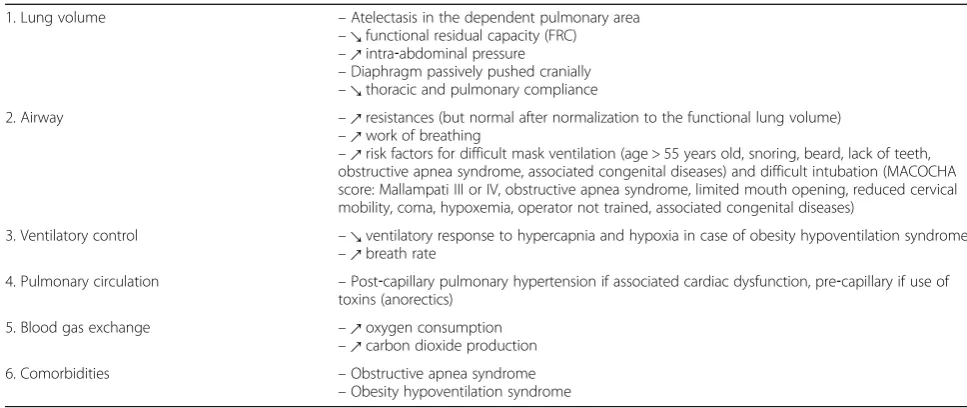
Obese patients can be admitted in a critical care setting forde novoacute respiratory failure,‘acute‐on‐chronic’ re-spiratory failure with an underlying disease, such as an obesity hypoventilation syndrome, or in the perioperative period. The main challenges for ICU clinicians are to take into account the pulmonary pathophysiological specific-ities of the obese patient (detailed in Table 1) to optimize airway management and non‐invasive or invasive mechan-ical ventilation.
Physiology
Oxygenation decreases with increase in weight, mostly because oxygen consumption and work of breathing are increased in obese patients [12]. At rest, oxygen con-sumption is 1.5 times higher in obese patients than in non‐obese patients [12]. Obese patients have an excess production of carbon dioxide (CO2), because of their
increased oxygen consumption and increased work of breathing, especially when there is an associated obesity hypoventilation syndrome, including a decreased respira-tory drive [13]. In several studies, the spontaneous breath rate was from 15 to 21 breaths per minute in morbidly obese patients (BMI > 40 kg/m2), whereas it was close to 10 to 12 in non‐obese patients [14]. Moreover, abdominal pressure is increased because of increased abdominal and visceral adipose tissue deposition. The capacity of the chest is reduced compared to non‐obese individuals, because the diaphragm is passively pushed cranially. Obese patients have decreased pulmonary and thoracic compliance, a re-duction in FRC, and an increased work of breathing, com-pared to non‐obese patients [15]. Airway resistance is increased, but not after normalization to the lung volume. The main change remains the decreased FRC, leading to more frequent atelectasis in obese than in non‐obese pa-tients after ventilation. Finally, as mentioned earlier, obesity is a major risk factor for obstructive apnea syndrome.
Noninvasive respiratory management Non‐invasive ventilation
Non‐invasive ventilation (NIV) may be applied to avoid intubation in obese patients with acute respiratory failure, without delaying intubation if needed. In hypercapnic obese patients, higher positive end‐expiratory pressure (PEEP) might be used for longer periods to reduce the hy-percapnia level below 50 mmHg [16]. NIV is as efficient in patients with obesity hypoventilation syndrome as in pa-tients with chronic obstructive pulmonary disease (COPD), in case of acute hypercapnic respiratory failure [17].
High‐flow nasal cannula oxygen
High flow nasal cannula oxygen (HFNC) could be par-ticularly interesting in obese patients. HNFC permits
continuously humidified and warmed oxygen to be deliv-ered through nasal cannula, with an adjustable fraction of inspired oxygen (FiO2). The flow administered can reach 60 l/min with 100% FiO2[18]. A moderate level of PEEP has been measured with this device [18] when the patient breaths with a closed mouth. In case of hypoxemia, HNFC could be performed between sessions of NIV.
Positioning
Optimization of body position can enhance respiratory function in patients requiring mechanical ventilation. In healthy spontaneously breathing obese subjects, a signifi-cant reduction in pulmonary compliance was shown in the supine position [19]. A sitting position should there-fore be privilegied in case of respiratory failure.
Airway management Pre‐oxygenation Facial mask
Following pre‐oxygenation, there is a reduction in the non‐hypoxic apnea time (length of apnea following anesthetic induction during which the patient has no oxygen desaturation) in obese patients [20]. Using classic bag‐mask ventilation as a method of pre‐oxygenation, desaturation during intubation thus occurs within 3 min on average, sometimes less than one minute in severe obesity. The end‐expiratory volume is reduced by 69% after anesthetic induction in the supine position, com-pared with baseline values [21]. The main cause of this rapid desaturation is the decrease in the FRC.
Non‐invasive ventilation
[image:2.595.54.538.97.301.2]Using a PEEP of 10 cmH2O during pre‐oxygenation is associated with a reduced atelectasis surface, improved
Table 1Pathophysiological specificities of the obese patient
1. Lung volume –Atelectasis in the dependent pulmonary area
–↘functional residual capacity (FRC) –↗intra‐abdominal pressure –Diaphragm passively pushed cranially –↘thoracic and pulmonary compliance
2. Airway –↗resistances (but normal after normalization to the functional lung volume)
–↗work of breathing
–↗risk factors for difficult mask ventilation (age > 55 years old, snoring, beard, lack of teeth, obstructive apnea syndrome, associated congenital diseases) and difficult intubation (MACOCHA score: Mallampati III or IV, obstructive apnea syndrome, limited mouth opening, reduced cervical mobility, coma, hypoxemia, operator not trained, associated congenital diseases)
3. Ventilatory control –↘ventilatory response to hypercapnia and hypoxia in case of obesity hypoventilation syndrome –↗breath rate
4. Pulmonary circulation –Post‐capillary pulmonary hypertension if associated cardiac dysfunction, pre‐capillary if use of toxins (anorectics)
5. Blood gas exchange –↗oxygen consumption
–↗carbon dioxide production
6. Comorbidities –Obstructive apnea syndrome
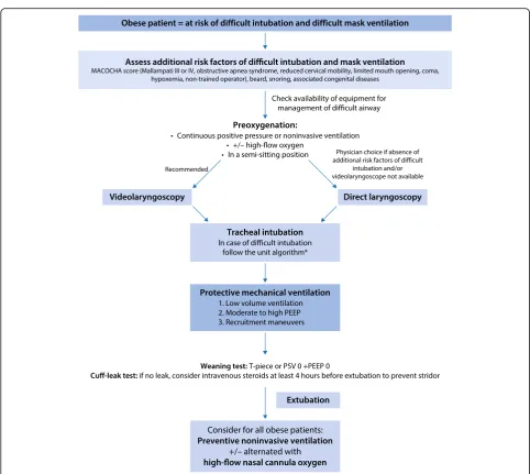
oxygenation and increased time of apnea without hypox-emia by one minute on average [22]. Pre‐oxygenation of 5 min with NIV, associating pressure support (PS) and PEEP, permits an exhaled fraction of oxygen (FeO2) > 90% to be reached more quickly [23]. In another study, the use of NIV limited the decrease in pulmonary volume and improved oxygenation compared to conventional pre‐ oxygenation with a face mask [24]. Continuous positive airway pressure (CPAP) or NIV are therefore the reference pre‐oxygenation methods (Fig. 1).
High‐flow nasal cannula oxygen
HFNC may also be considered for pre‐oxygenation of obese patients, including apneic oxygenation, enabling oxygen to be delivered during the apnea period (Fig. 1). This is particularly important in case of rapid sequence induction (RSI), where the obese patient does not re-ceive oxygen between removal of the NIV mask and ad-equate positioning of the tracheal tube into the trachea.
Positioning
A sitting position during pre‐oxygenation may decrease positional flow limitation and air trapping, limiting atel-ectasis and increasing oxygen desaturation during the in-tubation procedure (Fig. 1).
Intubation
Obesity and obstructive apnea syndrome, and a fortiori the combination of both, are risk factors for difficult in-tubation and difficult mask ventilation [3, 25]. Age > 55 years old, BMI > 26 kg/m2, snoring, beard and lack of teeth are independent risk factors for difficult mask ven-tilation. Most of these factors are directly related to obesity. In the same way, tracheal intubation is more dif-ficult in obese patients with obstructive apnea syndrome, with an incidence close to 15 to 20% (versus 2 to 5% in the general population), and associated with the severity of the obstructive apnea syndrome [26]. A recent study reported an increase in the incidence of difficult intub-ation in obese patients [3]. Moreover, in this study, ele-vated Mallampati score, limited mouth opening, reduced cervical mobility, presence of an obstructive apnea syn-drome, coma and severe hypoxemia (risk factors in-cluded in the MACOCHA score [27]) were associated with difficult intubation in obese patients. Each intubation in a morbidly obese patient should be considered as diffi-cult, and adequate preparation following an algorithm for difficult intubation performed (Fig. 1). Videolaryngoscopes are of particular interest in obese patients [28] and their use should be particularly emphasized when additional risk factors for difficult intubation are present.
Extubation
Obese patients are particularly at risk of post‐extubation stridor [29]. A cuff‐leak test [30] should be systematically performed in these patients, and in case of suspicion of laryngeal edema, prevention of stridor could be per-formed using a protocol of intravenous steroid adminis-tration, at least four hours before extubation, in the absence of contraindications [31].
Mechanical ventilation Protective ventilation Tidal volume
In obese patients, particularly at risk of atelectasis, the same rules can be applied. In spite of these recommen-dations, a recent study showed that obese patients were still ventilated in the perioperative period with tidal vol-umes that were too high [35]. In obese as in non‐obese patients, the optimal tidal volume is between 6 to 8 ml/ kg of IBW associated with PEEP to avoid atelectasis by alveolar closing (derecruitment). The tidal volume set-ting must be guided by the patient's height and not by his/her measured weight. The easiest formula for calculation of IBW to remember is the following: IBM (kg) = height (cm)−100 for a man and height (cm)−110 for a woman.
Positive end‐expiratory pressure
[image:4.595.56.539.81.512.2]cmH2O associated with a tidal volume of 6 to 8 ml/kg of IBW [24, 37]. However, it is necessary to remain on guard and always assess the hemodynamic effects of high PEEP: risk of decreased oxygenation because of an impact on cardiac flow and of hypotension because of compromised venous drainage. In case of auto‐PEEP, ap-plication of a PEEP will depend on the presence or not of a limitation in expiratory flow because of airway collapse during expiration. If this phenomenon exists, an extrinsic PEEP of 2/3 of the intrinsic PEEP should be applied.
The optimal level of PEEP in obese patients and the best means of titrating PEEP are still unknown. Some obese patients may benefit from higher levels of PEEP than others. Measuring transdiaphragmatic pressure seems crucial to determine the maximum pressure min-imizing alveolar damage, taking into account that the plateau pressure is related to both transthoracic and transalveolar pressures.
Recruitment maneuvers
To open alveoli once they are closed, recruitment ma-neuvers should be used, transitorily increasing the trans-pulmonary pressure. The impact of these maneuvers in the obese patient has been shown to improve arterial oxygenation and available lung volume [24].
The best recruitment maneuver has not been deter-mined in the obese patient. Recruitment maneuvers are mandatory to fully reopen the lung after anesthesia in-duction and a PEEP must be applied to prevent the pro-gressive closing of the lung leading to atelectasis. The optimal level of PEEP during protective ventilation re-mains to be determined, but many physiological studies suggest that PEEP levels of at least 5 cmH2O are neces-sary, in particular in obese patients. Levels of pressure needed to open the alveoli seem to be higher than in the non‐obese patient, mostly because of the increased transthoracic pressure. Questions persist regarding the type of recruitment maneuver to recommend. The refer-ence method is an expiratory pause with a PEEP level of 40 cmH2O during 40 s, but many alternatives exist, in-cluding progressive increase in PEEP until 20 cmH2O with a constant tidal volume within 35 cmH2O of plateau pres-sure, or a progressive increase in the tidal volume [38]. These recruitment maneuvers can be performed only if they are hemodynamically well tolerated. The ideal frequency for recruitment maneuvers has still not been determined.
Driving pressure
Driving pressure is the difference between inspiratory plat-eau pressure and end‐expiratory pressure. The concept of driving pressure assumes that functional lung size is better quantified by compliance than by predicted body weight. This concept explains why ventilator‐induced lung injury
(VILI), cyclic strain and survival may be better correlated with driving pressure than with tidal volume. Lower levels of driving pressure have been found to be associated with increased survival in ICU patients [39]. The ventilatory setting during mechanical ventilation, especially in obese patients, should be set to minimize driving pressure.
Respiratory rate
Concerning the setting or respiratory rate, obese patients have an excess production of CO2, because of their in-creased oxygen consumption and inin-creased work of breathing, especially when there is an associated obesity hypoventilation syndrome, with a decreased respiratory drive [13]. In four studies, the spontaneous breath rate was from 15 to 21 breaths per minute in morbidly obese patients (BMI > 40 kg/m2), whereas it was close to 10 to 12 in non‐obese patients [14]. Ventilation should, there-fore, be adapted, essentially increasing breath rate.
Ventilatory mode
Which ventilator mode is better in obese patients? The pressure modes deliver a constant pressure in the airway, decreasing the risk of barotrauma, with an insufflating pressure set at less than 30 cmH2O. In case of increase in airway resistance (bronchospasm, obstructed tube) or decrease in compliance of the respiratory system (obes-ity, atelectasis, selective intubation, surgical pneumoperi-toneum, pneumothorax …), the tidal volume decreases, leading to hypercapnia acidosis if alveolar ventilation is too low. It is consequently important to carefully check tidal volume, minute ventilation and capnography when using a pressure mode. The use of a volume mode car-ries the risk of an increase in the insufflation pressure to deliver the required tidal volume (risk of barotrauma), hence the importance of checking the alveolar pressure at the end of inspiration, i. e., the plateau pressure.
In obese patients, some teams recommend the pressure controlled mode because the decelerating flow should allow a better distribution of the flow in the alveoli. How-ever, studies comparing the two ventilatory modes report contradictory data: discordances can be explained by the different inclusion criteria and the methodological limita-tions of the studies [40]. In practice, the advantages and inconveniences of each mode must be known and the ventilatory mode that the physician prefers used.
ventilatory assist (NAVA), adaptive support ventilation (ASV), and proportional assist ventilation (PAV), with conventional pressure or volume controlled modes.
Positioning
In the supine position, positional flow limitation and air trapping impedes respiratory management particularly in obese patients [43]. A sitting position during mechanical ventilation is therefore advised. Prone positioning in obese ARDS patients enables an improvement in the partial arterial pressure of oxygen (PaO2)/FiO2 ratio more than in the non‐obese patient, and is not associ-ated with more complications [10].
Weaning from mechanical ventilation
A recent physiological study specifically investigated the inspiratory effort during weaning of mechanical ventila-tion in a populaventila-tion of critically ill, morbidly obese pa-tients [44]. The main result of this study was that for obese patients, T‐piece and PSV 0 + PEEP 0 cmH2O weaning tests were the tests that best predicted post‐ extubation inspiratory effort and work of breathing ([44]; Fig. 1). Following extubation, positive protective ventilation should be pursued, both in the ICU and in the recovery room. Postoperative CPAP or NIV might be extended to all obese patients, even those without ob-structive apnea syndrome.
Specific settings
Acute‐on‐chronic respiratory failure
Prevention of relapses of acute‐on‐chronic respiratory failure are essential and should be ensured by the inten-sivist. Positive airway pressure therapies can be imple-mented in the ICU and continued at home, with the support of home therapists. Sleep‐related breathing dis-orders, including obesity hypoventilation syndrome, should be followed by a specialist after ICU discharge, ideally in the setting of a multidisciplinary obesity team.
Perioperative management
In obese patients with obstructive apnea syndrome, noc-turnal CPAP should be initiated before surgery, espe-cially if the apnea hypopnea index (AHI) is more than 30 events per hour or if there is severe cardiovascular comorbidity. If CPAP or NIV were used prior to surgery, they should be pursued throughout the perioperative period, including the postoperative period.
The risk factors for postoperative respiratory failure include the severity of obstructive apnea syndrome, the intravenous administration of opioids, the use of sedatives, the site (close to the diaphragm) and the invasive nature of the surgical procedure, and the apnea onset during paradoxical sleep on the third or fourth postoperative day.
Some postoperative interventions that can decrease the risk of respiratory failure are a postoperative anal-gesia strategy sparing opioids, oxygenation by CPAP or NIV, careful patient positioning and monitoring. CPAP or NIV must be resumed in the recovery room [45]. Compliance to CPAP or NIV will be better if the pa-tients bring their own equipment to the hospital. In case of frequent or severe hypoxemias, start of CPAP or NIV should not be delayed. If possible, the supine position should be avoided in patients with an obstructive apnea syndrome at risk of postoperative pulmonary complica-tions, and a sitting position adopted. The prophylactic application of NIV after extubation decreases the risk of acute respiratory failure by 16% and reduces length of stay [45]. Moreover, in obese hypercapnic patients, the use of NIV following extubation is associated with de-creased mortality [46]. A randomized controlled trial performed in morbidly obese patients after bariatric sur-gery reported an improvement in ventilatory function when CPAP was immediately implemented after extuba-tion compared to CPAP started 30 min following extu-bation [47]. Hence, NIV associating pressure support and PEEP or CPAP alone must be used liberally in the postoperative period, in order to reduce the aggravation of atelectasis, a long period of oxygen dependence and con-sequently the patients’length of stay in the post‐surgical unit and in the hospital [45]. Among patients with hypox-emic respiratory failure following abdominal surgery, use of NIV compared with standard oxygen therapy reduced the risk of tracheal reintubation within 7 days [48]. These findings support the use of NIV in this setting.
Oxygen supplementation should be administered con-tinuously to all patients with obstructive apnea syn-drome at increased perioperative risk until they are able to maintain their baseline oxygen saturation on ambient air; oxygen saturations should be monitored after leaving the recovery room [49].
Respiratory physiotherapy and patient education of exercises, such as incentive spirometry or high volume respiration, also limit the reduction in lung volume in-duced by surgery.
Conclusion
prevent the complications related to the intubation‐pro-cedure (severe hypoxemia, arterial hypotension and car-diac arrest). Pre‐oxygenation should be optimized using positive‐pressure ventilation (CPAP or NIV) in a semi‐ sitting position, eventually added to apneic oxygenation using HFNC in the more severely obese patients. After tracheal intubation, to avoid both baro‐volutrauma and atelecto‐biotrauma, association of low tidal volume, moderate to high PEEP and recruitment maneuvers (lung protective ventilation) should be applied. The height of the lung being correlated to the height of the patient, tidal volume should be set according to IBW and not actual body weight, between 6 and 8 ml/kg IBW. In patients with ARDS, prone position is a safe procedure which permits respiratory mechanic improve-ments and oxygenation. Obstructive‐apnea syndrome and obesity‐hypoventilation syndrome should be investi-gated to introduce appropriate treatment, including im-plementation of positive airway pressure at home.
Acknowledgements
Not applicable.
Funding
Support was provided solely from institutional and/or departmental sources. Publication costs were funded by the "Centre Hospitalier Universitaire (CHU) Montpellier.
Availability of data and materials
Not applicable.
Authors‘contributions
ADJ, GC and SJ contributed to drafting the submitted article, and to provide final approval of the version to be published.
Competing interests
The authors declare that they have no competing interests.
Consent for publication
Not applicable.
Ethics approval and consent to participate
No applicable.
References
1. NCD Risk Factor Collaboration (NCD-RisC). Trends in adult body-mass index in 200 countries from 1975 to 2014: a pooled analysis of 1698 population-based measurement studies with 19.2 million participants. Lancet. 2016;387:1377–96. 2. Montravers P, Ribeiro-Parenti L, Welsch C. What's new in postoperative
intensive care after bariatric surgery? Intensive Care Med. 2015;41:1114–7. 3. De Jong A, Molinari N, Pouzeratte Y, et al. Difficult intubation in obese
patients: incidence, risk factors, and complications in the operating theatre and in intensive care units. Br J Anaesth. 2015;114:297–306.
4. Eichenberger A, Proietti S, Wicky S, et al. Morbid obesity and postoperative pulmonary atelectasis: an underestimated problem. Anesth Analg. 2002;95: 1788–92.
5. Pepin JL, Timsit JF, Tamisier R, Borel JC, Levy P, Jaber S. Prevention and care of respiratory failure in obese patients. Lancet Respir Med. 2016;4:407–18. 6. Flegal KM, Kit BK, Orpana H, Graubard BI. Association of all-cause mortality
with overweight and obesity using standard body mass index categories: a systematic review and meta-analysis. JAMA. 2013;309:71–82.
7. Hogue CW, Stearns JD, Colantuoni E, et al. The impact of obesity on outcomes after critical illness: a meta-analysis. Intensive Care Med. 2009;35:1152–70.
8. Akinnusi ME, Pineda LA, El Solh AA. Effect of obesity on intensive care morbidity and mortality: a meta-analysis. Crit Care Med. 2008;36:151–8. 9. De Jong A, Jung B, Chanques G, Jaber S, Molinari N. Obesity and mortality
in critically ill patients: another case of the simpson paradox? Chest. 2012; 141:1637–8.
10. De Jong A, Molinari N, Sebbane M, et al. Feasibility and effectiveness of prone position in morbidly obese patients with ARDS: A case-control clinical study. Chest. 2013;143:1554–61.
11. O'Brien Jr JM, Philips GS, Ali NA, Aberegg SK, Marsh CB, Lemeshow S. The association between body mass index, processes of care, and outcomes from mechanical ventilation: a prospective cohort study. Crit Care Med. 2012;40:1456–63.
12. Kress JP, Pohlman AS, Alverdy J, Hall JB. The impact of morbid obesity on oxygen cost of breathing (VO(2RESP)) at rest. Am J Respir Crit Care Med. 1999;160:883–6.
13. Pepin J, Borel JC, Janssens JP. Obesity hypoventilation syndrome: an underdiagnosed and undertreated condition. Am J Respir Crit Care Med. 2012;186:1205–7.
14. Chlif M, Keochkerian D, Choquet D, Vaidie A, Ahmaidi S. Effects of obesity on breathing pattern, ventilatory neural drive and mechanics. Respir Physiol Neurobiol. 2009;168:198–202.
15. Pelosi P, Croci M, Ravagnan I, Vicardi P, Gattinoni L. Total respiratory system, lung, and chest wall mechanics in sedated-paralyzed postoperative morbidly obese patients. Chest. 1996;109:144–51.
16. Gursel G, Aydogdu M, Gulbas G, Ozkaya S, Tasyurek S, Yildirim F. The influence of severe obesity on non-invasive ventilation (NIV) strategies and responses in patients with acute hypercapnic respiratory failure attacks in the ICU. Minerva Anestesiol. 2011;77:17–25.
17. Carrillo A, Ferrer M, Gonzalez-Diaz G, et al. Noninvasive ventilation in acute hypercapnic respiratory failure caused by obesity hypoventilation syndrome and chronic obstructive pulmonary disease. Am J Respir Crit Care Med. 2012;186:1279–85.
18. Chanques G, Riboulet F, Molinari N, et al. Comparison of three high flow oxygen therapy delivery devices: a clinical physiological cross-over study. Minerva Anestesiol. 2013;79:1344–55.
19. Naimark A, Cherniack RM. Compliance of the respiratory system and its components in health and obesity. J Appl Physiol. 1960;15:377–82. 20. De Jong A, Futier E, Millot A, et al. How to preoxygenate in operative room:
Healthy subjects and situations "at risk". Ann Fr Anesth Reanim. 2014;33:457–61. 21. Futier E, Constantin JM, Petit A, et al. Positive end-expiratory pressure
improves end-expiratory lung volume but not oxygenation after induction of anaesthesia. Eur J Anaesthesiol. 2010;27:508–13.
22. Gander S, Frascarolo P, Suter M, Spahn DR, Magnusson L. Positive end-expiratory pressure during induction of general anesthesia increases duration of nonhypoxic apnea in morbidly obese patients. Anesth Analg. 2005;100:580–4. 23. Delay JM, Sebbane M, Jung B, et al. The effectiveness of noninvasive
positive pressure ventilation to enhance preoxygenation in morbidly obese patients: a randomized controlled study. Anesth Analg. 2008;107:1707–13. 24. Futier E, Constantin JM, Pelosi P, et al. Noninvasive ventilation and alveolar
recruitment maneuver improve respiratory function during and after intubation of morbidly obese patients: a randomized controlled study. Anesthesiology. 2011;114:1354–63.
25. Langeron O, Masso E, Huraux C, et al. Prediction of difficult mask ventilation. Anesthesiology. 2000;92:1229–36.
26. Siyam MA, Benhamou D. Difficult endotracheal intubation in patients with sleep apnea syndrome. Anesth Analg. 2002;95:1098–102.
27. De Jong A, Molinari N, Terzi N, et al. Early identification of patients at risk for difficult intubation in the intensive care unit: development and validation of the MACOCHA score in a multicenter cohort study. Am J Respir Crit Care Med. 2013;187:832–9.
28. Andersen LH, Rovsing L, Olsen KS. GlideScope videolaryngoscope vs. Macintosh direct laryngoscope for intubation of morbidly obese patients: a randomized trial. Acta Anaesthesiol Scand. 2011;55:1090–7.
29. Frat JP, Gissot V, Ragot S, et al. Impact of obesity in mechanically ventilated patients: a prospective study. Intensive Care Med. 2008;34:1991–8. 30. Jaber S, Chanques G, Matecki S, et al. Post-extubation stridor in intensive
care unit patients. Risk factors evaluation and importance of the cuff-leak test. Intensive Care Med. 2003;29:69–74.
32. Petrucci N, De Feo C. Lung protective ventilation strategy for the acute respiratory distress syndrome. Cochrane Database Syst Rev. 2013;CD003844 33. Futier E, Constantin JM, Paugam-Burtz C, et al. A trial of intraoperative low-tidal-volume ventilation in abdominal surgery. N Engl J Med. 2013;369:428–37. 34. Hemmes SN, Gama de Abreu M, Pelosi P, Schultz MJ. High versus low
positive end-expiratory pressure during general anaesthesia for open abdominal surgery (PROVHILO trial): a multicentre randomised controlled trial. Lancet. 2014;384:495–503.
35. Jaber S, Coisel Y, Chanques G, et al. A multicentre observational study of intra-operative ventilatory management during general anaesthesia: tidal volumes and relation to body weight. Anaesthesia. 2012;67:999–1008. 36. Pelosi P, Croci M, Ravagnan I, et al. Respiratory system mechanics in
sedated, paralyzed, morbidly obese patients. J Appl Physiol. 1997;82:811–8. 37. Talab HF, Zabani IA, Abdelrahman HS, et al. Intraoperative ventilatory
strategies for prevention of pulmonary atelectasis in obese patients undergoing laparoscopic bariatric surgery. Anesth Analg. 2009;109:1511–6. 38. Constantin JM, Jaber S, Futier E, et al. Respiratory effects of different
recruitment maneuvers in acute respiratory distress syndrome. Crit Care. 2008;12:R50.
39. Amato MB, Meade MO, Slutsky AS, et al. Driving pressure and survival in the acute respiratory distress syndrome. N Engl J Med. 2015;372:747–55. 40. Aldenkortt M, Lysakowski C, Elia N, Brochard L, Tramer MR. Ventilation
strategies in obese patients undergoing surgery: a quantitative systematic review and meta-analysis. Br J Anaesth. 2012;109:493–502.
41. Spieth PM, Carvalho AR, Güldner A, et al. Pressure support improves oxygenation and lung protection compared to pressure-controlled ventilation and is further improved by random variation of pressure support. Crit Care Med. 2011;39:746–55.
42. Zoremba M, Kalmus G, Dette F, Kuhn C, Wulf H. Effect of intra-operative pressure support vs pressure controlled ventilation on oxygenation and lung function in moderately obese adults. Anaesthesia. 2010;65:124–9. 43. Lemyze M, Mallat J, Duhamel A, et al. Effects of sitting position and applied
positive end-expiratory pressure on respiratory mechanics of critically ill obese patients receiving mechanical ventilation. Crit Care Med. 2013;41:2592–9. 44. Mahul M, Jung B, Galia F, et al. Spontaneous breathing trial and
post-extubation work of breathing in morbidly obese critically ill patients. Crit Care. 2016;20:346.
45. Jaber S, Chanques G, Jung B. Postoperative noninvasive ventilation. Anesthesiology. 2010;112:453–61.
46. El-Solh AA, Aquilina A, Pineda L, Dhanvantri V, Grant B, Bouquin P. Noninvasive ventilation for prevention of post-extubation respiratory failure in obese patients. Eur Respir J. 2006;28:588–95.
47. Neligan PJ, Malhotra G, Fraser M, et al. Continuous positive airway pressure via the Boussignac system immediately after extubation improves lung function in morbidly obese patients with obstructive sleep apnea undergoing laparoscopic bariatric surgery. Anesthesiology. 2009;110:878–84. 48. Jaber S, Lescot T, Futier E, et al. Effect of noninvasive ventilation on tracheal reintubation among patients with hypoxemic respiratory failure following abdominal surgery: a randomized clinical trial. JAMA. 2016;315:1345–53. 49. Bolden N, Smith CE, Auckley D, Makarski J, Avula R. Perioperative
complications during use of an obstructive sleep apnea protocol following surgery and anesthesia. Anesth Analg. 2007;105:1869–70.