A COMPARATIVE ANALYSIS OF SURGICAL MANAGEMENT USING EXTERNAL FIXATION
AND INTERNAL FIXATION IN UNSTABLE COMMINUTED FRACTURE OF DISTAL RADIUS
DISSERTATION
SUBMITTED TO
THE TAMIL NADU DR. M.G.R.MEDICAL UNIVERSITY
IN PARTIAL FULFILMENT OF THE REQUIRMENTS FOR THE AWARD OF THE DEGREE
OF
M.S. ORTHOPAEDICS
BRANCH II (COURSE CODE 2202)
DECLARATION BY THE CANDIDATE
I hereby declare that this Dissertation / Thesis entitled : “A Comparative Analysis of Surgical Management Using External Fixation and Internal Fixation in Unstable Comminuted Fracture of Distal Radius” is a bonafide and genuine research work carried out by me under the guidance of Dr. S.Rama Guru, Professor Department of Orthopaedics, SreeMookambika Institute of Medical Sciences, during the period 2013 – 2016 in partial fulfillment of the requirements for the award of degree of M.S. in Orthopaedics, by the Tamil Nadu Dr. MGR Medical University, Chennai – 600 032.
Place :Kulasekharam
CERTIFICATE
This is to certify that this dissertation entitled “A Comparative Analysis of Surgical Management Using External Fixation and Internal Fixation in Unstable Comminuted Fracture of Distal Radius” is a bonafide research work done by Dr. T. Vishnu, under the guidance and supervision during the period 2013 – 2016 in partial fulfillment of the requirements for the award of degree of M.S. in Orthopaedics by the Tamil Nadu Dr. MGR Medical University, Chennai – 600 032.
Dr. K.C. Mathew, M.S. Dr. S.Ramaguru, M.S.
Professor and Head, Professor and Guide, Department of Orthopaedics, Department of Orthopaedics, SreeMookambika Institute of SreeMookambika Institute of
Medical Sciences, Medical Sciences,
Kulasekharam, Kulasekharam, Kanyakumari District, Kanyakumari District,
Tamil Nadu – 629 161 Tamil Nadu – 629 161
Dr. Rema. V. Nair, M.D., D.G.O.,
Director,
SreeMookambika Institute of Medical Sciences,
Kulasekharam,
PLAGIARIS
M
ACKNOWLEDGEMENT
My journey which has now attained its final countdown would not be complete if the following people are not acknowledge.
To begin with, I would like to dedicate my dissertation to my Alma Mata – SreeMookambika Institute of Medical Sciences from where I was nourished into a better human being and professional.
My deepest gratitude and respect to my beloved Chairman, Dr. C.K. Velayudhan Nair and Director, Dr. Rema. V Nair my mentor and inspiration for supporting me throughout and permitting me to utilize the hospital resources.
I am deeply grateful to my respect teacher and guide Prof. Dr. S.Ramaguru, for her valuable advice, constructive criticism, readiness to help and proper guidance without which this dissertation work has not been accomplished. He lent her full support in times of difficulties that I encountered during this study period.
It’s my privilege to have worked under the supervision of my respected teacher and Co-guide Professor & HOD Dr. K.C. Mathew, who vast knowledge in Orthopaedics has guided and inspired me to aspire for greater heights. His encouragement from the inception of this research to its culmination has been profound. I sincerely thank him for his constant encouragement and valuable support for achieving my goal.
I sincerely express my thanks to DR. SAHAYA JOSE, Assistant Professor, for supporting and guiding me in the period of my study.
My sincere thanks to Lab Technicians and Hospital Staff for their help and cooperation in my study.
I am indebted to My parents, My sisters and my friends for their unfaltering love, support and help in completing my study.
My deepest gratitude to all my Patients without whose whole hearted cooperation, this thesis would not have reached a conclusion.
CONTENTS
SL.NO. TITLE PAGE NO
I INTRODUCTION 1
II AIMS & OBJECTIVES 3
III REVIEW OF LITERATURE 4
IV METHODOLOGY 31
V RESULTS 48
VI DISCUSSION 77
VII CONCLUSION 81
BIBILOGRAPHY
ABSTRACT
TITLE
A comparative analysis of Surgical Management using External fixation and Internal fixation in unstable comminuted fracture of distal Radius.
BACKGROUND
Comminuted Intra-articular fractures of distal radius are one of the commonest fractures occurring in Adults.
Fractures of these type are difficult to manage by conservative Methods
Surgical Techniques such as External fixation and Internal fixation are used effectively in Management of comminuted unstable fractures of distal Radius.
External fixation Surgical procedure is done using Schanz pins and screws.
Open Reduction and Internal fixation Surgical procedure is done using K wire, buttress plates and locking compression plates.
AIMS AND OBJECTIVES
To compare the effects of External fixation and internal fixation in Surgical Management of unstable comminuted fracture of distal radius.
To evaluate functional results, advantages, disadvantages and complications of external and Internal fixation in treatment of unstable comminuted fracture of distal radius.
METHODS
68 patients were included in the study. 34 patients underwent Internal fixation and 34 patients underwent External fixation.
Functional outcome of the patient was assessed using DASH (Disability of the Arm, Shoulder and Hand) Method.
RESULTS
The difference in functional outcome of patients with comminuted unstable fractures of distal radius treated with surgical techniques such as External fixation and Internal fixation was not found to the statistically significant (P = 0.3955)
In our study, sex of patient, Age of patient, side involved, Mode of Injury, Type of fracture were also found to be statistically insignificant in determining functional outcome of patient.
CONCLUSION
The current study shows in surgical treatment of comminuted unstable fractures of distal radius, both external fixation and Internal fixation shows equally good results.
KEYWORDS
Comminuted unstable fracture of distal Radius Open Reduction and Internal fixation
External fixation Frykman classification
1
INTRODUCTION
Fractures of distal end of radius are commonest fractures presenting to
Orthopaedic outpatient Department and Emergency. It accounts for about One
Sixth of all fractures treated in emergency rooms1. Fractures of distal end of radius are caused due to fall on an outstretched hand. Unstable comminuted
fractures of these type occur due to high velocity Injuries.
It occurs commonly in both youngerand elderly population due to Road
traffic accidents and fall and in females common in postmenopausal period.
Uncomplicated fractures of this type can be treated with closed
reduction and immobilization in a cast, however, unstable comminuted fractures of radius occurs as a treatment challenge.
Review of Literature shows high incidence of unsatisfactory results in
treatment of unstable fractures by plaster cast method, It causes deformity up to
60% and unsatisfactory results in 32% of patients2.
In recent years, due to advancement of Surgical techniques, External
fixation and internal fixation are widely used as conservative methods fail to
maintain anatomical and functional stability.
External skeletal fixation uses minimally invasive procedures with
2
Both static and dynamic external fixators are used, depending upon difficulty in maintaining radial length and alignment.
Internal fixation are increasingly used as it directly controls and
maintains Anatomical and functional stability of wrist joint. Locking
compression plates K-Wires and volar buttress plates are widely used in this
method. Now a days latest methods like interlocking nailing, fragment specific
fixation using plates and clamps are used.
The purpose of this dissertation is directed towards evaluating functional outcome of patients with comminuted unstable fractures of distal radius treated
3
AIMS AND OBJECTIVES
To compare the effects of external and internal fixation in surgical
Management of Unstable comminuted fracture of distal Radius.
To evaluate functional results, advantages, disadvantages and complications of external and internal fixation in treatment of unstable
4
REVIEW OF LITERATURE
Abraham colles on (1814)3 first published on fracture of Carpal extremity of radius in Edinburgh Medical Journal. “This fracture takes place
about an inch and a half above carpal extremity of radius. If the surgeon holds his hands in that of patients and exerts even a moderate force the limbs
instantly return on extension on being removed”.
Till 1920 treatment of colles fracture was forceful \traction,
manipulation and immobilization of wrist in flexion. It lead to a high incidence
of Median nerve Neuropraxia.
After 115 years, Bohler4 in 1929 published Tran’s fixation technique with skeletal pins and plaster cast. The pin and plaster method showed good
results than old method.
In 1944 Roger Anderson and Gordon O’2 described new method of reduction through skeletal traction and counter traction and immobilization by
use of two slender rods instead of plaster of Paris cast.
Depalma (1952)5 described ulnar pinning where K-wire was introduced through ulna into reduced distal fragment of radius. It showed 18% unsatisfactory results. But with this method Dowling and sawyer (1961)
5
Ellis (1965)6 recommended open reduction and Internal fixation of unstable smith’s type (or) Volar Barton fractures. He devised ‘T’ shaped plate,
as volar buttress preventing deformity.
Green D.P. (1975)7 reported 86% good to excellent results with pin and plaster technique.
Cooney W.P. (1979)8 reviewed Roger Anderson external fixator by showing 90% excellent and 8% fair results in comminuted intra-articular
fractures. He found articular congruity and residual dorsal tilt were most significant criteria affecting results.
Charles Melone (1986)9 proposed open reduction of displaced intra-articular fractures of distal radius. He proved that maximal functional recovery
of fracture is dependent on accurate and stable restoration of articular surfaces.
These fractures have four basic components. Radial styloid, Radial shaft, dorsal
and volar Medial fragments.
The Medial fragments possess strong ligamentous attachments to carpus
and ulnar styloid, they together constitute Medial complex. On basis of
displacement of medial complex, he has classified four types. First two types
amenable to closed manipulation or skeletal traction. Type 3 and 4 are
associated with grossly volated fragments which are absolute indication for
6
Dennis Foster (1986)10 showed Hoffmann and Anderson showed equally good results. But 4% to 6% cases showed pin tract infection, 10% showed
persistent pain and 8% showed wrist weakness.
In 1986 jerry L. Knirk et al10 from Massachusetts Published a paper on Intra articular fractures of distal radius in young adults. All fractures were of
Frykman’s type VII or type VIII. They suggest external fixator is the treatment
of choice.
ClyburnT.A. (1987)11 showed new dynamic external fixation allows wrist movements and full movements of fingers. The results is early
rehabilitation. It is based on Principle of having ball type of joint on fixator in
par with physiological center of rotation (in proximal capitates) allows motion
and maintains distraction force. He showed good results when combined with
limited external fixation
esp for “diepunch” or radial styloid fragment.
Keating J.F., et al, (1994)12 studied 79 patients with volar displaced fractures of distal radius over 26 months with A0 T-plate and showed malunion
defined as more than 2mm of radial shortening more than 4mm of radial shift
more than 15 degrees of volar tilt, more than 2 mm of radial shortening, more
than 10mm of dorsal tilt, most patients achieved acceptable function with
7
Roger Anderson (1994)13 described prototype of External fixator used now for comminuted fracture of distal radius. He showed the causes of poor
results are shortened radius,maltilted fracture of distal radius. He showed
shortening was not only due to impaction and overriding but also due to
crushing of Juxta – articular cancellous bone and devised fixator that
maintained sustained fraction and maintained reduction. It showed good results
especially in early osteoporotic bones.
Frederick A. Kaempffe et al (2000)14 retrospectively studied 19 patients with distal radius fractures, treated with internal fixation and supplement K
wire fixation over 6 years. This is found to be good method of treating fracture
of distal radius.
Abbas Emami, et al (2000)15 treated 40 patients by insertion of external fixator half pins dorsally (other than dorso – radially) in diaphysis of radius and
showed it is safer position of pins and superficial radial nerve is Preserved.
Richard A. Rogachejsky et al (2001)16 showed comminuted intra- articular fracture of distal radius should be treated by open reduction and
combined internal and external fixation, supplemented by bone grafting and
plate fixation is satisfactory treatment.
David Ring et al (2004)17 studied 25 patients with A0C3 fractures treated with combined dorsal and volar plate fixation for 25 months after injury
8
ANATOMY
SURFACE ANATOMY
This is essential for diagnosis and management of wrist injuries. When
the wrist is flexed against resistance tendons that stand out prominently from
radial to ulnar side areflexorcarpiradialis, Palmaris longus, flexor
digitorumsuperficialis and flexorcarpiulnaris.
The Ulnar nerve and vessels are present between flexorcarpiulnaris and
flexordigitoriumsuperficialis. The radial styloid is ½ inch distal to ulnar styloid.
On the dorsum of lower radius is Lister’s tubercle, medial to which is tendon of
extensor pollicis longus.
ARTICULAR ANATOMY
The wrist joint is abiaxial type of Joint, grouped under ellipsoid variety.
The bones taking parts are distal end of radius and articular disc from above,
scaphoid, lunate and triquetral bone below, hence termed as a mid carpal joint.
Articular surface of radius and lower surface of triangular fibrocartilage from a
concave surface that is elliptical in shape. Inferior surface of radius has ridge that formtwoconcavities in the radius (ie) scaphoid and lunate fossa
respectively.The proximal articular surface of scaphoid and lunate fossa
respectively. The proximal articular surface of scaphoid, lunate and triquetral
bones form a smooth convex surface that articulate with concave surface of
9
The capsule covers all three bones and is reinforced by dorsal, volar, lateral and medial ligaments. The capsule is lined by synovial membrane. The
joint line corresponds to a line joining styloid process of radius and ulna and is
convex upwards.
Distal radioulnar joint : Uniaxial pivot joint between convex surface of
ulna and concave ulnar notch of radius. They are enclosed together and held by
articular disc. The capsule is lax superiorly, through which synovial out
pouching called recessussacriformis in front of lower part of interosseous membrane. The pronator quadratus has interosseous artery and carpal branches
of radial and ulnar artery. The nerve is derived from anterior and posterior
interosseous nerves.
OSTEOLOGY
Lower end of radius is expanded and is cancellous covered with a thin
layer of cortical bone. The bone at about 3/4thInch proximal to articular surface is weak and susceptible for fracture. On its anterior surface Pronator quadratus
muscle is attached. The posterior surface is ridged and has grooves to
accommodate wrist and finger extensor tendons.
Brachioradialis muscle is inserted little abovestyloid process of radius.
Medial surface has got a concave articular facet that articulates with ulna and
distal ridge, which gives attachment to base of triangular fibrocartilage. Distal
10
the ulnar side the concave surface is quadrilateral and rough and articulates with lunate07.The concavity on lateral aspect is triangular for scaphoid articulation. The plane of articular surfaces faces distally and slightly volarly.
Lower end of ulna is slightly expanded from the neck into small rounded
head. The distal surface is flat and articulates with disc. At the base of styloid is
apex of articular disc and tip of styloid gives attachment to ulnar collateral
ligament.
LIGAMENTS
There are two Major groups of ligaments of the wrist.
- Extrinsic group of ligaments
- Intrinsic group of ligaments
Extrinsic ligaments link carpal bones to radius, ulna and metacarpals.
Palmar wrist ligaments Originates laterally from radial palmar facet of
radial styloid and are directed in a distal ulnar direction where they meet
ligaments originating medially from triangular fibrocartilage and distal ulna. It
consist of two ‘V’ shaped ligamentous bands. One is proximal and connects
forearm to proximal carpal row and the other is distal and connects forearm to distal carpal row. The distal limb consists of radio scaphoid capitate ligaments
11
radioulnotriquetral and radio-scaphoid ligament laterally and ulnotriquetral ligament medially.
Dorsal wrist ligaments are radiotriquetral and scaphotriquetral ligament
which describe a ‘V’ shape from the dorsal aspect of radius near Lister’s
tubercle to triquetrum and then back to the dorsal scaphoid rim.
Dorsal ligaments are attached to proximal carpal row and volar
12 Intrinsic ligaments :
These are intra- articular intrinsic ligaments of wrist connecting adjacent
carpal bones. They are collections of relatively short fibers that bind to bones
of either proximal (or) distal carpal rows to each other.
Ulnar collateral ligament :
This is attached to ulnar styloid and divides into two slips; one slip is
attached to medial side of the triquetrum and the other to the pisiform.
Radial collateral ligament :
Extends from the tip of styloid crosses of radius to the radial side of
14 Functional anatomy
The distal end of radius is considered the anatomic foundations of wrist
joint. The main movements of wrist joint take place in transverse and antero –
posterior axis. Wrist flexion and extension occur at radiocarpal and intracarpal joints. Normal range is 75% each. Adduction and Abduction occur at
radiocarpal joints. Normal range 200 and 300 respectively. Supination and pronation take place at the distal radioulnar joint. Supination is greater than
pronation. The range of movements is 800 – 850 respectively.
Radiological anatomy
It forms the foundation of injury and outcome of treatment.
Standard X-ray views are Anteroposterior, lateral and oblique. Antero
posterior view shows the concave inferior articular surface of lower end of
radius extending down to tip of styloid process.
Lateral view 8
For extra-articular fracture, assess dorsal / palmar tilt, extent of
Metaphyseal comminution, carpal alignment, displacement of volar cortex and
position of Distal radial ulnar joint (DRUJ). For intra-articular fractures assess depression of palmar lunate facet, depression of central fragment and gap
15 Oblique view8 :
For extra-articular fractures, assess radial comminution. For
intra-articular fractures assess radial styloid for split or depression and depression of
dorsal lunate facet.
Dorsal / Palmar tilt8 :
On a true lateral view, a line is drawn connecting most distal points of
volar and dorsal lips of radius. The dorsal or palmar tilt is the angle created
16
Picture : 2
17 Volar Tilt / inclination :08
In sagittal view, a line is drawn connecting the distal most point of
dorsal and volar rims. The angle that this line creates with a line perpendicular
to the longitudinal axis of radius reflects the palmar inclination. Average
inclination is 110 (range from 40-220).
Radial Length 8 :
Measured on Anteroposterior view. It is distance in millimeters between
a line drawn perpendicular to long axis of radius and tangential to most distal
point of ulnar head and line drawn perpendicular to long axis of radius and at
18
Picture : 3
19 Ulnar Variance :
This is a measure of radial shortening. It is a vertical distance between
line parallel to medical corner of articular surface of radius and a line parallel
to most distal point of articular surface of ulnar head, which are perpendicular to long axis of radius.
s
20
FRACTURES OF DISTAL END OF RADIUS
The Avulsion theory : Suggested by Linhart (1852) and analyzed by
Lecomate (1861) says that ulna is probably alone (because of intimate contact
with humerus) absorbing impact of all on hand, the force being transmitted to radius via interosseus membrane and strong volar ligaments. Then the fracture
is produced by avulsion due to traction in strong volar radio carpal ligaments.
The Bending fracture theory :
The theory was put forward by Meyer (1925) and supported by Lewis
(1950) says that course of fracture is determined by three factors position of
hand, surface of impact and magnitude of force. The kinetic energy causes the
forward movement of body to continue, the wrist becomes hyperextended and
patient falls over the hand. This loads the volar ligaments and radius is pressed
against carpal articular surface, the force being stopped by scaphoid and lunate bones, it is then transmitted to radius, which fractures at its weakest point in
same manner as a beam that is loaded beyond the limits of elasticity, Lewis so
considered this fracture as a” bending fracture.”
Considerable force required to produce fracture – mean of 190 kg for
women and 282kg for men.
When a person falls on an out stretched hand, the radius through rigidly
21
strain is thrown upon palmar carpal ligaments and the line of force derives carpus upon radius. The radius first fractures on volar surface in tension. Then
the fracture propogates dorsally where the bending movement forces induce
compression stresses resulting in dorsal cortex comminution or the fracture line
producing 45° shear stress lines. The cancellous bone is compacted further
reducing dorsal stability. Charnley has shown that the dorsal comminution is a
cause of late collapse of radius during the period of immobilization.
The colles fracture occurs while the triangular fibrocartilagenous disc of inferior radio ulnar joint is still intact. Therefore, distal fragment rotates on this
hinge with center of rotation at ulnar styloid in direction of supination. If the
force is excessive and continues to act, the strain thrown upon the disc may
bring about the fracture of the ulnar styloid.
Hyperextension is common major force causing fractures. Intraarticular
fractures are prevalent among active persons whose wrists are exposed to violent multi component force comprising of compression, shearing, tension
and direct crush. The prominent among these forces is axial compression where
proximal carpus acting like a die punch impact disrupts distal radial articular
facets. So, the resultant articular fracture comprises of four components –
Metaphyseal or shaft, radial styloid, dorsal medial fragment is referred to die
22 FRACTURE ANATOMY
In colles fracture six displacements of distal fragment are Impaction,
Lateral displacement, lateral rotation, dorsal displacement, dorsal rotation and
supination. Distal fragment is compressed with shaft of radius and rotates dorsally. Dorsal Angulation and radial shortening results in “Dinner fork
deformity”
Melone identified most intra-articular fractures have four fracture
components, they are Radial shaft, Radial styloid, Dorsal medial and palmar
medial.
Two medial fragments along with ligamentous attachment to carpus and
ulnar styloid is termed as medial complex. Even minimal displacement of Medial fragment is likely to cause major disruption of radial and radioulnar
joint with compromise of articular fixation11.
De Palma demonstrated that even with most severely comminuted
fractures, ligaments of wrist remain intact. This is important to maintain
23 CLASSIFICATION
There are many classifications, proposed for Distal radius fracture, but
more accepted and recent ones are the following.
Gartland and Werley :
Proposed a classification that assessed three basic components of these
injuries.
Metaphyseal comminution, intra-articular extension and displacement
of fragments.
Group I : Simple colles fracture with no involvement of radial articular
surfaces.
Group II : Comminuted colles fractures with intra- articular extension
without displacement.
Group III : Comminuted colles fractures with intra-articular extension
with displacement.
24 Frykman classification :
It incorporated individual involvement of radioocarpal and radioulnar
joints. It is used in this study.
Type I : Extra- articular fracture
Type II : Extra- articular fracture with ulnar styloid fractures
Type III : Radiocarpal articular involvement
Type IV : Radiocarpal involvement with ulnar styloid fracture.
Type V : Radioulnar involvement
Type VI : Radioulnar involvement with ulnar styloid fracture
Type VII : Radioulnar and Radiocarpal involvement
Type VIII : Radiocarpal and Radioulnar involvement with ulnar
25
Picture : 4
26 Melone’sClassification :
He emphasized the effect of impaction of lunate on radial articular
surfaces to create four characteristic fragments.
Type I : Stable fracture without displacement. This pattern has characteristic fragments of radial styloid and a palmar and
dorsal lunar facet.
Type II : Unstable ‘die punch’ with displacement of characteristic
fragments and comminution of anterior and posterior
cortices.
Type II A : Reducible
Type II B : Irreducible central impaction fracture.
Type III : “Spike” fracture unstable Displacement of articular surface
and also of proximal spike of radius.
Type IV : “Spilt” fracture. Unstable Medial complex that is severely
comminuted with separation and or rotation of palmar and
distal fragments.
27
Picture : 5
28 OTA / Aoclassification :
It emphasizes increasing severity of bone injury.
Type A : Extra-articular fracture subgroups are based on direction of displacement and comminution.
Type B : partial articular fracture. Sub groups are based on lateral (radial styloid) palmar (or) dorsal fragments.
Type C : Complete articular subgroups are based on degree of comminution of articular surface and metaphysis.
Fernandez Classification :
In 1993, Fernandez proposed a mechanism based classification that would address potential for ligamentous injury and assist in treatment recommendations.
Type I : Metaphyseal bending fractures with inherent problem of loss of palmar tilt and radial shortening relative to ulnar (DRUJ Injuries)
Type II : Shearing fracture requiring reduction and of articular segment.
Type III : Compression of articular surface without characteristic fragmentation, also potential for significant interosseous ligament injury.
Type IV : Avulsion fracture or radiocarpal fracture dislocation.
29 Cooney (1990) Universal classification:
Type I : Extra-articular Undisplaced
Type II : Extra-articular displaced
Type III : Intra-articular undisplaced
30 Picture : 6
31
MATERIALS AND METHODS
This study is a prospective study, non randomized, study period includes
January 2014 to September 2015.
68 Patients were included in the study. 34 patients underwent internal fixation and 34 patients underwent external fixation.
All patients were treated by below elbow plaster slab after other life
threatening injuries were ruled out.
Definitive treatment was based on decision of the surgeon on a non –
randomized basis.
Treatment was either external fixation with Schanz pins and screws or internal fixation with K wire, Buttress plate, locking compression plate.
Assessment :
Done by non blinder assessment
Subjective assessment – pain, numbness, weakness of hand, stiffness.
Objective – Range of motion (flexion, extension, Radial deviation, ulnar
deviation, supination and pronation.)
Range of movements measured by hand held goniometer.
Functional outcome of patient was assessed using DASH (Disability of
32 Inclusion Criteria :
• Patients giving valid consent
• Comminuted fracture of distal end of radius of either side or both sides
• Age between 20 – 70 years
• Both Male and female
• Closed / open fractures
• Patients fit for surgery
Exclusion Criteria :
• Refusal by patient
• Compound fracture
33
SURGICAL TECHNIQUE (EXTERNAL FIXATION)
Anaesthesia :
Regional (Axillary block) or general anaesthesia is used.
.Patient placed in supine position with the affected limb abducted and placed
on a side table.
Procedure :
External fixator was applied in the operation theatre under sterile
conditions. The pins used for radius were 3.5 mm Schanz type and for that of
metacarpal were 2.5 mm schanz type. After painting and draping with or without pneumatic tourniquet a small incision was made on dorsolateral aspect
of forearm about 3-5 cm proximal to fracture site. Lateral cutaneous nerve of
forearm was identified, 2.7 mm drill bit was used for predrilling. 3.5 mm
Schanz pin (half pin) was inserted. Second pin site was selected beyond mid
forearm proximally, asgreater the distance from first pin in distal end of radius
3-5 cm proximal to fracture site, more stable is the fixation.
Two Schanz pins were passed to the 2nd metacarpal as follows. First Schanz pin 2.5mm was passed into base of second metacarpal to third
metacarpal base. The second schanz pin of 2.5 mm was passed into neck of
second metacarpal. Both these pins were passed on lateral surfaces. The radial
34
achieved under image intensifier, control and a rod to rod clamp or a third external rod was used when necessary to control angular element of deformity.
Best position is ulnar deviation of forearm.
Postoperatively, upper limb was elevated for 24 hours with monitoring
of neurovascular status. Early motion of digits, elbow and shoulder was
encouraged.
Patient was discharged and called for follow up every two weeks till 6
weeks, then every 3 months till one year.
During the follow up period, patients were advised about the exercises
of the elbow, digits, and shoulder and about the cleaning of the pin site with
saline and soap water. Early pin tract infection was treated with antibiotics.
Fixator removal was done after clinical and radiological evidence of fracture
healing.
After fixator removal, a removable splint or POP slab was given for another 3-6 weeks, that was to be removed during exercises. Range of motion
35
36
37
38
39
40
SURGICAL TECHNIQUE (INTERNAL FIXATION)
Anesthesia :
Regional (Axillary block) or general anesthesia used.
Buttress Plating
Procedure :
After painting and draping, a longitudinal incision about 7.5 cm long on
the radiovolar aspect of the distal forearm was made. The plane between the
flexorcarpiradialis and the Palmaris longus was developed. The flexor pollicis longus tendon was retracted towards radial side and the median nerve and other
tendons were retracted towards ulnar side. The fibres of pronator quadratus
were severed from their origin on the radius and the fracture was exposed.
Fracture was reduced and a buttress plate was contoured so that, when it
is applied and fixed to the proximal fragment, the distal transverse part will act
as a buttress and hold the fractures reduced. A minimum of two screws were
inserted in the proximal fragment.
Screws were inserted through the distal part of plate into the Fracture
were confi
in c arm. radius and
Pos
of neurov
encourage
firmed by d
Pronator q d wound wa
st operative
vascular st
d.
direct obse
quadratus w as closed. ely, upper tatus. Earl Ima 41 ervation an was replac
limb was e
y motion
age : 7
nd by anter
ced over th
elevated fo
of digits,
INCISION
roposterior
he plate to
or 24 hour
, elbow a
N
and latera
o its origin
s with mon
and should
al views
n on the
nitoring
Imag
42
Imagee : 9 EX
43
Immage : 10 AFTER O
44
45 FOLLOW UP
Patients were assessed which included objective impression of the
patient, objective grading of function and deformity, a comparison of final and
initial X-ray.
Subjective factors such as pain, functional limitations, occupational
consideration were taken into account.
Follow up intervals are 6 weeks, 3 months and 1 year. Objective
examination included inspection of the wrist for deformity, tenderness,
abnormal mobility of the distal radioulnar joint, measurement of range of
movements extending from shoulder to digits, grip strength, light touch and pin
prick sensitivity.
Complication of external fixation :
Superficial pin tract infection
Pin Breakage and loosening
Second metacarpal fracture
Reflex sympathetic dystrophy
Median nerve neuropraxia
Transient sensory impairment of radial nerve
Tendon and soft tissue tethering
46
Complications of Internal Fixation :
Early :
Inadequate Anaestheisa
Difficult reduction (or) reduction maintained only in extreme position
Depressed major Articular fragment
Distal radioulnar subluxation or dislocation
Median, ulnar, radial nerve stress, contusion or compression
Post reduction swelling – compartment syndrome
Tendon laceration especially Extensor pollicis longus
Pain dysfunction syndrome
Associated carpal bone injury.
Late :
Loss of reduction and secondary deformity
Radial shortening and angulation
Inadequate articular reduction
47 Shoulder hand syndrome
Carpal Tunnel syndrome
Radio carpal Osteoarthritis
48
RESULTS
Sixty Eight cases admitted is SMIMS hospital were considered for the
study.
STATISTICAL METHODS EMPLOYED
Chi square test was employed in the study.
Chi square test :
Chi Square test tabulates a variable into categories and computes a
chi-square statistic. The test compares observed and expected frequencies in each
category to test either all categories contain same proportion of values or each category contains user specified proportion of values.
The following observations were made from data collected during study
49
Table : 1
Sex Distribution :
Sex No. of cases %
Male 56 82.35%
Female 12 17.65%
Total 68 100%
Out of 68 cases, 56 were male and 12 were female.
Figure : 1 Sex Distribution
56
12
Male
[image:60.595.150.501.368.787.2]50
Table : 2 Sex Distribution:
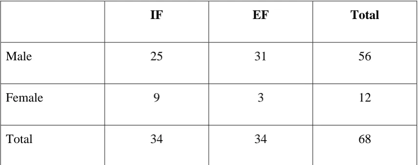
Results were corrected using chi square method. The Chi square value was found to be 6.25 and p value 0.1021 which shows that sex of patient did not
influence outcome of treatment.
Results Male Female
IF EF Total IF EF Total
Excellent 2 1 3 0 0 0
Good 21 24 45 7 0 7
Fair 2 5 7 2 3 5
Poor 0 1 1 0 0 0
51
Table : 3
Sex Distribution:
IF EF Total
Male 25 31 56
Female 9 3 12
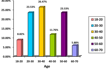
52 Table : 4
Age Distribution :
Age No. of Cases %
18 – 20 06 8.82%
20 – 30 16 23.53%
30-40 18 26.47%
40-50 08 11.76%
50 – 60 16 23.53%
60 – 70 4 5.88%
Total 68 100%
Figure : 2 0.00% 5.00% 10.00% 15.00% 20.00% 25.00% 30.00%
2 Age Di
% % % % % % %
18‐20
8.82%
istribution
20‐30
%
23.53%
53
n :
30‐40 40
26.47%
1
Age
0‐50 50‐6
11.76% 23.5
60 60‐70
53%
5.88%%
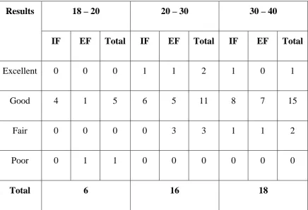
54 Table : 5 Age Distribution :
Results 18 – 20 20 – 30 30 – 40
IF EF Total IF EF Total IF EF Total
Excellent 0 0 0 1 1 2 1 0 1
Good 4 1 5 6 5 11 8 7 15
Fair 0 0 0 0 3 3 1 1 2
Poor 0 1 1 0 0 0 0 0 0
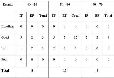
55 Table : 6 Age Distribution :
Results 40 – 50 50 – 60 60 – 70
IF EF Total IF EF Total IF EF Total
Excellent 0 0 0 0 0 0 0 0 0
Good 3 2 5 5 7 12 2 2 4
Fair 1 2 3 2 2 4 0 0 0
Poor 0 0 0 0 0 0 0 0 0
Total 8 16 4
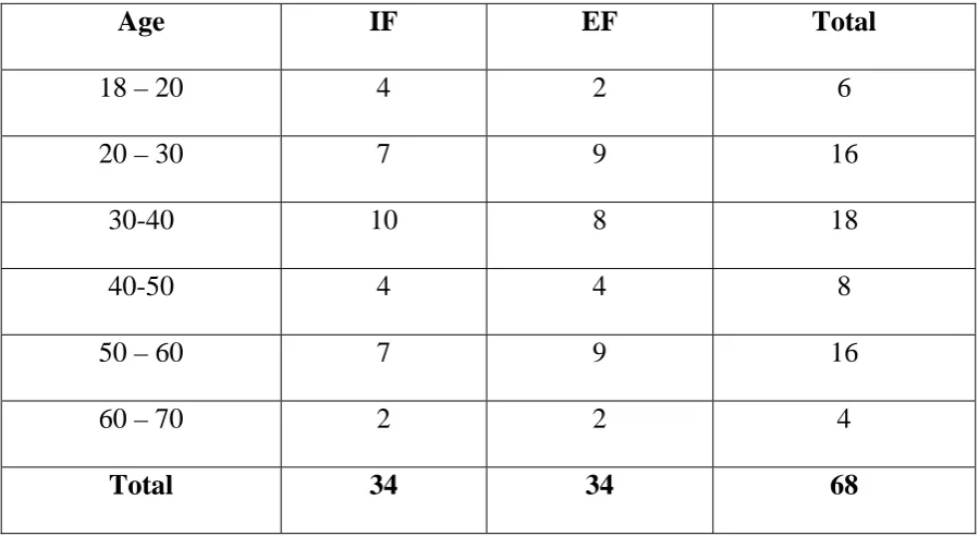
This chi square value was found to be 19.52 and p value 0.19117 which
56 Table : 7 Age Distribution :
Age IF EF Total
18 – 20 4 2 6
20 – 30 7 9 16
30-40 10 8 18
40-50 4 4 8
50 – 60 7 9 16
60 – 70 2 2 4
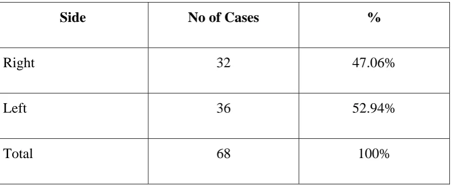
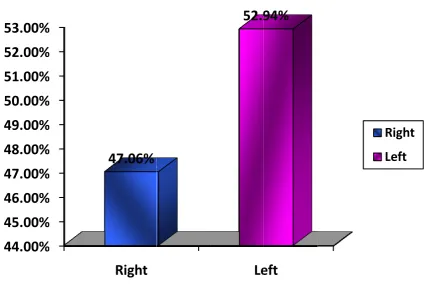
57 Table : 8 Side Involved :
Side No of Cases %
Right 32 47.06%
Left 36 52.94%
Total 68 100%
Figure : 3 Side Involv
44.00%
45.00%
46.00%
47.00%
48.00%
49.00%
50.00%
51.00%
52.00%
53.00%
ved :
59 Table : 9
Side Involved :
Result Right Left
IF EF Total IF EF Total
Excellent 0 1 1 2 0 2
Good 11 14 25 17 10 27
Fair 3 3 6 1 5 6
Poor 0 0 0 0 1 1
Total 32 36
60 Table : 10
Side Involved :
IF EF Total
Right 14 18 32
Left 20 16 36
Total 34 34 68
Table : 11
Mode of Injury :
Mode No of Cases %
RTA 39 51.35%
Fall 29 42.65%
Total 68 100%
In the study prominent cause of injury was high energy trauma due to
[image:71.595.101.546.482.630.2]Figure : 4
Mode of I
62
Table : 12
Mode of Injury:
Results RTA Fall
IF EF Total IF EF Total
Excellent 1 1 2 1 0 1
Good 21 10 31 7 14 21
Fair 3 3 6 1 5 6
Poor 0 0 0 0 1 1
Total 39 29
The Chi square value was found to be 1.83 and p value 0.6094 which shows
63 Table : 13
Mode of Injury:
IF EF Total
RTA 25 14 39
Fall 9 20 29
[image:74.595.104.531.196.369.2]Total 34 34 68
Table : 14
TYPE OF FRACTURE :
Type of Fracture No of Patients Percentage
Closed 64 94.12%
Open 4 5.88%
Total 68 100%
64
Table : 15
Type of Fracture :
Results Closed Open
Excellent 3 0
Good 51 1
Fair 9 3
Poor 1 0
Total 64 4
Figure : 5
Type of F
66 Table : 16
FRYKMAN’S CLASSIFICATION :
Classification No of Patients Type of Fixation
Type I 03 Internal fixation
Type II 00 -
Type III 13 Internal Fixation
Type IV 08
02
Internal Fixation External fixation
Type V 07
06
Internal Fixation External fixation
Type VI 03
05
Internal Fixation External fixation
Type VII 11 External Fixation
Type VIII 10 External Fixation
Table : 17
Method
IF
EF
[image:78.595.102.530.112.271.2]Total
Figure : 6
0%
5%
10%
15%
20%
25%
30%
35%
40%
45%
50%
METHd of Fixatio
METH
HOD OF FI
on
HOD OF FI
IF
50%
67 IXATION :
No of C
68 Table : 18.
Method of Fixation :
The chi square value was found to be 2.97 and p value 0.3955 which shows that method of fixation does not determine the outcome of treatment.
Results IF EF
Excellent 2 1
Good 28 24
Fair 4 8
Poor 0 1
69 Table : 19
Movements after 12 weeks compared with normal side :
Wrist Normal ROM Result (Average)
Dorsiflexion 750 700
Palmar Flexion 750 650
Ulnar deviation 300 250
Radial deviation 200 150
Forearm
Supination 800 700
70 Average Time of Fracture Union :
The Average time of fracture union was 5.76 months with a standard deviation of 0.50 in case of ORIF with Buttress plate and 3.76 months with standard deviation of 0.74 in case of fracture treatment with external fixator.
[image:81.595.101.546.321.640.2]Complication :
Table : 20
External Fixation
Complications No of Patients
Wrist Stiffness 1
Pin site infection 5
Pin Loosening 2
Tendon rupture 0
Compression Neuropathy 0
Sudeck’s osteodystrophy 0
Finger Stiffness 1
Iatrogenic rupture 0
71 Table : 21
Internal Fixation :
Complications No of Patients
Wrist Stiffness 3
Nerve Injuries 0
Vascular injuries 0
Tendon rupture 0
Compression Neuropathy 0
Sudeck’s osteodystrophy 0
Finger Stiffness 3
72 Table : 22
RESULTS
Results No of cases Percentage
Excellent 3 4.41%
Good 52 76.47%
Fair 12 17.65%
Poor 1 1.47%
Figu Wit functional r Exc Goo Fair Poo 0.00% 10.00% 20.00% 30.00% 40.00% 50.00% 60.00% 70.00% 80.00%
ure : 7
th help of results were cellent (3) od (52) r(12) or (1) % % % % % % % % % Excelle 4.41 Functional
f DASH (D e as follows
- 4.41 - 78.1 - 17.6 - 1.47 nt Goo 1% 76.4 73 Results
Disability o s : 1% 18% 65% 7% d Fai 7% 17
Age
of Arm, s
74 RESULTS
Table : 23
Internal Fixation :
Results No of cases Percentage
Excellent 2 5.88%
Good 28 82.35%
Fair 4 11.76%
Poor 0 0%
75 Table : 24
Results :
External Fixation :
Results No of cases Percentage
Excellent 1 2.94%
Good 24 70.59%
Fair 8 23.53%
Poor 1 2.94%
Figure : 8 Results : Internal Fi 0.00% 10.00% 20.00% 30.00% 40.00% 50.00% 60.00% 70.00% 80.00% 90.00%
ixation & E
Excellent 5.88% 8 2.94% External Fi Good 82.35% 1 % 70 76 ixation: Fair 11.76% .59% 23.53% Poor 0.00% % 2.944% I E
Internal Fi External F
ixation
77
DISCUSSION
Distal Radius fractures are one of the commonest fractures occurring in Adults. Comminuted unstable fractures of these type are often difficult to manage by conservative methods. Surgical techniques such as External fixation and Internal fixation are used in treatment of comminuted unstable fractures of distal radius.
External fixation surgical procedure is done with schanz pins and screws. It allows fracture fragments to fall in place and brings about reduction and maintains the distraction force during healing. It is used when fracture fragments are very small, extremely comminuted and open fractures are best treated by this method. Ligementotaxis is useful in restoring skeletal length and wrist position can be adjusted. Advantages of external fixator are its superior Mechanical efficiency, its capacity of fracture adjustment during the healing period.
Open reduction and Internal fixation surgical procedure uses better and smaller implants, K wire, Buttress plate and Locking compression plate. Even comminuted fractures with dorsal metaphyseal instability can be fixed with a volar plate. It has the advantages like early and better mobilization and function of hand, repair of ruptured tendons, less residual pain, stiffness, restriction and better chance of restoring joint congruity.
78
distal radius were employed in the study. 34 patients who underwent external fixation procedure and 34 patients who underwent Internal fixation were included in the study. The patient selection was based on factors such as Age, Fracture pattern, bone quality, Type of fracture, affordability of patient.
Of the patients employed in the study, 82% were males and 18% were females. In our study, sex of the patient is not statistically significant (p = 0.1021) in determining functional outcome of the patient. Previously zhuang cui et al19 conducted meta-analysis of unstable distal radius fractures treated with Internal fixation versus External fixation. It included pooled data from ten randomized controlled trials included 738 patients, orthopedic journals. It discussed that a prospective study of patient’s age more than 35 years with colles fracture at six centers in the united kingdom for a period of one year reported that the overall incidence of this fractures is found to be more in females than in males. Therefore, although there may effect modification due to mean and proportion of women, we could not determine this from available data.
79
fractures. However, age of the patient is statistically insignificant (P = 0.19117) in determining functional outcome of the patient.
In our series left sided fractures were common than right sided fractures. Besides, side of the fracture is not statistically significant (P = 0.7580) in determining the outcome of the patient.
In this study, mode of Injuries, Road Traffic Accident and fall were the causes of distal radius fractures. Road Traffic Accident were of major cause found than fall. However, Jerry Knirket al10 found on their series, fall from height to be the main cause of fractures. In our study, Mode of Injury is statistically insignificant (P = 0.6094) in determining functional outcome of the patient.
In our study, open fractures and closed fractures were included. Surprisingly, type of fracture was found to be statistically significant (P = 0.021845 and chi square value 9.6442) in determining functional outcome of the patient. Probably, the significance of type of Injury in functional outcome is due to less number of patients included in open fractures type, additionally complications such as infections are most common in open fractures than closed type which delay healing of fractures and ultimately the functional outcome.
80
al21 conducted a study in orthopedic department for a period of 5 years, they used frykman classification for assessment of fractures.
In the surgical procedure, External fixation 2.94% patients had Excellent results, 70.59% had good results, 23.53% had fair results and 2.94% had poor results. In the Internal fixation group 5.88% patients had excellent results, 82.35% had good results, 11.76% had fair results and no one had poor results. Our study showed that method of fixation is statistically not significant (P = 0.3955) in determining the functional outcome of the patient, though the study has certain limitations such as non-randomized, non-blinding techniques used and less number of patients were employed. Similarly Margaliotet al23 did a Meta – analysis of distal radius fractures treated with External fixation and Internal fixation. 46 articles were included in the study after careful serenity of Internal fixation and external fixation 917 patients were included in external fixation group and 603 were included in Internal fixation group. Outcomes were assessed using pooled grip strength, Range of motion, Radiographic assessment and physician related outcomes. The authors conclude that current literature does not recommend the superiority of one method over the other.
81
CONCLUSION
The prospective study was conducted to compare the functional outcome of patients with comminuted unstable distal radius fractures treated with External fixation and Internal fixation surgical procedures.
In the current study, in the external fixation group 2.94% patients had excellent results, 10.59% had good results, 23.53% had fair results and 2.94% had poor results. In the Internal fixation group 5.88% patients had excellent results, 82.35% had good results, 11.76% had fair results and no one had poor results.
In the current study, method of fixation is not statistically significant in determining functional outcome of the patient.
Similarly, sex of the patient, Age, mode of Injury were statistically insignificant in determining functional outcome in our study.
CLINICAL PICTURES
CASE : 1
50 YEARS OLD MALE CAME WITH HISTORY OF SLIP AND FALL ON AN OUTSTRETCHED HAND
2
ONE YEAFOLLOW- UP XRAYS
3 Image :13
RANGE OF MOTION
DORSI FLEXION PALMAR FLEXION
4 CASE :2
53 YEARS OF FEMALE HAD HISTORY OF SLIP AND FALL ON A OUT STRETCHED HAND
5
6
RANGE OF MOTIONS
7 CASE : 3
8
RANGE OF MOTIONS
9 CASE : 4
10
RANGE OF MOTIONS
11
BIBILOGRAPHY
1. Ark. J. Jupiter JB. The rationale for precise management of distal radius fractures. Orthoop clin North Am. 1993 Apr; 24(2) : 205 – 10.
2. Fernandez, DL., Jupiter, JB. Fractures of distal radius. A practical approach to management.Springer – verlag. Newyork ; 1995.
3. Colles A on fracture of the carpal extremity of the radius. Edinburgh Med. Surg J 1814; 10:182-86.
4. Bohler L. The treatment of fracture. New york, Grune and Stratton 1932; 90-96.
5. Depalma AF. Comminuted fracture of distal radius treated by ulnar pinning. JBone joint surg 1952; 34 : 614-2.
6. Ellis J. Smith’s and Baron’s fractures. A method of treatment. J Bone joint surg 1965; 47(B) : 724.
7. Green Dp. Pins and plaster treatment of comminuted fracture of distal end of radius. J Bone joint surg 1975; 57 (A) : 304.
8. Cooney WP. Linsched RL, Dobyns JH. External Pin fixation for unstable colles fracture. J Bone joint surg 1979; 6:842-5.
9. Melone CP, Articular fractures of distal radius. Orthop clin North Am 1984; 15:217-36.
12
11.Knirk J.L, Jupiter J.B. Intra-articular fractures of the distal end of radius in young adults. JBJS 68 (Am) 1986 : 697.
12.Clyburn T.A., MD. Hanston, Texas : Dynamic external fixation for comminuted inra-articular fractures of distal end of radius JBJS vol 69 A No .2: Feb 1987.
13.Kearing JF, Court – Brown CM, McQueen MM. Internal fixation of Volar displaced distal radius fractures. J Bone Joint surg Br. 1994; 76 (3): 401-5.
14.Anderson R, O’ Neil G. comminuted fractures of distal end of the radius S.G.D. 1994; 78:434.
15.Kaempffe FA, walker KM. External fixation of distal radius fracture: effects of distraction on outcome. Clin orthoop 2000; 1(380); 220-225.
16.Emami A, Mjoberg B. A safer Pin position for External fixation of radial fractures. Orthop clin N. Am 2000; 31 (9) : 749-750.
17.Rogachefsky Ra. Scott RL, Applegate B, Oucllete EA, savenor AM, McAuliffe JA, Treatment of severe comminuted, Intra-articular fracture of distal radius by open reduction and combined Internal fixation and external fixation. J Bone surg 2001; 83:509-19.
18.Ring D, Prommersberger L, Jupiter JB combined dorsal and volar plate fixation of
complex fractures of distal part of the radius. JBone joint surg 2004; 86-A(9) : 1646-1652.
19.David S.Rach and Margaret M. Queen. Distal Radius and ulna fractures. Rockwood Charles A Jr. David P. Green. Fractures in Adults. 7th edition. Vol I. Philadelphia Lippincot Williams and wilkins : 2010.
13
21.Kapoor M. Agarwal A. Dhaon BK. Displaced intra – articular fractures of distal radius. A comparative evaluation of results following closed reduction, external fixation and open reduction with Internal fixation.Injury.2000 ; 31 (2) : 75-9.
22.Wei DH, Raizman NM, Bottino CJ. unstable distal radius fractures treated with external fixation, a radial column plate, or a volar plate. A prospective randomized trail. J Bone joint surg Am. 2009 ; 91(7) : 1568-77.
23.Margaliot Z, Haase Sc. A Meta – analysis of outcomes of external fixation versus plate osteosynthesis for unstable distal radius fractures. J hand surg AM, 2005; 30 (6) : 1185-99.
24.Belloti JC, Santos JBGD, Atallah AN. Fractures of the distal radius (colles fracture) sao Paulo Med J 2007,125 (3) ; 132-8.
25.Blakeney W, webber L. Emergency department Management of colles – type fractures. Emerg Med Aust 2009 ; 21 (4) : 298-303.
26.Campbell’s operative orthopaedics, 12th edition Philadephia:Elseiver ; 2013.
27.Dionyssiotis Y, Dontos IA, Econompoulos D, Lyritis GP. Rehabilitation after falls and fractures. J Musculoskeletal Neuronal interact 2008 ; 8(3) : 244-50.
28.Frykman GK, comparison of eleven fractures. Journal of Hand Surgery ; 1989.
29.Gartland JJ, Welley CW. Evaluation of healed colles fracture. J. Bone joint surg 1957 ; 33 : 895-907.
30.Mc Queen MM, Hajducka C, courtbrown C M. Redisplaced unstable fractures of the distal radius. A prospective randomized comparison of four method of treatment.. J Bone surg 1996 ; 78: 404-9.
14
32.Smilovic J, Billc R, Meghan T. Treatment of extra – articular colles type fractures of the distal radius. Prospective study. Croat Med J. 2003 ; 44(6) : 740-5.
33.Smith D. Henry M. volar fixed angle plating of distal radius. Jam acad orthop surg 2005 ; 13: 28-36.
34.Smith EJ. Baron’s fracture – A method of treatment. J Bone Joint surg 1965 ; 47 : 124-5.
15
CASE RECORD FORM
Name :
Age :
Sex :
Ip/op No :
Address :
Phone No :
Date of Admission : Date of Surgery : Date of Discharge : Chief Complaints :
Pain :
Swelling :
Disability :
Mode of Injury :
RTA :
Assault :
Fall :
Domestic Accident : H/o presenting illness : Duration of injury :
Pain : Site of pain
16
Past H/o :
H/o Diabetes :
H/o Hypertension :
H/o CAD :
H/o other chronic disorder:
Family H/o :
Personal H/O :
H/O Smoking :
H/O Chronic Alcoholism : General Physical Examination
PR : BP:
Pallor : Icterus:
Cyanosis : Edema:
Systemic Examination
CVS :
RS :
PA :
CNS :
Local Examination
Side Involved : Right / Left
Type : Simple / Compound Swelling : Present / Absent
Associated Injury :
State of Wound : Clean / Contaminated / Infected Neurological Deficit : Present / Absent
17
Management
External Fixation : Type of Anaesthesia : Size of Schanz pins : No. of schanz pins used : Position of Wrist : Neutral : Palmar flexion :
Dorsiflexion :
Pronation :
Supination :
Ulnar deviation : External fixator Removed on:
Internal fixation Type of Anesthesia:
Implant used: Buttress plate/ Locking Compression plate Buttress plate:
Size:
No. of holes: No. of screws: Screw size:
Locking Compression plate:
Size :
Kwire :
No. of wires :
18 Sutures Removed on :
Any External Immobilisation : Cast / Slab / Fixator Follow up Evaluation :
Clinical Examination : Subjective
Pain :
Swelling :
Limitation of Movement :
Disability :
Restriction of Activity :
Other Complaints :
Objective: Dorsiflexion Palmar flexion Radial Deviation Ulnar Deviation Pronation Supination: Grip:
Tenderness in Distal Radioulnar Joint: Tenderness in Radiocarpal Joint:
Residual deformity: Radial Deviation Hand / Ulnar styloid Prominence / Dorsal Tilt
Complication :
Nerve Injury :
Infection :
19
Re displacement :
Pin Site Infection :
Loosening :
Rupture of Extensor pollicis Longus Tendon: Carpal Tunnel Syndrome :
20
CONSENT FORM
PART 1 of 2
INFORMATION FOR PARTICULARS OF THE STUDY’
Dear Participants,
We welcome you and thank you for your keen interest in participation in this research project. Before you participate in this study, it is important for you to understand why this research is being carried out. This form will provide you all the relevant details of this research. It will explain nature, Purpose, benefits, risks, discomforts, precautions and information about how these projects will be carried out. It is important that you read and understand the contents of form carefully. This form may contain certain scientific terms and hence if you have any doubts or if you want more information, you are free to ask study person or contact person mentioned before you give your consent and also at any time during the entire course of the project.
1. NAME OF PRINCIPAL INVESTIGATOR:
Dr.T. Vishnu,
Post graduate Student, Orthopedics,
SMIMS, Kulasekharam.
2. NAME OF THE GUIDE:
NAME : Dr.S.Ramaguru MS Ortho
DESIGNATION : Professor
21
3. NAME OF THE CO GUIDE:
NAME : Dr.K.C.Mathew MS Ortho
DESIGNATION : Professor
DEPARTMENT : Department of Orthopedics INSTITUTE & PLACE : SMIMS, Kulasekharam
NAME OF THE CO GUIDE (2):
NAME : Dr.M.Mohammed Sheriff MS Ortho
DESIGNATION : Associate Professor DEPARTMENT : Department of Orthopedics INSTITUTE & PLACE : SMIMS, Kulasekharam
4. INSTITUTE:
SreeMookambika Institute of Medical Sciences, Kulasekharam, Kanyakumari District, Tamilnadu – 629 161
5. TITLE OF THE STUDY:
A COMPARATIVE ANALYSIS OF SURGICAL MANAGEMENT USING EXTERNAL FIXATION AND INTERNAL FIXATION IN UNSTABLE COMMINUTED FRACTURE OF DISTAL RADIUS.
6. BACKROUND INFORMATION:
Fracture of distal radius is the most common fracture. Comminuted unstable fracture of distal radius is best treated by surgical methods such as external fixation and internal fixation.
7. AIMS AND OBJECTIVES:
22
8. SCIENTIFIC JUSTIFICATION STUDY:
Scientists – Cooney W.P, Ellis J, Charles Melone, Roger Anderson and many others described benefits of surgical intervention in management of unstable comminuted fracture of distal radius.
They have shown these techniques gave good anatomical and functional outcomes compared to conservation methods.
9. PROCEDURE FOR THE STUDY:
Patients with distal end of radius are clinically examined. X ray Investigation of wrist joint are taken in SMIMS.
If it shows comminuted unstable fractures of distal radius, patient is admitted in hospital, in orthopedics ward.
Preoperatively treated with analgesics and antibiotics provisional reduction and immobilization with plaster of paris.
They observe for any progressive swelling and neurovascular complications
Patients were divided into Group I and II randomly and analyzed for fitness for Surgery.
Group I Patients are treated by External fixation. Group II Patients are treated by Internal Fixation.
Post operatively patients were observed for acute complications.
Check X-ray taken and assumed for quality of reduction and then patient followed for every week, for month and then followed for 3 months, 6 months and 1 year post operative X –rays were taken in SMIMS.
23
10. EXPECTED RISK FOR PARTICIPANTS:
Risk of Anaesthesia, post operative complication such as Infection, failure of return of normal wrist joint function.
11. EXPECTED BENEFITS OF RESEARCH FOR
There may be any personal benefits but this study will be beneficial for betterment of health sector.
12. MAINTANANCE OF COFIDENTIALITY:
All data collected for the study will be kept confidentially and would reflect on general statistical evaluation only and would not reveal any personal details.
13. WHY I HAVE CHOOSED IN THIS STUDY:
As you are suffering from comminuted unstable fracture of distal radius, you are chosen for the study.
14. HOW MANY PEOPLE WILL BE IN THE STUDY:
68
15. AGREEMENT OF COMPENSATION TO THE PARTICIPANT:
Yes
16. ANTICIPATED PRORATED PAYMENT, IF ANY, TO THE PARTICIPANTS OF THE STUDY:
Nil