EVALUATION OF BRAINSTEM AUDITORY EVOKED POTENTIAL AND SERUM INTERLEUKIN-1BETA LEVELS IN PATIENTS WITH GENERALIZED TONIC CLONIC SEIZURES
Dissertation submitted to
THE TAMIL NADU DR. MGR MEDICAL UNIVERSITY
In partial fulfillment of the regulations for the award of the degree of
M.D. PHYSIOLOGY Branch V
INSTITUTE OF PHYSIOLOGY & EXPERIMENTAL MEDICINE
MADRAS MEDICAL COLLEGE AND GOVERNMENT
GENERAL HOSPITAL
CHENNAI – 600003
The Tamil Nadu Dr. MGR Medical University
Chennai – 600 032
CERTIFICATE
This is to certify that the dissertation entitled “Evaluation of Brainstem
auditory evoked potential and serum Interleukin-1beta levels in patients with
generalized tonic clonic seizures” by the candidate Dr. G. Savitha for M.D
Physiology is a bonafide record of the research done by her during the period
of study (2012-2015) in the Institute of Physiology and Experimental
medicine, Madras Medical College, Chennai-600003.
DEAN
Madras Medical College Chennai-600003
DIRECTOR AND PROFESSOR Institute of Physiology and
Experimental Medicine, Madras Medical College,
Chennai-600003
ACKNOWLEDGEMENT
I gratefully and sincerely thank Dr.R.Vimala, the Dean of Madras Medical College, Chennai-3 for granting me permission to carry out this study at the Institute of Physiology and Experimental medicine, Madras Medical College, Chennai.
I take this pleasant and unique opportunity to express my profound sense of gratitude, respect and sincere thanks to Prof. Dr. K. Padma, M.D., who with her expertise has provided unsurpassable guidance and encouragement not only during the preparation of this dissertation but also throughout my post graduation course.
I am thankful to Dr. Banu, Head of Department, Institute of Neurology, Madras Medical College for her unconditional help in granting permission to recruit generalized tonic clonic seizure patients from the department.
I am greatly indebted to Prof. Dr. R. Vijayalakshmi whose enthusiastic supervision and valuable guidance made this work possible.
I express my gratitude to Dr. Parimala for her guidance and valuable suggestions. I extend my thanks to Dr. Sathya for her motivation and advice throughout the study. I sincerely thank Dr. C. Thirupathi for his immense support to the study.
With immense sense of gratitude, I thank Dr. Rathna Manjushree, Dr.KanmaniKarthikeyan, Dr.AnanthaSubramaniam, Dr.SatyaNarayanan Dr.Shanthimalar, Dr.Gomathy, Dr.Kavitha, Dr.Subramaniam for their extreme support and guidance throughout the study.
I sincerely thank Prof. Dr. Mini Jacob, Head of Department, Department of Experimental medicine, The Tamilnadu Dr. MGR Medical University, Guindy, Chennai for granting permission to avail the laboratory facilities.
I express my profound sense of gratitude to Dr. Anitha, Department of Experimental Medicine for her unflagging interest and immense support in the lab procedures.
I acknowledge the immense faith of the volunteers and patients who have participated in this study and express my gratitude for their co-operation.
I feel extremely grateful to my entire family for their affection and prayers.
Above all, I thank the Almighty for blessing this endeavour.
ABSTRACT
EVALUATION OF BRAINSTEM AUDITORY EVOKED POTENTIAL AND SERUM INTERLEUKIN-1BETA LEVELS IN PATIENTS WITH GENERALIZED TONIC CLONIC SEIZURES
Degree for which submitted : Doctor of Medicine(MD) in Physiology
Supervisor and guide : Prof.Dr.K.Padma
Director and Head of the Department
Department : Institute of Physiology and Experimental Medicine
College : Madras Medical College Chennai-600003.
University : The Tamilnadu Dr.M.G.R.Medical University, Chennai-600032.
Year : 2014
BACKGROUND
Tonic Clonic Seizure(GTCS) patients even before the clinical manifestation of hearing impairment occurs so that proper measures to intervene the disease process at the earliest possible is achieved to provide a better quality of life for GTCS patients.
AIM OF THE STUDY
· To determine the functional integrity of auditory pathway in patients with GTCS by recording brainstem auditory evoked potential.
· To assess serum Interleukin -1 beta levels in these patients.
· To find the correlation between serum Interleukin-1 beta level and Brainstem auditory evoked potential in patients with GTCS.
MATERIALS AND METHODS
30 patients with GTCS of both sexes in the age group 20-40 years without any clinical evidence of hearing impairment were included in the study. Controls were age, sex and BMI matched healthy population. Both the controls and GTCS patients were subjected to BAEP and serum interleukin-1beta levels were also measured. The data were analyzed by Student ‘t’ test.
RESULT
CONCLUSION
There was significant prolongation of central conduction time in GTCS patients even though there was no clinical evidence of hearing impairment assessed by pure tone audiogram prior to the study. Hence BAEP can be utilized as an objective electrophysiological tool to evaluate the functional integrity of auditory pathway from the external ear to lower brainstem.
KEY WORDS
CONTENTS
LIST OF TABLES
LIST OF PHOTOGRAPHS AND FIGURES
LIST OF GRAPHS
ABBREVIATIONS
S. No. Title Page No.
1. INTRODUCTION 1
2. REVIEW OF LITERATURE 8
3. AIM & OBJECTIVES 46
4. MATERIALS AND METHODS 47
5. RESULTS 75
6. DISCUSSION 86
7. CONCLUSION 99
8. SUMMARY 101
BIBLIOGRAPHY
ANNEXURE
i) ETHICAL COMMITTEE APPROVAL
ii) CONSENT FORM
iii) PROFORMA
LIST OF TABLES
S.No Title Page
No.
1 Generators of BAEP waveforms 38
2 Normal waves of BAEP 63
3 Gender differences in normal BAEP waveforms 67
4 Age distribution in controls and GTCS patients 76
5 BMI distribution in controls and GTCS patients 76
6
Comparison of mean values of absolute and interpeak latencies between genders in the right ear of GTCS patients
77
7
Comparison of mean values of absolute and interpeak latencies between genders in the left ear of GTCS patients
77
8
Comparison of mean values of absolute and interpeak latencies between genders in the right ear of control group
78
9
Comparison of mean values of absolute and interpeak latencies between genders in the left ear of control group
78
10
Comparison of mean values of absolute latencies between GTCS females and control females in the right ear
79
11
Comparison of mean values of interpeak latencies between GTCS females and control females in the right ear
12 Comparison of mean values of absolute latencies between GTCS females and control females in the left ear
80
13
Comparison of mean values of interpeak latencies between GTCS females and control females in the left ear
80
14
Comparison of mean values of absolute latencies between GTCS males and control males in the right ear
81
15
Comparison of mean values of interpeak latencies between GTCS males and control males in the right ear
81
16
Comparison of mean values of absolute latencies
between GTCS males and control males in the left ear 82
17
Comparison of mean values of interpeak latencies
between GTCS males and control males in the left ear 82
18
Comparison of Interleukin-1 beta levels in controls
and GTCS patients 83
19
Correlation between IL-1 beta levels and ABR wave
III in the right ear of GTCS patients 84
20
Correlation between IL-1 beta levels and ABR wave
III in the left ear of GTCS patients 84
21
Correlation between IL-1 beta levels and ABR I-III
IPL in the right ear of GTCS patients 85
22
Correlation between IL-1 beta levels and ABR I-III
LIST OF PHOTOGRAPHS
Photo
No Title
Page No (between)
1 Computerised Neurostim-Medicaid systems for recording Brainstem auditory evoked potential
52-53
2 Recording of Brainstem auditory evoked
potential in normal persons 57-58
3 Interleukin-1β kit and serum samples,
ELISA reader 71-72
LIST OF FIGURES
Figure No Title Page No
1 Cross section of brain showing primary
generalized seizure 4
2 GABA in epileptogenesis 17
3 Classification of evoked potentials 29 4 Generators of auditory evoked potentials 33-34
(between)
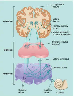
5 Auditory pathway 37
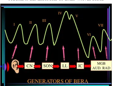
6 Generators of BAEP waveforms 38
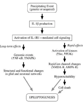
7 Interleukin-1β in epileptogenesis 43
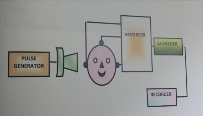
8 Apparatus for BAEP 52
LIST OF GRAPHS
Graph
No Title
Page No (Between)
1 Age distribution in controls and GTCS
patients 76-77
2 BMI distribution in controls and GTCS
patients 76-77
3 Comparison of mean values of absolute and interpeak latencies between genders in the right ear of GTCS patients
77-78
4 Comparison of mean values of absolute and interpeak latencies between genders in the left ear of GTCS patients
77-78
5 Comparison of mean values of absolute and interpeak latencies between genders in the right ear of control group
78-79
6 Comparison of mean values of absolute and interpeak latencies between genders in the left ear of control group.
78-79
7
Comparison of mean values of absolute and interpeak latencies between GTCS females and control females in the right ear
80-81
8 Comparison of mean values of absolute and interpeak latencies between GTCS females and control females in the left ear
80-81
9 Comparison of mean values of absolute and interpeak latencies between GTCS males and control males in the right ear
82-83
10 Comparison of mean values of absolute and interpeak latencies between GTCS males and control males in the left ear
82-83
11 Comparison of serum inteleukin-1 beta levels
GLOSSARY OF ABBREVIATIONS
ABR Auditory Brainstem Response AVCN Anterior Ventral Cochlear Nucleus AP Action potential
BERA Brainstem Evoked Response Audiometry BAEP Brainstem Auditory Evoked Potential CNS Central Nervous system
CPS Complex Partial Seizure CSF Cerebrospinal fluid DCN Dorsal Cochlear Nucleus dB decibel
EPSP Excitatory Post Synaptic potential GABA Gamma Amino Butyric Acid GTCS Generalized Tonic Clonic seizure ILAE International League Against Epilepsy IL-1β Interleukin -1 beta
IPL Inter Peak Latency
IPSP Inhibitory Post Synaptic Potential IBE International Bureau of Epilepsy MGB Medial Geniculate Body
ms milliseconds
PVCN Posterior Ventral Cochlear Nucleus pg picogram
NTS Nucleus Tractus Solitarius RMP Resting Membrane Potential SPS Simple Partial Seizure
SUDEP Sudden Death
1
1.
INTRODUCTION
A seizure (derived from Latin word Sacire meaning to take possession
of) is a paroxysmal event which occurs due to abnormal excessive or
synchronous neuronal activity in a discrete or generalized portion of brain.
Seizures can be provoked by acute brain insult or systemic diseases
like stroke or metabolic causes but if it occurs in the absence of an acute
provoked event, it is termed as unprovoked seizure1. Epilepsy is defined as
the tendency to have at least two episodes of unprovoked seizures separated
by a minimum period of 24 hours.
In 2005, International League Against Epilepsy (ILAE) and
International Bureau of Epilepsy (IBE) proposed the definition of epilepsy as
a disorder of brain by an enduring predisposition to generate epileptic
seizures and by the neurobiological, cognitive, psychological and social
consequences of this condition.
Efforts to classify epileptic seizures date back to the earliest of
medical literature. In 1964, the Commission on classification and
terminology of ILAE proposed the first official classification of epileptic
2
ILAE classification of epileptic seizures
I. Partial (focal, local) seizures
A. Simple partial seizures (consciousness not impaired)
-with motor symptoms
-with somatosensory or special sensory symptoms
-with autonomic symptoms
-with psychic symptoms
B. Complex partial seizures (with impairment of consciousness)
-with simple partial onset followed by impairment of consciousness
-with impairment of consciousness at onset
C. Partial seizures evolving to secondarily generalized seizures
-SPS evolving to generalized seizures
-CPS evolving to generalized seizures
3
II. Generalized seizures (convulsive or non -convulsive)
-Absence seizures
1. Typical absence seizures
2. Atypical absence seizures
-Myoclonic seizures
-Clonic seizures
-Tonic seizures
-Tonic clonic seizures
-Atonic seizures
III. Unclassified epileptic seizures
The ILAE described generalized seizures as “In seizures that are
generalized at onset, the abnormal activity probably originates in the central
mechanisms controlling cortical activation and then it spreads rapidly”. So
generalized seizures are rightly defined as originating at some point within
and rapidly engaging, bilaterally distributed networks which does not
necessarily include the entire cortex. They begin with simultaneous and
4
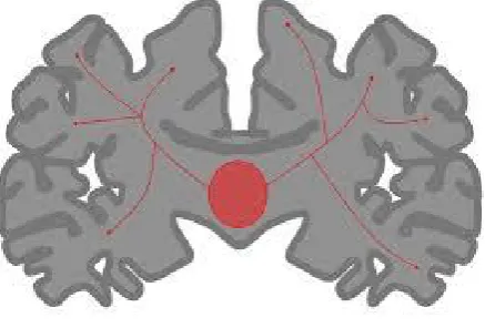
involve the deeper thalamic, subcortical and brainstem structures in a
[image:19.595.197.416.194.342.2]feedback loop to the cortices.
FIGURE 1: Cross section of brain showing primary generalized seizure
Generalized tonic clonic seizures are the most commonly encountered
seizures in both children and adults, the classic picture which the public
generically perceives as epilepsy. The glossary of descriptive terminology for
ictal semiology provided by ILAE taskforce describes a tonic clonic seizure
as “a sequence consisting of a tonic followed by a clonic phase .Variants such
as clonic-tonic –clonic may be seen”102.
A GTCS may be of generalized onset (primarily GTCS) or it may
begin focally, followed by secondary generalization (secondarily
generalized). The clinical manifestations observed in these seizures are
initiated by abnormal electrical discharges within the brain and depend on the
part of brain involved in epileptic neuronal discharge and the intensity of
5
psychic, autonomic and sensory phenomena with or without alteration in
consciousness and awareness.
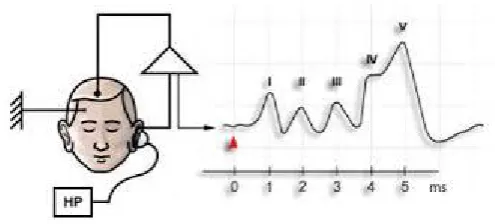
Brainstem auditory evoked potentials are electrophysiological
recordings of responses from within the auditory system that are activated by
sounds. They are recorded from the ear and vertex in response to a brief
auditory stimulation to assess the conduction through auditory pathways up
to midbrain. The human Brainstem auditory evoked potentials (BAEP)
consist of far field evoked potentials from the auditory nervous system. The
BAEP is the averaged surface recorded activity from multiple source neural
generators in the peripheral and lower central auditory nervous system and
represents the synchronous discharge activity of onset –sensitive single units
from first through sixth order neurons. The evoked transient responses can be
recorded up to 500 ms from time of onset of the sound stimulus. The evoked
potentials of the first 10 ms i.e.) short latency response (SLR) is popularly
known as Brainstem auditory evoked potentials (BAEP).
BAEP comprises of 5 or more waves within 10 ms of stimulus and 3
interpeak latencies. Each individual wave and interpeak latencies provide
information about an area of auditory pathway starting with cochlear nerve to
the level of inferior colliculi. These were first described by Jewett and
6
Epilepsy is a neurological disorder characterized by abnormal changes
in the brain’s electrical potentials. The dysfunction occurring at the cellular
level leads to excessive neuronal excitability and AEPs are expected to be
altered by such cellular dysfunction. Hence BAEPs have emerged as an
important clinical tool in studying the electrophysiological phenomena of
neural excitation, conduction and transmission across the auditory pathway
(Tandon OP et al 1990)4.
Cytokines are soluble potent glycoproteins secreted by the glial cells of
CNS and they function as immune system mediators. They mediate cell to
cell sigalling by binding to high affinity surface receptors and serve as a
biomarker for earlier detection of brain damage to prevent further
neurological complications. Abnormalities in the expression of cytokines and
immune cells is noted in epilepsy patients and in various animal models (Jobe
PC et al 1991)5 and hence the immune system and its associated
inflammatory reactions appear to play a major role in epileptogenesis and
aggravate brain damage (Holmes et al 2002)6. Epilepsy per se is capable of
producing elevated levels of cytokines. Such elevated levels as during
inflammation of brain or periphery decreases seizure threshold and
predisposes to epilepsy.
Interleukin-1beta (IL-1β) is one such pro inflammatory cytokine
released from glial cells during seizures. Many CNS diseases such as seizure
7
extravasation of CNS-foreign proteins like albumin and simultaneously the
excitotoxic damage produced by such diseases causes increased microglial
IL-1β expression. This leads to reduced seizure threshold and epilepsy which
depends on the amount of neuronal IL-1RI and IL-1RII.
The chronic expression of IL-1β during epileptogenesis
contributing to neuronal injury suggests that IL-1 β activated pathways play a
vital role in the genesis of spontaneous seizures, thus raising the possibility of
using IL-1 antagonist as a novel drug for seizure inhibition in clinical practice
(Randle et al 2001)103. IL-1 production and activity are regulated by many
factors like caspase-1, IL-1RI, IL-1RII, IL-Ira which implies there are many
ways to interfere with IL-1β activity, of which the anticonvulsive effects can
be explored. IL-1β also influence many central neurotransmitters including
GABA, 5-hydroxy tryptamine, noradrenaline and acetyl choline as well as
expression of a number of neuropeptides in several brain regions contributing
to changes in auditory evoked potentials of GTCS patients.
Hence considering the above factors, the present study is undertaken to
assess the functional integrity of auditory pathway using Brainstem evoked
response audiometry in patients with generalized tonic clonic seizures. We
also compare serum Interleukin-1β levels in patients with GTCS and normal
8
2. REVIEW OF LITERATURE
Seizure is one of the most dramatic example of the collective electrical
behaviour of the mammalian brain It is generally a chronic problem with
significant impact on personal, social and economic aspects, often affecting
the ability to hold jobs and drive. Generalised tonic clonic seizures are the
best recognised form of seizures often presenting with loss of consciousness
and a generalised tonic contraction evolving gradually into clonic activity.
BAEP is an objective neurophysiological test used to evaluate the
neural activity from the external ear to lower brainstem. This non-invasive
tool can be utilized in GTCS patients for early detection of neural conduction
irregularities in the auditory pathway. A few studies in this field of research
have shown an increase in the latency of BAEP waves and so with this
background , the present study has been taken up to assess the integrity of
auditory pathway in GTCS patients by recording brainstem auditory evoked
potentials.
As several researches have high-lighted the intrinsic role of
interleukin-1β in the process of epileptogenesis, the present study is aimed at
measuring serum Interlukin-1β levels in GTCS patients so that the
antagonists of this cytokine can be used in clinical practice for seizure
9
Epilepsy had been one of the earliest and commonly recognised
neurological disorders (Temkin O et al 1994)7. The earliest references to the
disorder dates back to second millennium BC where Mesopotamian writings
described it as antasabbu literally meaning the falling disease. Indian
Ayurvedic writings roughly belonging to the same period also contain
elaborate clinical description of epilepsy as ashepak or apasmara. In 400 BC,
epilepsy was called sacred disease because people believed that people
suffering from seizures were possessed by evil spirits or gods and should be
treated by the invocation of religious, occult and magical powers.
Hippocrates, the Father of Medicine made the notable conceptual
contribution saying epilepsy is an illness as any other disease and no more
considered divine or spiritual. He proposed that brain is the organ where the
site of seizure onset is located and also revealed the existence of genetic
basis in epileptic patients.
Galen, a Greek physician introduced the term aura to describe the
symptoms that preceded the onset of epilepsy.
Tissot recognised two types of ictal events, GTCS which he called
grands acces and absence seizures as petits acces.
10
monkeys that motor cortex and not medulla initiated the convulsive motor
activity.
John Hughlings Jackson (1835-1911), the father of epilepsy confirmed
the relationship that existed between the structure and physiology of the
nervous system to elucidate the pathophysiology associated with seizures. He
also supported the findings of David Ferrier regarding the site of seizure onset
and implicated that the ictal behaviour correlated well with the region of
functional anatomy. The potential therapeutic importance of his intellectual
conclusion was that the surgical treatment may be effective as an underlying
pathology or structural lesion was presumed to be associated with the site of
epileptogenesis.
Victor Horseley performed the first surgery for epilepsy in 1886 by
resecting a traumatic cortical scar in a patient with focal motor seizure
rendering him seizure free.
Hans Berger’s invention of electroencephalogram in 1929 made a
11
2.1.1. Epidemiology
The incidence of epilepsy is 0.3-0.5% in the world population which is
age dependent. Along with various studies, Hirtz D et al also have shown that
higher rates occur in infants younger than one year and a second peak is found
in people older than 60 years presenting with bimodal distribution8. Seizures
are so common to occur in 10%of the population at some point in their
lifetime (Berg et al 1991)9. Sex specific incidence rates are not usually
significant, although the incidence rates are almost always higher for males as
compared to females. The prevalence is estimated to be 5-10 persons /1000
which is higher in developing countries. Sridharan et al estimated the
prevalence in Indian population to be 1% which is higher in rural than urban
population 10.
Perhaps more important than incidence and prevalence in
understanding the impact of epilepsy as a worldwide health problem is the
global burden of the disease which is measured by Disability Adjusted Life
Years(DALYs) and number of life years lost due to disability or death(YLL).
According to WHO, epilepsy was estimated to account 0.5% of the global
12
2.1.2 Etiology
1. In 60-70% of patients with seizures, no specific cause is identified
which is commonly referred as idiopathic.
2. Genetic- risk is increased 2-3 times in individuals having first degree
relatives with epilepsy.
3. Infants and children- congenital malformations, perinatal injuries or
hypoxia, developmental neurological disorders, metabolic defects,
injuries and infections.
4. Young adults- head trauma, brain tumours, infections and arteriovenous
malformations.
5. Elderly – cerebrovascular disease, CNS degenerative diseases, brain
tumours.
6. Drugs- antihistamines, narcotic analgesics and iodinated contrast agents
like metrizamide (Messing RO et al 1984) 12.
2.1.3. Genetics and Epilepsy13
Most of the seizures are believed to be complex traits resulting from
interactions between non genetic and genetic factors, the latter thought to
provide minor contributions from multiple genes (oligogenic or polygenic)
and thus the patterns of inheritance in majority of seizures are complex and
13
Many generalised seizures in humans have revealed a genetic basis and
these almost become apparent before the age of 35 years. Recent studies
suggest that the mutations affecting the ion channel function and
chromosomal microdeletion may be the cause of epilepsy in a subset of
patients.
Tan NC et al14 have quoted many association studies that have been
undertaken to examine the influence of common genetic variation on disease
susceptibility in epilepsy, but unfortunately no genetic variants have been
proven to underlie any common epilepsy. Studies in twins have demonstrated
concordance rates as high as 70% in monozygotic and 10% in dizygotic pairs
implying that factors other than genetics play a role in the occurrence of
seizures (Treiman et al 1993)15.
Triggering factors for seizures include
-Sleep deprivation
-Physical and mental exhaustion
-Flickering lights including television and computer screens
-Alcohol (particularly withdrawal)
-Intercurrent infections or metabolic disturbances
14
2.1.4. Comorbidities in Epilepsy
Individuals with epilepsy are at risk for increased morbidity and
mortality as compared to general population. Patients with drug resistant
epilepsy and longer duration of the disorder contribute significantly to higher
morbidity in various surveys due to the result of stress and its adverse effects
on various systems. In patients with newly diagnosed epilepsy, 18% of them
present with additional dementia, 6% motor disabilities and 6% with severe
psychiatric disturbances. So about 1 in 15 of patients with seizures depend on
others for daily living due to associated handicaps. Poor epilepsy control and
seizure themselves can lead to significant cognitive and personality changes
as well as chronic depression. It results in social stigmatisation resulting in
isolation which further creates problem in education, employment, personal
relationships and family life.
Patients with epilepsy have 2-3 times greater risk of mortality than
expected in a matched population without epilepsy. SUDEP accounts for
about 1.7% deaths in epilepsy which when witnessed is most often associated
with GTCS near the time of death.
2.1.5. Pathophysiology of Seizures
The electro physiologic and molecular mechanisms that underlie the
15
maximal electroshock or chemoconvulsants such as pentylenetetrazol have
been utilised to probe into the pathophysiology of focal and generalised
seizures.
Seizures are linked at the lowest level to membrane potentials, ionic
fluxes and generation of action potentials. The RMP of the neuronal
membrane is approximately about -70mV. In neurons the action potential is
generated due to changes in the permeability of sodium, chloride, calcium
and potassium ions which enter and exit the neurons by voltage dependent
channels. At threshold voltage there is Na+ influx due to high permeability of
these ions, the membrane potential becomes dramatically positive (+60 mV)
which generates the action potential. After 1ms Na+ channels are inactivated
followed by K+ efflux and the coincident Cl-- influx result in membrane
hyperpolarisation and the termination of electrical activity at that point along
the cell membrane (Hille B et al 1984)16.
As the action potential reaches the axon terminal, voltage dependent
Calcium channels open, permitting Calcium influx into presynaptic terminals
which results in neurotransmitter release. Hence any changes or
abnormalities at any point along this electric cascade will have a significant
impact on the excitability and epileptogenecity of the individual neuron (Mc
16
The effects of neuronal activity are mediated through synaptic
connections existing between neocortical, thalamocortical and
corticothalamic projections. The impulses finally culminate in either
excitatory (EPSP) or inhibitory postsynaptic potentials (IPSP). Excitatory
inputs are transmitted by neurotransmitters like glutamate and aspartate
whereas inhibitory inputs are mediated by gamma aminobutyric acid
(GABA).
Seizure propagation is likely to occur in cells with intrinsic bursting
if the balance between excitatory and inhibitory inputs is altered. Neurons
begin to fire synchronously leading to a seizure in the absence of appropriate
inhibitory regulation (loss of IPSPs).
Goldensohn et al18 proposed a hypothesis that the neurons within
the epileptic focus of cortex undergo paroxysmal synchronous depolarisation
termed as paroxysmal depolarizing shift(PDS) resulting in an abnormal burst
of action potentials which continue in synchronous volleys without
appropriate inhibition.
2.1.6. Neurotransmitters
GABA is found to be a critical inhibitory modulator of neuronal
activity in the brain (Gale K et al 1992)19. It generates IPSPs mediating
17
regions of brain. Three distinct types of GABA receptors are identified.
GABA-A, B, C. GABA-A receptor, a pentameric complex is organised to
form a chloride channel through which Cl- ions can enter into the
postsynaptic region. When GABA binds with GABA-A receptor, there is
influx of Cl- ions resulting in generation of IPSP. Wallace et al20 found that
mutations of GABRG2 gene encoding for GABA-A subunit leads to
generalised and febrile seizures.
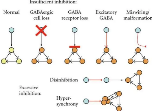
Lothman E et al21 suggest that disruption of GABAergic function may
be central to the molecular pathophysiology of seizures. Hence interruption
of integrated GABA mediated inhibition in cortex will result in a lowered
threshold for ictal discharges. Various animal models have supported this
mechanism which shows that GABA agonists prevent seizures while
[image:33.595.149.412.539.731.2]antagonists may provoke seizures.
FIGURE 2: GABA in epileptogenesis
18
Glutamate and aspartate are excitatory neurotransmitters which act by
binding to EAA receptors resulting in enhanced intracellular Calcium levels.
This causes activation of proteases and lipases and osmotic swelling of
neurons resulting in cell death.
Meldrum B et al proved in his study that glutamate and aspartate
contribute to the initiation, spread and maintenance of epileptic activity in
cortex22.
During MJ et al studied in epilepsy patients to confirm increased levels
of EAA compounds such as aspartate using micro dialysis catheters
immediately prior to the onset of seizure activity23.
2.1.7. Effects of Seizures on Brain
Ischemia /infarction, trauma and seizure are three major forms of acute
brain injury which leads to significant neuronal loss resulting in neuronal
dysfunction. There may be some common cellular mechanisms involved in
these pathologies, but the induction of cell death following seizure activity is
probably less well understood than other forms of brain injury (Liou AK et al
2003)24.
Delorenzo RJ et al observed that following a prolonged seizure, there is
19
aspartate(NMDA) receptors and voltage activated Calcium channels that
results in cellular influx of Calcium ions25. An increase in free calcium tends
to produce mitochondrial dysfunction with activation of various enzymes like
lipases, endonucleases, proteases etc. In addition, free radicals are generated
contributing to mitochondrial dysfunction26 and ultrastructural changes may
be observed after 30-60 min of seizures (Meldrum BS et al 2002)27. This
results in reversible or irreversible neuronal damage and CSF markers of
neuronal injury like neuron specific enolase increase following seizure
activity (Pitkanen S et al 2002)28.
Prolonged seizures may ultimately result in neuronal death by either
necrotic or apoptotic pathways. Based on classic morphological definitions,
cell necrosis appear to be the dominant mechanism, the exception being
granule cell death in the dentate gyrus including recently generated neurons
in this region which more readily show apoptosis (Ekdhal CT et al 2003)29.
However animal models suggested varying ratios of apoptic vs necrotic cell
death suggesting heterogeneity in the cellular effects of seizures. This state of
flux is found to be dependent on energy state of the cell and on the time
course and severity of the neurotoxic insult, cell necrosis being observed with
severe excitototoxic insults and apoptosis with milder insults to more resilient
neurons which allows the induction of more delayed energy dependent cell
20
Thus epileptic seizures can cause severe and long lasting events on
the brain architecture including neuronal cell death, accompanied
neurogenesis, reactive gliosis and mossy fibre sprouting.
2.1.8. Generalised Tonic Clonic Seizures
Gestaut and Broughton gave an elaborate description of the semiology
and pathophysiology of GTCS32. By stressing the stereotypical nature of
GTCS, they divided it into 4 distinct phases
1. Preictal manifestations
2.Ictal manifestations (with loss of consciousness)
a. Tonic phase (includes intermediate vibratory phase)
b. Clonic phase
c. (concurrent) Autonomic changes
3. Immediate postictal features
4. Late postictal features
According to Gestaut and Broughton, preictal manifestations are brief
with bilateral myoclonic contractions which immediately precedes the onset
21
with nonspecific symptoms such as headache, irritability, lethargy, mood
changes and sleep disturbances. These symptoms actually do not represent
epileptic aura but reflect the physiological changes that reduce the seizure
threshold. If aura occurs before a seizure, it indicates that the tonic clonic
seizure is secondarily generalised.
GTCS synonymous with the previous term grandmal epilepsy or
convulsions is the classical and best recognised form of seizures which
typifies epilepsy in public imagination. These seizures do not have an aura
but preceded by a prodrome that occurs for minutes to a few hours before a
seizure which includes inconsistent nonspecific premonitory symptoms like
ill-defined anxiety, instability, reduced concentration, headache or vague
uncomfortable feelings.
The seizure onset is abrupt often presenting with loss of consciousness
followed by the tonic phase. In this phase the patient tends to fall if he is
standing with bilateral tonic extension of trunk and extremities followed by
synchronous muscle jerks. This phase can have asymmetrical movement
which often varies from seizure to seizure and one such commonly observed
asymmetry is versive head turning which is not an evidence of focal onset. It
also includes upward deviation with partially opened eyes and mouth is also
22
Involvement of respiratory muscles produces epileptic cry which is
characterised by forced expiration that produces a loud guttural localisation.
Cyanosis may occur in this phase in association with apnoea. Autonomic
signs are present during this phase which includes tachycardia, hypertension,
cyanosis, salivation, sweating and incontinence of urine or stools.
Benarroch EE et al was able to elucidate that increase in heart rate and
blood pressure was either mediated directly through the ictal activation of
structures of the central autonomic network which includes insular cortex,
amygdala, hypothalamus, periaqueductal grey matter, para brachial complex,
NTS and ventrolateral medulla or reflecting the high metabolic demand of the
seizure.
Plum F et al34 reported that the rise in blood pressure evoked by seizure
usually causes a significant increase in cerebral blood flow to meet the
increased metabolic demands of brain.
Bateman LM etal observed that when diaphragm and
thoracoabdominal muscles are involved during the tonic phase, it results in
insufficient air exchange, which in turn may lead to alveolar hypoventilation
causing decrease in blood O2 saturation and cyanosis.
This stage lasts on an average of 10-30 sec and gradually evolves into
clonic activity. The transition can be initially of high frequency and low
23
As the cortical discharges diminish with frequency, the clonic phase
ensues where the limbs produce repetitive myoclonic jerks for a variable
time. With seizure progression, the frequency of clonic jerks reduces and the
amplitude which rises initially also decreases just before the seizure stops.
The final phase lasts for 2-30 min and is characterised by flaccidity of
muscles and diminished tendon jerks.
Confusion and unresponsiveness is invariable in the postictal period.
Respiration is loud and stertorous in nature. In this phase the patient feels
dazed with a severe headache and extremely unwell, he often lapses into a
deep sleep. On awakening within minutes or hours later, some residual
symptoms may persist which include headache, dysthymia, lethargy and
generalised muscle soreness. GTCS rarely last greater than 2 minutes and the
postictal state is found to be correlated with severity and duration of the
seizure episode.
2.1.9. Diagnosis of Epilepsy
In assessment of a patient with epilepsy, the history takes primacy
ideally in conjunction with an eye witness account. In a vast majority of cases
the diagnosis of epilepsy is clinical but a syndromic diagnosis is also possible
on the basis of clinical signs and symptoms.
24
confirming the diagnosis and type of epilepsy (focal vs generalised) and also
in localising the area of seizure onset. EEG must be performed, it is more
likely to be abnormal within first 2 days after a seizure31. Ictal EEG findings
at onset include high amplitude anteriorly dominant generalised spike wave
discharges, diffuse fast frequencies that evolve to generalised spike wave
discharges or polyspike wave discharges.
Once the seizure is clinically manifest, the muscle activity prevents the
determination of EEG changes. Post ictally the EEG often shows diffuse
slowing ie) slow spike wave discharges. Inter ictal EEG shows either a
normal background or runs of occipital delta activity and may show either
fragmented diffuse spike wave or polyspike wave discharges or frank
generalised spike wave discharges.
2.2. Evoked Potentials
An evoked potential is an electrical manifestation of the brain’s
reception of and response to an evoked stimulus. Evoked potentials have been
studied in patients with neurological diseases since early 1950s but it was
only in the early 1970s that evoked potentials began to have definite clinical
utility. These tests provide sensitive and quantitative extensions of the
clinical neurologic examination. Brainstem auditory evoked potential has
25
auditory pathway from the external ear to lower brainstem and has been
extensively used in patients with seizures.
2.2.1. BAEP and epilepsy
Epilepsy is a neurological disorder which is characterized by abnormal
changes in the brain’s electrical potentials. The excessive neuronal
excitability produced as a result of biophysical and biochemical cellular
dysfunction alters the BAEP which is noted in various studies.
- Cranfor JL et al128 has reported that central auditory impairment is
present in bi-hemispheric seizure disorders. The patients have shown
improvement in the measures of central auditory function following
successful surgery to control epilepsy, but the results are prone to
variability.
- Zhao JY et al analysed a Chinese family and proved that hearing loss
associated with epilepsy is due to a mitochondrial mutation. He
evaluated in clinical and genetic aspects along with sequential analysis
of mitochondrial genome in a three generation Chinese family and
identified that 7472 del C is likely to be a novel mitochondrial mutation
associated with hearing loss in epilepsy patients.
26
prolonged I-III and I-V IPLs and longer standard deviations as
compared to the normal controls. He noted that the number of different
seizure types in an epileptic patient was significantly related to the
changes in latency but type and duration of seizure do not show any
such correlation.
- Salah Soliman et al98 recorded auditory brain stem response and
middle latency response in 49 epileptic patients. He found that a
statistically significant number of epilepsy patients showed elevated
ABR (30.1%) and MLR (40.7%) in spite of these patients having
normal hearing sensitivity which was assessed by pure tone
audiograms. He observed that threshold elevation was more frequent in
patients with grandmal epilepsy when compared with temporal
epilepsy patients reflecting poor response in the former group.
Furthermore they also noted that chronicity of illness was significantly
related to the elevated ABR and MLR thresholds in patients with
grandmal epilepsy in contrast to temporal lobe epilepsy patients.
- Masayuki Ohishi et al125 selected 114 epileptic patients –both males
and females and recorded ABR in these patients. He observed that the
27
- Chayasirisobhon S, Rodin E et al studied the functions of brainstem in
81 epileptic patients using BAEPs. He acknowledged that the epileptic
patients had significantly longer latencies for all the wave components
and IPLs, especially I-III, I-V than the normal controls.
- Usha Panjwani et al83 studied the effects of antiepileptic drugs on
BAEPs in 32 female epileptic patients. They observed that drug free
epileptics had shortened wave V absolute latency and I-V IPL as
compared to normal controls.
The development in the field of recording evoked potentials is closely
linked to the discovery of electricity.
1752-Benjamin Franklin with his kite experiment charged his leyden jar by
using kite during electrical storms and postulated the presence of two
opposing forces of electricity that is positive and negative.
1791- Luigi Galvani discovered that nerves were good conductors of
electricity.
1850-Helmholtz was able to measure conduction velocity of nerve in frog.
1861- The method of electro diagnosis based on faradic and galvanic current
was introduced by Erb.
28
goes to Richard Canton, who reported that he had detected currents from
electrodes placed on the skull of exposed brain in rabbits and monkeys.
1929- Hans Berger recorded the first human electroencephalogram from the
electrodes placed on scalp.
1939- Davis was the first to record electric potentials on the human skull in
response to auditory stimuli. The potentials were generated in the cortex with
latencies that ranged from 50 to 500 ms. Some years later, thanks to
computers, faster and shorter amplitude responses were recorded called as
middle latency potentials between 10-80 ms.
1967-Sohmer and Feimesser were the first to record BAEP initially and
attributed their origin to brainstem structures37.
1971-Jewett and Williston were the first to describe BAEP waveforms.
1974-Hecox and Galambos showed that ABR could be used for threshold
estimation in adults and infants.
The clinical utility of evoked potentials is based on their ability to
- Demonstrate abnormal sensory system function when the history and
/or neurological examination are equivocal.
- Reveal the presence of clinically unsuspected malfunction in a sensory
29
and signs in another area of CNS.
- Help in defining the anatomic distribution of a disease process
- Monitor the changes objectively over time in a patient’s status
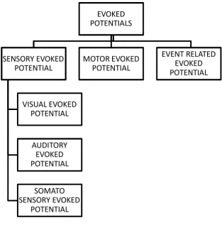
FIGURE 3: Classification of evoked potentials
Sensory evoked potentials are recorded from CNS following
stimulation of sensory organs. For example visual evoked potentials are
elicited by a flashing light or changing patterns on a monitor; AEPs by a click
stimulus presented through the ear phone and tactile or somatosensory
evoked potential elicited by tactile or electrical stimulation of a sensory or
[image:45.595.182.496.264.589.2]30
mixed nerve in the periphery. All these potentials are reliable diagnostic tests
providing an objective measure of function in their related sensory system
and tracts.
2.2.2. Brainstem Auditory Evoked Potential
The ground work for recording BAEP was laid in early 1930s when the
electronic amplifiers became available. In 1939, the electrical response to
auditory stimuli was first observed in raw EEG.
Recording BAEP is a simple non-invasive way of evaluating hearing
function and has been widely used for early detection of neural conduction
irregularities in the auditory pathway. Technical advances have lead to more
widespread use of BAEP in various fields like audiology, neurology,
anaesthesiology and neonatology.
Bluestone CD et al had acknowledged in their study that a normal ABR
finding does not guarantee hearing and not all normally hearing subjects have
normal ABR findings40.
In spite of many advantages, BAEP equally has limitations- requires an
experienced person to identify the waveforms accurately and the procedure is
time consuming (Graham JM et al 2007)41. Development of sophisticated
31
2.2.3. Principle of auditory evoked potential35
The evoked response audiometry is based on the principle that the
bioelectric response which is evoked by a sound stimulus always tend to
occur after the same time interval.
The auditory pathway extends from the middle ear structures through
the eighth cranial nerve, the brain stem, and finally to the auditory cortex.
Auditory stimuli either in the form of clicks or pure tones can be used to
assess the integrity of the auditory pathway.
Thus the auditory evoked potential is obtained by presenting auditory
stimuli to each ear resulting in a sequence of waveforms which bear a close
relationship to the structures in the auditory pathway and enables relatively
specific localisation in the auditory pathway, particularly in the eighth cranial
nerve and the brainstem. From the time of onset of the sound stimulus, the
auditory evoked transient response can be recorded up to 500 milliseconds.
2.2.4. Classification of Brainstem auditory evoked potentials
This tool of investigation which was first described by Jewett and
Williston in 1971 can be classified as short, middle and long latency
32
Short latency response
The normal Brainstem auditory evoked potentials occurring within first
10 ms give unique information about the brainstem functions is called the
early phase of transient response or short latency response. These potentials
are well known as Brainstem Evoked Response Audiometry (BERA) or
Brainstem Auditory Evoked Potentials (BAEP) and this has been utilised
extensively by the clinicians.
Advantages of BAEP include
1. This test is used to detect deafness in uncooperative patients like
infants and mentally retarded or malingering individuals and can also
be carried out correctly even in deeply sedated and anaesthetised
patients.
2. Objectively determines the nature of deafness (i.e. sensory or neural) in
difficult to test patients especially when they cannot respond
adequately to tests.
3. Identification of the site of lesion in retro cochlear pathologies- The
retro cochlear area is fairly a large area extending right from the spiral
ganglion of the cochlear nerve to the midbrain (level of inferior
colliculus). Unlike other tests used by the neurotologists which merely
33
helps to identify the approximate area in the retro cochlear pathway
where the lesion is present which is utilised in diagnosing conditions
like acoustic neuroma with accuracy.
4. Study of central auditory disorders- Evoked response audiometry is
useful in differentiating diseases of the auditory cortex from diseases
of the more peripheral organs.
5. Helps to analyse the maturity of the central nervous system especially
in new born, evaluating prognosis in comatosed patients, objective
identification of brain death etc.
Middle latency response (MLR)
Auditory evoked potentials occurring before 10-50 msec is called
MLR which reflects the activation of both subcortical structures(thalamus)
and auditory cortices(mainly primary auditory cortex)42.
Long latency response (LLR)
This response arises from multiple cortical generator sources (Neshige
et al1992)43 and have latencies greater than 50 msec distributed entirely over
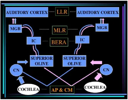
FIGURE 4: Generators of auditory evoked potentials
LLR - Long latency response
MLR - Middle latency response
BERA - Brainstem evoked response audiometry
AP - Action potential
CM - Cochlear microphonics
MGB - Medial geniculate body
34
2.2.5. Anatomical and Neurophysiological basis of brainstem auditory
evoked response audiometry37
The sound pressure waves causes displacement of tympanic membrane
which is transmitted through inner ear ossicles to the oval window. This
causes movement of perilymph present in the scala vestibuli and tympani and
secondarily of potassium rich endolymph contained in the ductus cochlearis
and hence the basilar membrane, spiral organ and tectorial membrane are
displaced.
So the spiral organ containing the hair cells produce auditory receptor
potentials when it is displaced by the movement of tectorial membrane. The
receptor potentials tend to trigger action potentials in the dendrites of afferent
nerve fibres of cochlear nerve due to the release of neurotransmitters. It is
approximately estimated that 20000 to 30000 hair cells are scattered over a
distance of 31.5mm in 2.5 spirals of cochlea. It is found that low frequency
sounds activate the apical portion of cochlea and high frequency sounds
generate receptor potentials in the basal portion. The click stimulus used in
BERA contains mainly high frequency tones which stimulates the basal
portion of cochlea.
The cochlear nerve neurons situated in the spiral ganglia are bipolar
35
This cochlear nucleus has 3 sub nuclei components:
1. Anterior ventral cochlear nucleus (AVCN)
2. Posterior ventral cochlear nucleus (PCVN)
3. Dorsal cochlear nucleus (DCN)
The output of AVCN runs through the ventral acoustic striae forming
the bulk of trapezoid body and terminates in the superior olivary nucleus and
inferior colliculus. The neurons in AVCN tend to discharge at short latency to
acoustic stimuli with a pattern similar to that of cochlear nerve.
Most of the output of PVCN goes through the ventral and middle
acoustic striae to terminate in the superior olivary nucleus and inferior
colliculus. Through the dorsal striae, the dorsal cochlear nucleus terminates
in the superior olivary nucleus and contralateral inferior nucleus. The
discharge from these neurons have a longer latency thus differing from
AVCN.
The cochlear nucleus thus terminates in the superior olivary nuclear
complex which includes 2 components-medial and lateral at the base of pons.
The medial superior olivary nucleus receives excitatory inputs from both
ipsilateral and contralateral AVCN. The lateral superior olivary nucleus also
36
inhibitory inputs from contralateral AVCN and PVCN via) trapezoid body.
From the olivary nucleus, the impulses travel to the ipsilateral and
contralateral lateral lemniscus and to inferior colliculi. The olivary nuclei are
the first site in the auditory pathways where the neurons are affected in a
nonlinear manner to binaural stimulation.
The lateral lemniscus nuclei and inferior colliculi converges the input
from contralateral cochlear nucleus and superior olivary nucleus. The
impulse from the inferior colliculi reaches the medial geniculate body where
the neurons form the acoustic radiation of internal capsule finally synapsing
in the Heschl gyrus of the primary auditory cortex (superior temporal gyrus
and upper bank of sylvian fissure including the frontal and parietal opercula),
the deeper mesial portion of which is activated by the high frequency tones
like clicks used in BERA.
The orderly orientation of the neurons in dorsal cochlear, medial
superior olivary and lateral superior olivary nucleus results in summation of
synaptic potentials to result in high amplitude electric fields.
The journey of the auditory impulses through this pathway generates
an electric activity which can be recorded by placing surface electrodes on the
scalp. This electrical activity is manifested as waveforms with discrete peaks
37
various parameters and this reveals the structural and functional integrity of
the auditory pathway.
Spiral ganglion in the cochlea àVentral and dorsal cochlear nuclei in
the brainstem àSuperior olivary complex in the midbrain àLateral
lemniscus in midbrain àInferior colliculus in midbrainàMedial geniculate
[image:54.595.160.455.340.721.2]body in thalamus àAuditory area in cortex.
38
2.2.6. Normal Waveforms of BAEP
There are 5 or more distinct waveforms recorded within 10millisec of
auditory stimuli (Jewett DL et al 1971)44. The analysis of these waveforms
with regard to latency, amplitude, wave morphology provides
[image:55.595.175.455.284.435.2]neurodiagnostic information on cochlear and retrocochlear auditory function.
TABLE 1: Generators of BAEP waveforms
WAVEFORM GENERATORS
I Eighth nerve(Cochlear nerve)
II Cochlear nucleus
III Superior olivary nucleus
IV Lateral lemniscus
V Inferior colliculi
[image:55.595.116.488.467.750.2]39
2.2.7. Clinical applications
- Neil Bhattacharya53 demonstrated that these evoked potentials can be
utilised as an effective screening tool in the evaluation of suspected
retro cochlear pathology such as vestibular schwannoma or acoustic
neuroma.
- Young G Bryan54 suggested that persistent abnormalities of BAEPs
reliably indicate the likelihood permanent vegetative state or death.
- J.K.Nousak et al55 showed that the BAEP latencies are accurate in
evaluating hearing threshold.
- Studies carried out by Avasthi R Subhendu56 have identified an
increase in absolute latencies of all the waves of BAEP in patients with
advanced hypertension and following treatment significant decrease in
the wave latencies were observed demonstrating their role in
prognostic follow up of these patients.
- Kurita A et al57 compared 20 normal controls with diabetic patients and
showed that diabetics had significantly longer latencies.
- Flint Boettcher A58 demonstrated that latencies were prolonged in
presbyacusis and can be used as an evaluating tool in elderly
40
- In a study conducted by Ikuta et al59, he showed that differences in
waveforms are seen in evoked potentials of schizophrenics, manic
depressives and epileptics as compared to healthy adults.
- Atis et al60 recorded BAEP in patients with COPD and attributed the
prolonged latencies to chronic hypoxic-hypercapnic status occurring in
the brainstem.
- Schwarz G et al61 studied BAEP in patients with respiratory
insufficiency following encephalitis and observed prolongation of all
waves and IPL due to proximity of respiratory control centre in the
brainstem.
- In a study done by Reyes Contreras et al62, he observed significant
differences in I-V IPL in HIV infected patients as compared with
controls and concluded that HIV infection may produce subclinical
pathologic changes in the cochlear nerve and brainstem which can be
recorded by BAEP recordings.
- Leocani et al63 in his study suggested that patients with multiple
sclerosis can have abnormal ABRs.
- Lew and Henry L64 demonstrated that BAEP recording can serve as an
objective tool for estimating hearing dysfunction in traumatic brain
41
2.3. Cytokines
They are low molecular weight regulatory proteins secreted by white
blood cells and various other cells of the body in response to multiple
stimuli65. In recent years an increasing body of evidence suggests that there is
a complex relationship existing between epilepsy and immune system.
Plata –Salaman CR66 observed abnormalities in the expression of
cytokines and immune cells in patients with epilepsy.
Kalueff AV et al67 recognised that the immune system and its
associated inflammatory reactions play an important role in the process of
epileptogenesis.
Steffensen SC et al68 implicated cytokines as mediators of spontaneous
seizures.
Fann MJ et al69 identified that these cytokines influence many central
neurotransmitters including Noradrenaline, Gamma amino butyric acid,
Acetyl choline, 5 Hydroxy tryptamine as well as the expression of various
neuropeptides in several brain regions.
2.3.1. Interleukin-1beta (IL-1β)
IL -1β which is synonymous with catabolin is a cytokine protein
42
pro inflammatory cyokine that activate additional cytokine cascade and
enhances the susceptibility of seizures. IL 1 cytokines are regularly expressed
at very low levels in human CNS (Ravizza T et al 2006)70. Seizures enhance
the expression of IL- 1βand its mRNA as well as IL 1Ra mRNA (De Simoni
MG et al 2000)71. Concentration of IL-1β in extracellular compartment is the
prime factor for determining it functional actions in the brain.
IL 1β augment nitric oxide formation to raise the seizure susceptibility
and also increase the neuronal excitability by directly inhibiting
GABA(A)receptors, enhancing NMDA receptor function and inhibiting K
efflux104. Viviani et al105 has shown that IL-1β increases the phosphorylation
of the NR2B subunit of NMDA receptor thereby enhancing Calcium influx
into the neurons. Through the activation of sphingomyelinase, IL-1β induces
the production of ceramide which in turn activates the Src family tyrosine
kinases leading to NR2B phosphorylation. Balosso et al106 suggest that the
activation of this pathway underlies the proconvulsant activity of IL-1beta.
43
FIGURE 7: Interleukin-1beta in epileptogenesis
During epileptogenesis, strong IL-1β and IL-1R immunoreactivity was
found also in perivascular astrocytic end feet impinging on blood vessels and
in endothelial cells of microvasculature. Ravizza et al 2008 associated these
changes with tissue extravasation of serum albumen. IL-1β can affect the
permeability properties of blood brain barrier by disruption of tight junctions
or nitric oxide production along with activation of metalloproteinases in
44
alterations in BBB permeability may favour the entry of the cells of adaptive
and innate immunity into the brain which perpetuates inflammation.
Van illet et al 2006 proved that the extent of BBB damage positively
correlates with the frequency of spontaneous seizures
IL-1β is cleaved from precursor protein (pro IL-1β) by IL-1β
converting enzyme (ICE) otherwise called as caspase 1. Black et al showed
that this cleavage is essential for the formation of active form of IL-1β.
Recently Ravizza et al has reported a novel anticonvulsant treatment strategy
which inhibits IL-1β production in brain using ICE inhibitors resulting in
reduced seizure duration as well as increased resistance to seizures in kainic
acid models.
Peltola et al92 suggests that the levels of various cytokines increase
transiently in the blood and CSF of patients with epilepsy after different types
of seizure and the cytokine concentration was found to higher in CSF than in
blood suggesting a brain origin
Vezzani et al in his experimental studies identified that IL-1β prolong
the duration of kainic acid induced seizures.
Rosenbaum KJ et al129 acknowledged that seizures themselves can
activate the sympathetic nervous system and induce the release of
catecholamine which mediates cytokine release from the peripheral blood
45
Lehtimaki et al101 have proved that that the levels of IL 1 beta, IL1ra
and IL6 were transiently elevated after electrographic seizures.
Gang Li et al suggests that the level of IL-1β, IL-6 and TNF alpha
increases quickly after either GTCS or complex partial seizures and return to
baseline after varying time intervals.
S.Sinha, S.A.Patil, V.Jeyalekshmy et al91 analysed serum cytokine
levels in 100 patients with epilepsy and new onset seizure in the immediate
postictal phase. They observed a highly significant increase in serum levels of
IL-1β, IL-2, 4, 6, TNF-alpha, IFN gamma in epilepsy patients as compared to
46
3. AIM AND OBJECTIVES
The aim of this study is to evaluate the Brainstem auditory evoked
potential in patients with Generalised Tonic Clonic Seizures in comparison
with age and sex matched controls.
The objectives of the study were
· To determine the functional integrity of auditory pathway in patients
with GTCS by recording brainstem auditory evoked potential.
· To assess serum Interleukin -1 beta levels in these patients.
· To find the correlation between serum Interleukin-1 beta level and
Brainstem auditory evoked potential in patients with Generalized
4. MATERIALS
47
4. MATERIALS AND METHODS
The study was conducted during the year 2013-2014 in the Institute of
Physiology and Experimental Medicine, Madras Medical College after
obtaining approval from the Institutional Ethics Committee, Madras Medical
College Chennai.
4.1 Patient selection
Patients of both sexes in the age group between 20-40 years diagnosed
as generalized tonic clonic seizures were included in the study. They were
selected from the Institute of Neurology, Rajiv Gandhi Government General
Hospital, Chennai - 3. 30 age and sex matched apparently healthy people
were selected as controls.
4.2 Inclusion criteria
Thirty patients , both men and women in the age group of 20-40 years
diagnosed as generalized tonic clonic seizures who were on treatment were
included in the study after confirming the normal hearing ability of these
48
4.3 Exclusion criteria
- Children and pregnant women
- Patients with diabetes and hypertension
- Subjects with congenital hearing loss and sensorineural deafness
- Tumours like acoustic neuroma and meningioma
- Acute brainstem stroke
- Demyelinating diseases like multiple sclerosis
- Subjects with head injury and infections like meningitis and
encephalitis
- Neurodegenerative diseases like dementia
- Febrile seizures
- Conditions that mimic GTCS like psychogenic nonepileptiform
seizures
- Subjects with neoplastic, hepatic, respiratory and any cardiovascular
49
4.4 Control group
Thirty age and sex matched controls were selected from technicians,
staffs and attenders of the patients.
With all these criteria, a total of 60 individuals were selected for the
study. Out of these 30 were apparently normal and termed as controls and the
remaining 30 persons were patients with GTCS who were called cases.
Informed verbal and written consent was obtained from the participants after
explaining the procedure.
STUDY DESIGN: Cross sectional study
TYPE OF STUDY: Comparative study
PLACE OF STUDY: Institute of Physiology and Experimental Medicine,
Madras Medical College, Chennai.
All subjects included in the study had no hearing deficit as reported
after thorough ENT examination which includes pure tone audiometry.
Specific ENT examination
Both the control and study group of individuals were subjected for
specific ENT examination in the Upgraded Institute of Otorhinolaryngology,