Copyright © 2003, American Society for Microbiology. All Rights Reserved.
Inoculation of Plasmids Encoding Japanese Encephalitis Virus PrM-E
Proteins with Colloidal Gold Elicits a Protective Immune
Response in BALB/c Mice
Zijiang Zhao,
1,2Takaji Wakita,
1and Kotaro Yasui
1*
Department of Microbiology and Immunology, Tokyo Metropolitan Institute for Neuroscience, Tokyo 183-8526, Japan,1and
Institute of Virology, Chinese Academy of Preventive Medicine, Beijing 100052, People’s Republic of China2
Received 26 July 2002/Accepted 10 January 2003
We established a simple and effective method for DNA immunization against Japanese encephalitis virus (JEV) infection with plasmids encoding the viral PrM and E proteins and colloidal gold. Inoculation of plasmids mixed with colloidal gold induced the production of specific anti-JEV antibodies and a protective response against JEV challenge in BALB/c mice. When we compared the efficacy of different inoculation routes, the intravenous and intradermal inoculation routes were found to elicit stronger and more sustained neutral-izing immune responses than intramuscular or intraperitoneal injection. After being inoculated twice, mice were found to resist challenge with 100,000 times the 50% lethal dose (LD50) of JEV (Beijing-1 strain) even
when immunized with a relatively small dose of 0.5g of plasmid DNA. Protective passive immunity was also observed in SCID mice following transfer of splenocytes or serum from plasmid DNA- and colloidal gold-immunized BALB/c mice. The SCID mice resisted challenge with 100 times the LD50of JEV. Analysis of
histological sections detected expression of proteins encoded by plasmid DNA in the tissues of intravenously, intradermally, and intramuscularly inoculated mice 3 days after inoculation. DNA immunization with colloidal gold elicited encoded protein expression in splenocytes and might enhance immune responses in intravenously inoculated mice. This approach could be exploited to develop a novel DNA vaccine.
Japanese encephalitis is a serious mosquito-borne viral dis-ease in southeastern and far eastern Asia. Every year, more than 35,000 cases and 10,000 deaths are reported. One third of these have occurred in China. Japanese encephalitis virus (JEV), the etiologic agent, belongs to theFlavivirusgenus of the Flaviviridae family. The majority of JEV infections are subclinical. However, among patients with clinical symptoms, fatality rates range from 10% to 50% (32).
Vaccination has been observed to protect against JEV in-fection in humans and domestic animals (15, 49, 52). Three kinds of Japanese encephalitis vaccine have been used in Asian countries with measurable success. One is a formalin-inacti-vated JEV vaccine purified from infected adult mouse brain. It was developed in Japan and is currently used worldwide, in-cluding India, Korea, and Taiwan (15, 24, 32, 27). Another formalin-inactivated vaccine and a live-attenuated vaccine des-ignated SA14-14-2also exist, both of which are prepared from
infected primary hamster kidney cells in the People’s Republic of China (52). Due to regulatory issues surrounding interna-tional standards, both vaccines are only used in mainland China (14). All vaccines have effectively decreased the mor-bidity of Japanese encephalitis. However, the inherent risk of using the live-attenuated viral vaccines and the potential for allergic reactions with the mouse brain-derived inactivated vac-cine make vaccination undesirable in some areas where the incidence of Japanese encephalitis is low (27, 35, 38, 42).
An-other major problem is that inactivated vaccines do not confer sufficient long-term immunity to provide effective protection (17, 24, 38). In addition, the minimum three-dose inoculation requirement makes vaccination programs costly (38). There-fore, it is imperative that a safer, more effective, and less expensive vaccine be developed to protect against JEV infec-tion worldwide.
Several recombinant baculovirus and vaccinia virus vectors containing PrM-NS2B flavivirus genes have been developed. Expression of their encoded proteins has been observed in infected cell cultures, and they have been found to elicit spe-cific immune responses, thereby conferring complete or partial immunity in murine models (2, 9, 22, 30, 31, 37, 44, 53). Un-fortunately, recombinant vector-based vaccines are potentially problematic in humans due to the fact that antivector immune responses have been detected in several systems (8, 41). Since intramuscular injection of plasmid DNA encoding the nucleo-protein of influenza virus under the control of a eukaryotic promoter elicited virus-specific humoral and cytotoxic T-cell immune responses (50), naked DNA vaccines, which do not pose the problem of antivector immunity, have been tested against a variety of viral pathogens.
Several investigations have reported inoculation of plasmids containing a flavivirus PrM, E, or NS1 gene to elicit specific immune responses in mice (4, 6, 7, 21, 23, 26, 36, 45). The gene gun system may induce stronger immune responses in mice than syringe injection (6). However, equipment requirements and the complexity of preparing cartridges have limited its widespread use. In this study, we constructed two plasmids encoding the JEV PrM and E proteins and established a sim-ple, more effective method for DNA immunization. Inocula-tion of these plasmids with colloidal gold resulted in rapid
* Corresponding author. Mailing address: Department of Microbi-ology and ImmunMicrobi-ology, Tokyo Metropolitan Institute for Neuro-science, 2-6 Musashidai, Fuchu-shi, Tokyo 183-8526, Japan. Phone: (81) 42 325 3881, ext. 4416. Fax: (81) 42 321 8678. E-mail: [email protected].
4248
on November 8, 2019 by guest
http://jvi.asm.org/
Downloaded from
on November 8, 2019 by guest
http://jvi.asm.org/
Downloaded from
on November 8, 2019 by guest
http://jvi.asm.org/
production of high titers of specific anti-JEV antibodies in BALB/c mice, above and beyond that achieved following im-munization with plasmid alone. Twice-inoculated mice were found to resist challenge with 100,000 times the 50% lethal dose (LD50) of JEV (Beijing-1 strain), even those inoculated
with doses as low as 0.5 g. A comparison of the different inoculation routes revealed that both intravenous and intra-dermal inoculation elicited stronger and more sustained neu-tralizing immune responses than intramuscular or intraperito-neal injection. Histological analysis found transfected target cells in the tissues of intravenously, intradermally, and intra-muscularly injected mice 3 days after inoculation.  -Galacto-sidase activity in adherent splenocytes isolated from BALB/c mice inoculated with plasmid pCAGLacZ was more than four times greater in colloidal gold-delivery DNA-immunized mice than in those immunized with plasmid alone.
MATERIALS AND METHODS
Cells and viruses.Vero cells were cultivated in Eagle’s minimal essential medium (MEM; Nissui Pharmaceutical Co. Ltd., Tokyo, Japan) supplemented with 5% heat-inactivated fetal bovine serum (FBS; Equitech-Bio Inc., Ingran, Tex.). COS-7 cells were grown in Dulbecco’s modified Eagle’s medium (Nissui Pharmaceutical Co. Ltd.) containing 10% heat-inactivated FBS. C6/36 cells (16) were grown at 28°C in the medium used for Vero cells except that 5% FBS and 0.1 mM nonessential amino acids were added.
The Beijing-1 strain of JEV was propagated in C6/36 cells for the plaque reduction neutralization test (PRNT) and purified from the supernatant of
infected Vero cells with polyethylene glycol (Mr6,000; Sigma, St. Louis, Mo.) for
enzyme-linked immunosorbent assay (ELISA). The Beijing-1 strain was also propagated twice by intracerebral inoculation into suckling BALB/c mouse brain for the animal challenge test.
Colloidal gold.A stock of 0.01% colloidal gold was made by a slight modifi-cation of the method described by Frens et al. (11). Ten milligrams of chloroauric acid (Sigma) was added to 100 ml of boiling distilled deionized water in a silicified triangular glass bottle. This solution was boiled and stirred vigorously. After 3 min, 2.5 ml of 1% trisodium citrate dihydrate (Sigma) was rapidly dropped into the boiling solution. While still being heated, the color of the solution changed from gold to faint blue within about 30 s, after which it turned dark blue. After 2 min, the color of the solution changed to a clear brilliant red, indicating the formation of monodisperse spherical particles. The solution was boiled for another 10 min to reach the reaction endpoint and then slowly cooled at room temperature. It was stored at 4°C in a tightly sealed silicified glass bottle.
Construction of plasmids expressing JEV PrM and E proteins. Plasmid pSLKJ12 was kindly provided by T. Sato (Biological Science Laboratory, Nippon Zeon Co. Ltd.). This plasmid contains the premembrane signal sequence as well as the premembrane (PrM) and envelope (E) genes of the Sagayama strain of JEV (44). The viral sequence, spanning nucleotides 408 to 2477, was retrieved
from this plasmid byPstI-EcoRI digestion. The fragment was cloned into the
eukaryotic expression vectors pcDL-SR␣296 (48) and pCAGGS (34), generously
provided by Y. Takebe (Laboratory of Molecular Virology and Epidemiology, AIDS Research Center, National Institute of Infectious Diseases, Shinjuku-ku, Tokyo, Japan) and J. Miyazaki (Division of Stem Cell Regulation Research, G6, Osaka University Medical School, Suita, Osaka, Japan), respectively. After am-plifying the fragments by reverse transcription-PCR and sequencing with an ABI Prism 310 genetic analyzer (PE Biosystems, Chiba, Japan), the new constructs
were designated pSR␣J12 and pCAGJ12, respectively (Fig. 1A). Plasmid DNA
was extracted and purified with the Qiagen Endo-free plasmid maxi kit (Qiagen, Tokyo, Japan). Purified plasmid DNA was dissolved in 1.7% NaCl and diluted to 1.0 mg/ml prior to use. In this study, the diluted plasmid was mixed with an equal volume of 0.01% colloidal gold for inoculation into BALB/c mice.
Mouse experiments.Three- or 6-week-old female BALB/c mice were pur-chased from SLC Co. (Tokyo, Japan). Six-week-old female SCID mice (C.B.-17/Icr Tac-scid) were obtained from Clea Co. (Tokyo, Japan). All mice were maintained in sterile cages in specific-pathogen-free environments. All experi-ments were conducted in accordance with the Guidelines for the Care and Use of Animals (Tokyo Metropolitan Institute for Neuroscience, 2000). Six-week-old female BALB/c mice were inoculated with the plasmid and colloidal gold mixture in a series of experiments designed to measure anti-JEV antibody production.
The mice were primed on day 0 and boosted on days 9 and 22. The immunized mice were bled via the periorbital route prior to inoculation and on days 57, 92,
and 144 after priming. Serum samples were stored at⫺80°C until used.
For the protection test, 3-week-old female BALB/c mice were inoculated with
either 50, 5, or 0.5g of the mixture on days 0 and 9 intravenously or
intrader-mally. On day 22, all immunized mice were challenged by an intraperitoneal
injection of 100,000 times the LD50of JEV (Beijing-1 strain, 0.15 ml), at which
time they were simultaneously inoculated intracerebrally with 25l of saline into
the right hemisphere of their brains with a 27-gauge one-stop needle (Top Injection Needle, Tokyo, Japan). Other groups of mice were also immunized with the inactivated Japanese encephalitis vaccine as described previously (6), with some modifications. A formalin-inactivated, mouse brain-propagated puri-fied JEV (Beijing strain) vaccine obtained from Fujisawa Pharmaceutical Co.,
Ltd. (Osaka, Japan), was injected with 50l or 100l (1/10th or 1/5th of the
recommended adult dose, respectively) into 3-week-old female BALB/c mice intraperitoneally. Mice were boosted on day 9. On day 22, immunized mice were
challenged with 100,000 times the LD50of JEV intraperitoneally, as described
above. Mice inoculated with 50g of empty vector DNA or 50l of colloidal
gold alone were used as controls. All challenged mice were observed for more than 3 weeks. Postchallenge blood samples were collected from mice that sur-vived the challenge in order to detect the production of neutralizing antibodies. Passive immunity of colloidal gold-delivery DNA-immunized BALB/c mice was detected by a previously described method (20). Groups of five 3-week-old female BALB/c mice were immunized via intravenous or intradermal injection of
[image:2.603.305.535.76.370.2]1g or 10g of plasmid pCAGJ12 along with colloidal gold. Mice were boosted
FIG. 1. Construction of plasmids pCAGJ12 and pSR␣J12 and identification of JEV PrM and E protein expression in vitro. (A) Sche-matic representation of the JEV (Sagayama strain) premembrane signal. PrM and E genes were cloned into the expression vectors pCAGGS and pcDL-SR␣296. (B) Western blotting was performed to detect JEV PrM and E protein expression in transfected COS-7 cells as described in Materials and Methods. Lane 1, JEV-infected COS-7 cells. Lane 2, COS-7 cells. Lane 3, COS-7 cells transfected with pCAGJ12. Lane 4, COS-7 cells transfected with pCAGLacZ. Lane 5, COS-7 cells transfected with pSR␣J12. Lane 6, COS-7 cells transfected with pcDL-SR␣296. M, molecular size markers.
on November 8, 2019 by guest
http://jvi.asm.org/
on days 9 and 22 with the same dose used for priming. Control mice were
inoculated with 50g of an empty vector plus gold colloid intravenously. Two
hundred and twenty-two days after priming, splenocytes from individual mice were harvested, and red blood cells were removed from the splenocytes with
0.144 M NH4Cl–0.017 M Tris-HCl by a standard method. After washing three
times with RPMI 5, cells were cultured at 37°C and 5% CO2for 3 h. Suspended
cells were transfused into five 6-week-old female SCID mice (approximately 1.7
⫻107splenocytes per mouse) intravenously. SCID mice transfused with
spleno-cytes from BALB/c mice inoculated with an empty vector were used as controls.
After 48 h, all transplanted mice were challenged with 100 times the LD50of the
Beijing-1 strain of JEV intraperitoneally and simultaneously inoculated
intrace-rebrally with 25l of saline into the right hemisphere of the brain with a
27-gauge one-stop needle. Mice were observed for more than 3 weeks postchal-lenge.
Protective ability of the antiserum was analyzed in vivo as described previously, with some modifications (20). Five 3-week-old female BALB/c mice were
inoc-ulated with 50g of pCAGJ12 plus gold colloid on days 0 and 9 intravenously.
On day 22, sera obtained from the immunized BALB/c mice were transferred into five 6-week-old male SCID mice (0.4 ml/mouse) intravenously. SCID mice transfused with the serum of BALB/c mice inoculated with empty vector were used as a control. Four hours after transfusion, serum-transfer SCID mice were
challenged with 100 times the LD50of JEV (Beijing-1 strain) intraperitoneally,
as described above. Mice were observed for 3 weeks postchallenge.
Serological assays.The ELISA was performed with a slight modification of a previously described method (18, 45). For production of JEV antigen, Vero cell
monolayer cultures grown in 175-cm2bottles with Eagle’s MEM containing 5%
FBS were infected with JEV (multiplicity of infection,⬇5). After 24 h, the
medium was replaced with 20 ml of MEM containing 5% heat-inactivated FBS. At 96 h postinfection, the culture fluid was harvested and centrifuged at 10,000
⫻gand 4°C for 60 min. The viral solution was concentrated by addition of 40%
polyethylene glycol in NTE buffer (0.12 M NaCl, 0.012 M Tris-HCl, 0.001 M disodium EDTA, pH 8.0) to a final concentration of 8% with stirring at 4°C for
18 h. The solution was recovered by centrifugation at 10,000⫻gand 4°C for 60
min. The pellet was drained and dissolved in borate-saline solution (0.05 M borate, 0.12 M NaCl, pH 9.0) to concentrate it 100-fold. This antigen solution
was stored at⫺80°C.
A round-bottomed 96-well polystyrene microtiter plate (Labsystems Oy,
Hel-sinki, Finland) was coated with 50l of a 1:100 antigen dilution in borate-saline
by incubation at 4°C overnight and rinsed twice with phosphate-buffered saline (PBS) containing 0.2% Tween 20 (PBS-T). The plate was blocked by addition of
200l of 5% nonfat dry milk (Sigma) in PBS at 37°C for 2 h. After two washes
with PBS-T, 50l of a twofold serial dilution of inactivated mouse serum (from
1:50) in PBS containing 2% nonfat dry milk was added to each well, followed by incubation at 37°C for 60 min. After washing the plate five times with PBS-T, bound proteins were detected with horseradish peroxidase-conjugated goat anti-mouse immunoglobulin G (IgG; Fc specific; 1:10,000 dilution; Sigma). After incubation at 37°C for 60 min, the plate was washed five times and further
incubated at 37°C for 30 min with 100l ofo-phenylenediamine solution (Sigma)
in each well. The reaction was stopped with 20l of 4 M H2SO4. The absorbance
at 490 nm was measured on a microplate reader (model 550; Bio-Rad Laboratory Co., Tokyo, Japan). Optical density cutoff values were established as the mean optical density plus three standard deviations for eight negative control wells containing sera from immunized control mice. A test serum was considered positive if its optical density value was greater than twice the optical density cutoff value. The endpoint titer was calculated as the reciprocal of the last dilution giving a positive optical density value.
The isotype of specific anti-JEV immunoglobulin G in the sera of immunized BALB/c mice was detected as previously described (10), with some modifica-tions. First, 96-well microtiter plates were coated with live JEV at 4°C overnight. After blocking with 5% nonfat dry milk, plates were incubated with a twofold dilution of inactivated BALB/c mouse serum (from 1:25) that was obtained on day 22 after priming, as described above. Goat anti-mouse IgG1 and IgG2a (heavy-chain specific, 1:1,000 dilution; Sigma) and horseradish peroxidase-con-jugated anti-goat IgG (whole molecule) developed in rabbit affinity-isolated antigen-specific antibody (1:8,000 dilution; Sigma) were used as detection re-agents. Absorbance at 490 nm was measured on a microplate reader (model 550; Bio-Rad Laboratory Co., Tokyo, Japan). Endpoint titers were determined as the highest serum dilution resulting in an absorbance value twice that of nonimmune serum plus three standard deviations for eight negative controls, as previously described. Samples below the limit of detection were assigned a value of zero.
PRNT.Neutralizing antibody in the sera of inoculated BALB/c mice was detected by the PRNT test, as previously described (19, 29). To perform the neutralization test, mouse sera were serially diluted in Eagle’s MEM containing
5% FBS. After heat inactivation at 56°C for 30 min, all dilutions were incubated at 37°C for 60 min with an equal volume of JEV solution (Beijing-1 strain) containing about 100 to 500 PFU/ml. Remaining infectivity in the samples was then assayed on a confluent Vero cell monolayer overlaid with Eagle’s MEM containing 5% FBS and 1.25% methyl cellulose on a 24-well plate (Corning Inc.).
After 5 days of incubation at 37°C with 5% CO2, cells were fixed with 10%
neutral buffered formalin and stained with 0.1% crystal violet in PBS containing 20% ethanol. The neutralizing antibody titer was expressed as the maximum
dilution of serum that yielded a 90% plaque reduction (PRNT90) in the virus
inoculum.
Western blotting.To detect JEV protein expression in vitro, COS-7 cells
grown on 60-mm-diameter dishes were transfected with 1g of pCAGJ12 or
PSR␣J12 plasmid with FuGene 6 (Roche). The transfected COS-7 cells were
incubated at 37°C and 5% CO2for 72 h. The cells were washed three times with
PBS and lysed with radioimmunoprecipitation assay (RIPA) buffer (0.05 M Tris, 0.15 M NaCl, 1% NP-40, 0.5% deoxycholate, 0.1% sodium dodecyl sulfate, and
0.1% aprotinin). Following centrifugation at 10,000⫻gfor 10 min at 4°C, the
lysates were subjected to sodium dodecyl sulfate-polyacrylamide gel electro-phoresis (SDS-PAGE, 15% polyacrylamide) and transferred onto a polyvinyli-dene difluoride membrane (Millipore Co.) with transfer buffer (0.05 M Tris, 0.192 M glycine and 20% methanol) at 50 mA and 4°C for 2 h.
For detection of JEV protein secreted from transfected COS-7 cells, culture supernatant was harvested at 48 and 72 h after transfection. After centrifugation
at 10,000⫻gfor 60 min, supernatant containing extracellular JEV PrM and E
proteins was incubated with 2% nonimmune mouse serum for 2 h on ice and
absorbed with 100l of protein G-Sepharose (Amersham Pharmacia Biotech
AB, Uppsala, Sweden) at 4°C overnight. This method was performed as previ-ously described, with some modifications (30). Unbound materials were incu-bated with mouse antiserum to JEV PrM and E proteins for 60 min at 4°C and then absorbed with protein G-Sepharose for 30 min. The reaction mixture was
centrifuged at 3,000⫻gfor 30 min. The pellet was washed three times with PBS
and then suspended in Laemmli electrophoresis loading buffer (25). After boiling and centrifugation, the supernatant was subjected to SDS–15% PAGE. The proteins were also transferred to a polyvinylidene difluoride membrane.
The membranes were incubated with 5% bovine serum albumin in PBS at 4°C for 2 h and then reacted with rabbit anti-JEV antiserum (53) (1:2,000 dilution in 2% bovine serum albumin washing buffer) for 60 min. After washing, the mem-branes were reacted with horseradish peroxidase-conjugated goat anti-rabbit IgG (1:2,000 dilution in 2% bovine serum albumin washing buffer; Cappel) at room temperature for 60 min. The signal was visualized with an ECL Western blot kit (Amersham Pharmacia Biotech United Kingdom Ltd., Buckinghamshire,
En-gland). COS-7 cells infected with JEV (multiplicity of infection,⬇5) or
trans-fected with 1g of empty vector in 60-mm dishes were used as positive and
negative controls, respectively. Cellular lysates and supernatant were boiled with loading buffer and subjected to electrophoresis, as described above.
Histological sectioning and immunostaining.In order to detect the expression
levels of target cells in vivo, 25g of both pCAGJ12 and pCAGLacZ mixed with
an equal volume of 0.01% gold colloid were coinoculated into 6-week-old female BALB/c mice via the intravenous, intradermal, or intramuscular route. After 72 h, spleens from mice who received intravenous injections, tails from mice who received intradermal injections, and muscle from mice who received
intramus-cular injections were analyzed after cryosectioning to a thickness of 10m. Some
of the slides were fixed with 1.5% glutaraldehyde-PBS solution at room
temper-ature for 15 min, followed by staining with a-galactosidase staining set (Roche)
at 37°C for 18 h in order to detect the-galactosidase protein encoded by the
lacZgene. Other slides were fixed with acetone-methanol (1:1, vol/vol) at⫺20°C
for 10 min in order to detect expression of JEV proteins with immune rabbit serum specific to JEV proteins with an indirect immunofluorescence assay (30, 53). Slides were blocked with 5% bovine serum albumin (fraction V; Sigma, St. Louis, Mo.) in PBS at 37°C for 2 h.
The rabbit antiserum to JEV was absorbed with mouse splenic acetone powder at 4°C overnight. The absorbed serum was diluted 1:100 with PBS containing 2% bovine serum albumin and combined with JEV antigen at 37°C for 60 min. After washing five times with PBS, the slides were reblocked with normal goat serum (Cappel) at 37°C for 2 h. They were then combined with fluorescein isothiocya-nate-conjugated goat anti-rabbit IgG (Cappel) at 37°C for 60 min. Expression of JEV antigen was detected by fluorescence microscopy (Zeiss microscope).
Expression of plasmid administered with and without colloidal gold in vivo.
To compare the expression efficacy of plasmids inoculated with and without colloidal gold, groups of 12 6-week-old female BALB/c mice were intravenously
inoculated with 50g of pCAGLacZ plus either 50l of 0.01% colloidal gold or
50l of distilled deionized water. Mice inoculated with 50l of 0.01% colloidal
gold or 50l of distilled deionized water were used as controls. After 4 days,
on November 8, 2019 by guest
http://jvi.asm.org/
splenocytes from individual mice were harvested and purified, as described
above. Isolated splenocytes were suspended in RPMI-5 to about 106cells/ml and
cultured at 37°C with 5% CO2for 3 h. After washing three times with serum-free
RPMI 1640, the adherent cells were cultured for another 16 h.
Cells were harvested with a cell scraper, and-galactosidase activity was
detected with 15 mM chlorophenol red–-galactopyranoside solution (CPRG;
Roche), following a standard method. After washing three times with PBS, cells
were lysed with 50l of RIPA buffer for 5 min on ice. The lysate was sonicated
and centrifuged at 4°C and 3,000⫻gfor 2 min. Then 10l of supernatant was
mixed with 100l of Z buffer [0.1 M Na2HPO4-NaH2PO4buffer (pH 7.5), 10
mM KCl, 1 mM MgSO4, 50 mM-mercaptoethanol (Sigma)]. After incubation
at 37°C for 5 min, 20l of 15 mM CPRG solution was added and incubated at
37°C for 60 min. The reaction was stopped with 50l of 1 M Na2CO3. TheA574
was measured with a microplate reader. The-galactosidase activity of each
sample was calculated from standard concentration curves of-galactosidase
activity (Promega, Madison, Wis.) and protein concentration.
Statistics.Statistical analysis was conducted with Student’sttest. Survival rates
of challenged mice were analyzed by the Kaplan-Meier method.Pvalues of
⬍0.05 were considered significant.
RESULTS
Analysis of JEV PrM and E protein expression in vitro.
COS-7 cells were transfected with pCAGJ12 or pSR␣J12 with the FuGene6 transfection reagent, after which the expression of viral proteins from each plasmid was confirmed. Viral pro-teins within the transfected cell lysate and supernatant were detected by Western blot, as described in the Materials and Methods section. Two significant bands, one with a molecular mass of 56 kDa, representative of protein E, and the other with a molecular mass of 26 kDa, representative of PrM protein, were detected in both the cellular lysate and supernatant of COS-7 cells transfected with either pCAGJ12 or pSR␣J12. No visible bands were detected at the same location in either the cellular lysate or the supernatant of normal COS-7 cells or COS-7 cells transfected with an empty vector (Fig. 1B). These results indicate that both plasmids express the JEV PrM and E proteins in vitro. Both proteins were properly processed, gly-cosylated, and released into the culture medium. The proteins were then combined with specific anti-JEV antibodies. After being transferred to a polyvinylidene difluoride membrane, both intracellular and secretory PrM and E proteins were rec-ognized by rabbit antiserum specific to JEV.
Detection of JEV protein and-galactosidase expression in tissues of colloidal gold-delivery DNA-immunized BALB/c mice.To detect JEV protein and-galactosidase expression in vivo, groups of four 6-week-old female BALB/c mice were injected with 50g of plasmid (containing 25g of pCAGJ12 plus 25g of pCAGLacZ) with 50l of colloidal gold by the intravenous, intradermal, or intramuscular route. Mice inocu-lated with colloidal gold alone were used as controls. After 72 h, tissues (spleen for the intravenous route, tail for the intradermal route, and muscle for the intramuscular route) were obtained and examined by both immunofluorescence as-say and-galactosidase staining, as described in Materials and Methods.
Cryosection slides from each organ were fixed for the im-munofluorescence assay with cool acetone-methanol or for -galactosidase staining with 1.5% glutaraldehyde–PBS. Slides that were fixed with acetone-methanol were combined with rabbit anti-JEV hyperimmune serum. Fluorescein-conjugated goat anti-rabbit immunoglobulin G (Cappel) was used as the secondary antibody. Slides fixed with 1.5% glutaraldehyde
were then stained with the-galactosidase staining kit (Roche) in order to detect-galactosidase activity. Positive signals were detected in the spleens of intravenously inoculated mice fol-lowing both the immunofluorescence assay and  -galactosi-dase staining. Most of the positive cells were located in the red pulp area of the marginal zones (Fig. 2A and G). Positive cells were also detected in muscle tissue near the site of intramus-cular injection (Fig. 2C and I). Epithelioid cells expressing JEV proteins and-galactosidase activity were detected in the tail of intradermally inoculated mice. (Fig. 2E and K). No visible staining was detected in the control tissue.
Comparison of immune response stimulation following in-jection of plasmid pCAGJ12 with and without colloidal gold in BALB/c mice.Groups of four 6-week-old female BALB/c mice were intravenously injected with 50 g of plasmid pCAGJ12 combined with an equal volume of either 0.01% colloidal gold or water. The mice were boosted on day 9. Mice inoculated with 50g of empty vector DNA and gold colloid were used as controls. The mice were bled by the periorbital route at ap-proximately 1-week intervals. Specific anti-JEV antibodies were detected by ELISA and the PRNT assay as described in Materials and Methods. After boosting, anti-JEV antibodies were first detected by ELISA in the serum of BALB/c mice inoculated with pCAGJ12 plasmid and colloidal gold with a 1:800 serum dilution on day 14 and later by PRNT with a 1:80 serum dilution on day 22. Administration of plasmid pCAGJ12 without colloidal gold resulted in detection of a measurable amount of anti-JEV antibody on day 22 by ELISA (1:400) and on day 35 by PRNT (1:40) (Fig. 3). Thirty-five days after priming, there were no significant differences with regard to serum antibody concentrations among mice administered plas-mid with colloidal gold and those who received plasplas-mid alone. These results demonstrate that coadministration of plasmid and colloidal gold speeds up the production of specific anti-JEV antibodies, especially neutralizing antibodies.
Determination of optimal route of inoculation.Groups of five 6-week-old female BALB/c mice were injected with 50g of pCAGJ12 and colloidal gold by either the intravenous, in-tradermal, intramuscular, or intraperitoneal route. Mice were primed on day 0 and boosted on days 9 and 22. Blood samples were obtained via the periorbital route prior to injection and 57, 92, and 144 days after priming. Anti-JEV neutralizing an-tibodies were detected by the PRNT test, as described above. Specific anti-JEV antibodies were detected in all inoculated mice. Both intravenous and intradermal inoculation led to a more rapid and pronounced induction of neutralizing antibody than was observed with other routes (Fig. 4). Moreover, the antibodies produced as a result of intravenous and intradermal injection had prolonged survival over those produced by other routes of inoculation. No significant differences with regard to efficacy were observed between them.
Intramuscular inoculation elicited neutralizing antibody pro-duction later than inoculation via the intravenous and intra-dermal routes. Only one mouse in the intraperitoneal inocu-lation group produced a detectable level of neutralizing antibody, and this was 35 days after the last boost. Significant differences were detected between the intravenous (intrader-mal) and intramuscular routes as well as between the intrave-nous (intradermal) and intraperitoneal routes (P⬍0.05). Spe-cific anti-JEV neutralizing antibodies were not detected in
on November 8, 2019 by guest
http://jvi.asm.org/
FIG. 2. Histological examination and immunostaining to detect antigen-expressing cells in tissues of inoculated BALB/c mice. Six-week-old female BALB/c mice were inoculated with 25g of pCAGJ12, 25g of pCAGLacZ, and an equal volume of 0.01% colloidal gold via the intravenous, intradermal, or intramuscular route. After 72 h, the organs of immunized mice (spleen from intravenously inoculated mice, tail from intradermally inoculated mice, and muscle from intramuscularly inoculated mice) were analyzed for protein expression following cryosectioning and-galactosidase staining or immunofluorescence assay. These procedures were performed as described in Materials and Methods. The results of-galactosidase staining are shown in panels A to F. Spleen, A and B (arrows show positive cells). Muscle, C and D. Tail, E and F. A, C, and E, plasmids plus colloidal gold; B, D, and F, distilled deionized water plus colloidal gold. The results of the immunofluorescence assay are shown in panels G to L. Spleen, G and H. Muscle, I and J. Tail, K and L. G, I, and K, plasmid plus colloidal gold; H, J, and L, distilled deionized water plus colloidal gold. Cap, capsule. CT, connective tissue. EP, epidermis. HF, hair follicles. N, nuclei. RP, red pulp. Seb, sebaceous glands. T, trabeculae. WP, white pulp.
on November 8, 2019 by guest
http://jvi.asm.org/
FIG. 2—Continued.
on November 8, 2019 by guest
http://jvi.asm.org/
control mice inoculated with the empty vector and colloidal gold. These results clearly demonstrate that both intravenous and intradermal inoculation are more effective at inducing and maintaining high levels of anti-JEV neutralizing antibody in BALB/c mice than intramuscular and intraperitoneal inocula-tion.
Determination of effective dose for inoculation with both plasmids via intravenous and intradermal routes in BALB/c mice. Groups of five 6-week-old female BALB/c mice were immunized with 50, 5, or 0.5g of pCAGJ12 and colloidal gold intravenously. The inoculated mice were boosted twice on days 9 and 22 after being primed. Blood samples were obtained on days 57, 92, and 144. Anti-JEV antibodies were detected with ELISA and PRNT as described above. Neutralizing antibodies in the sera of inoculated mice were first detected on day 22 and peaked on day 57 after priming. Antibodies continued to be detected until at least 144 days after priming. No significant differences in response were observed with any of the doses administered. Even 0.5g of plasmid resulted in the produc-tion of specific anti-JEV antibodies in BALB/c mice (Fig. 5). Similar results were obtained via the intradermal inoculation route following the same procedure (data not shown). With the same protocol, immunization with plasmid pSR␣J12 and col-loidal gold via the intravenous or intradermal route produced an immune response similar to that observed following
intra-venous or intradermal administration of pCAGJ12 and colloi-dal gold in BALB/c mice (data not shown).
Protection test.Groups of five 3-week-old female BALB/c mice were immunized with 50, 5, or 0.5 g of pCAGJ12 or pSR␣J12 with colloidal gold intravenously or intradermally and boosted on day 9. To compare the induction of protective immunity, a dose of 50l or 100 l (1/10th and 1/5th of the recommended adult human dose, respectively) of the inacti-vated Japanese encephalitis vaccine was administered to mice intraperitoneally, with boosting conducted on day 9. Mice in-oculated with an empty vector and colloid gold or colloidal gold alone were used as controls. On day 22 after priming, all inoculated mice were challenged with 100,000 times the LD50
of JEV (Beijing-1 strain), as described in Materials and Meth-ods. The challenged mice were observed for more than 3 weeks. Serum samples were collected prior to challenge and after the 3-week observation period from all mice that survived the challenge.
[image:7.603.44.284.72.295.2]Inoculating the mice twice elicited neutralization antibody titers of 1:20, irrespective of immunization route and prechal-lenge dose. All inoculated mice that received plasmid pCAGJ12 or pSR␣J12 survived the initial challenge and dis-played significantly increased neutralizing antibody titers (1: 400 to 1:560) at 3 weeks postchallenge. Although anti-JEV neutralizing antibodies were not detected in the prechallenge sera of inactivated Japanese encephalitis vaccine-inoculated BALB/c mice (below 1:10; Table 1), survival rates of 40% and 20% were observed in mice immunized with 100l or 50l of inactivated Japanese encephalitis vaccine, respectively. Even a
[image:7.603.304.534.74.285.2]FIG. 3. Comparison of immune responses in BALB/c mice intra-venously inoculated with pCAGJ12 with or without colloidal gold. Groups of four 6-week-old female BALB/c mice were intravenously immunized with 50g of pCAGJ12 with colloidal gold (DNA⫹GC) or with distilled water (DNA⫹DW). The mice were boosted on day 9 after priming with the same dose. Serum samples were obtained prior to injection and at 1-week intervals from days 14 to 56. Mice inoculated with the empty vector and colloidal gold (Vec⫹GC) were used as controls. Specific anti-JEV antibodies were detected by ELISA and PRNT, as described in Materials and Methods. Lines represent anti-JEV antibody titers as detected by ELISA. The bars show the specific neutralizing antibody titers detected by PRNT with Vero cells. The data are presented as means⫾standard deviations for four animals per time point.
FIG. 4. Anti-JEV neutralizing antibodies induced in BALB/c mice by inoculation via different routes. Groups of five 6-week-old female BALB/c mice were inoculated with 50g of pCAGJ12 and colloidal gold via intravenous (open bars), intradermal (dotted bars), intramus-cular (hatched bars). or intraperitoneal (solid bars) injection. The mice were boosted on days 9 and 22 and bled by the periorbital route prior to inoculation and on days 57, 92, and 144 after priming. Specific anti-JEV neutralizing antibodies (NT Abs) were detected by PRNT as described in the text. Bars present means⫾standard deviations for five animals in each group per time point. *,P⬍0.05.
on November 8, 2019 by guest
http://jvi.asm.org/
100-l inoculation of inactivated Japanese encephalitis vaccine did not protect the mice against JEV challenge as completely as DNA plus colloidal gold immunization did. Mice inoculated with the empty vector and colloidal gold or colloidal gold alone did not have detectable levels of neutralizing antibody and died after the challenge (Table 1). These results indicate that one boost of pCAGJ12 or pSR␣J12 with colloidal gold induced a complete protective immune response to the 105 LD
50 JEV
challenge in BALB/c mice. No significant differences in neu-tralizing antibody responses or survival rates were observed between intravenous and intradermal inoculation.
For analysis of the side effects of gold colloid-DNA inocu-lation in mice, BALB/c mice were immunized with 50 g of pCAGJ12 with colloidal gold intravenously. Mice were boosted on days 9 and 22 and bled on day 57. The phases of blood cells were calculated with an automatic electronic blood cell counter (PCE-170; ERMA Inc., Tokyo, Japan). No signif-icant differences in blood phases (such as blood cell count, platelet count, hematocrit, and hemoglobin count) were de-tected between the immunized mice and normal mice (data not shown). Furthermore, the levels of glutamate pyruvate transaminase in the sera of immunized mice were measured, and similar levels were detected in both the colloidal gold-DNA-immunized mice and normal mice (data not shown).
[image:8.603.48.279.71.283.2]Characterization of passive protective immunity of inocu-lated BALB/c mice.In order to assess passive immune func-tion, groups of five 3-week-old female BALB/c mice were in-oculated intravenously or intradermally with 10 or 1 g of pCAGJ12 with colloidal gold. Mice were then boosted on days 9 and 22. Two hundred days after the final boost, splenocytes from the inoculated BALB/c mice were isolated and transfused
FIG. 5. Dose-dependent induction of anti-JEV antibodies in BALB/c mice. Groups of five 6-week-old BALB/c mice were intrave-nously primed with 50, 5, and 0.5g of pCAGJ12 and colloidal gold. Mice were boosted on days 9 and 22 and bled by the periorbital route prior to inoculation and on days 57, 92, and 144 after priming. Specific anti-JEV antibodies were detected by ELISA and PRNT as described in Materials and Methods. The lines represent the anti-JEV antibody titers as determined by ELISA. The bars represent the neutralizing antibody titers as detected by PRNT. Data are presented as means⫾ standard deviations for five animals per time point in each group. There were no significant differences among immunized mice.
TABLE 1. Protection test of colloidal gold-delivered pCAGJ12- and pSR␣J12-immunized BALB/c mice challenged intraperitoneally with 100,000 LD50of JEV (Beijing-1 strain)
Inoculum Route Dose (g orl) No. of survivors/5 in groupa
Neutralizing antibody titer
Prechallenge Postchallenge
pCAGJ12 Intravenous 50 5/5ⴱ 1:20 1:400
5 5/5ⴱ 1:20 1:560
0.5 5/5ⴱ 1:20 1:480
Intradermal 50 5/5ⴱⴱ 1:20 1:400
5 5/5ⴱⴱ 1:20 1:400
0.5 5/5ⴱⴱ 1:20 1:480
Vector Intravenous 50 0/5 ⬍1:10
Intradermal 50 1/5 ⬍1:10 1:20
Colloidal gold Intravenous 50 0/5 ⬍1:10
Intradermal 50 0/5 ⬍1:10
pSR␣J12 Intravenous 50 5/5ⴱ 1:20 1:480
5 5/5ⴱ 1:20 1:560
0.5 5/5ⴱ 1:20 1:400
Intradermal 50 5/5ⴱ 1:20 1:400
5 5/5ⴱ 1:20 1:480
0.5 5/5ⴱ 1:20 1:560
Vector Intravenous 50 0/5 ⬍1:10
Intradermal 50 0/5 ⬍1:10
Colloidal gold Intravenous 50 0/5 ⬍1:10
Intradermal 50 0/5 ⬍1:10
Inactivated JEV vaccine Intraperitoneal 100 2/5 ⬍1:10 NDb
50 1/5*ⴱⴱ ⬍1:10 ND
aⴱ,P⬍0.01;ⴱⴱ,P⬍0.05 compared with vector-only controls;ⴱⴱⴱ,P⬍0.05 compared with pCAGJ12 or pSRaJ12 plus colloidal gold groups.
bND, not done.
on November 8, 2019 by guest
http://jvi.asm.org/
[image:8.603.48.540.454.708.2]into 6-week-old female SCID mice (about 1.7 ⫻107
spleno-cytes per mouse) intravenously. After 48 h, all transfused SCID mice were challenged with 100 times the LD50of JEV
(Bei-jing-1 strain, 0.15 ml) intraperitoneally and simultaneously in-oculated intracerebrally with 25 l of saline into the right hemisphere of the brain. All of the challenged mice were observed for more than 3 weeks.
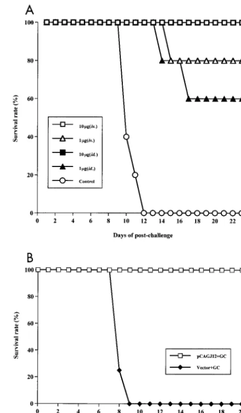
Significant differences in survival rates were discovered be-tween the immunized and control mice (P⬍0.05). Transfusion of splenocytes from mice inoculated both intravenously and intradermally with 10g provided complete protection (100% survived) against the 100-LD50JEV challenge in SCID mice.
Survival rates of 80% and 60% were observed for SCID mice that received splenocytes from mice inoculated with 1g in-travenously and intradermally, respectively. No protective re-sponses were observed in the control groups (Fig. 6A). These results indicate that the splenocytes of BALB/c mice immu-nized with 10g or 1g of DNA and colloidal gold conferred efficient protective immunity to transfused SCID mice against a 100-LD50 JEV challenge more than 6 months after DNA
immunization.
The protective ability of the sera of BALB/c mice immu-nized with pCAGJ12 plus colloidal gold was analyzed by intra-venous transfer into 6-week-old SCID mice. After 4 h, trans-fused SCID mice were subjected to challenge with 100 times the LD50of JEV intraperitoneally, as described in Materials
and Methods. At the end of the observation period, all SCID mice transfused with the immune sera remained alive. All mice that received the control sera were dead within 9 days post-challenge (Fig. 6B). A significant difference was observed be-tween these two groups (P⬍0.05). These results clearly dem-onstrate that, within 3 weeks, the immune antiserum confers sufficient neutralizing ability to transfused SCID mice to in-hibit a challenge with 100 times the LD50of JEV.
Study of isotypes of specific anti-JEV antibody in BALB/c mice intravenously immunized with pCAGJ12 with or without gold colloid.The different inoculation approaches and routes of antigen can result in different antibody subclasses and dif-ferent T helper (Th) cell types during the immune response (1, 33). In general, Th1 immune responses promote the produc-tion of IgG2a antibody, whereas Th2 immune responses en-hance the antibody production of IgG1. We analyzed the IgG isotypes produced by intravenous immunization with pCAGJ12 with or without colloidal gold in BALB/c mice. Similar titers of specific anti-JEV IgG were measured in both groups of immu-nized mice on day 22 after priming (data not shown). Regard-ing the IgG subclass profiles, the gold colloid delivery groups produced almost exclusively IgG1 anti-JEV antibody, whereas IgG2a antibody was only detected in one mouse with a lower titer (Table 2). In contrast, plasmid immunization generated higher levels of IgG2a antibody, with only low titers of IgG1 antibody induced (Table 2). These results suggest that the two methods of JEV DNA inoculation induced different helper T-cell responses.
[image:9.603.300.539.88.497.2]Measurement of -galactosidase activity in adherent splenocytes of immunized mice. To measure -galactosidase activity, groups of 12 6-week-old female BALB/c mice were intravenously inoculated with 50 g of pCAGLacZ with or without colloidal gold. Mice inoculated with colloidal gold alone were used as controls. The -galactosidase activity of
FIG. 6. Passive protection of SCID mice resulting from splenocyte or serum transfer from colloidal gold-DNA-immunized BALB/c mice against JEV infection. (A) Groups of five 3-week-old female BALB/c mice were inoculated with either 10 or 1g of pCAGJ12 and colloidal gold intravenously (iv.) or intradermally (id.). Mice were boosted twice on days 9 and 22 with the same doses and routes of administration that were used for priming. Mice inoculated with an empty vector plus colloidal gold were used as controls. Two hundred days after the last inoculation, splenocytes of immunized BALB/c mice were transfused into 6-week-old female SCID mice intravenously (1.7⫻107
spleno-cytes per mouse). After 48 h, the transfused SCID mice were chal-lenged with 100 times the LD50 of JEV (Beijing-1 strain, 0.15 ml)
intraperitoneally and simultaneously injected with 25l of saline in-tracerebrally as described in Materials and Methods. Survival in each group was monitored for more than 3 weeks after challenge. (B) Groups of five 3-week-old BALB/c mice were immunized with 50 g of pCAGJ12 with gold colloid (GC) intravenously and boosted on day 9. Mice inoculated with the vector plus gold colloid were used as a control. On day 22, sera from the immunized mice were collected and heated at 56°C for 30 min. Immune sera were transfused into 6-week-old male SCID mice intravenously. After 4 h, SCID mice were chal-lenged with 100 times the LD50of JEV as described above. Survival in
each group was monitored for 3 weeks postchallenge.
on November 8, 2019 by guest
http://jvi.asm.org/
each sample was detected as described above.-Galactosidase activity in the colloidal gold inoculation group was more than four times that of the group inoculated with plasmid alone. The lowest level of-galactosidase activity was measured in control mice (Fig. 7). These results clearly demonstrate that colloidal gold is able to enhance the expression efficiency of -galactosidase in vivo, particularly in antigen-presenting cells.
DISCUSSION
JEV is a single-stranded, positive-sense RNA virus, approx-imately 11 kb in length, containing an open reading frame encoding a polyprotein (39, 47). The polyprotein is cleaved into three structural proteins, a capsid (C), membrane (M) or precursor M (PrM), and envelope (E) protein, encoded by one-third of the open reading frame (near the 5⬘region) and at least seven nonstructural (NS) proteins, from NS1 to NS5, which are encoded in the remainder of the open reading frame
(3, 39). The E protein has been found to play an important role in inducing neutralizing antibodies and the protective immune response (18, 19). Transfer of neutralizing monoclonal anti-bodies to the JEV E protein to SCID mice can provide sus-tained protection against JEV infection (20). However, both the unglycosylated JEV E protein produced byEscherichia coli
and the denatured E protein produced from virions of the West Nile virus fail to induce production of neutralizing anti-bodies in mice, indicating that glycosylation or the correct three-dimensional form of the protein is essential for its func-tion (28, 51).
Inoculation of several eukaryotic expression systems encod-ing a series of JEV genes, rangencod-ing from PrM to NS1, elicited anti-JEV antibodies and variant protective immune responses in mice (4, 6, 23, 26, 53). Previous investigations suggest that a signal peptide, which is located upstream of the translation initiation site on the precursor gene, is responsible for intro-ducing the PrM-E polyprotein into the lumen of the endoplas-mic reticulum, where the polyprotein is then made into type I transmembrane protein (43) and secreted from the cell (4). The E proteins that are expressed combine to form twin-helix particles. In the present study, both the pSR␣J12 and pCAGJ12 constructs contained JEV PrM and E genes en-coded within a 690-amino-acid polyprotein (from amino acids 105 to 794), which also contained a few positively charged amino acids prior to the translation initiation site of the PrM gene. These may comprise the signal peptide (Fig. 1A). The secreted PrM and E proteins were recognized by specific anti-JEV antibodies in vitro (Fig. 1B). Inoculation of BALB/c mice with the pCAGJ12 plasmid resulted in expression of JEV pro-tein and recognition by specific anti-JEV antibodies in tissue (Fig. 2G, I, and K).
DNA immunization has recently become a popular method for induction of specific immune responses against infectious agents, such as bacteria and viruses. Either a cellular or hu-moral bias of the immune response following DNA inoculation has been reported and seems to depend on several factors, including the type and form of antigen, the route and method of immunization, and the animal species being studied (4, 5, 6, 7, 10, 11, 12, 21, 23, 26, 36, 45, 50). Two kinds of DNA delivery have been attempted in mice: direct injection of naked plasmid DNA with a syringe (50), and inoculation of gold-covered plasmid with a gene gun (5, 12,). Although the gene gun re-quires less DNA, special equipment is required to coat the particles with DNA and to inoculate it. This results in limited use of this method. Mice inoculated with plasmid DNA with the gene gun exhibit stronger immune responses than mice inoculated by syringe delivery (6, 10, 12). Although the mech-anism behind this is not well understood, it appears that gold cartridges might enhance immune responses in vivo.
[image:10.603.42.283.99.152.2]The present work is the first time that syringe delivery of plasmid DNA with colloidal gold has been used to vaccinate BALB/c mice. Intravenous inoculation of plasmids encoding the PrM and E genes with colloidal gold resulted in higher titers of neutralizing antibody than inoculation with plasmid alone (Fig. 3). Specific antibodies were also detected shortly after inoculation of plasmid (pCAGcore or pCAGE1-E2) en-coding the hepatitis C virus core or E1 and E2 genes with colloidal gold in BALB/c mice (Z. Zhao et al., unpublished data). Our results suggest that this is a feasible method by
FIG. 7. Measurement of -galactosidase activity in adherent splenocytes of immunized BALB/c mice in vitro. Groups of 12 6-week-old female BALB/c mice were inoculated with 50g of pCAGLacZ with or without gold colloid (GC) intravenously. Mice inoculated with 50l of colloidal gold and distilled water (DW) alone were used as controls. After 4 days, the adherent splenocytes were cultured at 37°C and 5% CO2for 16 h and lysed with RIPA buffer. The-galactosidase
[image:10.603.44.281.421.631.2]activity of adherent splenocytes was detected by a unified assay, as described in Materials and Methods. Bars present means⫾standard deviations for 12 animals in each group.
TABLE 2. Isotype of anti-JEV IgG titers in serum of BALB/c mice inoculated with 50g of pCAGJ12 with or without gold colloid
intravenously on day 22 post-intravenous priminga
Inoculation with pCAGJ12 plus:
Titer (log10)
IgG1/IgG2a
IgG1 IgG2a
Gold colloid 2.15⫾0.15 0.35⫾0.35 6.14 Distilled water 0.70⫾0.40 2.15⫾0.15 0.33
aThe data are presented as means⫾standard deviations for four animals in
each group.
on November 8, 2019 by guest
http://jvi.asm.org/
which to elicit a sustained specific antibody response against plasmid DNA encoding different proteins in BALB/c mice.
Several inoculation routes for syringe-mediated DNA im-munization have been reported (10, 12, 21, 50). In the present study, neutralizing antibody production following immuniza-tion by different routes was examined. Both intravenous and intradermal inoculation elicited production of higher titers of neutralizing antibody more rapidly than the other routes of inoculation. Intramuscular inoculation stimulated delayed pro-duction of lower titers of neutralizing antibody. Intraperitoneal inoculation induced the lowest titers of neutralizing antibody. The immune response also appeared later and was shorter-lived than that following inoculation via the other routes (Fig. 4). Histochemical analysis showed transfected muscle fiber cells by both-galactosidase staining and immunofluorescence assay near the intramuscular injection site (Fig. 2C and I). The form in which proteins are expressed might influence their ability to induce a neutralizing antibody response in vivo. In addition, although low titers of anti-JEV antibody were de-tected by ELISA in the sera of intraperitoneally immunized mice (data not shown), neutralizing antibodies were only de-tected in one of those mice on day 35 postinoculation (Fig. 4). In light of these results, it appears that intravenous and intra-dermal inoculation elicits greater production of neutralizing antibody than intramuscular or intraperitoneal inoculation in BALB/c mice.
To complement these data, we examined the efficacy of administering different doses via intravenous or intradermal injection in BALB/c mice. Specific JEV neutralizing anti-body in immunized mice was examined. After challenge, neu-tralizing antibody was greatly increased in all surviving mice, while lower antibody titers (1:20) were detected prior to chal-lenge. Even 0.5g of plasmid and colloidal gold elicited high titers of neutralizing antibody and protected BALB/c mice against challenge with 100,000 times the LD50of JEV
follow-ing both intravenous and intradermal injection (Fig. 5 and Table1). Mice were twice administered 1/5th or 1/10th the adult human dose of inactivated Japanese encephalitis vaccine with the same protocols as for DNA immunization, and al-though they exhibited undetectable prechallenge levels of neu-tralizing antibody, partial protective immunity against chal-lenge with 100,000 times the LD50 of JEV was observed.
Survival rates were, respectively, 40% and 20% at 3 weeks postchallenge (Table 1). Significant differences were detected between DNA-immunized mice and mice injected with a 1/10th dose of inactivated Japanese encephalitis vaccine (P⬍ 0.05), whereas no significant differences were observed be-tween the DNA-immunized mice and those receiving a 1/5th dose of Japanese encephalitis vaccine (P⫽0.051). However, a 1/5th dose of JEV vaccine did not completely protect mice from JEV challenge, while DNA plus colloidal gold immuni-zation offered complete protection (Table 1).
Two hundred days after the final inoculation, splenocytes were obtained from colloidal gold-DNA-immunized BALB/c mice and transferred into SCID mice. This transfer conferred effective passive immunity to the transfused SCID mice against challenge with 100 times the LD50of JEV (Fig. 6A). Significant
increases in neutralizing antibodies were detected in the sera of SCID mice receiving transfers that survived the initial chal-lenge. The antibody titers detected were about 1:4
prechal-lenge and reached about 1:100 and 1:400 on days 7 and 14 postchallenge, respectively. Furthermore, SCID mice trans-fused with the sera of DNA-immunized BALB/c mice that survived a challenge with 100 times the LD50of JEV clearly
demonstrate that neutralizing antibody is a major factor in inhibiting JEV infection in vivo (Fig. 6B). However, the im-portance of the cellular immune response should be investi-gated further.
Different routes or methods of DNA immunization elicited different isotypes of specific antibody and helper T-cell types during the immune response (6, 10). For example, the syringe delivered plasmids elicited stronger Th1 immune responses in both intramuscularly and intradermally inoculated mice, while the gene gun delivery cases showed activated Th2 cell re-sponses. In our study, the gold bead delivery DNA immuniza-tion by intravenous injecimmuniza-tion produced almost entirely IgG1 anti-JEV antibody (Table 2). The IgG1-to-IgG2a ratio was 6.14. In contrast, plasmid DNA immunization by the same route induced high levels of IgG2a anti-JEV antibody (Table 2). The IgG1-to-IgG2a ratio was 0.33. Because the Th1 im-mune response promotes the production of IgG2a antibody and the Th2 immune response promotes the production of IgG1 antibody (1, 31), our results demonstrate that intrave-nous DNA inoculation with gold colloid activated the Th2 immune response in immunized BALB/c mice, whereas immu-nization without gold colloid stimulated the Th1 immune re-sponse.
Examination of antigen-expressing cells in the tissues of intravenously inoculated BALB/c mice by both-galactosidase staining and immunofluorescence revealed the predominance of antigen-expressing cells in the spleen. We also compared the distribution of antigen-expressing cells in the spleens of BALB/c mice intravenously inoculated with plasmid alone or with plasmid and colloidal gold. Most antigen-expressing cells were located in the red pulp area of the marginal zone of the spleen in plasmid and colloidal gold-delivered mice (Fig. 2A and G). Irregular distribution of antigen-expressing cells was observed in the mice administered plasmid alone (data not shown). Because B lymphocytes are primarily located within the marginal zone of the spleen, this might explain why there is rapid production of specific antibodies following inoculation with plasmid combined with colloidal gold.
Recent studies have demonstrated that dendritic cells play a critical role in the inductive immune response, including CD8⫹
cytotoxic T-lymphocyte, CD4⫹Th1, and B-cell responses
fol-lowing DNA vaccination (10, 40, 46, 54). In this study, we detected antigen-expressing cells in adherent splenocytes of intravenously inoculated mice in vitro. These results indicate that plastic-adherent splenocytes are the primary antigen-ex-pressing cells and likely include both dendritic cells and mac-rophages. Measurement of-galactosidase activity in adherent splenocytes isolated from mice inoculated with colloidal gold-DNA showed several times greater -galactosidase activity than was observed in mice inoculated with plasmid alone (Fig. 7). These results demonstrate that colloidal gold-DNA mix-tures preferentially induce transgene expression in adherent splenocytes in order to elicit stronger immune responses in vivo.
This is the first report of inoculation of plasmid DNA en-coding the JEV PrM and E proteins with colloidal gold to
on November 8, 2019 by guest
http://jvi.asm.org/
illustrate induction of a sustained neutralizing antibody re-sponse and complete protection against challenge in immu-nized BALB/c mice. Comparison of different inoculation routes revealed that both intravenous and intradermal inocu-lation elicit high titers of specific anti-JEV antibody and pro-vided rapid protective immunity, even when only 0.5 g of DNA was administered. Two hundred days after the third inoculation, transfusion of splenocytes conferred passive im-munity to SCID mice against a challenge with 100 times the LD50of JEV. Examination of antigen-expressing cells in the
spleens of intravenously immunized mice revealed that most antigen-expressing cells were located in the red pulp areas of the marginal zone. Most of the antigen-expressing cells were found to be plastic-adherent splenocytes in vitro. Although the function and safety of colloidal gold is still not clearly under-stood, inoculation of plasmid DNA with colloidal gold does not require any special apparatus and elicits continuous produc-tion of high titers of antibody in mice. Thus, this method has been shown to be a very simple and efficient method of DNA immunization.
ACKNOWLEDGMENTS
We are grateful to T. Sato, Biological Science Laboratory, Nippon Zeon Co. Ltd., Japan, for providing the pSLKJ12 clone of JEV and J. Miyazaki, Division of Stem Cell Regulation Research, G6, Osaka University Medical School, Suita, Osaka, Japan, for supplying the pCAGGS vector. We also thank Y. Takebe, Laboratory of Molecular Virology and Epidemiology, AIDS Research Center, National Insti-tute of Infectious Diseases, Shinjuku-ku, Tokyo, Japan, for providing pcDLSR␣296. In addition, we thank S. Koike, J. Mukaigawa, and M. Miyamoto, Department of Microbiology and Immunology, Tokyo Metropolitan Institute for Neuroscience, for discussion and technical assistance.
This work was partially supported by a Grant-in-Aid from the Japan Health Sciences Foundation.
REFERENCES
1. Abbas, A. K., K. M. Murphy, and A. Sher.1996. Functional diversity of
helper T lymphocytes. Nature383:787–793.
2. Bray, M., and C.-J. Lai.1991. Dengue virus premembrane and membrane
proteins elicit a protective immune response. Virology185:505–508.
3. Chambers, T. J., C. S. Hahn, R. Galler, and C. M. Rice.1990. Flavivirus genome, organization, expression and replication. Annu. Rev. Microbiol.
44:649–668.
4. Chang, G. J., A. R. Hunt, and B. Davis.2000. A single intramuscular injec-tion of recombinant plasmid DNA induces protective immunity and prevents
Japanese encephalitis in mice. J. Virol.74:4244–4252.
5. Chen, D., R. L. Enders, C. A. Erickson, K. F. Weis, M. W. Mcgregor, Y. Kawaoka, and L. G. Payne.2000. Epidermal immunization by a needle-free powder delivery technology: immunogenicity of influenza vaccine and
pro-tection in mice. Nat. Med.6:1187–1190.
6. Chen, H. W., C. H. Pan, M. Y. Liau, R. Jou, C. J. Tsai, H. J. Wu, Y. L. Lin, and M. H. Tao.1999. Screening of protective antigens of Japanese enceph-alitis virus by DNA immunization: a comparative study with conventional
viral vaccines. J. Virol.73:10137–10145.
7. Colombage, G., R. Hall, M. Pavy, and M. Lobigs.1998. DNA-based and alpha virus-vectored immunization with PrM and E proteins elicits long-lived and protective immunity against the flavivirus Murray Valley encephalitis
virus. Virology250:151–163.
8. Cooney, E. L., A. C. Collier, P. D. Greenberg, R. W. Coombs, J. Zarling, D. E. Argitti, M. C. Hoffman, S. L. Hu, and L. Corey.1991. Safety of and immu-nological response to a recombinant vaccinia virus vaccine expressing HIV
envelope glycoprotein. Lancet337:567–572.
9. Falgout, B., M. Bray, J. J. Schlesinger, and C.-J. Lai.1990. Immunization of mice with recombinant vaccinia virus expressing authentic dengue virus nonstructural protein NS1 protects against lethal dengue virus encephalitis.
J. Virol.64:4356–4363.
10. Feltquate, D. M., S. Heaney, R. G. Webster, and H. L. Robinson.1997. Different T helper cell types and antibody isotypes generated by saline and
gene gun DNA immunization. J. Immunol.158:2278–2288.
11. Frens, G.1973. Controlled nucleation for the regulation of the particle size
in monodisperse gold suspension. Nat. Phys. Sci.241:20–22.
12. Fynan, E. F., R. G. Webster, D. H. Fuller, J. R. Haynes, J. C. Santoro, and H. L. Robinson.1993. DNA vaccines: Protective immunizations by
paren-teral, mucosal, and gene-gun inoculations. Proc. Natl. Acad. Sci. USA90:
11478–11482.
13. Geoghegan, W. D., and G. A. Ackerman.1977. Adsorption of horseradish peroxidase, ovomucoid and anti-immunoglobulin to colloidal gold for the indirect detection of concanavalin A, wheat germ agglutinin and goat anti-human immunoglobulin G on cell surfaces at the electron microscopic level:
a new method, theory and application. J. Histochem. Cytochem.25:1187–
1200.
14. Hennessy, S., Z. Liu, T. F. Tsai, B. L. Stron, C. M. Wan, H. L. Liu, T. X. Wu, H. J. Yu, Q. M. Liu, N. Karabatsos, W. B. Bilker, and S. B. Halstead.1996. Effectiveness of live-attenuated Japanese encephalitis vaccine (SA14–14–2):
a case-control study. Lancet347:1583–1586.
15. Hoke, C. H., A. Nisalak, N. Sangawhipa, S. Jatanasen, T. Laorakapongse, B. L. Lnnis, S. Kotchasenee, J. B. Gringrich, J. Latendresse, K. Fukai, and D. S. Burke.1988. Protection against Japanese encephalitis by inactivated
vaccines. N. Engl. J. Med.319:608–614.
16. Igarashi, A.1978. Isolation of a Singh’sAedes albopictuscell clone sensitive
to dengue and Chikungunya viruses. J. Gen. Virol.40:531–544.
17. Juang, R. F., Y. Okuno, T. Fukunaga, M. Tadano, K. Fukai, K. Baba, N. Tsuda, A. Yamada, and H. Yabuuchi.1983. Neutralizing antibody responses
to Japanese encephalitis vaccine in children. Biken J.26:25–34.
18. Kimura-Kuroda, J., and K. Yasui.1983. Topographical analysis of antigenic determinants on envelope glycoprotein V3(E) of Japanese encephalitis virus,
with monoclonal antibodies. J. Virol.45:124–132.
19. Kimura-Kuroda, J., and K. Yasui.1986. Antigenic comparison of envelope protein E between Japanese encephalitis virus and some other flaviviruses
with monoclonal antibodies. J. Gen. Virol.67:2663–2672.
20. Kimura-Kuroda, J., and K. Yasui.1988. Protection of mice against Japanese encephalitis virus by passive administration with monoclonal antibodies.
J. Immunol.141:3606–3610.
21. Kochel, T., S. Wu, K. Raviprakash, P. Hobart, S. Hoffman, K. Porter, and C. Hayes.1997. Inoculation of plasmids expressing the dengue-2 envelope gene
elicit neutralizing antibodies in mice. Vaccine15:547–552.
22. Konishi, E., S. Pincus, B. A. Fonseca, R. E. Shope, E. Paoletti, and P. W. Mason.1991. Comparison of protective immunity elicited by recombinant vaccinia viruses that synthesize E or NS1 of Japanese encephalitis virus.
Virology185:401–410.
23. Konishi, E., M. Yamaoka, K.-S. Win, I. Kurane, and P. W. Mason.1998. Induction of protective immunity against Japanese encephalitis in mice by immunization with a plasmid encoding Japanese encephalitis virus
premem-brane and envelope genes. J. Virol.72:4925–4930.
24. Ku, C. C., C. C. King, C. Y. Lin, H. C. Hsu, L. Y. Chen, Y. Y. Yueh, and G. J. Chang.1994. Homologous and heterologous neutralization antibody re-sponses after immunization with Japanese encephalitis vaccine among
Tai-wan children. J. Med. Virol.44:122–131.
25. Laemmli, U. K.1970. Cleavage of structural proteins during the assembly of
the head of bacteriophage T4. Nature227:680–685.
26. Lin, Y. L., L. K. Chen, C. L. Liao, C. T. Yeh, S. H. Ma, J. L. Chen, Y. L. Huang, S. S. Chen, and H. Y. Chiang.1998. DNA immunization with Jap-anese encephalitis virus nonstructural protein NS1 elicits protective
immu-nity in mice. J. Virol.72:191–200.
27. Liu, Z. L., S. Hennessy, B. L. Strom, T. F. Tsai, C. M. Wan, S. C. Tang, C. F. Xiang, W. B. Bilker, X. P. Pan, Y. J. Yao, Z. W. Xu, and S. B. Halstead.1997. Short-term safety of live attenuated Japanese encephalitis vaccine (SA14– 14–2): results of a randomized trial with 26,239 subjects. J. Infect. Dis.
176:1366–1369.
28. Mason, P. W., J. M. Dalrymple, M. J. Fournier, and T. L. Mason.1987.
Expression of Japanese encephalitis virus antigens inEscherichia coli.
Virol-ogy158:361–372.
29. Mason, P. W., S. Pincus, M. J. Fournier, T. L. Mason, R. E. Shope, and E. Paoletti.1991. Japanese encephalitis virus-vaccinia recombinants produce particulate forms of the structural membrane proteins and induce high levels
of protection against lethal JEV infection. Virology180:294–305.
30. Matsuura, Y., M. Miyamoto, T. Sato, C. morita, and K. Yasui.1989. Char-acterization of Japanese encephalitis virus envelope protein expressed by
recombinant baculoviruses. Virology173:674–682.
31. McCown, J., M. Cochran, R. Putnak, R. Feighny, J. Burrous, E. Henchal, and C. Hoke.1990. Protection of mice against lethal Japanese encephalitis
with a recombinant baculovirus vaccine. Am. J. Trop. Med. Hyg.42:491–499.
32. Monath, T. P., and F. X. Heinz.1996. Flaviviruses, p. 961–1034.InB. N. Fields, D. M. Knipe, and P. M. Howley (ed.), Field’s virology, 3rd ed. Lippincott-Raven Publishers, Philadelphia, Pa.
33. Mosmann, T. R., and R. L. Coffman.1989. Heterogeneity of cytokine
secre-tion patterns and funcsecre-tions of helper T cells. Adv. Immunol.46:111–147.
34. Niwa, H., K. Yamamura, and J. Miyazaki. 1991. Efficient selection for
high-expression transfectants with a novel eukaryotic vector. Gene108:193–
200.
35. Nothdurft, H. D., T. Jelinek, A. Marschang, H. Maiwald, A. Kapaun, and T. Loscher.1996. Adverse reactions to Japanese encephalitis vaccine in
travel-lers. J. Infect.32:119–122.