Whose Proteins Regulate HCV RNA Replication and Virus Assembly
and Egress
Urtzi Garaigorta, Markus H. Heim,* Bryan Boyd, Stefan Wieland, and Francis V. Chisari
Department of Immunology and Microbial Science, The Scripps Research Institute, La Jolla, California, USA
Stress granules (SGs) are cytoplasmic structures that are induced in response to environmental stress, including viral infections.
Here we report that hepatitis C virus (HCV) triggers the appearance of SGs in a PKR- and interferon (IFN)-dependent manner.
Moreover, we show an inverse correlation between the presence of stress granules and the induction of IFN-stimulated proteins,
i.e., MxA and USP18, in HCV-infected cells despite high-level expression of the corresponding MxA and USP18 mRNAs,
suggest-ing that interferon-stimulated gene translation is inhibited in stress granule-containsuggest-ing HCV-infected cells. Finally, in short
hairpin RNA (shRNA) knockdown experiments, we found that the stress granule proteins T-cell-restricted intracellular antigen
1 (TIA-1), TIA1-related protein (TIAR), and RasGAP-SH3 domain binding protein 1 (G3BP1) are required for efficient HCV
RNA and protein accumulation at early time points in the infection and that G3BP1 and TIA-1 are required for intracellular and
extracellular infectious virus production late in the infection, suggesting that they are required for virus assembly. In contrast,
TIAR downregulation decreases extracellular infectious virus titers with little effect on intracellular RNA content or infectivity
late in the infection, suggesting that it is required for infectious particle release. Collectively, these results illustrate that HCV
exploits the stress granule machinery at least two ways: by inducing the formation of SGs by triggering PKR phosphorylation,
thereby downregulating the translation of antiviral interferon-stimulated genes, and by co-opting SG proteins for its replication,
assembly, and egress.
H
epatitis C virus (HCV) is a major human pathogen. Over 170
million people are chronically infected, many of whom
de-velop chronic liver disease and hepatocellular carcinoma (
1
).
There is no vaccine against HCV, and the most widely used
ther-apy, pegylated alpha interferon (IFN-
␣
) combined with ribavirin
with or without small-molecule protease inhibitors, is not
univer-sally curative and has significant side effects (
46
).
HCV, the sole member of the genus
Hepacivirus
within the
Flaviviridae
family (
33
), is an enveloped, single-stranded,
posi-tive-sense RNA virus (
10
). The HCV genome contains a long open
reading frame (ORF) that encodes a single polyprotein of
approx-imately 3,000 amino acids (
10
). The ORF is flanked by 5=
and 3=
nontranslated regions (NTR) that regulate RNA translation and
replication (
14
,
15
,
21
). Polyprotein translation is driven by a
highly structured internal ribosome entry site (IRES) located in
the 5=
NTR (
21
). The polyprotein is co- and posttranslationally
processed by cellular and viral proteases, leading to the expression
of the structural (core, E1, and E2) and nonstructural (p7, NS2,
NS3, NS4A, NS4B, NS5A, and NS5B) proteins (
37
). HCV triggers
the autophosphorylation of protein kinase R (PKR), an
interfer-on-induced, double-stranded RNA-activated protein kinase (
47
)
that phosphorylates the
␣
subunit of eukaryotic translation
initi-ation factor (eIF2
␣
), thereby inhibiting initiation of translation of
capped cellular mRNA (
42
) and triggering the formation of stress
granules (
9
).
Stress granules (SGs) are large (50 to 200 nm), dynamic
cyto-plasmic structures that are induced in response to environmental
stress, including viral infections (
8
). These granules contain stalled
translation preinitiation complexes characterized by the presence
of cellular mRNAs, translational initiation factors (i.e., eIF4E,
eIF4G, eIF4A, eIF4B, eIF3, and the small subunit of the ribosome),
and RNA binding proteins, including T-cell-restricted
intracellu-lar antigen 1 (TIA-1), the homologous TIA1-related protein
TIAR, and RasGAP-SH3 domain binding protein 1 (G3BP1) (
2
,
3
,
9
,
28
).
Although the physiological function of stress granules is
uncer-tain, it has been suggested that during environmental stresses, they
serve as a “way station” through which nontranslated mRNAs pass
before being translated or degraded (
4
). Nonetheless, there is
growing evidence that several viruses modulate SG assembly (
8
,
43
). For example, Semliki Forest virus (
34
), rotavirus (
36
), West
Nile virus (
12
,
29
), and dengue virus (
12
) inhibit SG formation
presumably to blunt their ability to interfere and/or limit viral
growth. Poliovirus modulates the composition of SGs by cleaving
G3BP1 (
49
) without interfering with their assembly (
38
). In
con-trast, respiratory syncytial virus (
20
,
31
,
32
), mouse hepatitis
coro-navirus (
40
), and reovirus (
39
) transiently induce SGs and, in
some cases, benefit from them (
31
). Using an exogenously
ex-pressed enhanced green fluorescent protein (EGFP)-G3BP1
con-struct and live-imaging technology, Jones et al. have recently
shown that the assembly and disassembly of G3BP1 containing
Received13 December 2011Accepted23 July 2012
Published ahead of print1 August 2012
Address correspondence to Francis V. Chisari, [email protected], or Urtzi Garaigorta, [email protected].
* Present address: Markus H. Heim, Hepatology Laboratory, Departments of Biomedicine and Gastroenterology and Hepatology, University Hospital Basel, Basel, Switzerland.
This is paper number 21557 from the Scripps Research Institute. Copyright © 2012, American Society for Microbiology. All Rights Reserved.
doi:10.1128/JVI.07101-11
on November 7, 2019 by guest
http://jvi.asm.org/
granules is a dynamic process in HCV-infected cells (
23
). Ariumi
et al. have reported the relocalization of G3BP1 protein as well as
P-body components around lipid droplets in HCV-infected cells
(
6
). Finally, Yi et al. have shown that G3BP1 interacts with the
HCV NS5B protein and is required for RNA replication in the
HCV life cycle (
51
). Despite these observations, the mechanism by
which stress granules are induced during HCV infection and the
function of stress granule proteins in the HCV life cycle remain
largely unknown.
In this study, we addressed some of those issues using the
in
vitro
HCV cell culture infection model (
30
,
48
,
52
). Our results
indicate that HCV induces bona fide stress granule formation in
infected cells by a PKR kinase-dependent process that is
aug-mented by IFN, since PKR itself is an interferon-stimulated gene
product. We also demonstrate that interferon-stimulated proteins
like MxA are inversely correlated with the presence of SGs at the
single-cell level in IFN-treated HCV-infected cells. Finally, we
confirmed that G3BP1 protein is required for efficient HCV
infec-tion, extended that requirement to TIA-1 and TIAR proteins, and
demonstrated that TIA-1, TIAR, and G3BP1 play a dual role in the
HCV life cycle: first at the level of HCV RNA replication and later
at the level of HCV particle assembly (TIA-1 and G3BP1) and
egress (TIAR).
MATERIALS AND METHODS
Cells, plasmids, antibodies, and reagents.The origins of Huh-7 (52), Huh-7.5.1 (52), Huh-7.5.1 subclone 2 (Huh-7.5.1c2) (17), and HEK-293T (19) cells have been described previously. All cells were maintained in Dulbecco’s modified Eagle’s medium (DMEM) (Cellgro; Mediatech, Herndon, VA) supplemented with 10% fetal calf serum (FCS) (Cellgro), 10 mM HEPES (Invitrogen, Carlsbad, CA), 100 U/ml penicillin, 100 mg/ml streptomycin, and 2 mML-glutamine (Invitrogen) in 5% CO2at 37°C. The subgenomic and full-length JFH-1 stable replicon Huh-7 cell lines have been previously described (17) and were cultured in medium supplemented with G418 (400 and 250g/ml, respectively). The plasmids containing the JFH-1 (25) and H77S genomes were kindly provided by T. Wakita and S. Lemon, respectively. Lentiviral vectors encoding short hair-pin RNAs (shRNAs) targeting PKR (16), G3BP1, TIA-1, TIAR, and the nontargeting control (shCtrl) were commercially available (Sigma-Al-drich, St. Louis, MO), and their sequences are shown below. Vectors en-coding compatible packaging proteins and vesicular stomatitis virus gly-coprotein (VSV-G) were kindly provided by Inder Verma (Salk Institute, La Jolla, CA). Rabbit polyclonal antibodies for the detection of cellular MxA and PKR proteins, goat polyclonal antibodies for the detection of cellular TIA-1 and TIAR proteins, and the mouse monoclonal antibody against the HCV core protein were purchased from Santa Cruz Biotech-nology (Santa Cruz, CA). Polyclonal antibodies for the detection of USP18, glyceraldehyde 3-phosphate dehydrogenase (GAPDH), and phospho-eIF2␣(Ser 51) proteins were purchased from Cell Signaling Technology (Danvers, MA). Monoclonal antibodies against G3BP1 and EEA1 were obtained from BD Transduction Laboratories (Franklin Lakes, NJ), and antibody against-actin protein was purchased from Sigma-Aldrich. Recombinant human IgG E2 and rabbit MS5 anti-NS5A antibodies were kindly provided by D. Burton (The Scripps Research Institute, La Jolla, CA) and M. Houghton (Chiron), respec-tively. Recombinant human beta interferon 1a (IFN-) was purchased from PBL Interferon Source (Piscataway, NJ). Sodium arsenite and 3-[4,5-dimethylthiazol-2-yl]-2,5-diphenyltetrazolium bromide (MTT) were purchased from Sigma-Aldrich. Protease and phosphatase inhibitors were purchased from Roche (Indianapolis, IN).
Preparation of viral stocks and infections.The original JFH-1 virus was generated by transfection of anin vitro-transcribed full-length JFH-1 HCV RNA into Huh-7 cells, and viral stocks were produced by
inoc-ulation of Huh-7 cells at a multiplicity of infection (MOI) of 0.01 as described previously (52). High-titer virus stocks of cell culture-adapted JFH-1 day 183 virus (D183) (53) were produced by inocula-tion of the highly susceptible Huh-7.5.1c2 at a low MOI and used for single-step virus infection experiments (MOI⫽0.2 or 5) as described previously (17). Cell extracts containing intracellular infectious HCV particles were prepared by 4 freeze-thaw cycles of infected cells as described previously (18). Infectivity titers of cell culture supernatants and cell extracts were determined by endpoint dilution using Huh-7.5.1 cells as described previously (52).
Lentiviral particle production and Huh-7 cell transduction. Lentivi-ral particles were produced in HEK-293T cells by cotransfection of plas-mids encoding short hairpin RNAs (shRNAs) targeting the indicated gene products (see below) and the plasmids necessary for vesicular stomatitis virus glycoprotein-pseudotyped lentivirus production as described previously (16). Cell supernatants were collected at 40 h posttransfec-tion, filtered through 0.45-m filters, aliquoted, and kept at⫺80°C till needed. Twofold serial dilutions of lentiviral particles were used to inoculate Huh-7 cells, and the downregulation of target proteins was determined 7 days posttransduction by Western blotting. Proliferation of the cells (measured by cell counting) was monitored, and cell via-bility was confirmed by MTT cytotoxicity assays. Lentiviruses display-ing no cytotoxic effect were selected for further experiments, and their sequences are as follows: for PKR, TCCTGGCTCATCTCTTTATTC; for shCtrl (nontargeting control shRNA), sequence not available; for TIA-1, no. 1, GCCGTTGTTTACTTAAAGATT, no. 3, GCCAGTATATGCCTA ATGGTT, and no. 4, CGGAAGATAATGGGTAAGGAA; for TIAR, no. 1, CCCATATTGCTTTGTGGAATT, no. 2, CCACAACAGTATGGACAG TAT, and no. 6, ATTCATGATAGGCTTCGATTT; and for G3BP1, no. 1, GCCTGTAAGAAATACAGGATT, and no. 3, CCACCTCATGTTGTTA AAGTA.
Protein analyses.For confocal immunofluorescence experiments, Huh-7 cells were grown in glass bottom 96-well plates (Nunc; Thermo Scientific, Rochester, NY) and treated or infected as indicated in the figure legends. At various time points, cells were washed with phosphate-buff-ered saline (PBS) and fixed for 20 min with 4% paraformaldehyde (PFA) at room temperature. After extensive washing with PBS, blocking buffer (1⫻PBS, 3% bovine serum albumin [BSA], 10% fetal bovine serum [FBS], 0.3% Triton X-100) was added for 1 h at room temperature, after which the cells were incubated for 1 h at room temperature with a mix of primary antibodies at the appropriate dilutions in binding buffer (1⫻ PBS, 3% BSA, 0.3% Triton X-100). After the cells were washed with PBS, binding buffer containing a mixture of fluorophore-conjugated second-ary antibodies (1:1,000 dilution) (Invitrogen) and Hoechst dye (0.5 mg/ ml) (Invitrogen), for nucleus staining, was added for 1 h at room temper-ature. For lipid droplet staining, LipidTOX reagent (Invitrogen) was added at a 1:1,000 dilution in PBS and images were obtained using a Zeiss LSM 710 laser scanning confocal microscope. For the combinedin situ
hybridization and conventional immunofluorescence experiments, Huh-7 cells were infected with HCV JFH-1 D183 virus at a high multi-plicity of infection (MOI⫽5). Forty-eight hours later, the infected and uninfected cells were seeded in glass bottom 96-well plates at a density of 15,000 cells in each well. The next day, the cells were treated with 1,000 U/ml of IFN-for 7 h, after which they were fixed in 4% paraformalde-hyde and processed for the detection of MxA, USP18, or GAPDH mRNA using sequence-specific probes provided by Affymetrix (Santa Clara, CA) that were used according to the manufacturer’s instructions. MxA, USP18, or GAPDH and G3BP1 proteins were detected by conventional immunofluorescence as described above. The Image-Pro Plus package software was used to perform single-cell quantitation analysis of confocal images followed by statistical analysis (Pearson correlation coefficients andPvalues) using GraphPad Prism software.
For Western blot analysis, cell extracts were prepared in radioimmu-noprecipitation assay (RIPA) buffer supplemented with protease and phosphatase inhibitors, and after protein quantitation by bicinchoninic
on November 7, 2019 by guest
http://jvi.asm.org/
acid (BCA) assay, equivalent amounts of total protein for each sample (typically 30g) were separated by polyacrylamide-SDS gel electropho-resis and transferred to Immobilon (Millipore; Billerica, MA) mem-branes. The membranes were blocked for 1 h at room temperature with PBS-5% milk and incubated with the primary antibodies diluted in 1% milk-0.1% Tween 20 in PBS at room temperature for various periods of time, depending on each antibody. After being washed at least 4 times for 15 min each with 0.1% Tween 20 in PBS, the filters were incubated with a dilution of goat anti-rabbit, goat anti-mouse, or mouse anti-goat IgG conjugated to horseradish peroxidase in 1% milk-0.1% Tween 20 in PBS. After being washed at least 4 more times with 0.1% Tween 20 in PBS, the filters were developed using the SuperSignal-West-Pico or -Femto sub-strate purchased from Thermo Scientific (Rockford, IL). Images were ob-tained in a Molecular Imager ChemiDocXRS⫹ and processed with ImageLab software (Bio-Rad, Hercules, CA). Densitometry of nonsatu-rated Western blots was performed using Image J analysis software (NIH), and the results are expressed relative to those for control cells using EEA1 or-actin protein expression for normalization and as loading controls. For immunoprecipitation experiments, Huh-7 cells were grown in 10-cm dishes and were infected with JFH-1 D183 virus at a high multi-plicity of infection (MOI⫽5). Seventy-two hours after infection, when the cultures reached 80% confluence, cells were washed twice with 10 ml of ice-cold PBS and were lysed in 1 ml of lysis buffer (50 mM Tris-HCl [pH 7.4], 150 mM NaCl, and 0.5% NP-40, supplemented with a cock-tail of serine and cysteine protease inhibitors [catalog number 04 693 132 001; Roche] and a cocktail of phosphatase inhibitors [catalog number 04 906 845 001; Roche]). After cell debris was removed by centrifugation, the cell lysates were incubated with antibodies to G3BP1, TIA-1, and TIAR or with the corresponding isotype control antibodies at 4°C. After an overnight incubation, equal amounts of protein G-agarose beads (Roche) were added to each tube and were incubated for another 4 h at 4°C, after which the tubes were centri-fuged and the supernatants containing the unbound material were collected. The beads were washed 3 times with lysis buffer, boiled at 100°C for 15 min in 2⫻loading buffer, and finally analyzed by SDS-PAGE and Western blotting as indicated above.
RNA analyses.Total RNA was extracted from the cells using the gua-nidinium isothiocyanate extraction method (52) after 20g of glycogen (Roche) per sample was added as a carrier. HCV, MxA, USP18, and GAPDH (for normalization) RNA levels were measured by reverse tran-scription–real-time quantitative PCR (RT-qPCR) as described previously (52) using the following primers: for HCV, 5=-TCTGCGGAACCGGTGA GTA-3=and 5=-TCAGGCAGTACCACAAGG-3=; for GAPDH, 5=-GAAG GTGAAGGTCGGAGTC-3=and 5=-GAAGATGGTGATGGGATTTC-3=; for MxA, (5=-AGAGGACCATCGGAATCTTG-3=and 5=-CCCTTCTTC AGGTGGAACAC-3=); and for USP18, 5=-CTCAGTCCCGACGTGGAA CT-3=and 5=-ATCTCTCAAGCGCCATGCA-3=.
Downregulation experiments in persistently infected cells.Huh-7 cells were infected with JFH-1 virus at a low multiplicity of infection (MOI⫽0.1) as previously described (52). After 3 weeks of infection, the cells were inoculated with the corresponding lentiviruses. Cells were split on days 2 and 4 posttransduction. Downregulation of the corresponding target proteins was confirmed by Western blotting. On day 5, the cells were washed extensively with PBS and the medium was replaced. Forty-eight hours later, the supernatants were collected and extracellular infec-tivity was determined by endpoint dilution using Huh-7.5.1 cells as pre-viously described (52). Cell extracts were harvested and the intracellular infectivity and HCV RNA were analyzed by titration and RT-qPCR, re-spectively, as described previously (18).
Downregulation experiments in acutely infected cells.Huh-7 cells were inoculated with the corresponding lentiviruses. On day 7 the down-regulation of each target protein was determined by Western blotting. Downregulated cells were infected with JFH-1 D183 virus at a multiplicity of infection of 0.2 (low MOI) or 5 (high MOI) as described before (52). Five hours after inoculation of the virus, the cells were washed twice with
PBS and the medium was replaced. At the indicated times after infection, the supernatants were collected and extracellular infectivity was deter-mined by endpoint dilution using Huh-7.5.1 cells as previously described (52). Cell extracts were harvested and the intracellular infectivity, HCV RNA, and viral proteins were analyzed by titration, RT-qPCR, and West-ern blotting, respectively, as described previously (18).
Electroporation experiments within vitro-transcribed RNAs.The H77S, Jc1, and Jc1 GND mutant RNAs were produced using the T7 MEGAscript (Ambion) kit by following the manufacturer’s instructions.
In vitro-transcribed RNAs were introduced into Huh-7.5.1c2 cells by elec-troporation as previously reported (52). At various time points, the cells were fixed and processed for immunofluorescence analysis or harvested for HCV RNA quantitation by RT-qPCR as described above.
RESULTS
HCV induces stress granule (SG) formation in HCV-infected
cells.
It was previously reported that cells expressing an ectopic
EGFP-G3BP construct would relocalize the EGFP-G3BP protein
into discrete cytoplasmic aggregates upon HCV infection (
23
),
suggesting that HCV induces the formation of SGs. In order to
determine the nature of these cytoplasmic foci, we first examined
the localization of endogenously expressed SG markers (G3BP1,
TIA-1, and TIAR) in persistently HCV-infected Huh-7 cells and
uninfected cells. As expected, endogenous G3BP1 displayed
dif-fuse cytoplasmic distribution in uninfected Huh-7 cells, while
TIA-1 and TIAR were located both in the nucleus and cytoplasm
(
Fig. 1A
, Mock). Notably, all three markers relocalized in
cyto-plasmic granules in a subset of HCV-positive cells (
Fig. 1A
,
HCV-infected), suggesting that HCV infection triggers the formation of
SGs in
⬃
14% of the infected cells. Further confocal analysis
re-vealed that G3BP1 colocalized with TIA-1 and TIAR in some of
the infected cells, confirming the stress granule nature of those foci
(
Fig. 1B
). Next, Huh-7 cells were infected with the cell
culture-adapted JFH-1 day 183 virus (D183) at a high multiplicity of
in-fection (MOI
⫽
5), and the number of total infected cells and
SG-positive infected cells was quantitated at various time points
after infection. As shown in
Fig. 1C
, the fraction of HCV
E2-positive cells increased over the time course of the experiment,
from
⬃
18% at 24 h to
⬃
85% at the end of the experiment.
Inter-estingly, the fraction of HCV E2-positive cells that contain SGs
increased as the infection proceeded and peaked at 72 h
postinfec-tion, when
⬃
15% of the HCV E2-positive cells contained SGs in
their cytoplasm. Importantly, SG formation was never observed in
HCV-negative cells. To rule out the possibility that the
SG-nega-tive cells might be constituSG-nega-tively unable to respond to any stress
stimulus, HCV-infected and uninfected Huh-7 cells were treated
with sodium arsenite, a widely used and well-known SG inducer
(
5
,
22
,
50
), at different times after infection, and the formation of
SGs was monitored and quantitated. As shown in
Fig. 1D
, sodium
arsenite induced SGs in most of the infected and uninfected cells,
suggesting that the lack of response in the majority (85%) of
HCV-infected cells (
Fig. 1C
) is not due to an intrinsic defect on the
stress response pathway. These results are compatible with recent
results suggesting that SG formation is an oscillatory
phenome-non during HCV infection (
23
). To address whether SG
forma-tion is virus genotype specific, we electroporated the highly
per-missive Huh-7.5.1c2 cells with
in vitro
-transcribed H77S
(genotype 1a), Jc1 (intragenotypic chimeric genotype 2a), and
Jc1-GND mutant (nonreplicative) RNAs and monitored the
formation of stress granules and the intracellular HCV RNA
content at several times thereafter. As expected, both H77S and
on November 7, 2019 by guest
http://jvi.asm.org/
FIG 1HCV triggers stress granule (SG) formation in infected cells. (A) Persistently HCV-infected (at an MOI of 0.1 for 3 weeks) and uninfected control (Mock) Huh-7 cells were seeded in glass bottom 96-well plates, fixed with 4% PFA, and processed for immunofluorescence analysis. In green are stress granule markers (G3BP1, TIA-1, and TIAR); in red, viral E2 or NS5A proteins; and in blue, nuclei stained with Hoechst. Images displayed are examples of 2-m sections of confocal microscope 40⫻field snapshots. These results were confirmed in 3 independent experiments. (B) Colocalization of G3BP1 with TIA-1 and TIAR in persistently HCV-infected (at an MOI of 0.1 for 3 weeks) cells. Left panels show staining of TIA-1 (upper panel) and TIAR (lower panel) in green, middle panels show staining of G3BP1 in red, and right panels show the merge of the corresponding images. Yellow dots represent colocalization of G3BP1 with TIA-1 or TIAR protein in SG structures. Images displayed are projections of 5 consecutive 0.4-m z-series images taken with a confocal microscope. These results were confirmed in 3 independent experiments. (C) Quantitation of SG induction kinetics in HCV-infected Huh-7 cells. Huh-7 cells seeded in a glass bottom 96-well plate format were infected at an MOI of 5 with JFH-1 D183 virus and fixed at the indicated times after infection. After the last time point, all the wells were processed for the detection of cellular G3BP1 and viral E2 proteins by immunofluorescence and pictures were taken in a confocal microscope. Three to five 40⫻ fields were randomly selected, and the presence or absence of SGs was determined in E2-positive cells. The results display the percentage of total cells that are E2 positive (blue bars) and the percentage of E2-positive cells that are also SG positive (red bars). Data are presented as averages and standard deviations (n⫽3 to 5). Similar results were obtained in another two independent experiments. (D) Quantitation of sodium arsenite-induced SG formation at different times after HCV infection. Huh-7 cells were infected as described for panel C or left uninfected as a control. At the indicated times after infection, the cells were treated with 0.5 mM sodium arsenite for 45 min and the induction of SGs was quantitated by immunofluorescence as described above. The same treatment of persistently infected cells was done in parallel for comparison. The results are displayed as percentages of cells containing SGs (mean and SD;n⫽5). These results are representative of two independent experiments, each one performed in duplicate. (E) Quantitation of SG induction and the intracellular HCV RNA levels in H77S, Jc1, or Jc1 GND RNA-containing cells at the indicated times after electroporation. Huh-7.5.1c2 cells were electroporated as described in Materials and Methods, and the presence of HCV-replicating cells and SGs was determined by staining with antibodies against E2 and G3BP1, respectively. Results are displayed as SG-containing E2-positive cells (leftyaxis; bars). Asterisks indicate absence of detection. The HCV RNA content at each time point was determined by RT-qPCR and normalized by the GAPDH mRNA levels (rightyaxis; lines). RNA results are displayed as averages and standard deviations of HCV RNA copy numbers per microgram of total RNA (means and SDs;n⫽3). These results are representative of two independent electroporation experiments, each one performed in duplicate.
on November 7, 2019 by guest
http://jvi.asm.org/
[image:4.585.135.451.65.509.2]Jc1, but not Jc1-GND, RNA-electroporated cells were able to
sustain HCV RNA replication (
Fig. 1E
, lines), and SGs were
induced in a fraction of the H77S and Jc1 (
Fig. 1E
, bars). Note
that electroporation of Jc1-GND RNA did not trigger any SG
formation, suggesting that active HCV RNA replication is
re-quired for SG formation.
HCV-induced stress granule formation is dependent on PKR
and is greatly enhanced by interferon.
We have previously
re-ported that HCV infection triggers PKR and eIF2
␣
phosphoryla-tion, leading to a decrease in the cellular protein synthesis without
inhibiting HCV translation or replication (
16
). Since SG
induc-tion is triggered by translainduc-tional inhibiinduc-tion (
9
), HCV-induced
PKR activation could be responsible for the SG formation
ob-served in HCV-infected cells. To test this hypothesis, cells in which
PKR had been downregulated and control cells were infected with
JFH-1 D183 virus at a high multiplicity of infection (MOI
⫽
5)
and the presence of SGs was monitored and quantitated at
differ-ent times after infection. As expected (
16
), HCV infection
trig-gered the phosphorylation of eIF2
␣
in a PKR-dependent manner
(
Fig. 2A
). Importantly, PKR downregulation almost completely
abolished the induction of SG in HCV-infected cells (
Fig. 2B
),
with no effect on the stress granule protein (G3BP1, TIA-1, and
TIAR) content (
Fig. 2A
) or HCV RNA accumulation (data not
shown) over the time course of the experiment. Together, these
results indicated that PKR is the key kinase involved in
HCV-induced SG formation.
We have also previously reported that interferon (IFN)
treat-ment of HCV-infected cells strongly enhances HCV-induced
phosphorylation of PKR and eIF2
␣
, thereby inhibiting
de novo
cellular protein synthesis, including translation of antiviral
inter-feron-stimulated genes, without inhibiting the translation of HCV
proteins in the cell (
16
,
41
,
44
,
45
). Since PKR and eIF2
␣
phos-phorylation appear to be required for SG induction in
HCV-in-fected cells, we predicted that IFN would increase SG formation in
HCV-infected cells. To test this hypothesis, cells in which PKR had
been downregulated and control cells were infected with JFH-1
D183 virus at a high multiplicity of infection (MOI
⫽
5), and 72 h
later, the cells were treated with 1,000 U/ml of IFN-

for 7 h, after
which SG formation was detected by immunofluorescence. As
shown in
Fig. 2C
and
D
, the number of HCV-infected
SG-con-taining cells was greatly enhanced by IFN treatment (
⬃
65% in
IFN-treated versus 15% in untreated cells) in a PKR-dependent
manner (
⬃
6% in shPKR-treated cells versus 65% in
shCtrl-treated cells). This effect was not due to an induction in the
expression of any of the stress granule proteins G3BP1, TIA-1,
and TIAR by IFN (
Fig. 2E
), suggesting that IFN induced the
formation of SGs in the infected cells without increasing the
intracellular content of these SG proteins. This increase was
specific for HCV-infected cells, since uninfected cells did not
contain any SGs even after IFN treatment, while IFN treatment
enhanced SG formation in subgenomic replicon-containing
Huh-7 cells (
⬃
25% in IFN-treated versus 5% in untreated
cells) and full-length replicon-containing Huh-7 cells (
⬃
40%
in IFN-treated versus 7% in untreated cells), although to a
lower extent (data not shown). Collectively, these results
dem-onstrate that IFN greatly enhances HCV-induced stress granule
formation in a PKR-dependent manner.
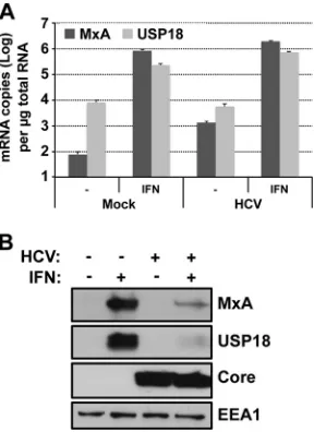
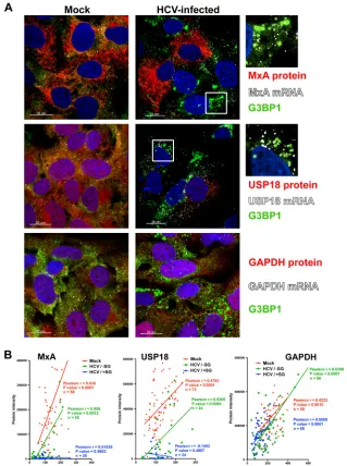
Interferon-stimulated protein expression is suppressed in
stress granule-positive HCV-infected Huh-7 cells.
We have
pre-viously shown that interferon-stimulated protein expression is
FIG 2Stress granule induction in HCV-infected cells is dependent on PKR and is increased by IFN treatment. (A) Huh-7 cells were transduced with lentiviruses expressing shRNAs against PKR (shPKR) or a nontargeting shRNA control (shCtrl). Seven days after transduction, the cells were infected (⫹) or not (⫺) with JFH-1 D183 virus at an MOI of 5 and PKR, phospho-eIF2␣, HCV core, G3BP1, TIAR, and TIA-1 protein expression was deter-mined 72 h later. EEA1 expression is shown as a loading control. (B) Huh-7 cells that were transduced and infected as described for panel A were fixed with 4% PFA for 30 min at the indicated times after infection. After the last time point, all the wells were processed for the detection of cellular G3BP1 and viral E2 proteins by immunofluorescence, and pictures were taken in a confocal microscope. Three to five 40⫻fields were randomly selected, and the cells present in those fields (n⬎200) were scored for the presence or absence of stress granules. Results are displayed as the percentage of infected cells con-taining SG. Data are displayed as averages and standard deviations (mean and SD;n⫽3 to 5). Arrows point to the reduced SG formation in cells in which PKR had been downregulated. The results displayed are representative of three independent experiments. (C) Interferon enhances SG induction in HCV-infected cells. Huh-7 cells transduced as for panel A were HCV-infected with JFH-1 D183 virus at an MOI of 5 and 72 h later were treated with 1,000 U/ml of interferon(IFN). Seven hours after IFN treatment, the cells were fixed and processed for immunofluorescence for the detection of G3BP1 and viral NS5A proteins. In green is G3BP1, in red NS5A, and in blue the nuclei stained with Hoechst. Images displayed are representative examples of 2-m-thick sections of confocal microscope 40⫻field snapshots. (D) Three to five fields were randomly selected, and the E2-positive cells present in those fields (n⬎200) were scored for the presence or absence of stress granules (based on G3BP1 staining). Results shown in the graph are displayed as percentages of infected cells containing SGs. Data are displayed as averages and standard deviations (mean and SD;n⫽3 to 5). Arrows indicate the reduced SG formation in cells in which PKR had been downregulated. These results were confirmed in three independent experiments. (E) Western blotting of G3BP1, TIA-1, and TIAR expression in Huh-7 cells that were infected or not and treated or not with IFN as for panel C. HCV core and NS5A and cellular EEA1 protein expression is shown as an infection marker and a loading control, respectively.
on November 7, 2019 by guest
http://jvi.asm.org/
[image:5.585.299.539.68.379.2]suppressed in IFN-treated HCV-infected cultures relative to that
in IFN-treated uninfected cells (
16
). Since stress granules are
known to contain stalled translation preinitiation complexes (
26
,
27
) and are strongly induced by IFN in HCV-infected cells (see
Fig. 2C
and
D
), we examined the induction of
interferon-stimu-lated mRNA and protein expression in HCV-infected cells at the
population (
Fig. 3
) and single-cell (
Fig. 4
) levels. JFH-1
D183-infected (MOI
⫽
5) and uninfected (mock) control Huh-7 cells
were treated with IFN for 7 h, after which MxA and USP18 mRNA
and protein content were determined by RT-qPCR and Western
blotting, respectively. As we have previously reported (
16
), IFN
treatment induced the expression of MxA and USP18 mRNAs to
similar degrees at the population level in uninfected and
HCV-infected cells (
Fig. 3A
). In contrast, MxA and USP18 proteins were
highly induced in uninfected cells but poorly or not induced in
HCV-infected cells (
Fig. 3B
). Confocal microscopic analysis (
Fig.
4A
) of parallel wells was used to study the expression of
interfer-on-stimulated gene (MxA [upper panels] and USP18 [middle
panels]) and GAPDH (lower panels) mRNAs (in white), their
corresponding proteins (in red), and G3BP1 (in green) protein, as
a stress granule marker, by fluorescence in situ hybridization
(FISH) and conventional immunofluorescence, respectively. As
shown in
Fig. 4A
, MxA and USP18 proteins (red stain) were highly
induced by IFN in uninfected cells (Mock) and SG-negative
HCV-infected cells. However, SG-positive HCV-HCV-infected cells showed
reduced expression of MxA and USP18 proteins despite high-level
expression of the corresponding mRNA (white dots in
Fig. 4A
). As
shown in
Fig. 4B
, single-cell quantitative analysis of MxA, USP18,
and GAPDH mRNA dots and protein staining revealed a highly
significant positive correlation between the amount of mRNA and
corresponding protein in uninfected (Mock [in red]) and
SG-negative HCV-infected (HCV/
⫺
SG [in green]) cells. The same
analysis performed in G3BP1-positive SG-containing
HCV-in-fected (HCV/
⫹
SG [in blue]) cells showed a significant positive
correlation for GAPDH, while there was no correlation at all for
MxA or USP18. These results suggest that
de novo
translation of
short-lived mRNAs, such as the potentially antiviral
interferon-stimulated gene mRNAs, is impaired in stress granule-containing
HCV-infected cells.
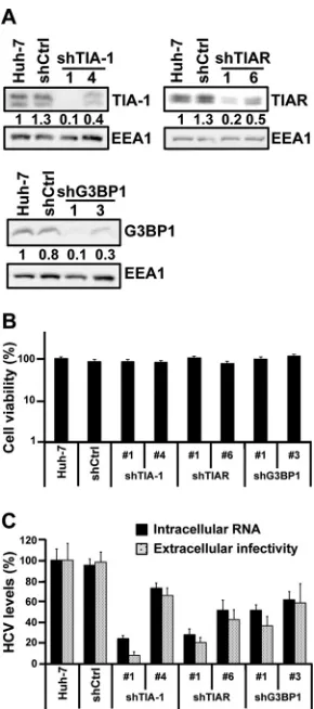
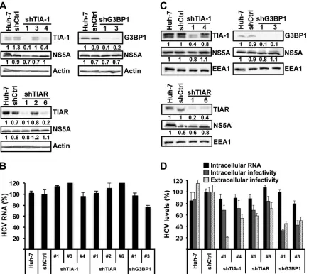
Stress granule proteins TIA-1, TIAR, and G3BP1 are
re-quired for efficient HCV infection.
It was recently shown that the
stress granule protein G3BP1 is required for efficient HCV
infec-tion (
51
). To extend those findings, we wished to examine if other
stress granule proteins, e.g., TIA-1 and TIAR, are also required for
HCV infection. We transduced Huh-7 cells with lentiviruses
ex-pressing shRNAs targeting TIA-1, TIAR, and G3BP1 and a
non-targeting shRNA control (shCtrl). One week after transduction,
the downregulation of the target proteins was confirmed by
West-ern blotting (
Fig. 5A
), and the absence of cytotoxic effects was
demonstrated by MTT assay (
Fig. 5B
). In order to study the SG
protein requirement on HCV infection in a more sensitive system
(multiple rounds of infection), downregulated and control cells
were inoculated with JFH-1 D183 virus at a low multiplicity of
infection (MOI
⫽
0.2). Six days after infection, the intracellular
HCV RNA content and extracellular infectivity were determined
by RT-qPCR and titration, respectively. Downregulation of each
of the three proteins had a negative impact on HCV infection,
reducing both intracellular HCV RNA and the extracellular
infec-tivity (
Fig. 5C
). These results confirmed the requirement of
G3BP1 and demonstrated that TIA-1 and TIAR are also required
for efficient HCV infection.
Viral proteins colocalize and/or directly interact with TIA-1,
TIAR, or G3BP1 in HCV-infected cells.
To gain insight into the
mechanism by which stress granule proteins TIA-1, TIAR, and
G3BP1 can regulate HCV infection, colocalization and
coimmu-noprecipitation studies were performed. Huh-7 cells were
in-fected with JFH-1 D183 virus at a high multiplicity of infection
(MOI
⫽
5), and 72 h later, the cells were processed for
immuno-fluorescence and confocal analysis. As shown in
Fig. 6A
, all three
SG proteins (in orange) colocalized with the HCV NS5A (in red)
protein to some extent (see colocalization mask at the extreme
right side of
Fig. 6A
). Interestingly, NS5A–TIA-1, NS5A-TIAR,
and NS5A-G3BP1 colocalization takes place predominantly in
as-sociation with lipid droplets (in green), a very important organelle
involved in viral assembly (
35
). These observations confirm and
extend a prior report demonstrating the colocalization of the
HCV core protein and G3BP1 in lipid droplets (
6
). These results
suggest that the stress granule proteins might play a direct role in
HCV infection by interacting with NS5A, core, and possibly other
HCV proteins. To address this hypothesis, G3BP1, TIA-1, and
TIAR proteins were immunoprecipitated from HCV-infected
cells using specific and isotype control antibodies followed by
Western blot analysis for the presence of HCV proteins (core,
NS4A, NS4B, NS5A, and NS5B) in the immunoprecipitate and
supernatant. As shown in
Fig. 6B
, each of the specific antibodies
immunoprecipitated the corresponding target protein very
effi-ciently, while the isotype controls did not. Importantly, a small
but readily detectable fraction of the HCV NS5A and NS5B
pro-teins was specifically immunoprecipitated by antibodies to TIA-1
FIG 3ISG mRNA and protein induction by IFN in HCV-infected cells. Huh-7 cells were infected (HCV-infected) or not (Mock) with JFH-1 D183 virus at a high multiplicity of infection (MOI⫽5). Forty-eight hours later, cells were treated or not with 1,000 U/ml of IFN-for 7 h. Shown are results of MxA and USP18 mRNA (A) and protein (B) analysis by RT-qPCR and Western blotting, respectively. RNA results were normalized to GAPDH mRNA and are displayed as copy numbers per microgram of total RNA. Data are displayed as the average and standard deviation (mean and SD;
n⫽3). Core and EEA1 protein expression is shown as an infection marker and a loading control, respectively. These results are representative of 3 independent experiments.
on November 7, 2019 by guest
http://jvi.asm.org/
[image:6.585.92.236.67.265.2]and TIAR, but not G3BP1 or the corresponding isotype control
antibodies.
TIA-1, TIAR, and G3BP1 are not required to maintain HCV
RNA replication once it has been established.
The multiple-cycle
experiments (i.e., low-MOI infections) whose results are shown in
Fig. 5
revealed that TIA-1, TIAR, and G3BP1 are required for
efficient HCV infection, and the colocalization and
coimmuno-precipitation studies suggested a direct interaction between SG
and viral proteins, but they did not identify the step in the HCV
life cycle at which they are required. Since TIA-1, TIAR, and
G3BP1 are RNA binding proteins, we hypothesized that they
could be required for HCV RNA replication. To test this
hypoth-esis, we took advantage of an Huh-7 cell line that stably replicates
an HCV JFH-1 subgenomic RNA that encodes all of the
nonstruc-tural HCV proteins and contains all of the
cis
elements required
for HCV RNA replication and translation. JFH-1 subgenomic
rep-licon cells were transduced with lentiviruses expressing shRNAs
targeting TIA-1, TIAR, and G3BP1 proteins as described above.
FIG 4Interferon-stimulated protein expression is suppressed in stress granule-positive HCV-infected cells. Huh-7 cells were infected (HCV-infected) or not (Mock) with JFH-1 D183 virus at a high multiplicity of infection (MOI⫽5). Forty-eight hours later, the cells were treated or not with 1,000 U/ml of IFN-for 7 h. (A) Visualization of MxA (upper panels), USP18 (middle panels), and GAPDH (lower panels) mRNAs (white dots) and the corresponding proteins (in red) as well as G3BP1 protein (in green) by fluorescencein situhybridization (FISH) and conventional immunofluorescence after IFN treatment of HCV-infected and uninfected control (Mock) cells. Images displayed are of single 0.3-m z-sections taken in a confocal microscope at a magnification of⫻40. Nuclei are displayed in blue. Pictures on the very right are enlargements of the boxed areas showing a partial localization of MxA or USP18 mRNAs in stress granules. (B) Single-cell quantitation analysis of the number of MxA (left graph), USP18 (middle graph), and GAPDH (right graph) mRNA dots and the corresponding protein intensity per cell in samples that were treated with IFN as described above. Three data sets are displayed in each graph: in red, uninfected cells; in green, stress granule-negative HCV-infected cells; and in blue, stress granule-positive HCV-infected cells. Each data point in the graphs represents a single cell. Pearson correlation coefficients (r), theirPvalues, and the number of cells analyzed (n) are displayed for each data set. The results displayed are representative of 2 (USP18 and GAPDH) or 3 (MxA) independent experiments.
on November 7, 2019 by guest
http://jvi.asm.org/
[image:7.585.132.451.67.495.2]Eight days after transduction, cells were harvested, and the
intra-cellular content of the target proteins and the HCV RNA and
NS5A protein content were determined by RT-qPCR and Western
blotting. As shown in
Fig. 7A
and
B
, downregulation of TIA-1,
TIAR, and G3BP1 had no impact on the steady-state levels of HCV
RNA or NS5A protein, suggesting that these SG proteins are not
required to maintain HCV RNA replication once it has been
es-tablished.
TIA-1, TIAR, and G3BP1 are required at early and late steps
in the HCV life cycle.
Next, we analyzed the impact of TIA-1,
TIAR, and G3BP1 protein downregulation on HCV RNA
replica-tion and infectious virus producreplica-tion and secrereplica-tion in two different
HCV infection models: persistently (ongoing infection) and
acutely (
de novo
infection) infected cells. First, we used the
persis-tently HCV JFH-1-infected Huh-7 cell model, which allows us to
analyze not only HCV RNA replication but also intracellular and
extracellular infectivity in an ongoing infection. Downregulation
of TIA-1, TIAR, and G3BP1 proteins in persistently infected cells
had little or no impact on the steady-state levels of intracellular
HCV RNA (
Fig. 7D
) or NS5A protein (
Fig. 7C
) compared to the
levels in nontargeting shRNA control cells. These results confirm
the subgenomic replicon results shown in
Fig. 7B
, and they suggest
that TIA-1, TIAR, and G3BP1 are not required to maintain HCV
RNA replication once it has been established. Interestingly,
down-regulation of the three SG proteins reduced the production of
both intracellular and extracellular infectious virus (
Fig. 7D
)
sug-gesting that TIA-1, TIAR, and G3BP1 might regulate late steps in
the HCV life cycle.
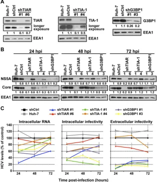
Next, we determined the impact of TIA-1, TIAR, and G3BP1
protein downregulation in acutely infected cells. This system
al-lows us to manipulate the expression level of those proteins prior
to infection and analyze its impact on HCV infection at different
times postinfection. Huh-7 cells were transduced as described
above, and downregulation of the target proteins was examined by
Western blotting 5 days later (
Fig. 8A
). Cells in which the proteins
had been downregulated and control cells were infected at a high
multiplicity of infection (MOI
⫽
5) with JFH-1 D183 virus, and
the accumulation of intracellular HCV RNA, the accumulation of
HCV NS5A and core proteins, and intracellular and extracellular
viral infectivity were determined by RT-qPCR, Western blotting,
and virus titration, respectively, at various times after infection. As
shown in
Fig. 8B
, the accumulation of HCV NS5A and core
pro-teins was reduced in cells in which TIAR, TIA-1, and G3BP1 had
been downregulated compared to control cells early after
infec-tion (24 hours postinfecinfec-tion [hpi]) and was restored to nearly
normal levels in TIA-1 and G3BP1 downregulated cells late in the
infection (72 hpi). Similarly, as shown in
Fig. 8C
, the intracellular
HCV RNA content was also reduced in TIAR, TIA-1, and G3BP1
downregulated cells early in the infection (24 hpi) and recovered
to normal levels in almost all cells in which the proteins had been
downregulated over the time course of the experiment (72 hpi).
The reduction in HCV NS5A and core protein expression and
HCV RNA content correlated with the downregulation
effi-ciency achieved by each shRNA. These results are consistent
with previous reports describing a functional requirement of
G3BP1 protein in HCV RNA replication (
6
,
51
) and suggested
that TIAR and TIA-1 proteins are also required at early steps in
the HCV life cycle. Moreover, our results demonstrate that the
negative effect on the HCV RNA and protein content observed
early after infection is overcome at later time points, suggesting
that there is a delay in viral replication.
Next we examined the impact of TIAR, TIA-1, and G3BP1
downregulation on late steps of infection such as assembly and
release of HCV infectious virions. Both intracellular and
extracel-lular infectivity titers were reduced at 24 hpi in cells in which
TIAR, TIA-1, and G3BP1 had been downregulated (
Fig. 8C
), most
probably reflecting the reduced intracellular HCV RNA and viral
protein content achieved in the early stage of infection.
Interest-ingly, the extracellular infectivity titers did not increase
propor-tionally to the expansion of HCV RNA in the downregulated cells
FIG 5Stress granule proteins TIA-1, TIAR, and G3BP1 are required for effi-cient HCV infection. Huh-7 cells were transduced with lentiviruses that ex-press shRNAs targeting TIA-1, TIAR, or G3BP1 or a nontargeting shRNA control (shCtrl). (A) After 7 days, the downregulation of each protein was confirmed by Western blotting and the relative quantitation was determined by densitometry (shown below each gel). The intensity of TIA-1, TIAR, and G3BP1 proteins relative to EEA1 protein (loading control) in Huh-7 cells was set as 1 and used to calculate the relative amount of each protein. (B) Down-regulated and control cells were seeded in 96-well plates at 5,000 cells per well. Four days later, cytotoxicity assays (MTT) were performed by following the manufacturer’s instructions. Results are displayed as percentages of the control (shCtrl). Assays were run with 6 replicate wells per cell type in two independent experiments. (C) Downregulated cells were infected with JFH-1 D183 virus at a low multiplicity of infection (MOI⫽0.2), and the intracellular HCV RNA levels and extracellular infectivity were determined on day 6 postinfection by RT-qPCR and titration assays, respectively. GAPDH mRNA quantitation of the same cellular extracts was used for normalization of the HCV RNA levels. Results are displayed as percentages of the control (Huh-7 cells). Data are displayed as averages and standard deviations (mean and SD;n⫽3). These results were confirmed in 2 inde-pendent experiments performed in triplicate.
on November 7, 2019 by guest
http://jvi.asm.org/
[image:8.585.91.236.64.391.2]at the 48- and 72-h time points. These results suggested that TIAR,
TIA-1, and G3BP1 proteins are also required at late steps in the
HCV life cycle, most likely in assembly or release. To distinguish
between assembly and release, we determined the effect of TIAR,
TIA-1, and G3BP1 downregulation on the accumulation of
intra-cellular infectious virus. The level of intraintra-cellular infectious virus
increased proportionally to the expansion of HCV RNA in cells in
which TIAR (blue and red lines) had been downregulated,
sug-gesting that TIAR protein is required for the release of infectious
virus but not its assembly. However, the level of intracellular
in-fectious virus did not increase proportionally to the expansion of
HCV RNA in cells in which TIA-1 (green and orange lines) and
G3BP1 (purple and yellow lines) had been downregulated over
time. These results suggest that TIA-1 and G3BP1 are required for
the assembly of infectious virus. Although different pairs of
shRNAs had different degrees of effect on the HCV infection, they
correlate with the degree of downregulation of the specific target
protein achieved by each shRNA.
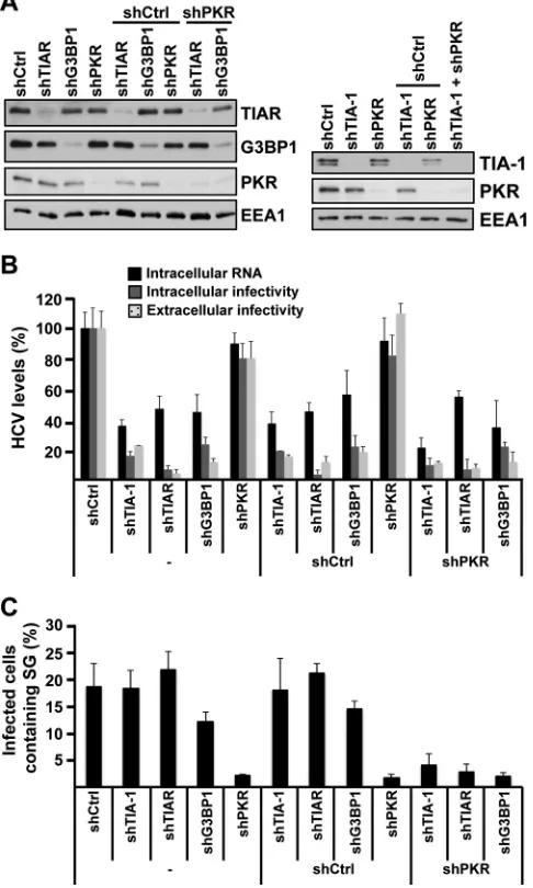
Finally, to test whether TIA-1, TIAR, and G3BP1 must be
con-tained in SGs in order to promote efficient HCV infection, their
expression was downregulated in PKR-downregulated cells in
which SGs are not induced by HCV infection (see
Fig. 2
). After
confirming the degree of downregulation of each target protein by
Western blotting (
Fig. 9A
), the cells were infected with JFH-1
D183 virus at a high multiplicity of infection (MOI
⫽
5) and the
intracellular HCV RNA and infectivity and the extracellular
infec-FIG 6Colocalization and coimmunoprecipitation of stress granule proteins with HCV viral proteins during infection. (A) Huh-7 cells were infected with JFH-1 D183 virus at a high multiplicity of infection (MOI⫽5). Seventy-two hours later, the cells were fixed and processed by immunofluorescence for the detection of G3BP1, TIA-1, or TIAR (in orange) and viral NS5A (in red) proteins. Lipid droplets (in green) were stained using LipidTOX from Invitrogen. Images displayed are representative pictures of single 0.35-m z-sections taken in a confocal microscope at a magnification of⫻63. Nuclei are displayed in blue. The staining of each channel and the merge and the colocalization mask channel between stress granule and NS5A proteins are shown. These results were confirmed in at least 2 independent experiments. (B) Huh-7 cells were infected as described for panel A and were subjected to immunoprecipitations using G3BP1-, TIA-1-, and TIAR-specific antibodies and isotype controls 72 h later, as indicated in Materials and Methods. Detection of target proteins as well as viral core, NS4A, NS4B, NS5A, and NS5B proteins was carried out in the immunoprecipitated material (IP) and the unbound material in the supernatant (S). Gels were loaded using 5% of the starting material (Input), 20% of the immunoprecipitated material (IP), and 5% of the unbound supernatant (S). These results were confirmed in 2 independent experiments.
on November 7, 2019 by guest
http://jvi.asm.org/
[image:9.585.136.451.68.463.2]tivity were analyzed 24 h after infection. As shown in
Fig. 9B
,
TIA-1, TIAR, and G3BP1 downregulation had a negative effect on
HCV RNA and infectivity irrespective of the presence or absence
of PKR. Quantification of SG formation in all those cells
demon-strated a profound reduction of SGs in all PKR downregulated cell
types (
Fig. 9C
). These results demonstrate that TIA-1, TIAR, and
G3BP1 proteins are required for efficient HCV infection in the
absence of SG formation.
Collectively, the experiments performed in acutely infected
cells suggest that TIAR, TIA-1, and G3BP1 proteins regulate early
steps that affect HCV RNA and protein accumulation and that
TIA-1 and G3BP1 are also required for efficient infectious virus
particle assembly, while TIAR is required for infectious virus
par-ticle release.
DISCUSSION
The recent development of an
in vitro
HCV infection system that
recapitulates all the steps of the HCV life cycle permits
identifica-tion and characterizaidentifica-tion of cellular factors and pathways that
regulate the infection. Using this infection model, we have
previ-ously identified cellular factors required for HCV assembly and
secretion (
17
), and we have shown that autophagy proteins are
FIG 7TIA-1, TIAR, and G3BP1 proteins are not required for maintenance of HCV RNA replication, but they are required at a postreplication step in persistently infected cells. (A and B) Huh-7 cells bearing a subgenomic JFH-1 replicon were transduced with lentiviruses expressing shRNAs targeting TIA-1, TIAR, or G3BP1 or a nontargeting shRNA control (shCtrl). Eight days after transduction, the cells were harvested and the expression and relative quantitation of target proteins and HCV NS5A protein were determined by Western blotting (A). Densitometry results are shown under each gel image.-Actin was used for normalization. (B) The intracellular HCV RNA content of the same samples was determined by RT-qPCR. GAPDH mRNA quantitation was used for normalization. Results are shown as percentages of the control (shCtrl). Data are displayed as averages and standard deviations (mean and SD;n⫽3). These results were confirmed in 3 independent experiments performed in triplicate. (C and D) Persistently infected cells were transduced with lentiviruses expressing shRNA that target TIA-1, TIAR, or G3BP1 or an irrelevant shRNA control (shCtrl). Seven days after transduction, the supernatants were collected and the cells were harvested for further analysis. (C) The expression and relative quantitation of target proteins and HCV NS5A protein were determined by Western blotting. Densitometry results are shown under each gel image. EEA1 protein was used as a loading control. (D) The intracellular HCV RNA and infectivity titer as well as the extracellular infectivity titer were determined by RT-qPCR and titration assays, respectively. GAPDH mRNA quantitation of the same samples was used for normalization of the HCV RNA levels. Black bars, intracellular HCV RNA; gray bars, intracellular infectivity; light gray bars with black dots, extracellular infectivity. Results are shown as percentages of the control (shCtrl). Data are displayed as averages and standard deviations (n⫽3). These results are representative of 3 independent experiments, each one performed in triplicate.
on November 7, 2019 by guest
http://jvi.asm.org/
[image:10.585.77.512.66.452.2]required for the initiation of HCV replication by controlling the
initiation of translation of incoming genomic RNA (
11
). In this
study, we showed that stress granules (SGs) are induced in
HCV-infected cells, we characterized their mechanism of induction, and
we showed that SG proteins G3BP1, TIA-1, and TIAR are required
at both early and late steps during HCV infection.
Confocal microscopic analysis demonstrated the formation of
cytoplasmic structures containing SG proteins (G3BP1, TIA-1,
and TIAR) in a fraction of HCV-infected cells (
Fig. 1
). These
re-sults are consistent with previous observations by Jones et al. in
which dynamic assembly and disassembly of ectopically expressed
EGFP-G3BP-containing foci were observed by live-cell imaging
(
23
). In this study, we confirmed the formation of SGs by looking
at endogenously expressed G3BP1 protein and extended the
ob-servation to two other SG proteins, i.e., TIA-1 and TIAR. Bona
fide SGs are characterized by the presence of cellular mRNAs,
RNA binding proteins (e.g., G3BP1, TIA-1, and TIAR), and
trans-lation initiation factors. Using fluorescence
in situ
hybridization
and conventional immunofluorescence analysis, we showed that
cellular MxA and USP18 mRNAs partially colocalized with
G3BP1-containing granules in IFN-treated HCV-infected cells,
suggesting that HCV triggers the formation of bona fide SGs (
Fig.
4A
). A recent report by Ariumi et al. described the transient
in-duction of G3BP1 containing SGs early after HCV infection (36 to
48 hpi) and G3BP1 relocalization around lipid droplets at later
time points (
6
). Although we did observe relocalization of G3BP1,
TIA-1 or TIAR protein around structures resembling lipid
drop-lets in some of the infected cells late in the infection (
Fig. 6A
), in
other infected cells G3BP1 was still localized in SG (
Fig. 1
),
sug-gesting a dynamic and/or variable localization of these proteins
during HCV infection.
It has been previously shown that SG formation is triggered by
FIG 8TIA-1, TIAR, and G3BP1 proteins are required in early and late steps in the HCV life cycle. Huh-7 cells were transduced with lentiviruses that express shRNAs targeting TIA-1, TIAR, or G3BP1 or a nontargeting shRNA control (shCtrl). (A) After 5 days, the downregulation of the corresponding proteins was confirmed by Western blotting. EEA1 protein expression is shown as a loading control and was used to perform relative quantitation of each band by densitometry analysis. Longer-exposure images are shown for TIAR and TIA-1 gels. (B and C) Downregulated cells were infected with JFH-1 D183 virus at a high multiplicity of infection (MOI⫽5), and the accumulation of NS5A and core protein (B) and the accumulation of the intracellular HCV RNA and intracellular and extracellular infectivity (C) were determined at the indicated times postinfection by Western blotting, RT-qPCR, and titration assays, respectively. Relative quantitation of NS5A and core proteins is shown below each gel. EEA1 protein expression was used as a loading control. GAPDH mRNA quantitation of the same cellular extracts was used for normalization of the HCV RNA levels. The RNA and infectivity results are displayed as percentages of the control (shCtrl) at each time point. Data are displayed as averages and standard deviations (mean and SD;n⫽3). These results are representative of 3 independent experiments performed in triplicate.
on November 7, 2019 by guest
http://jvi.asm.org/
[image:11.585.135.449.69.433.2]events that inhibit cellular mRNA translation (
9
). We (
16
) and
others (
7
,
24
) have previously reported that HCV infection
trig-gers PKR activation and subsequent eIF2
␣
phosphorylation,
lead-ing to a small decrease in global protein synthesis that is further
suppressed by treatment with type I interferon (IFN). Therefore,
we asked if PKR was required for the induction of SGs in HCV
infected cells. PKR downregulation almost completely abolished
the formation of SGs, indicating that PKR plays a critical role in
HCV-induced SG formation (
Fig. 2
). Consistent with the higher
PKR expression level and the increased PKR and eIF2
␣
phosphor-ylation levels in HCV-infected IFN-treated cells (
16
), IFN greatly
enhanced the number of SG containing HCV-infected cells.
How-ever, IFN did not trigger SG formation in uninfected cells despite
higher levels of PKR expression (data not shown), suggesting that
the PKR activation state rather than the total PKR protein level
itself is important for SG induction. Quantitation of the
immuno-fluorescence and immuno-fluorescence
in situ
hybridization images
sug-gests that HCV induces translational inhibition in SG-containing
cells, thereby reducing interferon-stimulated protein expression
without decreasing the corresponding mRNA content (
Fig. 4
).
Analysis of a non-interferon-stimulated gene, such as that for
GAPDH, did not show any reduction of GAPDH protein
accumu-lation in SG-containing HCV-infected cells as was observed for
MxA and USP18 proteins. This difference can be explained by
the relatively long half-life (
⬃
72 h) and abundance of
preexist-ing GAPDH protein (
13
) compared to the typical absent or low
interferon-stimulated protein content under basal conditions
in Huh-7 cells (
Fig. 3
). Collectively, these results suggest that
SG formation in HCV-infected cells reflects the ability of HCV
to induce PKR and eIF2
␣
phosphorylation and translational
inhibition at the single-cell level. The dynamic oscillatory
pro-cess of SG assembly and disassembly described by others (
23
)
and the function of PKR in the formation of SG described
herein might reflect a balance between the ability of HCV to
trigger PKR phosphorylation and the capacity of the cell to
dephosphorylate PKR in order to maintain the cellular
trans-lation. Alternatively, transient SG induction might allow the
survival of the cells that otherwise could die as a consequence of
prolonged inhibition of translation. This could indirectly
pro-mote viral persistence, since it has been previously
demon-strated that SG induction prevents apoptosis (
5
).
In this study, we also obtained evidence that SG proteins
G3BP1, TIA-1, and TIAR are required for efficient HCV infection
affecting both early (initiation of HCV RNA replication) and late
(virus particle assembly and release) events in the HCV life cycle
(
Fig. 5
to
9
), confirming and extending results of a recent study in
which G3BP1 was shown to be required for HCV infection at the
level of HCV RNA amplification (
51
). Our results show that
G3BP1 is required to initiate viral RNA replication (
Fig. 8
) in the
early stages of infection but not to maintain it (
Fig. 7
), and they
suggest that G3BP1 also regulates the assembly of HCV infectious
virions (
Fig. 7
and
8
) later in the infection. Interestingly, the
par-tial relocalization of G3BP1 around lipid droplets observed by
others (
6
) and us (
Fig. 6A
) is compatible with a late function
during infection, since lipid droplets have been shown to be an
important platform during HCV assembly (
35
).
Like that of G3BP1, the downregulation of TIA-1 and TIAR
displayed differential effects in acutely and persistently infected
cells. Both proteins were required for efficient HCV RNA and
protein accumulation early after
de novo
infection, but they were
dispensable in ongoing infections, suggesting that initiation and
maintenance of HCV RNA replication are differentially regulated.
Consistent with this idea, we have previously reported the
require-ment of autophagy proteins for the establishrequire-ment of HCV
repli-cation but not for maintenance of the infection (
11
). Alternatively,
differences in the requirement for G3BP1, TIA-1, and TIAR in
persistently infected and acutely infected cells and stable
replicon-containing cells could also reflect selection or adaptation of the
FIG 9The requirement of TIA-1, TIAR, and G3BP1 proteins in HCV infec-tion is independent of their presence in stress granules. (A) Huh-7 cells were transduced with one or two lentiviruses as indicated, and 5 days later, the expression of target proteins was determined by Western blotting. (B) After downregulation was confirmed, the cells were infected with JFH-1 D183 virus at a high multiplicity of infection (MOI⫽5), and the intracellular HCV RNA and infectivity titers and extracellular infectivity titers were determined 24 h later as indicated in Materials and Methods. The results shown are averages and standard deviations (mean and SD;n⫽3) (C) Cells infected in parallel were fixed 72 h after inoculation, and the number of HCV-infected cells con-taining stress granules was determined by immunofluorescence and counting as described forFig. 2B. The results displayed were confirmed in 2 independent experiments, each one performed in triplicate. The shRNA sequences used in these experiments were shTIA-1 no. 1, shTIAR no. 1, shG3BP1 no. 1, shPKR, and shCtrl.
on November 7, 2019 by guest
http://jvi.asm.org/
[image:12.585.43.286.72.477.2]cells or the virus under these different conditions. In agreement
with this idea, we have previously reported the coevolution of cells
and virus during persistent infection
in vitro
(
53
), Nonetheless,
irrespective of their ability to regulate the initiation of the
infec-tion, our results demonstrate that TIA-1 and G3BP1 are also
quired for virus particle assembly, while TIAR is required for
re-lease of infectious particles, suggesting that these SG proteins have
dual functions during the HCV life cycle. Consistent with this dual
function, we found that small fraction (around 1%) of the NS5A
and NS5B proteins coimmunoprecipitate with TIA-1 and TIAR
during the infection. Since these viral proteins are involved in
HCV RNA replication and NS5A also plays a role in viral particle
assembly, it is conceivable that TIA-1 and TIAR might regulate
those steps by directly binding to NS5A and NS5B. A previous
report by Yi et al. showed that NS3, NS5A, and NS5B
coimmuno-precipitated with an overexpressed myc-G3BP1 construct in
HCV-infected cells, and they suggested that G3BP1 is present in
replication complexes (
51
). In contrast to those results, we were
not able to detect any specific coimmunoprecipitation of core,
NS4A, NS4B, NS5A, or NS5B protein with the endogenous G3BP1
protein, suggesting either that G3BP1 does not bind to any of
those viral proteins or that the binding was too transient or weak
to be detected under our experimental conditions. Consistent
with the latter hypothesis, we showed that G3BP1 as well as TIA-1
and TIAR proteins colocalized with NS5A protein in some of the
infected cells.
Since SGs are induced upon HCV infection and since G3BP1,
TIA-1, and TIAR proteins are required for efficient HCV
infec-tion, it would be interesting to know if the effects of these proteins
are related to their presence in stress granules. Our experiments in
cells with double downregulation of PKR and stress granule
pro-teins that do not produce SGs upon HCV infection suggest that
the effects of G3BP1, TIA-1, and TIAR may be independent of
their aggregation within SGs.
In conclusion, we have shown that HCV triggers the formation
of stress granules in a PKR-dependent manner and that IFN
greatly increases the number of SG-containing cells. We have
con-firmed that G3BP1 is required for HCV infection, and we
identi-fied two new host factors, TIA-1 and TIAR, that regulate HCV
infection by affecting both early (i.e., initiation of HCV RNA
rep-lication) and late (i.e., virus particle assembly and release) steps in
the HCV life cycle.
ACKNOWLEDGMENTS
We are grateful to Takaji Wakita (National Institute of Infectious Dis-eases, Tokyo, Japan) for providing the infectious JFH-1 molecular clone and replicon constructs, Stanley Lemon (University of North Carolina, Chapel Hill, NC) for the H77S molecular clone, Charles Rice (Rockefeller University, New York, NY) for the Huh-7.5 cells from which Huh-7.5.1 cells and Huh-7.5.1c2 cells were derived, Dennis Burton (The Scripps Research Institute, La Jolla, CA) for recombinant human IgG anti-E2, Michael Houghton (Chiron) for MS5 anti-NS5A antibody, and Inder Verma (Salk Institute, La Jolla, CA) for lentiviral plasmids. We are grateful to Josan Chung and Christina Whitten for excellent technical assistance and to Marlene Dreux, Ariel Rodriguez, Lorena Ver, and Cristina Godio for helpful discussions, advice, and critical reading of the manuscript.
Urtzi Garaigorta was supported by The Irvington Institute Fellowship Program of the Cancer Research Institute. This work was supported by grant AI-079043 from the NIH.
REFERENCES
1.Alter HJ, Seeff LB.2000. Recovery, persistence, and sequelae in hepatitis C virus infection: a perspective on long-term outcome. Semin. Liver Dis. 20:17–35.
2.Anderson P, Kedersha N.2006. RNA granules. J. Cell Biol.172:803– 808. 3.Anderson P, Kedersha N. 2009. Stress granules. Curr. Biol.19:R397–
R398.
4.Anderson P, Kedersha N.2002. Stressful initiations. J. Cell Sci.115:3227– 3234.
5.Arimoto K, Fukuda H, Imajoh-Ohmi S, Saito H, Takekawa M.2008. Formation of stress granules inhibits apoptosis by suppressing stress-responsive MAPK pathways. Nat. Cell Biol.10:1324 –1332.
6.Ariumi Y, et al.2011. Hepatitis C virus hijacks P-body and stress granule components around lipid droplets. J. Virol.85:6882– 6892.
7.Arnaud N, et al.2010. Hepatitis C virus controls interferon production through PKR activation. PLoS One 5:e10575. doi:10.1371/ journal.pone.0010575.
8.Beckham CJ, Parker R.2008. P bodies, stress granules, and viral life cycles. Cell Host Microbe3:206 –212.
9.Buchan JR, Parker R.2009. Eukaryotic stress granules: the ins and outs of translation. Mol. Cell36:932–941.
10. Choo QL, et al.1991. Genetic organization and diversity of the hepatitis C virus. Proc. Natl. Acad. Sci. U. S. A.88:2451–2455.
11. Dreux M, Gastaminza P, Wieland SF, Chisari FV.2009. The autophagy machinery is required to initiate hepatitis C virus replication. Proc. Natl. Acad. Sci. U. S. A.106:14046 –14051.
12. Emara MM, Brinton MA.2007. Interaction of TIA-1/TIAR with West Nile and dengue virus products in infected cells interferes with stress gran-ule formation and processing body assembly. Proc. Natl. Acad. Sci. U. S. A. 104:9041–9046.
13. Franch HA, Sooparb S, Du J, Brown NS.2001. A mechanism regulating proteolysis of specific proteins during renal tubular cell growth. J. Biol. Chem.276:19126 –19131.
14. Friebe P, Boudet J, Simorre JP, Bartenschlager R.2005. Kissing-loop interaction in the 3=end of the hepatitis C virus genome essential for RNA replication. J. Virol.79:380 –392.
15. Friebe P, Lohmann V, Krieger N, Bartenschlager R.2001. Sequences in the 5=nontranslated