Open Access
Short Report
Heminested reverse-transcriptase polymerase chain reaction
(hnRT-PCR) as a tool for rabies virus detection in stored and
decomposed samples
Danielle B Araújo
1, Helio Langoni
1, Marilene F Almeida
2and Jane Megid*
1Address: 1UNESP, School of Veterinary and Animal Science, Department of Veterinary Hygiene and Public Health, Botucatu, SP, Brazil and 2Center
of Zoonosis Control, CCZ, São Paulo, SP, Brazil
Email: Danielle B Araújo - [email protected]; Helio Langoni - [email protected]; Marilene F Almeida - [email protected]; Jane Megid* - [email protected]
* Corresponding author
Abstract
Background: The use of methods, both sensitive and specific, for rabies diagnosis are important tools for the control and prophylaxis of the disease. Reverse-Transcriptase Polymerase Chain Reaction (RT-PCR) has been used in rabies diagnosis with good results, even in decomposed materials. Additionally, molecular techniques have been used for epidemiological studies and to gain a better knowledge of viral epidemiology.
Findings: The aim of this work was to evaluate the RT-PCR and hnRT-PCR for rabies virus detection in original tissues stored at -20°C for different periods considering their use for rabies virus detection in stored and decomposed samples. RT-PCR and hnRT-PCR were evaluated in 151 brain samples from different animal species, thawed and left at room temperature for 72 hours for decomposition.
The RT-PCR and hnRT-PCR results were compared with previous results from Direct Fluorescent Antibody Test and Mouse Inoculation Test. From the 50 positive fresh samples, 26 (52%) were positive for RT-PCR and 45 (90%) for hnRT-PCR. From the 48 positive decomposed samples, 17 (34, 3%) were positive for RT-PCR and 36 (75%) for hnRT-PCR. No false-positives results were found in the negatives samples evaluated to the molecular techniques.
Conclusion: These results show that the hnRT-PCR was more sensitive than RT-PCR, and both techniques presented lower sensibility in decomposed samples. The hnRT-PCR demonstrated efficacy in rabies virus detection in stored and decomposed materials suggesting it's application for rabies virus retrospective epidemiological studies.
Findings
Rabies is a viral, zoonotic and fatal disease, which causes encephalomyelitis in humans and animals. The annual number of deaths worldwide caused by rabies is estimated to be 55 000 and about 10 million people receive
post-exposure treatments each year after being exposed to rabies-suspect animals [1].
The disease is caused by a RNA virus that belongs to the
Lyssavirus genus in the Rhabdoviridae family [2]. All
mam-Published: 4 June 2008
BMC Research Notes 2008, 1:17 doi:10.1186/1756-0500-1-17
Received: 12 February 2008 Accepted: 4 June 2008
This article is available from: http://www.biomedcentral.com/1756-0500/1/17
© 2008 Araújo et al; licensee BioMed Central Ltd.
mals are susceptible to the rabies virus, mainly the Car-nivora and Chiroptera orders.
Developed during the late 1950s, the Direct Fluorescent Antibody Test (DFA) remains the gold standard test in rabies diagnosis because it is fast, with low cost and a great sensitivity [3]. As a single negative test does not rule out the possibility of infection, inoculation tests should be carried out simultaneously with Mouse Inoculation Test (MIT). Alternatively, a monolayer culture of susceptible cells is inoculated with the same material as used for mice [4].
Even so DFA is the gold standard test for rabies diagnosis the sensitivity decreases in decomposed samples [5-8].
MIT is also highly sensitive, but the virus must be viable and the results are achieved after several days of incuba-tion [9]. Addiincuba-tionally, samples in decomposiincuba-tion may present bacteria and/or toxins that disabling the use of the inoculation test [10].
The use of molecular techniques, mainly Polymerase Chain Reaction (PCR) are useful tools in rabies diagnosis [11]. Many works are being made with the PCR for rabies diagnosis, showing that this technique presents highly sensitivity and specificity [12-14].
RT-PCR and hnRT-PCR can detect the rabies virus genome in highly decomposed samples, even when DFA and MIT present negative results, a common situation in countries with tropical weather like Brazil [15-22].
The hnRT-PCR technique allows a sensitive, specific and fast diagnosis for rabies virus, even when samples are in a decomposed state. Additionally, the amplified products can be used in techniques such as sequencing, RFLP, SS-PCR and Multiplex SS-PCR for epidemiological characteriza-tion of the virus [20,22]. However, the recommendacharacteriza-tion for RNA is the storage at -80°C until the moment of its extraction [23], but samples of cerebral tissue are rou-tinely stored at -20°C, in this condition viral RNA can be degraded disabling retrospectives studies.
The aim of this study was to evaluate the Reverse-tran-scriptase Polymerase Chain Reaction (RT-PCR) and Hem-inested RT-PCR (hnRT-PCR) techniques for the detection of rabies virus genome in brain samples stored at -20°C (average freezers) for distinct periods and after decompo-sition for 72 hours in room temperature.
Methods
It has been analyzed 151 brain samples from different ani-mal species, all suspected of rabies, previously diagnosed for DFA and MIT and stored at -20°C. Samples were 20%
homogenate in sterile physiological solution and part of these samples were left at ambient temperature (20– 30°C) during 72 hours resulting in decomposed material.
Challenge virus standard (CVS) was used as positive con-trol and non-inoculated mouse brain as negative concon-trol.
Total RNA was extracted from the samples by the TRIzol®
(Invitrogen) method according to manufacturer's instruc-tions. Reverse transcription was performed with 7 μL of the extracted RNA added to a "mix" containing: 01 mM of dNTP mix, 20 pmols of externals primers P510 and P937, 1 × RT Buffer, 01 mM of DTT and 200 U (units) of MuLV RT® 200 U/μl (Invitrogen) enzyme in a final volume of 20
uL. The mixture was incubated for 1 h at 42°C.
The followed RT-PCR and hnRT-PCR protocol and oligo-nucleotides (table 1) were used according to Soares et al. [18] designed to amplify segments located in the middle of the N gene of rabies virus.
A primary amplification was performed in 2.5 μL of the cDNA added to a "mix" containing 0.2 mM of dNTP mix, 25 pmols of external primers P510, and P942, 1.5 mM of MgCl2, 1 × PCR buffer, 02 U(units) of Thermus aquaticus
DNA polymerase (Taq DNA polymerase)® 5 U/μL
(Invit-rogen) and ultra pure water in a final volume of 50 μL. The amplification was performed on a MJ Research PCT-200 Thermal Cycler. The following cycling conditions were adopted: initial heating at 95°C/3 min, 35 cycles at 94°C/30 sec., 55°C/30 sec., 72°C/30 sec and a final extending step at 72°C/5 min. The heminested amplifica-tion was performed in 2.5 μL of the primary amplification template and external primer P510 and internal primer P784, and the thermal cycles for the heminested assay are the same. PCR products were run in 1.5% agarose gel elec-trophoresis stained with ethidium bromide and observed under UV light with and photographed. Fragments of 455 base pairs(bp) for RT-PCR and 299 bp for hnRT-PCR were observed.
Statistical analysis was performed by the Kappa coefficient (K) to determinate the concordance between the tests con-sidering the MIT as gold standard test. Sensitivity (S) and specificity (E) were also calculated.
Table 1: Oligonucleotides used for the RT-PCR (Reverse-trancriptase Polymerase Chain Reaction (RT-PCR) and heminested RT-PCR techniques.
Primer Sequence Sense
Results
Out of the 151 analyzed samples, 50 were previously pos-itive for DFA and MIT originating from 18 equines and 32 bovines. Considering the thawed samples, 26 were posi-tive for RT-PCR, with 52% of sensitivity and Kappa coeffi-cient 0.59; and 45 (90% of sensitivity and Kappa coefficient 0.92) when analyzed to hnRT-PCR. Consider-ing the 48 positive decomposed samples, 17 were positive for RT-PCR, with 35% of sensitivity and Kappa coefficient 0.43; and 36 (75% of sensitivity and Kappa coefficient 0.80) with hnRT-PCR.
All previously negative samples (101 samples) for DFA and MIT were negative for RT-PCR and hnRT-PCR (100% of specificity).
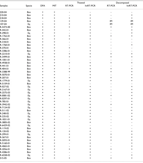
Results of the 50 previously positive samples thawed and 48 positive samples decomposed for DFA, MIT, RT-PCR and hnRT-PCR are shown in table 2.
Figure 1 represents the electrophoresis of samples evalu-ated to RT-PCR and hnRT-PCR.
The sensitivity of RT-PCR on thawed samples was 52%, while the sensitivity with the association of hnRT-PCR was 90%. These results corroborate previous studies with nested technique for rabies diagnosis; Kamolvarin et al. [14] found 38% of sensitivity for RT-PCR and 100% when considering the nested step.
Considering the Kappa coefficient values, the concord-ance in thawed samples was 0.59 for RT-PCR and 0.92 for the association with hnRT-PCR; in decomposed samples was 0.43 and 0.80 for RT-PCR and hnRT-PCR respectively, confirming the greater sensitivity of heminested when compared with RT-PCR alone. These better results can be explained by the fact that in heminested assay a second amplification of the template increases the sensitivity being capable of detecting minimum quantities of virus [10].
The analyzed samples were stored from 1999 to 2006. These samples were also evaluated considering the time of storage in order to determine the influence of the freezing time in the observed results. However, no significant dif-ferences were observed; in samples from 2001, 50% of the positive ones were positive for RT-PCR, while in the ones from 2002, 33,3% were positive in the same situation. Lopes et al. [20] found a sensitivity of 83,3% in samples stored between the years of 1997 and 2002, the discrep-ancy in the values may be explained by the temperature of storage between -20 and -70°C, while in the present study this temperature was only of -20°C.
Negative results in the RT-PCR were obtained in 12 decomposed materials, previously FAT and MIT positive. All samples used in this study are from field and were not submitted to viral multiplication; a low viral titer may explain these false-negatives results in the RT-PCR [15,19].
Sensitivity in decomposed samples was lower for both techniques. Decomposed samples were tested to adverse conditions which must be considered in the interpreta-tion of these results, because RNA viruses are much more sensitive to ambient conditions than DNA viruses [11]. The great sensitivity of RNA to RNAses found in the envi-ronment and set free during cellular lysis [24] may explain the lower sensitivity in decomposed samples.
The 100% of concordance observed in the 101 negative samples with the results of DFA and MIT demonstrates the high specificity of PCR technique related in many studies for rabies diagnosis [10,12-14].
RT-PCR and hnRT-PCR can detect the rabies virus genome in samples highly decomposed, even when DFA and MIT present negative results [16,17]. When comparing the val-ues founded in the present study with others evaluating samples in the same state of decomposition, hnRT-PCR presented greater sensitivity than DFA and MIT [16,17]. BRITO [8] founded 43,48% and 39,13% of sensitivity for DFA and MIT respectively in samples decomposed for 72 hours, in the present study the sensitivity for hnRT-PCR was 75% for samples in the same conditions.
These results are in concordance with others in decom-posed samples with positive results for molecular tech-niques and negative for DFA and MIT [18,21,22]. Although DFA and MIT are the recommended techniques for rabies diagnosis, RT-PCR can be used as an important tool when samples are not suitable for the standard tech-niques due to decomposition.
The use of hnRT-PCR can be very useful not only for diag-nosis purposes but also using the amplified products for epidemiological classification of the virus [16,18].
Table 2: Comparative results of Direct Fluorescent Antibody Test (DFA), Mouse Inoculation Test (MIT), Reverse-transcriptase Polymerase Chain Reaction (RT-PCR) and Heminested RT-PCR techniques for rabies diagnosis in thawed and decomposed positive samples.
Thawed Decomposed
Samples Specie DFA MIT RT-PCR hnRT-PCR RT-PCR hnRT-PCR
220-04 Bov + + - - -
-210-04 Bov + + - + - +
218-04 Bov + + + + - +
139-04 Bov - + + + IM IM
107-04 Eq + + + + IM IM
R-5473-00 Bov + + + + + +
R-442-01 Bov + + - + - +
R-298-01 Eq + + - + - +
R-1762-01 Bov + + + + + +
R-366-01 Bov + + + + -
-R-318-01 Bov + + - - -
-R-1760-01 Bov + + - + - +
R-370-01 Bov + + + + -
-R-3386-01 Bov + + - + -
-R-2210-01 Eq + + + + + +
R-3499-01 Bov + + + + + +
R-1001-01 Bov + + - - -
-R-4938-01 Bov + + + + + +
R-441-01 Bov + + - + -
-R-404-01 Bov + + + + + +
R-3388-99 Bov + + + + + +
R-5070-01 Bov + + - + - +
R-207-01 Bov + + + + + +
R-1779-01 Eq + + + + + +
R-5159-01 Bov + + - - -
-R-027-02 Eq + + - + - +
R-2167-01 Eq + + + + - +
R-2575-03 Eq + + + + - +
R-0081-02 Eq + + + + + +
R-0297-01 Eq + + - + - +
R-785-01 Eq + + - + - +
R-3942-02 Eq + + + + + +
R-7124-03 Eq + + + + - +
R-511-02 Bov + + + + + +
R-248-02 Eq + + - + - +
R-235-02 Eq + + - + - +
R-1831-01 Eq + + - + - +
R-3805-02 Bov + + - + -
-R-6429-02 Eq + + - - -
-R-119-02 Bov + + - + -
-R-120-02 Bov + + + + - +
R-299-01 Eq + + + + + +
R-367-01 Bov + + + + + +
R-5093-01 Bov + + + + + +
R-5160-01 Bov + + - + - +
R-4065-01 Bov + + - + - +
R-2056-01 Bov + + + + + +
R-4286-01 Bov + + - + -
-R-6558-02 Eq + + - + - +
515-05 Bov + + + + + +
Competing interests
The authors declare that they have no competing interests.
Authors' contributions
DBA participated in samples analysis and drafted the manuscript, JM participated in design and coordination of study and helped to draft the manuscript.
Acknowledgements
DBA and JM tanks to Fundunesp for financial support.
References
1. WHO Expert Commitee on Rabies: Rabies: Fact Sheet N°99. 2006 [http://www.who.int/mediacentre/factsheets/fs099/en]. Geneva: WHO Access: 20 abr. 2007.
2. Pringle CR: The order mononegavirales. Arch Virol 1991, 117:137-140.
3. Bourhy H, Rollin PE, Vincent J, Sureau P: Comparative field evalu-ation of the fluorescent-antibody test, virus isolevalu-ation from tissue culture and enzyme immunodiagnosis for rapid labo-ratory diagnosis of rabies. J Clin Microbiol 1989, 27:519-523. 4. Organisation Mondiale de la Santé Animale: Manual of diagnostics
tests and vaccines for terrestrial animals. 5th edition. 2004 [http://www.oie.int/eng/normes/mmanual/a_00044.htm]. Paris: OIE – World Organisation for Animal Health Access: 9 abr. 2007. 5. Valentini EJG, Albas A, Augusto VL, Ito FH: Immunofluorescence
performed in brain of mice, infected with CVS strain of the
rabies virus in different stages of decomposition. Rev Inst Med Trop São Paulo 1991, 33:181-186.
6. Albas A, Ferrari CI, Queiroz LH, Bernardi F, Ito FH: Influence of canine brain decomposition on laboratory diagnosis of rabies. Rev Soc Bras Med Trop 1999, 32:19-22.
7. Arslam A, Saglam YS, Temur A: Detection of rabies viral antigens in non-autolysed and autolysed tissues by using immunoper-oxidase technique. Vet Rec155:550-552.
8. Brito CJC: Avaliação do teste de Imunofluorescência Direta com conjugados antiglicoproteína viral e antinucleocapsídeo viral em material fresco e em decomposição para o diagnós-tico da raiva. In Dissertação (Mestrado) Faculdade de Medicina Vet-erinária e Zootecnia da Universidade Estadual Paulista, Botucatu; 2002.
9. Chabra M, Bhardwaj M, Ichhpujani RL, Lal S: Comparative evalua-tion of commonly used laboratory tests for post-mortem diagnosis of rabies. Indian J Pathol Microbiol 2005, 48:190-193. 10. Picard-Meyer E, Bruyére V, Barrat J, Tissot E, Barrat MJ, Cliquet F:
Development of a hemi-nested RT-PCR method for the spe-cific determination of European Bat Lyssavirus. Comparison with other rabies diagnostic methods. Vaccine 2004, 22:1921-1929.
11. Belák S, Ballagi-Pordány A: Application of the polymerase chain reaction (PCR) in veterinary diagnostic virology. Vet Res Com-mun 1993, 17:55-72.
12. Ermine A, Larzul D, Ceccaldi PE, Guesdon JL, Tsiang H: Polymerase chain reaction amplification of rabies virus nucleic acids from total mouse brain RNA. Mol Cell Probes 1989, 4:189-191. 13. Sacramento D, Bourhy H, Tordo N: PCR technique as an
alter-native method for diagnosis and molecular epidemiology of rabies. Mol Cell Probes 1991, 5:229-240.
14. Kamolvarin N, Tirawatnpong T, Rattanasiwamoke R, Tirawatnpong S, Panpanich T, Hemachudha T: Diagnosis of rabies by polymerase
Reverse-transcriptase Polymerase Chain reaction (RT-PCR) and Heminested RT-PCR (hnRT-PCR) results for detection of rabies virus genome in animal brain samples
Figure 1
Reverse-transcriptase Polymerase Chain reaction (RT-PCR) and Heminested RT-PCR (hnRT-PCR) results for detection of rabies virus genome in animal brain samples. Line 01-C1 – 100 base pairs (bp) ladder; C2 – positive con-trol for RT-PCR with 455 pb; C3 – negative sample (R-441-01); C4 e C5 – positive samples (R-404-01 e R-3388-99); C6 – neg-ative sample (R-5070-01); C7 e C8 – negneg-ative controls. Line 02-C1 – 100 base pairs (bp) ladder; C2 – positive control for hnRT-PCR with 299 pb; C3 e C4 – negative samples (R-1001-01 E R-441-01); C5 – positive sample (R-5070-01); C6 a C8 – negative controls.
C1 C2 C3 C4 C5 C6 C7 C8
455pb
Publish with BioMed Central and every scientist can read your work free of charge "BioMed Central will be the most significant development for disseminating the results of biomedical researc h in our lifetime."
Sir Paul Nurse, Cancer Research UK
Your research papers will be:
available free of charge to the entire biomedical community
peer reviewed and published immediately upon acceptance
cited in PubMed and archived on PubMed Central
yours — you keep the copyright
Submit your manuscript here:
http://www.biomedcentral.com/info/publishing_adv.asp
BioMedcentral
chain reaction with nested primers. J Infect Dis 1993, 167:207-210.
15. Heaton PR, Johnstone P, McElhinney LM, Cowley R, O'Sullivan E, Whitby SE: Heminested PCR assay for detection of six geno-types of rabies and rabies related-viruses. J Clin Microbiol 1997, 35:2762-2766.
16. Whitby JE, Johnstone P, Sillero-Zubiri C: Rabies virus in the decomposed brain of an Ethiopian wolf detected by nested reverse transcription-polymerase chain reaction. J Wildl Dis
1997, 33:912-915.
17. David D, Yakobson B, Rotanberg D, Dveres N, Davidson I, Stram Y: Rabies virus detection by RT-PCR in decomposed naturally infected brains. Vet Microbiol 2002, 87:111-118.
18. Soares RM, Bernardi F, Sakamoto SM, Heinemann MB, Cortez A, Alves LM, Meyer AD, Ito FH, Richtzenhain LJ: A heminested polymerase chain reaction for the detection of Brazilian rabies isolates from vampire bats and herbivores. Mem Inst Oswaldo Cruz 2002, 97:109-111.
19. Rojas AE, Loza-rubio E, Banda RVM, Hernández BE: Use of reverse transcription-polymerase chain reaction to determinate the stability of rabies virus genome in brains kept at room tem-perature. J Vet Diagn Invest 2006, 18:98-101.
20. Lopes MC, Veditti LLR, Iamamoto K, Carvalho C, Queiroz LHQ: Via-bilidade de detecção do genoma do vírus rábico em amos-tras armazenadas por longos períodos de tempo. XVIII Congresso de iniciação científica da Unesp, Marília 2006.
21. Loza-Rubio E, Rojas-Anaya E, Banda-Ruiz VM, Nadin-Davis AS, Cortez-Garcia B: Detection of multiple strains of rabies virus RNA using primers designed to target Mexican vampire bat variants. Epidemiol Infect 2005, 135:927-934.
22. Favoretto SR, Martorelli LF, Elkhoury MR, Zargo AM, Durigon EL: Rabies virus detection and phylogenetic studies in samples from a exhumed human. Cli Infect Dis 2005, 41(3):413-414. 23. Azevedo MO, Felipe MSS, Brigido MM: Técnicas básicas em biologia
molecular Brasília: Ed. Universidade de Brasília; 2003.
24. Sambrook J, Fitsch EJ, Maniatis T: Molecular cloning: a laboratory manual