Abstract: Image enhancement is a pre-processing process to enhance the quality and information content of original data. This paper investigates two methods of image augmentation that is deployed to remove noise and improve radiographic images. The first method is image filtering, which consists of smoothing, sharpening and edge enhancement (Sobel & Prewitt) operations. The filtering method emphasizes certain characteristics or eliminates other details. While the second method is morphological technique that utilizes the opening and closing operation, which employed to removed distorted noise and imperfection on the processed images. Each method and operation applied to the image is evaluated subjectively based on the enhance image quality. The image quality measured using MSE (Mean Square Error) and PSNR (Peak Signal to Noise Ratio) which is a full reference metrics. The image quality results are compared to give a wide picture on the performance of the enhanced images. The image processing operations accomplished by using MATLAB image processing toolbox.
Keywords: digital radiography; image enhancement; image detection; image quality
I. INTRODUCTION
Radiography is one of the Non-Destructive Testing (NDT) techniques to find out internal incoherence existing in the material or component. This method has a lot of advantages over the conventional radiographic film which includes the ability to digitally transfer image, immediate image preview, cost reduce due to film processing steps and time efficiency. Ekinci S and Ron PINCU [1][2] reviewed the use of digital radiography in the evaluation of defects in welds. However, there are some problems in digital radiography system must have a problem which effect the imaging quality. Chen and Lu [3] reviewed the problems occurred in the digital radiographic system. Most of the digital radiography image is in poor quality. Suitable technique is important to improve image quality for better inspection.
Revised Manuscript Received on October 05, 2019.
Puteri Zirwatul Nadila M.Z, Maritime Engineering Technology (MET) Section, Universiti Kuala Lumpur-Malaysian Institute of Marine Engineering Technology (MIMET), Lumut, Perak, Malaysia.
Norfadhlina Khalid, Maritime Engineering Technology (MET) Section, Universiti Kuala Lumpur-Malaysian Institute of Marine Engineering Technology (MIMET), Lumut, Perak, Malaysia.
Roszaiman Abd Khalid, Universiti Kuala Lumpur-Malaysian Institute of Marine Engineering Technology (MIMET), Lumut, Perak, Malaysia.
M.K. Puteri Zarina, Student Development (SD) Section, Universiti Kuala Lumpur-Malaysian Institute of Marine Engineering Technology (MIMET), Lumut, Perak, Malaysia.
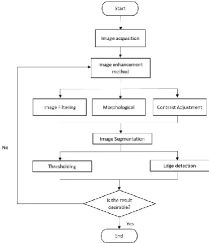
[image:1.595.323.531.380.622.2]Image processing method has a technique such as noise elimination, intensity adjustment, edge recognition and other means. The main goal of these approaches is to extract particulars of image or to enlarge the contrast in a low contrast image and it can be done by adjusting the concentration of the pixel of the input image [4]. The image processing method reviewed by many authors to improve the radiography image and shows better interpretation of weld defect [5][6][7]. Image augmentation and image segmentation is selected to enhance the digital radiography image contains three defects in piping. Figure 1 exhibits the process flow chart of image processing via image processing toolbox in MATLAB. Image enhancement allow removing of noise, sharpening, or adjusting an image's contrast. Meanwhile for detection process, methods such as thresholding and edge detection is selected to detect object boundaries within pictures.
Fig. 1 Flowchart of image processing II. IMAGEACQUISITION
In this research, image is taken using NDT Analyzer Model: m 225D from GE Phoenix X-ray equipped with digital image chain for improved contrast and greater resolution. The μ-focused digital radiography has 9” triple-, 6” dual- and 6” single-field image intensifier for full digital imaging. The piping is captured by using microfocused
Puteri Zirwatul Nadila M. Z, Norfadhlina Khalid, Roszaiman Abd Khalid, M. K. Puteri Zarina
Image Processing Method on Radiographic
Image of Piping using MATLAB: Enhancement
digital radiography. The current is 500 µA and voltage is 190 kV. Exposure time to acquire the image is 200 µs. The sampling is placed in the µ-focused digital radiography machine as displayed in Figure 2 below.
Fig. 2 The arrangements in the micro focus x-ray machine
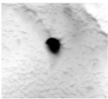
[image:2.595.56.280.115.263.2]The image in Figure 3 below shows radiographic image of piping obtained by exposure technique of single wall single image (SWSI). The weld defects are blow hole, porosity and lack of blending. Lack of root fusion or incomplete root fusion in Figure 4 happens when the weld metal does not produce a solid bond with the base metal or when the weld metal fails to cover the base metal to the desired depth, which causes inadequate throat thickness. Porosity and blow hole are voids or apertures caused by gas and non-metallic material entrapment in molten metal during solidification.
[image:2.595.336.518.296.457.2]Fig. 3 Original radiographic image
Fig. 4 Incomplete root fusion III. ENHANCEMENTPROCESS
Image enhancement processes in MATLAB provide variable procedures that seek to enhance the visual outlook of an image or to alter the image to a form that suits the human analyst to run the analysis or used for other computerized image processing procedures. The image improvement
process comprises three categories: filtering, morphology, and adjustment of contrast. Figure 5, Figure 7 and Figure 9 indicate the outcome after augmentation method and three weld defects obviously displayed in the image.
[image:2.595.54.278.428.693.2]Fig. 5 Processed image after image enhancement and incomplete root fusion weld defect
Fig. 6 Original image before enhancement and blow hole weld defect
[image:2.595.332.520.491.662.2]Fig. 8 Original image before enhancement and porosity weld defect
Fig. 9 Processed image after image enhancement and porosity weld defect
IV. SEGMENTATIONPROCESS
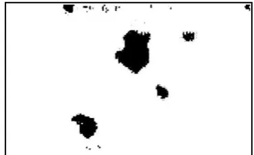
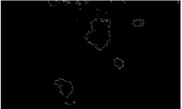
Image segmentation is a method of dividing the center from the background or grouping pixel areas based on color or shape resemblances. The method of image segmentation includes histogram thresholding and edge detection. This method is reviewed by many authors to achieved successful detection of different kinds of image [8][9][10][11][12]. Image detection method implemented after processing and enhancing an image. Image breakdown is one of the digital image processing procedures, which partition of image is analyzed into multiple parts or regions, often based on the facets of the pixels in the image. Image thresholding is shown in Figures 10, 12, and 14. Meanwhile canny edge detection result is shown in Figures 11, 13 and 15. Canny edge detection achieves enhancement and compression without the loss of any important edge with detecting discontinuities in brightness.
[image:3.595.341.503.202.363.2]Fig. 10 Incomplete root fusion segmented by histogram Thresholding
[image:3.595.62.277.207.336.2]Fig. 11 Incomplete root fusion segmented by Canny edge detection
Fig. 12 Blow hole segmented by histogram Thresholding
Fig. 13 Blow hole segmented by Canny edge detection
[image:3.595.344.503.395.527.2] [image:3.595.333.518.578.691.2] [image:3.595.57.279.597.741.2]Fig. 15 Porosity segmented by Canny edge detection V. RESULTANDDISCUSSION
[image:4.595.55.276.577.715.2]The result for image enhancement and image detection proved that the weld defect in radiographic image improved compared to original image. The methods discussed shows improvement by measuring the image quality after removing the noise and enhanced by using filtering technique. Table 1 has shown PSNR, MSE and NAE results, which connecting to elimination of noise and reducing of error in the picture.
Table. 1 Image Quality Measurement Result Weld Defect Quality Performance Incomplete root
fusion
MSE = 950.3119 PSNR = 18.3521 NAE = 0.1910 Blow hole MSE = 6245.7420
PSNR = 10.1750 NAE = 0.5842 Porosity MSE = 1906.2669
PSNR = 15.3290 NAE = 0.2589
MSE embodies the collective squared error between the compressed and the original image. PSNR, on the other hand, characterizes a value of the error of the peak. Peak Signal to Noise Ratio (PSNR) is normally deployed to examine the image, sound and video files quality in dB (decibels). A lower value for MSE means less error and greater value of PSNR is adequate because this suggests that the signal to noise ratio is greater [7].
n-10 y 2 * 1 -m 0 x
y)]
(x,
f
y)
[f(x,
1
mn
MSE
(1)PSNR = 20log10 MAX 2
MSE (2)
1 0 1 0 1 -M 0 j * 1 -N 0 i,
y)]
(x,
f
y)
[f(x,
.
1
m x n yj
i
f
M
N
NAE
(3) where:mn= image dimension in row, m and column, n f(x,y)= the pixel value at (x,y) of the original image f’(x,y)= the pixel value at (x,y) of the processed image MAX = maximum value of image pixel value.
Normalized Absolute Error (NAE) is a measure of the distance of the rebuilt image from the original one, with the value of zero being the perfect fit. High value of NAE shows a unsatisfactory quality image [13][14]. Table 1 below shows the quality performance results for three weld defects based on Figure 5, Figure 7 and Figure 9.
VI. CONCLUSION
The defect on the radiographic image of a pipe were identified and have been classified by using two method of image processing which are image enhancement and detection. The image is enhanced thus giving a clear vision of its defect which is then used and analyze by image detection process. As the current industry is growing rapidly through the years, the needs for the latest product are in high demands. In the welding inspection, the use of digital radiographic testing in non-destructive testing (NDT) has become more relevant as the industry develops but the high cost for the latest digital radiographic x-ray device are becoming a major drawback to some company. Additionally, the maintenance cost of a digital radiographic image is too high to be affordable by the small and average company in the country. Although the industry is going through a major revolution with the industry, there still some limitation in the current industry that needs to be consider such as the use of radiography film is still popular among the companies in the country.
REFERENCES
1. Ekinci S. “Examination of Welds by Digital Radiography” Proceeding of the Third Eurasian Conference "Nuclear Science and its Application", October 5-8,2004.
2. Ron PINCU, “Digital Radiography and its Advantages in Field NDT Inspections Today”, 17th World Conference on Nondestructive Testing, 25-28 Oct 2008, Shanghai, China.
3. Chen Shuyue and Lu Hongnian, “Noise Characteristic and its Removal in digital Radiographic System”, 15.WCNDT, Roma 2000.
4. X. Xiaodong, et al, 2009, “An Adaptive Image Enhancement Technique Based on Image Characteristic,” 2nd International Congress on Image and Signal Processing, pp. 1-5.
5. Shahidan Mohamad and Suhaila Abd Halim,” Image Enhancement Process on Digital Radiographic Image with Weld Discontinuities”, Journal of Mechanical Engineering, Vol SI 5(4), 275-292, 2018. 6. A.P.Nagarajan., Christy V.Vazhappilly, Sukesh O.P., Vivek Rajdhan J, Rajavelu.S., Prashanthkumar.S, “Improvement of Weld Images using
MATLAB –A Review”, International Journal of Engineering And Science, Vol.5, Issue 4 (April 2015), PP 16-22, Issn (e): 2278-4721, Issn (p):2319-6483
7. Suhaila Abd Halim, Yupiter HP Manurung, Shahidan Mohamad, Mohamad Firhan Morni, “The Effect of CLAHE and Gamma Correction in Enhancement of Digital Radiograph ic Imag e for Weld Imperfection Detection”, International Journal of Engineering & Technology, 7 (4.36) (2018) 1588-1592.
8. Roumen Kountchev, Vladimir Todorov, Roumiana Kountcheva, “Defects detection in X-ray images and photos”, ISBN: 978-960-474-276-9.
9. Dr.S.Vijayarani, Mrs.M.Vinupriya, “Performance Analysis of Canny and Sobel Edge Detection Algorithms in Image Mining”, International Journal of Innovative Research in Computer and Communication Engineering (An ISO 3297: 2007 Certified Organization) Vol. 1, Issue 8, October 2013.
Author-1 Photo
11. Kirti Agrahari1, Diksha Srivastava and Purnima Tripathi, “Image Enhancement Using Matlab”, International Journal of Advance in Science and Engineering, Vol No 6, Issue No 04, April 2017. 12. Sanjana C.Shekar and D.J.Ravi, “Image Enhancement and Compression using Edge Detection Technique”, International Research Journal of Engineering and Technology (IRJET), Volume: 04 Issue: 05, May -2017 13. Mayuresh Gulame, K. R. Joshi & Kamthe R. S, “A Full Reference Based Objective Image Quality Assessment”, ISSN (Print): 2278-8948,
Volume-2, Issue-6, 2013.
14. V.S.Vora, Prof. A.C.Suthar, Y.N.Makwana, S.J. DavdaJ. Wang, “Analysis of Compressed Image Quality Assessments”, International Journal of Advanced Engineering & Application, Jan. 2010
AUTHORSPROFILE
Puteri Zirwatul Nadila Megat Zamanhuri is a Lecturer in the Maritime Engineering Technology (MET) Section at Universiti Kuala Lumpur-Malaysian Institute of Marine Engineering Technology (MIMET) Lumut, Perak, Malaysia. She received a Master Degree in Mechanical Science from the Universiti of Teknologi Mara (UiTM) and worked as a research assistant in the Advanced Manufacturing Technology Excellent Centre before she joined University Kuala Lumpur-MIMET in 2012. She is an established teacher in welding process, welding inspection and operation management. Her research interests include of image processing of weld defect using MATLAB, development of application tool using MATLAB, weld inspection method and product design development. She actively participated in product competition and won medals for her final year project student. She has published a Scopus journals and participated in conferences.
Norfadhlina Khalid is a Lecturer in the Maritime Engineering Technology (MET) Section at Universiti Kuala Lumpur-Malaysian Institute of Marine Engineering Technology (MIMET) Lumut, Perak, Malaysia. She received a Master Degree in M. Eng. in Engineering Management (Manufacturing) from the Universiti Putra Malaysia (UPM) in 2011. She has a Bachelor Degree (Hons) in Product Design from the Universiti Kuala Lumpur – IPROM in 2009. She has been working with Universiti Kuala Lumpur-MIMET since January 2011. She has performed management duty as a Head of Section, Programme Coordinator and academic. Her research and teaching activities include of the design and product development, machining, manufacturing, project planning and management. Her research interests include of Computer Aided Design (CAD), Computer Aided Manufacturing (CAM) and CNC Technology. She has attended and presented papers in number of conferences and published a few papers. She is one of the editorial members in Marine Frontier-MIMET Technical Journal since 2012. She received the certification from Australian Institute of Technology Transfer (AITT) as a competent Trainer in TAE40110 Certificate IV and registered as a graduate technologist with Malaysia Board of Technologists (MBOT).
Roszaiman Abd Khalid is a student in Malaysian Institute of Marine Engineering Technology (MIMET) Lumut, Perak, Malaysia. He take course Bachelor in Naval Architecture (Hons) at UNIKL MIMET. He currently in final semester.
Puteri Zarina Megat Khalid is currently attached to Universiti Kuala Lumpur Malaysian Institute of Marine Engineering Technology (UniKL MIMET), Lumut as a senior lecturer-cum-Deputy Dean of Student Development & Campus Lifestyle. Among her research interests are language teaching and learning, modality analysis, pragmatics, corpus linguistics, English for Specific Purposes (ESP), genre analysis and Systemic Functional Linguistics. She received her Ph. D. in English Language from University of Glasgow, Scotland. She is also an appointed member of UniKL Research Journal Editorial Board and Editorial Board Member for UniKL MIMET Research Bulletin “Marine Frontier@UniKL” and several other publications. A member of International Systemic Functional Linguistics Association (ISFLA), Malaysian Society for Engineering & Technology (MySET) and Institute of Marine, Science and Technology (IMAREST).