model system
.
White Rose Research Online URL for this paper:
http://eprints.whiterose.ac.uk/1255/
Article:
Peers, C. (2004) Interactions of chemostimuli at the single cell level: studies in a model
system. Experimental Physiology, 89 (1). pp. 60-65. ISSN 0958-0670
https://doi.org/10.1113/expphysiol.2003.002657
[email protected] https://eprints.whiterose.ac.uk/ Reuse
See Attached
Takedown
If you consider content in White Rose Research Online to be in breach of UK law, please notify us by
Experimental Physiology –
Hot Topic Review
Festschrift for R. G. O’Regan – Sensing and adaptation to alterations in respiratory gases: oxygen and carbon dioxide
Interactions of chemostimuli at the single cell level:
studies in a model system
Chris Peers
Institute for Cardiovascular Research, University of Leeds, Leeds LS2 9JT, UK
The responses of afferent chemosensory fibres of the carotid body to individual chemostimuli have long been established. However, the mechanisms underlying the multiplicative interactions of these stimuli (i.e. how the combined effects of hypoxia and hypercapnia exert a greater effect on afferent nerve discharge than the sum of their individual effects) have not been elucidated. Using the membrane hypothesis for carotid body chemoreception, in which chemostimuli inhibit type I cell K+ channels, leading to depolarization, voltage-gated Ca2+ entry and hence the
triggering of exocytosis, this article considers data acquired in isolated type I carotid body cells and model chemoreceptor (PC12) cells to attempt to explain stimulus interactions. Whilst stimulus interactions are not clearly evident at the level of K+ channel inhibition or rises of
[Ca2+]
i, they are apparent at the level of transmitter release. Thus, it is clear that individual
chemoreceptor cells can sense multiple stimuli, and that interactions of these stimuli can produce greater than additive effects in terms of transmitter release.
(Received 23 October 2003; accepted 1 November 2003)
Corresponding authorC. Peers: Institute for Cardiovascular Research, University of Leeds, Leeds LS2 9JT, UK. Email: [email protected]
Introduction
For decades the carotid body has been recognized as the major peripheral arterial chemoreceptor, sensing changes in blood gas and pH levels and responding, when appropriate, by altering the firing frequency of afferent chemoreceptors (Gonzalezet al.1994; Gonzalez et al. 1992). In this way, the carotid body informs the central respiratory centres of arterial blood gas status, and allows the initiation of corrective cardiorespiratory reflexes (Marshall, 1994; Lopez-Barneo, 1996). Afferent chemosensory recordings, performed over many years, have established the closely controlled relationship between stimulus intensity and afferent nerve activity, and have shown that excitation of these nerves can be brought about by each physiological stimulus (hypoxia, hypercapnia and acidity) applied independently. More interestingly, perhaps, these stimuli can interact at the
Presented at a meeting of the Physiological Society at Trinity College Dublin in July 2003.
level of afferent chemosensory discharge in a multiplicative manner (Fitzgerald & Parks, 1971; Lahiri & DeLaney, 1975). In other words, the effects of hypoxia and hypercapnia applied together are greater than the sum of these two stimuli when applied separately to the carotid body. This is illustrated in Fig. 1, which plots the frequency of discharge of afferent chemoreceptor fibres as a function of CO2levels, whilst the background O2 levels are varied
from hyperoxia to severe hypoxia. Clearly, as the O2
levels decline, the relationship between nerve activity and CO2 becomes increasingly steep. This is indicative of
multiplicative stimulus interaction, as if these stimuli were simply additive, the slope of the relationship between nerve discharge and CO2would not be altered.
article is aimed at reviewing available evidence in order to examine a cellular basis for this phenomenon of stimulus interactions.
Chemoreception in single type I cells
[image:3.595.52.292.454.646.2]Since the development of viable preparations of isolated type I cells, numerous groups have sought to provide an answer to the question of how type I cells sense and respond to chemostimuli. Several lines of evidence had indicated that chemosensing by the carotid body was totally dependent on the presence of type I cells (reviewed by Gonzalezet al.1994), and that neurotransmitter release from these cells was an absolute requirement for excitation of afferent nerves of the carotid sinus nerve. The nature of the neurotransmitters involved has long been contentious, but the most compelling evidence published in recent times would indicate that hypoxia releases (amongst others) excitatory transmitters acetylcholine and ATP (Zhang et al. 2000), along with dopamine (Obesoet al. 1992). The role of dopamine in chemoreception remains to be fully established, but it is clear that stimulus evoked transmitter release from type I cells on to afferent nerve endings is a key step in carotid body excitation. This generally accepted mechanism begs the question of how stimuli cause type I cells to release neurotransmitters, a question that several groups have approached since the late 1980s. Whilst there remain some points of contention (which are outside the scope of this article), it seems reasonable to assume today that the general consensus of
Figure 1.
Plot of the relationship between afferent chemosensory nerve discharge and CO2levels, recorded from the cat carotid body
sinus nerve preparation. CO2levels were varied at different
backgroundPO
2levels, as indicated. Note the increased slope of
the relationship as background O2levels decline. Reproduced
with permission from Lahiri & DeLaney, 1975.
opinion is that hypoxia, hypercapnia and acidosis evoke transmitter release according to what has become known as the membrane hypothesis for chemotransduction.
The key initial step of the membrane hypothesis for chemotransduction is the stimulus-induced inhibition of K+ channels (Lopez-Barneo et al. 1988; Peers, 1990;
Stea & Nurse, 1991; Buckler, 1997). These channels normally maintain a hyperpolarizing influence on resting membrane potential and thereby control cell excitability. Their inhibition therefore leads to cell depolarization. This in itself is sufficient to open voltage-gated Ca2+channels,
and the consequent rise of [Ca2+]
i (Buckler &
Vaughan-Jones, 1994) is the trigger for transmitter release. Whilst this mechanism is not the only one to have been proposed to account for stimulus–secretion coupling in type I cells (Biscoe & Duchen, 1990; Rocheret al.1991), it is clearly the best supported, and it is with this sequential series of events we can address possible mechanisms to account for stimulus interactions within the carotid body.
Effects of hypoxia and acidosis on K+channel activity
Whilst a number of groups have reported that hypoxia and hypercapnia/acidosis can inhibit K+ channels and cause
depolarization of type I cells, the question of whether an individual type I cell can respond to both stimuli remained unanswered until 1997. In that year, Pepper & Kumar (1997) reported that K+ currents (most likely
high conductance, Ca2+ sensitive (maxiK) currents) in
individual rat type I cells could be inhibited both by hypoxia and by intracellular acidosis (in this case evoked by extracellular application of sodium propionate). This was the first report that a type I cell could respond to both stimuli. However, coapplication of both stimuli failed to cause channel inhibition that was quantitatively greater than either stimulus alone. Thus, stimulus interaction did not occur at the level of K+channel activity within type I
cells. To date, this remains the only study aimed directly at addressing this issue. It will be of interest to examine whether TASK channels, also present in these cells (Buckler et al.2000), or the O2sensitive, inactivating voltage-gated
K+ channels of rabbit type I cells (Lopez-Lopez & Peers,
1997), respond in a similar manner to those reported by Pepper and Kumar.
Effects of hypoxia and acidosis on [Ca2+]
i
in type I cells
Following K+ channel inhibition caused by hypoxia or
hypercapnia/acidosis in type I cells, a rise of [Ca2+]
i
is observed due to Ca2+ entry via voltage-gated Ca2+
channels. The question of whether rises of [Ca2+]
idisplay
C
any form of interaction was addressed in detail by Dasso et al.(2000). Under carefully controlled conditions, these workers examined the rises of [Ca2+]
i in response to
graded hypoxia/anoxia with CO2levels maintained either
at the normal, eucapnic level of 5% or when raised to hypercapnic levels of 10% or 20%. The key observations are summarized in Fig. 2. Whether or not one can conclude from these data that stimulus interactions occur at the level of [Ca2+]
iis not clear. Using 10% CO2 as the additional
stimulus (Fig. 2A), rises of [Ca2+]
i seen in response to
hypoxia are generally elevated, but not in a manner that could be described as multiplicative. Indeed, some of the additional rises of [Ca2+]
i seen when hypoxia is applied
together with hypercapnia are only slightly greater than those observed in response to hypoxia alone. Using a stronger CO2 stimulus of 20% (Fig. 2B), multiplicative
[image:4.595.47.286.297.658.2]interactions are detectable at some, but not all, levels of
Figure 2.
Plot of the mean relationship between [Ca2+]
i(measured
fluorimetrically from type I cells using the [Ca2+]
iindicator
Indo-1) andPO
2under eucapnic conditions, and when CO2was
raised to either 10% (A) or 20% (B). Taken from (Dassoet al. 2000) with permission.
hypoxia. These findings, whilst providing some evidence for multiplicative stimulus interactions, are not in full agreement with earlier studies of afferent chemoreceptor discharge (Fig. 1), which indicate that stimulus interaction occurs over a wide range of O2and CO2levels.
Effects of chemostimuli on catecholamine secretion
Direct measurements of stimulus-evoked transmitter release from individual type I cells are still in their infancy, and to date have been confined to the study of catecholamines released from type I cells (Hatton & Peers, 1997; Carpenter et al. 2000; Pardal et al. 2000). This is in large part due to the technical demands of monitoring release of various transmitters from isolated cells. Fortunately, catecholamines are amenable to such studies since they are easily oxidized and so can be monitored amperometrically (Chow & Von Ruden, 1995). The technique of amperometry allows placement of polarized carbon fibre microelectrodes close to individual cells, so that any released catecholamine diffusing to the electrode tip is instantly oxidized. This allows detection of exocytosis as a real-time electrical event. Amperometry has to date only been used to study catecholamine release from type I cells in a limited number of reports (see above), and the question of whether stimulus interaction occurs at the level of transmitter release from type I cells has yet to be addressed. However, progress has been made using the PC12 cell line, pioneered by Millhorn and colleagues as a model system for studying chemoreception at the single cell level (Zhuet al.1996; Conforti & Millhorn, 1997).
PC12 cells are a well-established cell line having proved useful as a model system for studying a wide variety of cellular functions. They are derived from rat adrenal chromaffin tissue and synthesize, store and release catecholamines. Zhu et al. (1996) first demonstrated that these cells could be used to study chemoreception by demonstrating that they possess O2 sensitive K+
channels, and that hypoxia caused cell depolarization and a subsequent rise of [Ca2+]
i. We used these cells to show that
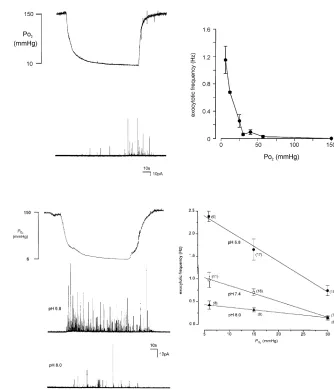
hypoxia could evoke quantal catecholamine release in a graded manner (Fig. 3, adapted from Taylor & Peers, 1998). The relationship betweenPO2 and catecholamine release
was qualitatively reminiscent of the relationship between PO2and rises of [Ca
2+]
i(Fig. 2) and afferent chemosensory
discharge. Subsequent studies established that hypoxia-evoked catecholamine secretion was entirely dependent on Ca2+influx through voltage-gated Ca2+channels (Taylor
transmitter release was also entirely dependent on voltage-gated Ca2+ entry (Tayloret al. 1999). Crucially, we also
found evidence that stimulus interactions occurred at the level of catecholamine release (Fig. 4). Thus hypoxia was only a weak secretagogue when the background pH was alkaline, but when pH was reduced, hypoxia was far more effective. Analysis of exocytotic frequency as a function of PO2 with varying pH levels indicated that
pH affected the slope of the relationship between PO2
and catecholamine secretion (Taylor et al. 1999). Thus, evidence of multiplicative stimulus interactions was found at the single cell level.
Since no compelling evidence for stimulus interactions was found at the level of K+ channel inhibition or at
the level of rises of [Ca2+]
i, the question arises of how
such interactions were apparent at the level of transmitter release. The answer may lie in two key observations. First, while interactions were not apparent at the level of [Ca2+]
i
Figure 3.
Hypoxia-evoked catecholamine secretion from a single PC12 cell. Left, upper trace showsPO
2levels measured in the same
position of a perfusion chamber as where a PC12 cell was placed for study. Lower trace shows amperometric recording from a single cell. Note the appearance of spike-like events as thePO
2declines. Each
spike corresponds to the oxidation of the released contents of a single vesicle of catecholamine. Right, plot of the relationship between mean spike frequency andPO
2, determined from
[image:5.595.214.548.331.721.2]experiments such as those shown on the left. Taken from (Taylor & Peers, 1998) with permission.
Figure 4.
Left, upper trace indicatesPO
2levels as in
Figure 3. Below are shown two
amperometric traces from individual PC12 cells in response to hypoxia either under acidic (middle trace) or alkaline (lower trace) conditions, as indicated. Right, plot of the relationship between mean spike frequency andPO
2at three different pH
levels. Note the increased slope of the relationship as background pH levels become more acidic. Taken from (Taylor et al.1999) with permission.
rises (Fig. 2), there clearly were larger rises of [Ca2+]
iin
response to hypoxia when CO2 levels were raised (Dasso
et al. 2000). These larger rises were generally modest, but the fact that they were seen at all is crucial. The second observation is not taken from studies of the carotid body, but instead comes from much earlier work in which the release of acetylcholine as a function of extracellular [Ca2+] was examined in motor nerve terminals, using the
classic frog neuromuscular junction preparation (Dodge & Rahamimoff, 1967). In these studies, it was established that the relationship between [Ca2+] and acetylcholine release
(measured as an end-plate potential) was not linear, but was in fact a power relationship (in this case, acetylcholine release was proportional to [Ca2+]3.9
). This means that only a small additional rise of [Ca2+]
iis required to observe
a substantial increase in transmitter release. Such small additional rises were reported in type I cells when CO2was
raised during hypoxia (Dassoet al.2000), and are likely
C
to account for the large rises in catecholamine exocytosis observed in PC12 cells.
Clearly, much further work is required before these ideas can be established as accounting for multiplicative interactions of chemostimuli in the carotid body. Primarily, the experiments reported in the model chemoreceptor PC12 cell need to be repeated in type I cells, and additional mechanisms within the carotid body (such as, for example, dysinhibition of retrograde tonic nitric oxide or carbon monoxide release (Prabhakar, 1999)). However, the long-standing possibility that the relationship between [Ca2+]
i and transmitter release is
supralinear in the carotid body type I cell, as it is in other synaptic preparations, is worthy of further consideration.
References
Biscoe TJ & Duchen MR (1990). Cellular basis of transduction in carotid chemoreceptors.Am J Physiol258,
L271–L278.
Buckler KJ (1997). A novel oxygen-sensitive potassium channel in rat carotid body type I cells.J Physiol498,
649–662.
Buckler KJ & Vaughan-Jones RD. (1994). Effects of hypoxia on membrane potential and intracellular calcium in rat neonatal carotid body type I cells.J Physiol476, 423–428.
Buckler KJ, Williams BA & Honore E (2000). An oxygen-, acid-and anaesthetic-sensitive TASK-like background potassium channel in rat arterial chemoreceptor cells.J Physiol525, 135–142.
Carpenter E, Hatton CJ & Peers C (2000). Effects of hypoxia and dithionite on catecholamine release from isolated type I cells of the rat carotid body.J Physiol523,
719–729.
Chow RH & Von Ruden L (1995). Electrochemical detection of secretion from single cells. InSingle Channel Recording, ed. Sakmann B & Neher E, pp. 247–275. Plenum,
London.
Conforti L & Millhorn DE (1997). Selective inhibition of a slow-inactivating voltage-dependent K+channel in rat PC12 cells by hypoxia.J Physiol502, 293–305.
Dasso LL, Buckler KJ & Vaughan-Jones RD (2000). Interactions between hypoxia and hypercapnic acidosis on calcium signaling in carotid body type I cells.Am J Physiol279, L36–L42.
Dodge FA & Rahamimoff R (1967). Co-operative action a calcium ions in transmitter release at the neuromuscular junction.J Physiol193, 419–432.
Fitzgerald RS & Parks DC (1971). Effect of hypoxia on carotid chemoreceptor response to carbon dioxide in cats.Resp Physiol12, 218–229.
Gonzalez C, Almaraz L, Obeso A & Rigual R (1992). Oxygen and acid chemoreception in the carotid body
chemoreceptors.Trends Neurosci15, 146–153.
Gonzalez C, Almaraz L, Obeso A & Rigual R (1994). Carotid body chemoreceptors: from natural stimuli to sensory discharges.Physiol Rev74, 829–898.
Hatton CJ & Peers C (1997). Electrochemical detection of K+-evoked quantal secretory events from isolated rat type I carotid body cells.Exp Physiol82, 415–418.
Lahiri S & DeLaney RG (1975). Stimulus interaction in the responses of carotid chemoreceptor single afferent fibers. Resp Physiol24, 249–266.
Lopez-Barneo J (1996). Oxygen-sensing by ion channels and the regulation of cellular functions.Trends Neurosci19, 435–440.
Lopez-Barneo J, Lopez-Lopez JR, Urena J & Gonzalez C (1988). Chemotransduction in the carotid body: K+current modulated by PO2in type I chemoreceptor cells.Science241, 580–582.
Lopez-Lopez J & Peers C (1997). Electrical properties of chemoreceptor cells. InThe Carotid Body Chemoreceptors, ed. Gonzalez C, 65–78. Landes Bioscience, Austin, TX, USA.
Marshall JM (1994). Peripheral chemoreceptors and cardiovascular regulation.Physiol Rev74, 543–594.
Obeso A, Rocher A, Fidone S & Gonzalez C (1992). The role of dihydropyridine-sensitive Ca2+channels in stimulus- evoked catecholamine release from chemoreceptor cells of the carotid body.Neuroscience47, 463–472.
Pardal R, Ludewig U, Garcia-Hirschfeld J & Lopez-Barneo J (2000). Secretory responses of intact glomus cells in thin slices of rat carotid body to hypoxia and
tetraethylammonium.Proc Natl Acad Sci U S A97, 2361–2366.
Peers C (1990). Hypoxic suppression of K+currents in type-I carotid-body cells – selective effect on the Ca2+-activated K+ current.Neurosci Lett119, 253–256.
Pepper DR & Kumar P (1997). Inhibition of adult rat carotid body type I cell K+currents by combined hypoxic and acidotic stimuli.J Physiol504.p, 202P.
Prabhakar NR (1999). NO and CO as second messengers in oxygen sensing in the carotid body.Respir Physiol115, 161–168.
Rocher A, Obeso A, Gonzalez C & Herreros B (1991). Ionic mechanisms for the transduction of acidic stimuli in rabbit carotid body glomus cells.J Physiol433,
533–548.
Stea A & Nurse CA (1991). Whole-cell and perforated-patch recordings from O2-sensitive rat carotid body cells grown in short- and long-term culture.Pflugers Arch418,
93–101.
Taylor SC, Roberts ML & Peers C (1999). Acid-evoked quantal catecholamine secretion from rat phaeochromocytoma cells and its interaction with hypoxia-evoked secretion.J Physiol 519, 765–774.
Zhang M, Zhong H, Vollmer C & Nurse CA (2000). Co-release of ATP and ACh mediates hypoxic signalling at rat carotid body chemoreceptors.J Physiol525,
143–158.
Zhu WH, Conforti L, Czyzyk-Krzeska MF & Millhorn DE (1996). Membrane depolarization in PC12 cells during
hypoxia is regulated by an O2-sensitive K+current.Am J Physiol271, C658–C665.
Acknowledgements
My own contributions to this topic, deriving from studies of exocytosis from PC12 cells, were conducted with Shafeena Taylor and Michael Roberts. Financial support was from the Wellcome Trust.
C

![Figure 2.Plot of the mean relationship between [Ca2+]i (measuredfluorimetrically from type I cells using the [Ca2+]i indicatorIndo-1) and PO2 under eucapnic conditions, and when CO2 wasraised to either 10% (A) or 20% (B)](https://thumb-us.123doks.com/thumbv2/123dok_us/8090881.231437/4.595.47.286.297.658/figure-plot-relationship-measureduorimetrically-indicatorindo-eucapnic-conditions-wasraised.webp)