JOURNAL OFVIROLOGY, May 2008, p. 5109–5114 Vol. 82, No. 10 0022-538X/08/$08.00⫹0 doi:10.1128/JVI.00060-08
Copyright © 2008, American Society for Microbiology. All Rights Reserved.
Variable Patterns of Programmed Death-1 Expression on Fully
Functional Memory T Cells after Spontaneous
Resolution of Hepatitis C Virus Infection
䌤
David G. Bowen,
1† Naglaa H. Shoukry,
2† Arash Grakoui,
3Michael J. Fuller,
1Andrew G. Cawthon,
1Christine Dong,
4Dana L. Hasselschwert,
5Kathleen M. Brasky,
6Gordon J. Freeman,
7Nilufer P. Seth,
8Kai W. Wucherpfennig,
8Michael Houghton,
4and Christopher M. Walker
1,9*
Center for Vaccines and Immunity, The Research Institute at Nationwide Children’s Hospital, Columbus, Ohio1; University of
Montreal Hospital Centre (CHUM) and Department of Medicine, University of Montreal, Montreal, Quebec, Canada2;
Emory University, Atlanta, Georgia3; New Iberia Research Center, New Iberia, Louisiana5; Southwest Foundation for
Biomedical Research, San Antonio, Texas6; Department of Medical Oncology7 and Department of
Cancer Immunology and AIDS,8Dana-Farber Cancer Institute, Harvard Medical School, Boston,
Massachusetts; Novartis Corporation, Emeryville, California4; and College of Medicine and
Public Health, Ohio State University, Columbus, Ohio9
Received 9 January 2008/Accepted 3 March 2008
The inhibitory receptor programmed death-1 (PD-1) is present on CD8ⴙT cells in chronic hepatitis C virus
(HCV), but expression patterns in spontaneously resolving infections are incompletely characterized. Here we
report that PD-1 was usually absent on memory CD8ⴙT cells from chimpanzees with resolved infections, but
sustained low-level expression was sometimes observed in the absence of apparent virus replication. PD-1-positive memory T cells expanded and displayed antiviral activity upon reinfection with HCV, indicating conserved function. This animal model should facilitate studies of whether PD-1 differentially influences effector and memory T-cell function in resolved versus persistent human infections.
The hepatitis C virus (HCV) is a major etiological agent of hepatic cirrhosis and hepatocellular carcinoma (3). Although the mechanism(s) facilitating chronic infection is not fully un-derstood, HCV-specific cytotoxic T lymphocytes (CTLs) that persist during the chronic phase of infection have an immature phenotype (1), impaired proliferative capacity, and reduced cytotoxic function and cytokine secretion (9, 16, 19, 20). It is therefore probable that the dysfunction of HCV-specific CD8⫹ T cells plays a role in HCV persistence.
Much recent interest has centered upon the inhibitory T-cell receptor programmed death-1 (PD-1) as a marker and medi-ator of impaired CTL function in persistent viral infections. Studies in lymphocytic choriomeningitis virus (LCMV)-in-fected mice have demonstrated that in persistent infection virus-specific T cells express PD-1, while in resolved infection, LCMV-specific cells lose expression of this molecule (2). In addition, the in vivo blockade of PD-1 interaction with one of its ligands, programmed death-ligand 1 (PD-L1), improved proliferation and cytokine secretion by LCMV-specific CD8⫹ T cells and reduced viral titers in persistently infected mice (2). More recently, studies have shown that in human immunode-ficiency virus (HIV)-infected individuals, HIV-specific T cells express PD-1 (4, 12, 17) and that the levels of expression of this molecule correlate with viremia and with CD4⫹T-cell count (4, 17). HCV-specific CD8⫹T cells from the blood and liver of
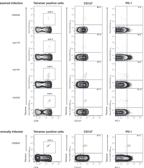
humans with chronic hepatitis C also express PD-1, and the in vitro blockade of PD-1–PD-L1 interaction enhanced antigen-driven proliferation and cytokine secretion (8, 11, 13, 18). Furthermore, in one study of acute human infection, the loss of PD-1 expression by HCV-specific CD8⫹T cells was shown to correlate with viral clearance (18). These data support the hy-pothesis that the PD-1-positive phenotype is associated with the impairment of virus-specific CD8⫹T-cell function, and as such, PD-1 expression is a correlate of the outcome of HCV infection. The goal of this study was to further evaluate the relation-ship between PD-1 expression and virus persistence, using the only animal model of human HCV infection. We analyzed the phenotype of HCV-specific CD8⫹T cells in chimpanzees that had spontaneously resolved HCV infection 5 to 7 years prior. Flow cytometric analysis of peripheral blood mononuclear cells (PBMCs) by use of major histocompatibility complex (MHC) class I peptide tetrameric complexes (tetramers) revealed HCV-specific CD8⫹memory T cells to be present at frequen-cies between 0.04% and 0.08% of CD8⫹T cells (Fig. 1). The majority expressed CD127, the interleukin-7 receptor, which is a marker of memory T cells and their precursors (10, 21). Few of these HCV-specific CD8⫹T cells were positive for PD-1 (Fig. 1). PD-1-negative memory CD8⫹T cells were functional. For instance, reinfection of animal CBO556 with HCV re-sulted in vigorous expansion and gamma interferon (IFN-␥) production by the Patr-A*0701-restricted CD8⫹T cells spe-cific for epitope P7756(reference 15 and data not shown). In
contrast, the majority of HCV-specific CD8⫹T cells circulating in the blood of an animal after 12 years of persistent infection were also CD127 positive but coexpressed PD-1 (Fig. 1). These findings in chimpanzees with acute resolving and persistent
* Corresponding author. Mailing address: Center for Vaccines and Immunity, WA4011, The Research Institute at Nationwide Children’s Hos-pital, 700 Childrens Dr., Columbus, OH 43205. Phone: (614) 722-2735. Fax: (614) 722-3680. E-mail: [email protected].
† D.G.B. and N.H.S. contributed equally to this work.
䌤Published ahead of print on 12 March 2008.
5109
on November 8, 2019 by guest
http://jvi.asm.org/
FIG. 1. Phenotype of HCV-specific CD8⫹T cells in four chimpanzees resolving infection (CB0556, 4x0179, 4x0197, and 4x0320) versus a persistently infected animal (CB0603) several years postinfection. Plots show flow cytometric analysis of PBMCs. Percentages in far left panels indicate the proportion of peripheral blood CD8⫹T cells comprised by the HCV-specific tetramer indicated (Patr-A*0701/p7758-SLAGTHGLV
SFL-769and Patr-A*0901/NS41962-QWISSECTTPC-1972). Percentages in the middle and far right panels indicate the proportion of
tetramer-positive CD8⫹T cells positive for CD127 (middle panels) or PD-1 (far right panels). The far left panels are gated on forward and side scatter appropriate for lymphocytes and live CD4⫺CD14⫺CD16⫺CD19⫺cells. The middle and far right panels are gated on forward and side scatter appropriate for lymphocytes and live CD4⫺CD14⫺CD16⫺CD19⫺CD8⫹cells. Quadrant gates are derived from appropriate fluorescence minus one controls.
5110 NOTES J. VIROL.
on November 8, 2019 by guest
http://jvi.asm.org/
infections suggest that the broad patterns of PD-1 and CD127 expression on virus-specific CD8⫹ T cells generally mirror those in HCV-infected humans (8, 11, 13, 18).
While differential PD-1 expression on CD8⫹T cells during chronic versus resolved HCV infection in both species suggests its involvement in the functional silencing of the immune re-sponse, there may be unexplained exceptions to this pattern in some individuals. We previously described the evolution of T-cell immunity in an animal (CBO572) that spontaneously resolved two consecutive HCV infections separated by an in-terval of 7 years. Primary infection with the HCV-1/910 strain was cleared within 3 months of inoculation (15). Consistent with a previous analysis (15), tetramer staining of peripheral blood demonstrated CD8⫹ T cells specific for the Patr-B*2301-restricted epitope E2445-457 at 8 weeks postinfection,
peaking at 17 weeks at 0.3% of CD8⫹T cells (Fig. 2). At 7 years after resolution of primary HCV infection, 0.1% of cir-culating CD8⫹T cells were still specific for this HCV epitope (Fig. 2). Further phenotypic analysis revealed that the majority of CD8⫹T cells collected at the earliest time points of acute infection were CD127 negative but became positive by week 29 and remained so through 7 years of follow-up (Fig. 2). Most E2445-457-specific CD8⫹T cells also expressed PD-1
through-out the acute phase of infection, even though the mean fluo-rescence intensity (MFI) of staining fell from 2,227 at week 8 to 626 at week 29 postinfection (Fig. 2), consistent with the notion that it can serve as a marker of activation. Surprisingly,
91% of memory E2445-457-specific CD8⫹T cells remained
pos-itive for PD-1 at the 7-year time point (Fig. 2), with an MFI of 929, even though virus was never detected on multiple occa-sions during prolonged follow-up using a highly sensitive (6) HCV RNA transcription-mediated amplification assay (Ver-sant HCV RNA qualitative assay; Bayer Diagnostics, Tarry-town, NY) that detects as few as 50 viral genomes per milliliter of plasma. This observation was unexpected in an animal that had spontaneously resolved infection, given that constitutive PD-1 expression has been associated with impaired CD8⫹ T-cell function and virus persistence in human infections.
Reinfection of chimpanzee CB0572 at 7 years post-primary infection facilitated an analysis of PD-1 expression and mem-ory CD8⫹T-cell function after a reencounter with the antigen. Viremia was successfully terminated by 14 days post-secondary infection as described previously (15). Repeat HCV infection was associated with the loss of CD127 expression by E2445-457
-specific memory CD8⫹T cells (Fig. 3), consistent with obser-vations for mice serially infected with LCMV (7). However, despite a predominant PD-1-positive phenotype prior to rein-fection and the maintenance of this marker during resolution of viremia during viral rechallenge, E2445-457-specific CD8⫹T cells were capable of expansion, reaching a peak frequency of 1.1% of CD8⫹T cells in peripheral blood 2 weeks postinocu-lation (Fig. 3). This rapid, marked expansion of the E2445-457
[image:3.585.45.543.83.368.2]-specific CD8⫹ T-cell population makes it likely that it was derived from the majority PD-1-positive population, rather
FIG. 2. Phenotype of E2445-HKFNSSGCPERL-456-specific CD8⫹T cells through the course of acute HCV infection and 7 years post-HCV
clearance in chimpanzee CB0572. Plots show flow cytometric analysis of PBMCs. Weeks or years indicated in plots are time-point post-primary inoculation with the 1/910 strain of HCV. Plots are gated as described in the legend for Fig. 1, and percentages indicated in plots are as per the legend for Fig. 1. Quadrant gates are derived from appropriate fluorescence minus one controls.
VOL. 82, 2008 NOTES 5111
on November 8, 2019 by guest
http://jvi.asm.org/
than from PD-1-negative cells, although derivation from this very minor subpopulation cannot be absolutely excluded. Three weeks after rechallenge, Patr-B*2301/E2445-457 tet-ramer-positive CD8⫹ T cells also displayed the activation marker CD69 and secreted IFN-␥in response to in vitro stim-ulation with cognate peptide (Fig. 3). Proliferation and cyto-kine secretion by these activated memory CD8⫹T cells indi-cated that at least in this circumstance, a PD-1-positive phenotype was not associated with functional exhaustion.
HCV-specific CD4⫹T helper cells were also directly visual-ized after primary and secondary infection of CBO572 using a Patr-DRB5*0310 class II tetramer that incorporated the pre-viously described epitope NS31376derived from the HCV non-structural 3 (NS3) protein (14, 22). Chimp MHC class II tet-ramers were generated by peptide exchange from precursors with the low-affinity CLIP peptide, as previously described for human MHC class II molecules (5). The kinetics of NS31376 -specific CD4⫹T-cell expansion in blood was identical to that of
the E2445-457CD8⫹T-cell population after both primary and secondary HCV challenge (Fig. 4). These CD4⫹T cells also expressed PD-1 during the period of viremia and for several weeks after apparent clearance of the primary and secondary infections. PD-1 expression as measured by MFI was stable during primary viremia and after apparent resolution of the infection (Fig. 4), despite significant detectable proliferative responses against this epitope during that period (data not shown and reference 15) indicating that these cells were func-tional. By comparison, PD-1 expression dropped rapidly and sharply from the peak observed after reinfection, perhaps be-cause viral antigens that could stimulate these CD4⫹T cells were cleared more efficiently. The frequency of circulating NS31376-specific CD4⫹T cells ultimately dropped below the
limit of detection using the Patr class II tetramer, so it is unclear if long-lived memory populations retained PD-1 ex-pression, as observed for E2445-457-specific CD8⫹T cells.
[image:4.585.50.538.79.427.2]Our findings suggest that at least in the chimpanzee model
FIG. 3. Phenotype and function of E2445-HKFNSSGCPERL-456-specific CD8⫹T cells through the course of a homologous HCV rechallenge
in chimpanzee CB0572. (A) Flow cytometric analysis of PBMCs at various time points after repeat inoculation as indicated above plots. Plots are gated as described in the legend for Fig. 1, and percentages indicated in plots are as per the legend for Fig. 1. Quadrant gates are derived from appropriate fluorescence minus one controls. (B) Intracellular cytokine staining for IFN-␥production by E2445-specific CD8⫹T cells 3 weeks
post-homologous viral rechallenge. Plots show surface expression of CD69 versus intracellular staining for IFN-␥following 6 h in vitro incubation at 37°C in the presence of brefeldin A, without peptide (top panel) versus incubation with E2445-457peptide (bottom panel). Plots are gated on
forward and side scatter appropriate for lymphocytes and CD8⫹Patr-B*2301/E2445-457-positive cells. Percentages indicate the proportion of CD8⫹
Patr-B*2301/E2445-457-positive cells in each quadrant.
5112 NOTES J. VIROL.
on November 8, 2019 by guest
http://jvi.asm.org/
of HCV infection, functional memory CD8⫹T cells can ex-press PD-1 and that it is not always a marker of exhaustion as described in published studies of human infections (8, 11, 13, 18). While our experiments do support a model where PD-1 is
[image:5.585.41.543.68.594.2]usually lost from memory T cells as HCV infections resolve, they also suggest that this phenotype is not universal and pre-dict that some human subjects will also harbor memory CD8⫹ T cells that constitutively express this coinhibitory molecule.
FIG. 4. Phenotype of NS31376-YGKAIPLEVI-1385-specific CD4⫹T cells through the course of acute HCV-primary infection (A) and subsequent
homologous viral rechallenge (B) in chimpanzee CB0572. Plots show flow cytometric analysis of PBMCs. Weeks or years indicated in plots are time-point post-primary (A) or -secondary (B) inoculation with the 1/910 strain of HCV. A control tetramer stain using Patr-DRB5*0310 complexed with the CLIP peptide is shown in panel A. Plots of CD4 versus tetramer are gated on forward and side scatter appropriate for lymphocytes and live CD8⫺CD14⫺ CD16⫺CD19⫺cells. For CD4 versus CD127 and PD-1 plots, plots are gated on forward and side scatter appropriate for lymphocytes and live CD8⫺ CD14⫺CD16⫺CD19⫺CD4⫹cells. Quadrant gates are derived from appropriate fluorescence minus one controls.
VOL. 82, 2008 NOTES 5113
on November 8, 2019 by guest
http://jvi.asm.org/
It is unlikely that PD-1 signaling acts in isolation on CD8⫹T cells, and the balance between costimulation and coinhibition may regulate antiviral function during acute hepatitis C infec-tion. Alternatively, differences in the levels of PD-1 expression on CD8⫹T cells in resolved versus persistent infections might influence the quality of the antiviral response, perhaps by gov-erning the threshold for the transduction of inhibitory signals. Indeed, although E2445-457-specific memory CD8⫹T cells were
PD-1 positive, levels of receptor expression fell from those observed early during acute primary infection. It is thus likely that relative expression levels of this receptor form a more useful marker of CD8⫹T-cell function rather than the pres-ence or abspres-ence of this molecule. The association of the ex-pression levels of this molecule with viremia and CD4⫹T-cell counts in HIV infection, as well as the observed decline in PD-1 expression during successful therapy for HIV infection (4, 17), would support this hypothesis. Our finding that a pos-itive PD-1 phenotype does not invariably imply CD8⫹T-cell dysfunction may also help to explain the apparent discrepancy between the recent finding of elevated PD-1 expression on cytomegalovirus-specific CD8⫹ T cells in chronically HCV-infected individuals (8) and the fact that generalized immune defects are not clinically observed with this population in the absence of advanced liver disease.
The expression of PD-1 by HCV-specific CD8⫹T cells re-mains potentially important as a prognostic marker of infection outcome, as well as a possible therapeutic target. However, while the frequency of functional memory CD8⫹T-cell popu-lations with sustained PD-1 expression remains to be fully delineated, our findings indicate that the relationship between a successful outcome of infection and the loss of PD-1 expres-sion is not absolute. Data presented here also suggest that the chimpanzee infection model is well suited to address these questions and to explore how PD-1 signaling might be inter-rupted to treat human chronic hepatitis C.
The authors thank the NIH tetramer core facility, Emory University, Atlanta, GA, and Russell Durbin and Priyani Fonseka for the synthesis of tetramers.
This work was supported by Public Health Service grants RO1 A147367 and U19 AI48231 to C. M. Walker and a grant from the Foundation for the National Institutes of Health through the Grand Challenges in Global Health Initiative (to C. M. Walker and G. J. Freeman). Work at New Iberia Research Center Animal was sup-ported by grants 5U42RR15087, 1CO6RR1643-01, and 1C06RR 014491-01 from the NCRR. D. G. Bowen was supported by a C. J. Martin Fellowship from the NHMRC, Australia, and a Postdoctoral Research Fellowship from the American Liver Foundation. N. H. Shoukry holds a New Investigator Award from the Canadian Institutes of Health Research (CIHR). A. G. Cawthon was the recipient of a fellowship from the U.S. Public Health Service (F32 AI055193). A. Gra-koui was supported by a Cancer Research Institute Investigator Award, the Woodruff Health Sciences Fund, the Yerkes Research Center Base Grant RR-00165, and Public Health Service grant AI070101.
REFERENCES
1.Appay, V., P. R. Dunbar, M. Callan, P. Klenerman, G. M. Gillespie, L. Papagno, G. S. Ogg, A. King, F. Lechner, C. A. Spina, S. Little, D. V. Havlir, D. D. Richman, N. Gruener, G. Pape, A. Waters, P. Easterbrook, M. Salio, V. Cerundolo, A. J. McMichael, and S. L. Rowland-Jones.2002. Memory
CD8⫹T cells vary in differentiation phenotype in different persistent virus
infections. Nat. Med.8:379–385.
2.Barber, D. L., E. J. Wherry, D. Masopust, B. Zhu, J. P. Allison, A. H. Sharpe, G. J. Freeman, and R. Ahmed.2006. Restoring function in exhausted CD8 T
cells during chronic viral infection. Nature439:682–687.
3.Bowen, D. G., and C. M. Walker.2005. Adaptive immune responses in acute
and chronic hepatitis C virus infection. Nature436:946–952.
4.Day, C. L., D. E. Kaufmann, P. Kiepiela, J. A. Brown, E. S. Moodley, S. Reddy, E. W. Mackey, J. D. Miller, A. J. Leslie, C. DePierres, Z. Mncube, J. Duraiswamy, B. Zhu, Q. Eichbaum, M. Altfeld, E. J. Wherry, H. M. Coova-dia, P. J. Goulder, P. Klenerman, R. Ahmed, G. J. Freeman, and B. D. Walker.2006. PD-1 expression on HIV-specific T cells is associated with
T-cell exhaustion and disease progression. Nature443:350–354.
5.Day, C. L., N. P. Seth, M. Lucas, H. Appel, L. Gauthier, G. M. Lauer, G. K. Robbins, Z. M. Szczepiorkowski, D. R. Casson, R. T. Chung, S. Bell, G. Harcourt, B. D. Walker, P. Klenerman, and K. W. Wucherpfennig.2003. Ex vivo analysis of human memory CD4 T cells specific for hepatitis C virus
using MHC class II tetramers. J. Clin. Investig.112:831–842.
6.Desombere, I., H. Van Vlierberghe, S. Couvent, F. Clinckspoor, and G. Leroux-Roels.2005. Comparison of qualitative (COBAS AMPLICOR HCV 2.0 versus VERSANT HCV RNA) and quantitative (COBAS AMPLICOR HCV monitor 2.0 versus VERSANT HCV RNA 3.0) assays for hepatitis C virus (HCV) RNA detection and quantification: impact on diagnosis and
treatment of HCV infections. J. Clin. Microbiol.43:2590–2597.
7.Fuller, M. J., D. A. Hildeman, S. Sabbaj, D. E. Gaddis, A. E. Tebo, L. Shang, P. A. Goepfert, and A. J. Zajac.2005. Cutting edge: emergence of CD127high
functionally competent memory T cells is compromised by high viral loads
and inadequate T cell help. J. Immunol.174:5926–5930.
8.Golden-Mason, L., B. Palmer, J. Klarquist, J. A. Mengshol, N. Castelblanco, and H. R. Rosen.2007. Upregulation of PD-1 expression on circulating and
intrahepatic HCV-specific CD8⫹T cells associated with reversible immune
dysfunction. J. Virol.81:9249–9258.
9.Gruener, N. H., F. Lechner, M. C. Jung, H. Diepolder, T. Gerlach, G. Lauer, B. Walker, J. Sullivan, R. Phillips, G. R. Pape, and P. Klenerman.2001.
Sustained dysfunction of antiviral CD8⫹T lymphocytes after infection with
hepatitis C virus. J. Virol.75:5550–5558.
10.Kaech, S. M., J. T. Tan, E. J. Wherry, B. T. Konieczny, C. D. Surh, and R. Ahmed.2003. Selective expression of the interleukin 7 receptor identifies
effec-tor CD8 T cells that give rise to long-lived memory cells. Nat. Immunol.4:1191–
1198.
11.Penna, A., M. Pilli, A. Zerbini, A. Orlandini, S. Mezzadri, L. Sacchelli, G. Missale, and C. Ferrari.2007. Dysfunction and functional restoration of HCV-specific CD8 responses in chronic hepatitis C virus infection.
Hepa-tology45:588–601.
12.Petrovas, C., J. P. Casazza, J. M. Brenchley, D. A. Price, E. Gostick, W. C. Adams, M. L. Precopio, T. Schacker, M. Roederer, D. C. Douek, and R. A. Koup.2006. PD-1 is a regulator of virus-specific CD8⫹T cell survival in HIV
infection. J. Exp. Med.203:2281–2292.
13.Radziewicz, H., C. C. Ibegbu, M. L. Fernandez, K. A. Workowski, K. Obideen, M. Wehbi, H. L. Hanson, J. P. Steinberg, D. Masopust, E. J. Wherry, J. D. Altman, B. T. Rouse, G. J. Freeman, R. Ahmed, and A. Grakoui.2007. Liver-infiltrating lymphocytes in chronic human hepatitis C virus infection display an exhausted phenotype with high levels of PD-1 and
low levels of CD127 expression. J. Virol.81:2545–2553.
14.Shoukry, N. H., D. G. Bowen, N. P. Seth, K. W. Wucherpfennig, and C. M. Walker.2004. Abstr. 11th Int. Symp. Hepat. C Relat. Viruses, abstr. 170, Heidelberg, Germany, 3 to 7 October 2004.
15.Shoukry, N. H., A. Grakoui, M. Houghton, D. Y. Chien, J. Ghrayeb, K. A. Reimann, and C. M. Walker.2003. Memory CD8⫹T cells are required for
protection from persistent hepatitis C virus infection. J. Exp. Med.197:1645–
1655.
16.Spangenberg, H. C., S. Viazov, N. Kersting, C. Neumann-Haefelin, D. Mc-Kinney, M. Roggendorf, F. von Weizsacker, H. E. Blum, and R. Thimme.
2005. Intrahepatic CD8⫹T-cell failure during chronic hepatitis C virus
infection. Hepatology42:828–837.
17.Trautmann, L., L. Janbazian, N. Chomont, E. A. Said, S. Gimmig, B. Bessette, M. R. Boulassel, E. Delwart, H. Sepulveda, R. S. Balderas, J. P. Routy, E. K. Haddad, and R. P. Sekaly.2006. Upregulation of PD-1
expres-sion on HIV-specific CD8⫹T cells leads to reversible immune dysfunction.
Nat. Med.12:1198–1202.
18.Urbani, S., B. Amadei, D. Tola, M. Massari, S. Schivazappa, G. Missale, and C. Ferrari.2006. PD-1 expression in acute hepatitis C virus (HCV) infection
is associated with HCV-specific CD8 exhaustion. J. Virol.80:11398–11403.
19.Urbani, S., C. Boni, G. Missale, G. Elia, C. Cavallo, M. Massari, G. Rai-mondo, and C. Ferrari.2002. Virus-specific CD8⫹lymphocytes share the same effector-memory phenotype but exhibit functional differences in acute
hepatitis B and C. J. Virol.76:12423–12434.
20.Wedemeyer, H., X. S. He, M. Nascimbeni, A. R. Davis, H. B. Greenberg, J. H. Hoofnagle, T. J. Liang, H. Alter, and B. Rehermann.2002. Impaired effector
function of hepatitis C virus-specific CD8⫹T cells in chronic hepatitis C
virus infection. J. Immunol.169:3447–3458.
21.Wherry, E. J., and R. Ahmed.2004. Memory CD8 T-cell differentiation
during viral infection. J. Virol.78:5535–5545.
22.Woollard, D. J., A. Grakoui, N. H. Shoukry, K. K. Murthy, K. J. Campbell, and C. M. Walker.2003. Characterization of HCV-specific Patr class II
restricted CD4⫹T cell responses in an acutely infected chimpanzee.
Hepa-tology38:1297–1306.
5114 NOTES J. VIROL.