Received 4 Aug 2015
|
Accepted 16 Nov 2015
|
Published 18 Dec 2015
Efficient backbone cyclization of linear peptides
by a recombinant asparaginyl endopeptidase
Karen S. Harris
1,
*, Thomas Durek
2,
*, Quentin Kaas
2
, Aaron G. Poth
2
, Edward K. Gilding
2
, Brendon F. Conlan
1,
w
,
Ivana Saska
2
, Norelle L. Daly
2,
w
, Nicole L. van der Weerden
1
, David J. Craik
2
& Marilyn A. Anderson
1
Cyclotides are diverse plant backbone cyclized peptides that have attracted interest as
pharmaceutical scaffolds, but fundamentals of their biosynthetic origin remain elusive.
Backbone cyclization is a key enzyme-mediated step of cyclotide biosynthesis and confers a
measure of stability on the resultant cyclotide. Furthermore, cyclization would be desirable for
engineered peptides. Here we report the identification of four asparaginyl endopeptidases
(AEPs), proteases implicated in cyclization, from the cyclotide-producing plant
Oldenlandia
affinis.
We recombinantly express
Oa
AEP1
band find it functions preferably as a cyclase by
coupling C-terminal cleavage of propeptide substrates with backbone cyclization.
Interestingly,
Oa
AEP1
bcannot cleave at the N-terminal site of
O. affinis
cyclotide precursors,
implicating additional proteases in cyclotide biosynthesis. Finally, we demonstrate the broad
utility of this enzyme by cyclization of peptides unrelated to cyclotides. We propose that
recombinant
Oa
AEP1
bis a powerful tool for use in peptide engineering applications where
increased stability of peptide products is desired.
DOI: 10.1038/ncomms10199
OPEN
1Department of Biochemistry and Genetics, La Trobe Institute for Molecular Science, La Trobe University, Melbourne, Victoria 3086, Australia.2Division of
P
roteases are abundant throughout nature and are essential
for a wide range of cellular processes. They typically
serve to hydrolyse polypeptide chains, resulting in either
degradation of the target sequence or maturation to a biologically
active
form.
Less
frequently,
proteases
can
also
ligate
polypeptides, producing new or alternatively spliced variants.
This unusual function has been reported for processes such
as the maturation of the lectin concanavalin A
1, peptide
presentation by major histocompatibility complex class I
molecules
2, and anchoring of bacterial proteins to the cell wall
3.
Recently, this enzymatic transpeptidation has also been
implicated
in
the
backbone
cyclization
of
ribosomally
synthesized cyclic peptides
4–10.
Cyclotides are a well-studied class of gene-encoded cyclic
peptides that are expressed in plants and exhibit a range of
bioactivities including insecticidal, nematicidal and molluscicidal
activity against agricultural pests
11–14. Structurally, they are
characterized by a cyclic cystine knot motif that confers
exceptional stability. Importantly, this stable framework can be
used as a pharmaceutical scaffold, and bioactive sequences have
been successfully grafted into cyclotides
15. Backbone cyclization
can also endow peptides with oral bioavailability, suggesting that
this modification might find broad application in peptide drug
engineering
16–18. However,
in vitro
cyclization of synthetic
peptides is challenging and the limited availability of enzymes
capable of this process is a hurdle to large-scale production
19,20.
Furthermore, expression yields of cyclotides in transgenic
plants that are not native cyclotide producers is poor, impeding
transfer of agriculturally relevant bioactivities to other plants
8,21.
The
mechanism
of
enzymatic
cyclization
intrinsic
to
cyclotide biosynthesis is poorly understood. Elucidating it will
be important for the realization of the pharmaceutical
and agricultural potential of cyclotides and for increasing
the cyclization efficiency of unrelated ‘designed’ bioactive
peptides.
Cyclotides are produced as precursors in which the cyclotide
sequence is flanked by N- and C-terminal propeptides (Fig. 1).
It is thought that enzymatic removal of the N-terminal
propeptide precedes the final maturation step of C-terminal
propeptide cleavage and ligation of the free N- and C-termini
8,21.
Only four native cyclases have been identified to date and the best
characterized of these is the serine protease PatG, which cyclizes
the bacterial cyanobactins
4–7. In plants, the serine protease PCY1
cyclizes the segetalins; cyclic peptides from the Caryophyllaceae
4.
However, in the two other classes of plant-derived cyclic peptides
(cyclotides and the PawS-derived cyclic peptides), strong Asx
sequence conservation at the C-terminal P1 site implicates as
possible cyclases the asparaginyl endopeptidases (AEPs), a group
of cysteine proteases also known as vacuolar processing enzymes
or Legumains, and this hypothesis is supported by studies in
transgenic plants
8,9,21,22.
Recently, an AEP (butelase 1) was isolated from the
cyclotide-producing plant
Clitoria ternatea
and shown to cyclize a modified
precursor of the prototypical cyclotide, kalata B1 (kB1) from
Oldenlandia affinis
, however, recombinant expression of
func-tionally active butelase 1 has not been achieved, limiting its
application
5. Only one AEP with any cyclizing ability has been
produced recombinantly, and this enzyme was highly inefficient,
producing mainly hydrolysed substrate
10. Here we report the
identification, recombinant production and characterization of an
O. affinis
AEP that preferentially functions as a cyclase. The
enzyme can cyclize native kalata substrate precursors and the
unrelated anti-malarial peptide, R1, at close to 100% efficiency.
This AEP releases the C-terminal propeptide of kB1, but it does
not mediate the N-terminal processing event, which must occur
first if efficient cyclization is to take place. Moreover, its specificity
for model peptides mirrors the sequence requirements for
cyclization of kB1 in transgenic plants, supporting a native
function in the maturation of
O. affinis
cyclotides
8,21.
Results
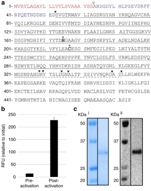
Identification and recombinant expression of
O. affinis
AEPs
.
Three expressed AEP isoforms were identified in an
O. affinis
complementary DNA library (
Oa
AEP1-3) and a fourth sequence,
with a single nucleotide change from
Oa
AEP1 (resulting in a
Glu
371Val
variant),
was
identified
from
genomic
DNA
(
Oa
AEP1
b) (Fig. 2a; Supplementary Fig. 1). The four isoforms
share at least 77% identity at the protein level, as determined by
pairwise protein alignments. When compared with butelase 1,
64–69% identity was observed, whereas identity with human
legumain was 49–53%.
Oa
AEP1
bwas expressed in
Escherichia coli
as a
His6-ubiquitin-AEP1
bfusion protein (Supplementary Fig. 2a). AEPs are usually
produced as zymogens that are self-processed at low pH to their
mature, active form
23–25. Consistent with this processing, activity
of r
Oa
AEP1
bagainst an internally quenched fluorescent (IQF)
peptide representing the native C-terminal processing site in kB1
(Table 1; wildtype (wt)) was markedly increased following
incubation at pH 4.5 (Fig. 2b). After purification, a dominant
band of
B
32 kDa was evident by reducing SDS–polyacrylamide
gel electrophoresis (PAGE) and confirmed to be r
Oa
AEP1
bby
Western blotting (Fig. 2c; Supplementary Fig. 2b). The average
total protein yield from two independent experiments was
B
1.8 mg l
1after activation and purification, however batch to
batch variation in purity was observed. Although glycosylation of
some AEPs has been reported
26, the production of an active form
in
E. coli
confirms that this is not a requirement for activity of
O. affinis
AEP1
b.
Mass spectrometry (MS)/MS sequencing of peptide fragments
generated from tryptic, chymotryptic or endoGlu-C digestion of the
activated enzyme identified several peptide fragments with
non-canonical cleavage sites, suggesting that they may be derived from
r
Oa
AEP1
bauto-processing events (Supplementary Fig. 2). This
allowed Asp52 to be assigned as the likely N-terminal
auto-processing site and Asp328, Asn329, Asp334, Asn336, Asp349 and/
or Asp351 as potential C-terminal processing sites (Fig. 2a;
Supplementary Fig. 2). No peptides downstream of Asp351 were
identified, indicating that the activation was essentially complete and
that the C-terminal domain (Leu352–Pro474) was removed during
the post-activation purification step. The theoretical mass of the
processed forms (30.4–32.8 kDa) is in good agreement with that
determined by SDS–PAGE/Western blotting (Fig. 2c).
Consistent with cysteine proteases of this class, r
Oa
AEP1
bwas
inhibited by iodoacetamide (1 mM), but was not affected by
E64 (250
m
M) or pepstatin A (10
m
M) (Supplementary Fig. 3).
Ac-YVAD-CHO (500
m
M), a caspase-1 inhibitor reported to also
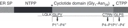
ER SP NTPP Cyclotide domain (Gly1-Asn29) CTPPCTRN GLPS
P4–P1P1′–P4′ LQLK GLPV
[image:2.595.60.275.604.651.2]P1′′–P4′′
Figure 1 | Schematic representation of theOak1gene.The precursor
protein encoded by theOak1 gene is proteolytically processed to mature
kB1..indicates the N- and C-terminal processing sites. rOaAEP1btargets
the C-terminal processing site. The C-terminal P1/P10- P4/P40sites are
indicated. P100-P400denote the N-terminal residues that replace the P10-P40
inhibit AEPs
27, was a poor inhibitor of the recombinant enzyme
suggesting that at least some P
0residues are important for active
site targeting.
Substrate specificity
. The activity of r
Oa
AEP1
bagainst IQF
peptides representing wt and mutant versions of the native kB1
C-terminal cleavage site was determined (Table 1; Supplementary
Fig. 4a). Along with the strict P1 Asx specificity characteristic of
AEPs
24, r
Oa
AEP1
bexhibited strong P2
0selectivity since after
Leu
31Ala substitution within the IQF peptide barely any
hydrolysis was observed. This observation is consistent with the
lack of cyclic product generated when the corresponding
mutation was introduced
in planta
21. Kinetic parameters (
V
max,
K
mand
k
cat) are reported where applicable (Table 1). The
turnover rates (
k
cat) reported here (
B
0.06–1.6 min
1) are much
slower than that reported for recombinant human legumain
assayed against a small substrate (
B
8 s
1) (ref. 28). This is not
unexpected given that r
Oa
AEP1
bprefers to carry out cyclization,
rather than the hydrolysis being measured here. Supporting the
observed P2
0selectivity, r
Oa
AEP1
bwas unable to cleave the
generic AEP substrate Z-AAN-MCA (Supplementary Fig. 5).
The substrate specificity of r
Oa
AEP1
bwas compared with that
of recombinant human legumain (rhuLEG; Supplementary
Fig. 4b)
29. A stringent P1 Asx requirement was again observed;
however, in contrast to r
Oa
AEP1
b, rhuLEG cleaved the Leu
31Ala
substrate at a rate similar to the wt substrate, demonstrating that
P2
0specificity is not a feature of all AEPs.
Cyclization of kB1 precursors
. To explore the cyclization ability
of r
Oa
AEP1
b, processing of correctly folded (as determined by
NMR) synthetic kB1 precursors was assessed by MS. When
incubated with the wt kB1 precursor carrying the native
C-terminal pro-hepta-peptide (GLPSLAA), the active enzyme
produced a peptide of 2,891.2 Da (monoisotopic, [M
þ
H]
þ),
consistent with the expected mass of mature, cyclic kB1 (Fig. 3a).
This product was confirmed to be identical to native kB1
by reversed phase-high performance liquid chromatography
(RP-HPLC) co-elution (Supplementary Fig. 6) and one- and
two-dimensional-NMR experiments (Supplementary Fig. 7).
Kinetic parameters (
±
s.e.m.) for the processing of the wt kB1
precursor were 0.53 (
±
0.1) s
-1for
k
cat
, 212 (
±
76)
m
M for
K
mand 2,500 M
1s
1for
k
cat/
K
mas determined from a
Michaelis–Menten plot (Supplementary Fig. 8). While the
turn-over rate (
k
cat) is lower than that reported for the plant-derived
250
200
150
100
50
0
RFU (relativ
e to initial)
Pre-activation
Post-activation
1-
41-
81-
121-
161-
201-
241-
281-
321-
361-
401-
441-KDa i KDaii
50 50
37 37
25 25
20 20
a
[image:3.595.44.285.52.357.2]b
c
Figure 2 | Expression of activerOaAEP1binE. coli.(a) Sequence of
OaAEP1bpredicted fromO. affinisgenomic DNA. Predicted ER signal
sequence shown in red; N-terminal propeptide shown in blue; the putative
signal peptidase cleavage site is indicated byrand autocatalytic
processing sites by.. The matureOaAEP1 cyclase domain is underlined
and the C-terminal auto-processing region is indicated with broken
underline. The putative catalytic dyad is shown in bold and labelled with*.
(b) An rOaAEP1-containing anion exchange fraction pre-and post-activation
at pH 4.5 (5 h, 37°C) was tested for activity against the wt IQF peptide
(14mM). Baseline fluorescence from a no substrate control has been
subtracted and the relative fluorescence intensity (RFU) att¼90 min is
reported. The average of two technical replicates is shown and error bars
report the range (c) Activated rOaAEP1bpurified by cation exchange was
analysed by SDS–PAGE and (i) Instant blue staining or (ii) Western blotting
with anti-OaAEP1b(residues D47–P474) polyclonal rabbit serum.
Table 1 | Kinetic parameters of IQF peptide cleavage by r
Oa
VPE1
b.
IQF peptide Sequence* Vmax(nmoles min-1mg-1protein) (±s.e.m.) w
Km(lM) (±s.e.m.) w
kcat(min-1) (±s.e.m.) w,z
wt Abz-STRNkGLPS-Y(3NO2) 51.3 (±5.8) 55.0 (±6.4) 1.6 (±0.2)
R28A Abz-STANkGLPS-Y(3NO2) 6.9 (±0.6) 13.0 (±2.4) 0.2 (±0.02)
R28K Abz-STKNkGLPS-Y(3NO2) 29.3 (±3.5) 42.0 (±4.0) 0.9 (±0.1)
N29A Abz-STRAkGLPS-Y(3NO2) NA
y
N29Q Abz-STRQkGLPS-Y(3NO2) NA
y
N29D Abz-STRDkGLPS-Y(3NO2) B2|| ND|| B0.06||
G30A Abz-STRNkALPS-Y(3NO2) 51.0 (±2.0) 29.0 (±1.4) 1.6 (±0.07)
G30S Abz-STRNkSLPS-Y(3NO2) 35.5 (±2.7) 31.4 (±2.5) 1.1 (±0.08)
L31A Abz-STRNkGAPS-Y(3NO2) NA
y
L31I Abz-STRNkGIPS-Y(3NO2) ND
z
NDz NDz
IQF, internally quenched fluorescent; NA, no activity; ND, not determined.
*IQF peptide residues are numbered according to their position within the native kB1 precursor, where the mature cyclotide incorporates Gly1-Asn29; native Cys26is substituted with Ser to avoid unpaired
Cys residues.
w
NZ3;±standard error of the mean (s.e.m.) zk
catis a conservative estimate assuming that the total concentration of active enzyme is equal to the total protein concentration in the enzyme preparation and an enzyme mass of 32 kDa.
y
No activity detected under the conditions tested (up to 80mM substrate; up to 6 h incubation). ||LowV
maxprecluded accurate estimation of kinetic parameters.
zK
[image:3.595.45.277.225.354.2] [image:3.595.48.547.547.675.2]butelase 1 (17.08 s
1; ref. 5), it is far higher than that of the
recombinantly expressed cyclase PatG (1 per day; ref. 7).
To determine if r
Oa
AEP1
bcould also carry out the N-terminal
processing required for cyclotide maturation, a kB1 precursor was
tested that contained the folded cyclotide domain flanked by four
residues from each of the N- and C-terminal propeptides
(Fig. 3b). No N-terminal processing was observed, indicating
that this processing is conducted by an enzyme other than
Oa
AEP1
b. Although the bulk of the precursor remained intact
after 20.5 h, the predominant processing product was a linear
peptide lacking the C-terminal propeptide, demonstrating that
correct N-terminal processing must occur before cyclization.
Interestingly, a mass corresponding to a cyclized version of the
C-terminally processed peptide (that is, C-terminal propeptide
residues released, N-terminal propeptide residues remaining) was
also observed, although this was the least abundant product.
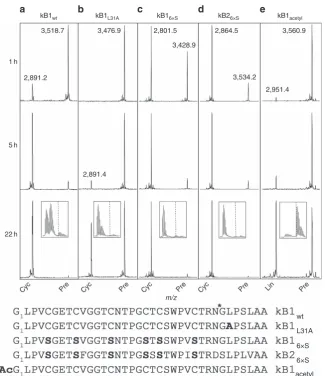
Processing of modified cyclotide precursors
. To further probe
cyclization
requirements,
we
tested
r
Oa
AEP1
b-mediated
processing of modified kB1 and kB2 precursors over time (Fig. 4).
When presented within an IQF peptide, the Leu
31Ala substrate
analogue was not hydrolysed by r
Oa
AEP1
b(Table 1). However,
the same substitution within the kB1 precursor did not preclude
cyclization by r
Oa
AEP1
b, although this version was cyclized far
more slowly than the wt precursor (Fig. 4a,b). Surprisingly, the
presence of disulfide bonds in the cyclotide precursors is not a
requirement for cyclization since a kB1 substrate analogue in
which all six cysteines were substituted with serines was also
efficiently cyclized (Fig. 4c). The absence of a defined kB1-like
structure in the kB1
6xSmutant was confirmed by NMR
spectroscopy (Supplementary Fig. 7c). Similarly, a kB2 linear
precursor with the same Cys
-
Ser substitutions was also
efficiently cyclized, confirming that enzyme activity is not specific
to individual cyclotides (Fig. 4d).
In cysteine protease-mediated peptide bond hydrolysis,
nucleophilic attack of a water molecule is required to resolve
the acyl-enzyme thioester intermediate. However, during peptide
cyclization (or transpeptidation) the substrate’s N-terminal amine
is postulated to function as a competing nucleophile, facilitating
aminolysis of the reactive thioester intermediate
30. Accordingly, a
kB1 precursor with an acetyl-capped N-terminal amine was
processed only to a linear peptide lacking the C-terminal
propeptide (Fig. 4e). This hydrolysis occurred at a slower rate
than cyclization of the wt precursor (compare with Fig. 4a).
Water can therefore access the active site of r
Oa
AEP1
b, but
cyclization is favoured over hydrolysis in the presence of an
appropriately positioned nucleophile.
Water is excluded during cyclization
. An alternative ligation
mechanism,
distinct
from
transpeptidation,
was
recently
proposed for huLEG
31). In that mechanism, initial hydrolysis of
the C-terminal propeptide is followed by a separate ligation event
requiring a C-terminal Asn residue in the substrate. To
distinguish
between
these
mechanisms
in
the
case
of
r
Oa
AEP1
b, reactions were carried out in the presence of
18O-labelled water and the products were analysed by high-resolution
MS. An isotopic shift consistent with the incorporation of
18O
was evident following enzymatic hydrolysis of the N-terminal
acetylated kB1 precursor to give a linear product (Fig. 5a).
However, there was no isotopic shift after processing of the wt
precursor to a cyclic product, suggesting that hydrolysis is
unlikely to play a role in cyclization by r
Oa
AEP1
b(Fig. 5a).
Cyc(2,891.2)
+6 Da
2,800 2,900 3,000 3,100 3,200 3,300 3,400 3,500 Pre
m/z
3,200 3,300 3,400 3,500 3,600 3,700 3,800 3,900 m/z Pre
(3,792.6)
Lin –99 Da
Lin (3438.6)
Cyc (3420.6)
a
[image:4.595.139.455.52.315.2]b
Figure 3 | Enzymatic processing products of linear kB1 precursors.(a) MALDI MS profile of a kB1 precursor (kB1wt) containing the C-terminal propeptide
in the presence of rOaAEP1b(22 h incubation). Theþ6 Da peak was observed only in the presence of reducing agent and corresponds to the reduced form
of cyclic kB1. (b) MALDI MS profile of a kB1 precursor (kB1C&N) containing four C-terminal propeptide residues and four N-terminal propeptide residues in
the presence of rOaAEP1b(20.5 h incubation). A side product originating from chemical synthesis likely represents a Val deletion (99 Da). Data are
representative of at least two technical replicates. X, benzoylphenylalanine.*denotes rOaAEP1bcleavage site. Observed monoisotopic masses (Da;
rOaAEP1
bcan cyclise an unrelated peptide
. We also
investigated
cyclization
of
other
substrates
structurally
unrelated to cyclotides by r
Oa
AEP1
b, focussing on the
anti-malarial peptide R1 (refs 32,33). This peptide was efficiently
cyclized following the addition of N- and C-terminal AEP
recognition sequences (Fig. 6a). Sequential trimming of the
added recognition residues revealed that cyclization could be
achieved following the addition of only a C-terminal Asn–Gly–
Leu motif (although some linear product was also produced
from this precursor) (Figs 6a–d). Lys and Gln were also
accepted in place of Gly at the N terminus (Figs 6e–f) with little
impact on yield at the time point tested. No processing of
either the native R1 peptide or a modified R1 carrying the
N-terminal Gly–Leu motif with only an Asn at the C terminus
was observed (Supplementary Fig. 9). Subsequent digestion
with endoGlu-C confirmed that, in all cases, r
Oa
AEP1
bprocessing produced cyclic peptide (Supplementary Fig. 10).
Evidence
of
an
additional,
linear,
r
Oa
AEP1
b-generated
cleavage product was only observed for the R1 variant without
any N-terminal flanking residues (Fig. 6d; Supplementary
Fig. 10c).
kB1wt kB1L31A kB16×S kB26×S kB1acetyl
1 h
2,891.2
3,476.9 2,801.5
3,428.9
2,864.5 3,560.9
2,951.4 3,534.2
5 h
2,891.4
Cyc Pre Cyc Pre Cyc Pre Cyc Pre Lin Pre
wt
L31A
6×S
6×S
acetyl 22 h
m/z
a
b
c
d
e
[image:5.595.137.463.47.424.2]3,518.7
Figure 4 | Modified linear kB1 and kB2 precursors are cyclized at different rates.MALDI MS spectra of (a) kB1wt, (b) kB1L31A, (c) kB16xS, (d) kB26xSand
(e) kB1acetylcyclotide precursors at 1, 5 and 22 h post-enzyme addition. Data are representative of three technical replicates.*denotes rOaAEP1bcleavage
site. Observed monoisotopic masses (Da; [MþH]þ) for dominant peaks are listed. Boxed inset at the 22 h time point zooms in on the region containing
the processing product. Approximate positions of the monoisotopic mass of processed products is indicated byy. Cyc, cyclic product; Lin, linear product;
Pre, linear precursor.
Hydrolysed product
2,950.2 2,955.2
m/z + H218O – H2
18 O
m/z 2,890.1
Cyclic product
2,895.1
[image:5.595.62.278.525.656.2]a
b
Figure 5 | Enzymatic cyclization excludes water.MALDI MS profile of the
enzymatic processing products of (a) kB1acetyland (b) kB1wtlinear
precursors in the presence and absence of18O-labelled water. An isotope
shift indicative of18O incorporation only occurs during hydrolysis. Observed
masses of two isotopic peaks (Da; [MþH]þ) are indicated. Data are
Discussion
This study reports the cloning of four AEPs from the
cyclotide-producing plant
O. affinis;
one of which was recombinantly
expressed. The recombinant enzyme required self-processing to
produce the active product: a cyclase that preferentially and
efficiently couples C-terminal processing with C- and N-terminal
ligation of linear
O. affinis
cyclotide precursors. Furthermore, this
cyclizing ability was highly efficient when transferred to an
unrelated anti-malarial peptide, demonstrating broad
applicabil-ity in peptide engineering.
Consistent with other auto-inhibited proteases, r
Oa
AEP1
brequired proteolytic activation to achieve maximum activity
(Fig. 2b). The observed N-terminal auto-processing site (Asp52)
is consistent with other experimentally validated N-terminal
auto-processing sites identified in jack bean AEP
34, butelase 1
(ref. 5) and human legumain
28,35(Fig. 2a, Supplementary Fig. 1).
In contrast, six potential C-terminal auto-processing sites
(Asp328/334/349/351, Asn329/336) were observed within a
region particularly rich in Asn/Asp residues (324–351). This
finding is in agreement with the multiple C-terminal maturation
steps recently described for rhuLEG
28,35. Regardless of which of
these sites is relevant
in planta
, the instability of active AEPs
above pH 6 (refs 28,29,36) will likely preclude direct production
of active enzyme in
E. coli
. Activated r
Oa
AEP1
bproteolytically
removed the C-terminal (but not N-terminal) propeptide of a
kB1 precursor and resolved the acyl-intermediate in a
hydrolysis-independent manner, generating a backbone cyclized
product (Figs 3 and 5). r
Oa
AEP1
bcould also hydrolyse
precursors lacking a free N-terminal amine to produce linear
products, albeit at a slower rate. Although it is unknown if
Cyc (3,075.1)–16 Da
Pre (3,360.4)
3,000 3,100 3,200 3,300 3,400
2,500 2,600 2,700 2,800 2,900
2,500 2,600 2,700 2,800 2,900 Cyc (2,633.7)
+22 Da
Pre (2,821.9)
Pre (2,708.8)
+22 Da
2,500 2,600 2,700 2,800 2,400
Cyc (2,463.5)
Lin (2,481.5)
Pre (2,651.7)
+22 Da
2,600 2,700 2,800 2,900 3,000 Cyc (2,704.8)
Pre (2,893.0)
2,600 2,700 2,800 2,900 3,000 Cyc (2,704.8)
+22 Da
Pre (2,893.0)
m/z
a
b
c
d
[image:6.595.131.464.48.503.2]e
f
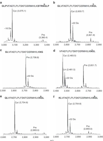
Figure 6 | Flanking sequence requirements for cyclization of a model peptide by rOaAEP1.(a–f). MALDI MS spectra of the R1 peptide
(VFAEFLPLFSKFGSRMHILK) with various flanking sequences 22 h post addition of rOaAEP1b. Bold residues, flanking sequences.*denotes rOaAEP1b
cleavage site. Observed monoiosotopic masses (Da; [MþH]þ) are listed.þ22 Da and16 Da peaks present in some precursor and product spectra are
likely to represent Naþadducts and a synthesis-derived modification respectively. Data are representative of three technical replicates. Cyc, cyclic product;
Oa
AEP1
bperforms both these cyclase and protease activities
in vivo,
the observed preference for cyclization over hydrolysis
suggests that it probably functions predominantly as a cyclase.
Dual protease/ligase capabilities have been reported for PatG
37,
a serine protease involved in cyclic peptide production in
cyanobacteria, and more recently for human legumain
31.
Separate mechanistic pathways for hydrolysis and ligation have
been proposed for human legumain: proteolysis proceeds via
hydrolysis of a cysteinyl-thioester enzyme intermediate, whereas
peptide ligation occurs via activation of the free peptidyl-
a
-carboxylate through transient formation of an enzyme-linked
anhydride intermediate that is subsequently resolved via
aminolysis
31.
The
catalytically
critical
residue
for
this
mechanism of ligation, Asp188, is conserved in
Oa
AEP1
b, but
several lines of evidence preclude a role for this pathway in
Oa
AEP1
b-mediated peptide cyclization. First, r
Oa
AEP1
bcannot
cyclize peptides carrying a free C-terminal Asn, the minimal
proposed substrate requirement in the alternative pathway
(Supplementary Fig. 9). Second, our H
218O experiments
demonstrate the absence of
18O incorporation into the cyclized
product, which strongly indicates that cyclization does not follow
a hydrolysis/ligation mechanism as proposed in the alternative
pathway (Fig. 5). Third, our MS/MS data for r
Oa
AEP1
bshow no
evidence of a reactive succinimide enzyme intermediate required
for formation of the substrate–enzyme anhydrides
31. Hence, our
mechanistic data are in agreement with the traditional concerted
mechanism, in which some of the energy from the (exergonic)
cleavage of the C-terminal Asn-propeptide bond is preserved in
the form of a thioester intermediate and used to overcome the
energetically unfavourable (endergonic) peptide bond formation
(cyclization) in the second step. However, alternative mechanisms
may still play a role in cyclization mediated by other AEPs or with
alternative substrates.
In the context of this established mechanism, C- and
N-terminal proximity was thought to be crucial for cyclization
to
be
favoured
over
hydrolysis
8.
Here
we
show
that
pre-organization of C- and N-termini in the substrate is not
required by r
Oa
AEP1
bsince unconstrained cyclotide precursors
lacking the characteristic disulfide-bonded structure are efficiently
cyclized (Fig. 4). Furthermore, r
Oa
AEP1
bcan cyclize an
anti-malarial peptide that is structurally and functionally
unrelated to cyclotides following the addition of short flanking
sequences (Fig. 6). These findings are consistent with the limited
structural and/or sequence requirements imposed by other native
cyclases on their substrates
4,5,7Conceivably, polypeptides of
diverse composition and length may be cyclized by r
Oa
AEP1
b,
provided that association of the C- and N-termini is not sterically
hindered.
The application of this technology is limited to peptides that
can retain activity following incorporation of the additional
residues required for AEP-mediated processing. While r
Oa
AEP1
bcan cyclize a model peptide with only a single non-native residue
incorporated to the mature peptide, this is at the cost of
cyclization efficiency (Fig. 6d). Understanding the interplay
between the sequence requirements for efficient cyclization and
retention of bioactivity for a given target peptide will be crucial to
realizing the potential of AEP-mediated cyclization. Importantly,
the estimated turnover rate of r
Oa
AEP1
b(
k
cat, 0.53 s
1;
Supplementary Fig. 8) is multiple orders of magnitude higher
than the recombinantly produced cyclase PatG (1 d
1; ref. 7),
supporting its widespread application in peptide engineering.
In a previous study, a conserved tripeptide motif C terminal to
both the N- and C-terminal cyclotide processing sites was
identified through cyclotide sequence analysis
8. This motif is
Gly–Leu–Pro in the kB1 sequence, and its importance for efficient
cyclization is supported by mutagenesis studies in transgenic
plants
8,21. The protease specificity of r
Oa
AEP1
breported here
against IQF peptides mirrors these requirements (Table 1). At the
C-terminal processing site, the P1 Asn and P2
0Leu are
particularly well-conserved, and both were crucial for both
in
planta
cyclization and
in vitro
cleavage of model peptides.
However, over the longer incubation period of the cyclization
assays, a kB1 precursor with a Leu
31Ala mutation was still
enzymatically processed to a cyclic product (Fig. 4). It was
initially proposed that the conserved Leu residue at the P2
0position of cyclotides was important for preventing water from
accessing the active site during cyclization. However the observed
cyclization of the kB1 Leu
31Ala mutant suggests that the role of a
conserved bulky hydrophobic residue at the P2
0position is only to
promote appropriate enzyme–substrate interaction. Congruent
with this hypothesis, the absence of a P2
0residue renders
substrates poor targets of both r
Oa
AEP1
b(cyclization and
hydrolysis; Fig. 6c; Supplementary Fig. 5) and butelase 1
(at least for hydrolysis)
5. In summary, our results suggest a
cyclization model in which the cleaved C-terminal propeptide
retains sufficient affinity to remain bound to the active site until it
is displaced by the incoming N terminus of the peptide, finally
leading to cyclization by resolving the acyl intermediate
8,38.
This P2
0requirement is not characteristic of all AEPs
9,34,39and
might be a predictor of cyclization ability within this protease
family. Indeed, in extracts from
C. ternatea
, protein fractions that
were active against the generic AEP substrate Z-AAN-MCA
(which does not contain a P2
0residue) did not contain the
cyclizing enzyme and, conversely, the butelase 1 containing
fraction did not display activity against this substrate
5. Here we
report the presence of four unique AEP sequences in
O. affinis
(Fig. 2a; Supplementary Fig. 1), and demonstrate that one
(
Oa
AEP1
b) is capable of cyclizing
O. affinis
cyclotide precursors.
Further work will investigate whether all, or a subset of,
O. affinis
AEPs (at least two more of which should exist to explain all the
AEP contig sequences observed) exhibit this function and
whether their substrate specificity is an accurate predictor of
cyclization ability.
The constraints on the sequence of the incoming N terminus
may not be very stringent. At least two residues with different
properties are accepted in place of Gly at the P1
00position by
r
Oa
AEP1
bwith comparable yields of cyclic product under the
conditions tested (Fig. 6e,f). Butelase 1 and Pat G also exhibit
promiscuity in this region
5,37. Interestingly, Gly
1is highly
conserved across cyclotides from different plant species, raising
the possibility that selection at this position is not driven by AEP
cyclase specificity. In transgenic plants, more stringent requisites
were observed and no cyclic product was made from kB1
precursors with a conservative Gly
1Ala mutation
8. Because AEP
cannot liberate the N-terminal propeptide, we hypothesize that
selection at Gly
1might be driven by the putative N-terminal
processing enzyme. Further analysis will be necessary to
determine if this reflects differences in the enzyme homologues
being assayed (that is, AEPs from the model plant
Nicotiana
benthamiana
compared with AEPs from native cyclotide
producers) or experimental conditions.
In conclusion, this study unequivocally demonstrates the
involvement of an AEP in maturation of native
O. affinis
cyclotides, advancing our understanding of the biosynthesis of
this important class of cyclic peptides. Furthermore, the
promiscuous yet highly efficient nature of this recombinantly
produced enzyme highlights its exciting potential value as a
biological tool for cyclization of a range of bioactive peptides.
Methods
Peptide substrates and inhibitors
.
IQF peptides containing an N-terminalwere synthesized by Genscript at490% purity. Control IQF peptides representing the predicted cleavage products of the wt peptide (Abz-STRN; GLPS-Y(3NO2)
were also synthesized by Genscript at490% purity. All IQF peptides were solu-bilized in 25% (v/v) acetonitrile:water. The fluorogenic peptide substrate Z-AAN-MCA (where Z is carboxybenzyl; Z-AAN-MCA is 7-amido-4-methylcoumarin) and the caspase inhibitor Ac-YVAD-CHO (where Ac, acetyl; CHO, aldehyde) were sup-plied by the Peptide Institute and solubilized in dimethyl sulfoxide. The linear cyclotide precursor peptides kB1wt, kB1C&N, kB1L31Aand kB1acetylwere chemically
synthesized in-house by standard Fmoc solid-phase peptide synthesis. Folding and disulfide formation was carried out by incubating the reduced peptides in folding buffer (100 mM ammonium bicarbonate, 50% isopropanol, 2 mM reduced glutathione, 1 mM oxidized glutathione, pH 8.2) for 3 days40. The products were isolated by RP-HPLC at495% purity and characterized by high-resolution MS and NMR spectroscopy. Peptides kB16xSand kB26xSas well as R1 and its
derivatives were supplied by Genscript at485% purity, as determined by RP-HPLC and MS. Peptides were dissolved in ultrapure water before analysis.
O. affinistranscriptome
.
Total RNA was extracted fromO. affinisroot, leaf and seedling tissues using a phenol extraction method. Plant material was frozen in liquid nitrogen and ground to a fine powder, which was then resuspended in buffer (0.1 M Tris-HCl pH 8.0, 5 mM EDTA, 0.1 M NaCl, 0.5% SDS, 1% 2-mercap-toethanol), extracted twice with 1:1 phenol:chloroform and precipitated by addition of isopropanol. The pellets were dissolved in 0.5 ml water and RNA was pre-cipitated overnight at 4°C by addition of 4 M lithium chloride. The extracted RNA of each tissue was analysed by GeneWorks using the Illumina GAIIx platform. In total, 69.3 million 75 bp paired-end reads were generated. Reads were filtered with a phred confidence value of Q37 and assembled into contigs using Oases41with k-mer ranging from 41–67. The assemblies were merged using cd-hit-est42,resulting in 270,000 contigs. Statistics on the depth of sequencing were made by aligning the reads of each tissue on the contigs using BWA43. All the sequences, including one AEP, previously identified from an EST library ofO. affiniswere present among the contigs44. Homologues of this AEP sequence were searched using BLAST45in the contig library using a maximum E-value of 1e-20, resulting in the identification of 371 putative AEP transcripts. These sequences could then be clustered in 13 groups sharing at least 90% sequence identity using cd-hit42.
OaAEP1-3 cloning
.
Full-length AEP transcripts from theO. affinistranscriptome assembly were used to design a set of primers. A single degenerate forward primer (OaAEPdegen-F, 50-ATG GTT CGA TAT CYC GCC GG-30) was sufficient to amplify all sequences since variability within the extreme 50region of each full-length transcript was limited to a single nucleotide position. Three reverse primers (OaAEP1-R, 50-TCA TGA ACT AAA TCC TCC ATG GAA AGA GC-30; OaAEP2-R, 50-TTA TGC ACT GAA TCC TTT ATG GAG GG-30;OaAEP3-R 50-TTA TGC ACT GAA TCC TCC ATC G-30) were designed with the aid of Primer 3 (ref. 46). Each primer set successfully amplified an AEP sequence.To clone expressedOaAEPs, total RNA was extracted fromO. affinisleaves and shoots using TRIzol (Life Technologies) and was reverse transcribed with SuperScript III reverse transcriptase (Life Technologies) according to the manufacturer’s instructions11. Target sequences were amplified from the resulting complementary DNA using Phusion High Fidelity Polymerase (New England BioLabs) and the primers described above under the recommended reaction conditions. Gel extracted PCR products were dA-tailed by incubation with Invitrogen Taq Polymerase (Life Technologies) and 0.5ml 10 mM dA in the supplied buffer. The processed products were cloned into pCR8-TOPO (Life Technologies) and transformed intoE. coli. Purified DNA from clones that were PCR positive for an AEP insert were sent for Sanger sequencing at the Australian Genome Research Facility (www.agrf.org). Coding sequences have been deposited in Genbank (accession codes:OaAEP1 (KR259377),OaAEP2 (KR259378),
OaAEP3 (KR259379)).
In an alternative approach, genomic DNA was extracted fromO. affinisleaf tissue using a DNeasy Plant Mini Kit according to the manufacturer’s instructions. PCR amplification from this DNA used primers specifically targeting theOaAEP1 nucleotide sequence. Gel extracted product was dA-tailed as above, cloned into the TOPO vector and transformed intoE. coli.Sequencing of PCR-positive clones identified a fourth sequence with a single amino acid change fromOaAEP1 (OaAEP1b).
Antibodies
.
Polyclonal anti-OaAEP1brabbit serum was generated by immunizinga New Zealand White rabbit with a denatured, inactive form ofOaAEP1b(residues
D47–P474) that was produced recombinantly inE. coli. The rabbit received three
doses, four weeks apart, of 150mg of antigen in 50% (v/v) PBS and Freund’s incomplete adjuvant. Serum was obtained 2 weeks after the final dose and used at a 1:2,000 dilution for Western blotting.
Recombinant expression ofO. affinisAEP1b(rOaAEP1b)
.
Initial trials toproduce activeO. affinisAEP1bbased on predicted N- and C-terminal processing
sites (residues D47–D420) were unsuccessful and subsequent expression attempts
incorporated both N- and C-terminal prodomains. DNA encoding full-length
O. affinisAEP1bwithout the putative signalling domain (residues A24-P474) was
inserted into the pHUE vector47to give a His6-ubiquitin-OaAEP1bfusion protein
construct (Supplementary Fig. 2) and introduced into T7 shuffleE. colicells (New England BioLabs). Transformed cells were grown at 30°C in superbroth (3.5% tryptone (w/v), 2% yeast extract (w/v), 1% glucose (w/v), 90 mM NaCl, 5 mM NaOH) to mid-log phase; the temperature was then reduced to 16°C and expression was induced with isopropyl–D-1-thiogalactopyranoside (0.4 mM) forB20 h. Cells were harvested by centrifugation and resuspended in non-denaturing lysis buffer (50 mM Tris-HCl, 150 mM NaCl, 0.1% triton X 100, 1 mM EDTA, pH 7). Lysis was promoted by a total of five freeze/thaw cycles and the addition of lysozyme (hen egg white; 0.4 mg ml1). DNase (bovine pancreas; 40mg ml1) and MgCl2(0.4 M) were also added. Cellular debris was removed by
centrifugation and the lysate was stored at 80°C until required.
Purification and activation of rOaAEP1b
.
Lysate containing expressed rOaAEP1bwas filtered through a 0.1mm glass fibre filter before being diluted 1:8 in buffer A (20 mM bis-Tris, 0.2 M NaCl, pH 7) and loaded onto two 5 ml HiTrap Q Sepharose high performance columns connected in series (GE Healthcare; 1.6–3.1 ml undiluted lysate per millilitre resin). Bound proteins were eluted with a continuous salt gradient (0–30% buffer B (20 mM bis-Tris, 2 M NaCl, pH 7); 15 column volumes) and AEP-positive fractions identified by Western blotting (anti-OaAEP1b
rabbit serum (1:2,000); peroxidase-conjugated anti-rabbit IgG (GE Healthcare NA934; 1:5000)). To self-activate rOaAEP1b, EDTA (1 mM) and
Tris(2-carbox-yethyl)phosphine hydrochloride (0.5 mM) were added, the pH was adjusted to 4.5 with glacial acetic acid and the protein pool was incubated for 5 h at 37°C. Protein precipitation at this pH allowed removal of the bulk of the contaminating proteins by centrifugation. The remaining protein was filtered (0.22mm), diluted 1:8 in buffer A2 (50 mM acetate, pH 4) then captured on a 1 ml HiTrap SP Sepharose high performance column (GE Healthcare). Bound proteins were eluted with a salt gradient (0–100% buffer B2 (50 mM acetate, 1 M NaCl, pH 4); 10 column volumes) and AEP-positive fractions were pooled. The final product was analysed by MS and reducing SDS–PAGE followed by Western blotting and staining with Instant blue (Expedeon). The total concentration of protein was estimated by bicinchoninic acid assay according to the manufacturer’s instructions.
Identification of the auto-processing sites of rOaAEP1b
.
Aliquots of rOaAEP1b(5ml) were diluted 1:1 with either 100 mM ammonium bicarbonate pH 8.0 (trypsin, chymotrypsin) or 100 mM ammonium phosphate pH 8.0 (endoGlu-C) and enzymatically digested with endoGlu-C, trypsin or chymotrypsin (100 ngml1). Cleavages were conducted over 16 h at 37°C (endoGlu-C, trypsin) or 30°C (chymotrypsin). Injections of each digest (5ml) were introduced to a Shimadzu nanoLC delivering a linear acetonitrile gradient at a flow rate of 500 nl min1for
reversed-phase separation on a C18 Zorbax column (Agilent 300SB-C18, 3.5mm particle size, 150 mm100mm). Column eluate was interfaced directly with a 5600 TripleTOF LC-MS/MS instrument (AB SCIEX, Canada) equipped with a nanoelectrospray ionization source.
Tandem MS data were generated in Information Dependent Acquisition experiments, wherein full-scan TOF-MS spectra were acquired for 250 ms overm/z
350–1,800, and the 20 most intense signals with charge state þ2 to þ5 were selected for product ion scans of duration 50 ms overm/z80–1,400. Data were acquired and processed using the Analyst TF 1.6 software (AB SCIEX, Canada). Automated protein identification was performed in ProteinPilot 4.0 using the Paragon algorithm with ‘no enzyme’ settings to interrogate anE. coliK12 proteome database (Uniprot) concatenated with sequences of rOaAEP1band those of the
three cleavage enzymes, with false discovery rate set atPo0.05. Spectral data for peptides software matched to rOaAEP1bwere confirmed via manual interpretation
in Analyst TF 1.6.
Assaying protease activity against fluorescent peptides
.
To assay activity of rOaAEP1bagainst both internally quenched and other fluorescent peptides,substrate and enzyme were diluted as appropriate in activity buffer (50 mM sodium acetate, 50 mM NaCl, 1 mM EDTA, 0.5mM Tris(2-carboxyethyl)phosphine hydrochloride, pH 5). To assay activity of rhuLEG (R&D systems) against the same substrates, the enzyme was first activated by incubation in 50 mM sodium acetate, 100 mM NaCl, pH 4 (4ml buffer/1ml enzyme) for 2 h at 37°C. Substrates and activated rhuLEG were diluted in 50 mM MES, 250 mM NaCl, pH 5 as required. Diluted enzyme and substrate were added to black, flat bottomed microtiter plates in a total assay volume of 110–200ml. The change in fluorescence intensity over time was monitored on a SpectraMax M2 (Molecular Devices) using excitation/ emission wavelengths of 320/420 nm (IQF peptides) or 360/460 nm (other fluorescent peptides). Substrate and enzyme concentrations in each assay and the time point presented are as indicated in the figure legends.
To determine the kinetics of rOaAEP1bactivity against IQF peptides, each
the assumption that the total protein concentration reflected active enzyme. At each substrate concentration, initial velocities were calculated from the linear portion of the progress curve.KmandVmaxwere estimated using the Michaelis–
Menten equation and the curve-fitting program GraphPad Prism (GraphPad Software, San Diego).
The high peptide concentrations required for estimating kinetic parameters necessitated the use of a correction factor to account for the inner filter effect; a phenomenon where high relative concentrations of the quenching group impede detection of the signal from the fluorescent donor even after substrate hydrolysis48. This was achieved as described previously48. The output generated by the
fluorescent hydrolysis product (Abz-STRN) was measured in the presence of each concentration of non-hydrolysed substrate. The correction factor was the ratio between the expected and observed fluorescence signal at each substrate concentration. The corrected signal for each data point was then converted to amount of product by comparison to a standard curve of the fluorescent hydrolysis product.
Inhibition assays
.
To investigate the impact of inhibitors on enzyme activity against the wt IQF peptide, rOaAEP1b(4.4mg ml–1total protein) was incubatedwith the indicated concentration of E64, Ac-YVAD-CHO, pepstatin A and iodoacetamide for 40 min before addition to the substrate (11mM). Enzyme activity against the wt IQF peptide was then assessed as described above.
Cyclization assay
.
Linear target peptides (280mM) were incubated with rOaAEP1b(12mg ml1total protein unless otherwise indicated) in activity buffer.The reaction was allowed to proceed for up to 22 h at room temperature and was analysed by matrix-assisted laser desorption/ionization MS (MALDI MS), RP-HPLC or NMR as appropriate.
To confirm the presence of cyclic product, R1 derivatives processed by rOaAEP1bwere subsequently digested with endoGlu-C (25mg ml1) in reaction
buffer (50 mM Tris-HCl, 0.5 mM Glu–Glu, pH 8) such that the final dilution of the cyclization mix was 1:4. The reaction was allowed to proceed for 18 h at 37°C before analysis by MALDI MS.
In heavy water experiments, isotopically labelled water (97 atom %18O) was
used in place of unlabelled water. Linear target peptides (70mM) were incubated with rOaAEP1b(6mg ml1total protein) in a non-reducing activity buffer (50 mM
sodium acetate, 50 mM NaCl, 1 mM EDTA, pH 5) for 22 h at room temperature. The final H218O concentration in the assay was 81%.
MS to track cyclization of linear peptides
.
Cyclization of linear target peptides was monitored by MALDI MS. The reaction mixture (10–20ml) was desalted using C18 zip tips and eluted in 4ml 50% acetonitrile, 0.1% trifluoroacetic acid (TFA). A saturated MALDI matrix solution (a–cyano-4-hyroxycinnamic acid) prepared in 95% acetonitrile, 0.1% TFA was diluted 1:22 such that the final matrix solution comprised 90% acetonitrile, 0.1% TFA and 1 mM NH4H2PO4. Eluted samples weremixed 1:4 with the MALDI matrix, spotted onto a MALDI plate and analysed by an Ultraflex III TOF/TOF (Bruker) in positive reflector mode.
Purification of kB1 following in vitro cyclization
.
The crude cyclization mixture was loaded to an Agilent Zorbax C18 reversed-phase column (4.6250 mm, 300 Å) and separated on a Shimadzu Prominence system using a linear gradient of 5–55% buffer B (90% acetonitrile, 10% H2O, 0.05% TFA) in buffer A (0.05%TFA/H2O) over 60 min. Fractions were collected manually, analysed by MALDI
MS essentially as described above and lyophilized. Analytical HPLC and co-elution studies with chemically synthesized kB1 were carried out as described above.
Nuclear magnetic resonance spectroscopy
.
All peptides were dissolved in 90% H2O/10% D2O at a concentration ofB2.0 mg ml1(0.5–0.75 mM). kB1 obtainedfromin vitrocyclizations was dissolved at 0.3 mg ml1(B0.1 mM). Spectra were
recorded on a Bruker Avance 600 MHz spectrometer equipped with a cryoprobe at 298 K. Phase-sensitive mode using time-proportional phase incrementation for quadrature detection in thet1dimension was used for all two-dimensional spectra.
Excitation sculpting with gradients was used to achieve water suppression. NMR experiments included TOCSY using a MLEV-17 spin lock sequence with an 80 ms mixing time, and NOESY with a 200 ms mixing time. Spectra were recorded with 4,096 data points in theF2dimension and 512 increments in theF1dimension. The
t1dimension was zero-filled to 1,024 real data points, and theF1andF2dimensions
were multiplied by a sine-squared function before Fourier transformation. The spectra were referenced to the water signal at 4.77 p.p.m. at 298 K. All spectra were processed using TopSpin (Bruker) and manually assigned with CCPNMR using the sequential assignment protocol.
Cyclization kinetics
.
To determine the kinetics of rOaAEP1bactivity against thewt kB1 precursor, the substrate was assayed at room temperature at a range of concentrations between 75 and 250mM in a total volume of 20–160ml of activity buffer. The total protein concentration of the enzyme preparation added to the kinetic assays was 19.7mg ml1. The reaction was quenched after 5 min with 0.1%
TFA and the volume adjusted to 800ml. A volume of 700ml was loaded onto a
reversed-phase C18 analytical column (Agilent Eclipse C18, 5mm, 4.6150 mm) and peptides were separated by HPLC (19 min linear gradient of 12–60% acet-onitrile, 0.1% TFA at 1 ml min1). The identity of eluted peaks was confirmed
using MALDI MS. The area under the curve corresponding to the precursor peptide was quantitated by comparison to a standard curve and initial velocities were calculated by converting this tommoles product formed. Kinetic parameters were estimated using the Michaelis–Menten equation and the curve-fitting pro-gram GraphPad Prism (GraphPad Software, San Diego). As for the IQF peptides, it was not possible to precisely determine the concentration of active enzyme due to impurities remaining in the preparation and the absence of an inhibitor appro-priate for active site titration. However, a conservative turnover rate (kcat) was
estimated based on a mass of 32 kDa and the assumption that the total protein concentration reflected active enzyme. Differences in enzyme preparations means these parameters are not directly comparable to those determined for the IQF peptides.
References
1. Sheldon, P. S., Keen, J. N. & Bowles, D. J. Post-translational peptide bond formation during conconavalin A processing in vitro.Biochem. J.320,865–870 (1996).
2. Hanada, K., Yewdell, J. W. & Yang, J. C. Immune recognition of a human renal cancer antigen through post-translational protein splicing.Nature427,1–5 (2004).
3. Mazmanian, S. K., Liu, G., Ton-That, H. & Schneewind, O. Staphylococcus aureus Sortase, an enzyme that anchors surface proteins to the cell wall.Science
285,760–763 (1999).
4. Barber, C. J. S.et al.The two-step biosynthesis of cyclic peptides from linear precursors in a member of the plant family Caryophyllaceae involves cyclization by a serine protease-like enzyme.J. Biol. Chem.288,12500–12510 (2013).
5. Nguyen, G. K. T.et al.Butelase 1 is an Asx-specific ligase enabling peptide macrocyclization and synthesis.Nat. Chem. Biol.10,732–738 (2014). 6. Luo, H.et al.Peptide macrocyclization catalyzed by a prolyl oligopeptidase
involved ina-amanitin biosynthesis.Chem. Biol.21,1610–1617 (2014). 7. Lee, J., Mcintosh, J., Hathaway, B. J. & Schmidt, E. W. Using marine natural
products to discover a protease that catalyzes peptide macrocyclization of diverse substrates.J. Am. Chem. Soc.131,2122–2124 (2009).
8. Gillon, A. D.et al.Biosynthesis of circular proteins in plants.Plant J.53,
505–515 (2008).
9. Saska, I.et al.An asparaginyl endopeptidase mediates in vivo protein backbone cyclization.J. Biol. Chem.282,29721–29728 (2007).
10. Bernath-Levin, K.et al.Peptide macrocyclization by a bifunctional endoprotease.Chem. Biol.22,571–582 (2015).
11. Jennings, C., West, J., Waine, C., Craik, D. & Anderson, M. Biosynthesis and insecticidal properties of plant cyclotides: the cyclic knotted proteins from
Oldenlandia affinis.Proc. Natl Acad. Sci. USA98,10614–10619 (2001). 12. Plan, M. R., Saska, I., Cagauan, A. G. & Craik, D. J. Backbone cyclised peptides from plants show molluscicidal activity against the rice pest Pomacea canaliculata (golden apple snail ).J. Agric. Food Chem.56,5237–5241 (2008).
13. Colgrave, M. L.et al.Cyclotides: natural, circular plant peptides that possess significant activity against gastrointestinal nematode parasites of sheep.
Biochemistry47,5581–5589 (2008).
14. Colgrave, M. L.et al.Anthelmintic activity of cyclotides: In vitro studies with canine and human hookworms.Acta Trop.109,163–166 (2009).
15. Poth, A. G., Chan, L. Y. & Craik, D. J. Cyclotides as grafting frameworks for protein engineering and drug design applications.Biopolymers100,480–491 (2013).
16. Clark, R. J.et al.Engineering stable peptide toxins by means of backbone cyclization: stabilization of the alpha-conotoxin MII.Proc. Natl Acad. Sci. USA
102,2–7 (2005).
17. Clark, R. J.et al.The engineering of an orally active conotoxin for the treatment of neuropathic pain.Angew. Chem. Int. Ed. Engl.49,6545–6548 (2010). 18. Chan, L. Y.et al.Cyclization of the antimicrobial peptide gomesin with native
chemical ligation: influences on stability and bioactivity.Chembiochem14,
617–624 (2013).
19. Craik, D. J. Host-defense activities of cyclotides.Toxins (Basel)4,139–156 (2012).
20. Stanger, K.et al.Backbone cyclization of a recombinant cystine-knot peptide by engineered Sortase A.FEBS Lett.588,4487–4496 (2014).
21. Conlan, B. F.et al.Insights into processing and cyclization events
associated with biosynthesis of the cyclic Peptide kalata B1.J. Biol. Chem.287,
28037–28046 (2012).
22. Mylne, J. S.et al.Albumins and their processing machinery are hijacked for cyclic peptides in sunflower.Nat. Chem. Biol.7,257–259 (2011).
23. Hiraiwa, N., Nishimura, M. & Hara-Nishimura, I. Expression and activation of the vacuolar processing enzyme in Saccharomyces cerevisiae.Plant J.12,
24. Hiraiwa, N., Nishimura, M. & Hara-Nishimura, I. Vacuolar processing enzyme is self-catalytically activated by sequential removal of the C-terminal and N-terminal propeptides.FEBS Lett.447,213–216 (1999).
25. Kuroyanagi, M.et al.Activation of Arabidopsis vacuolar processing enzyme by self-catalytic removal of an auto-inhibitory domain of the C-terminal propeptide.Plant Cell Physiol.43,143–151 (2002).
26. Kembhavi, A. A., Buttle, D. J., Knight, G. & Barrett, A. J. The two cysteine endopeptidases of legume seeds: purification and characterization by use of specific flurometric assays.Arch. Biochem. Biophys.303,208–213 (1993). 27. Hatsugai, N.et al.A plant vacuolar protease, VPE, mediates virus-induced
hypersensitive cell death.Science305,855–858 (2004).
28. Dall, E. & Brandstetter, H. Mechanistic and structural studies on legumain explain its zymogenicity, distinct activation pathways, and regulation.Proc. Natl Acad. Sci. USA110,10940–10945 (2013).
29. Chen, J.et al.Cloning, Isolation, and Characterization of Mammalian Legumain, an Asparaginyl Endopeptidase.J. Biol. Chem.272,8090–8098 (1997).
30. Conlan, B. F.et al.Circular proteins and mechanisms of cyclization.
Biopolymers94,573–583 (2010).
31. Dall, E., Fegg, J. C., Briza, P. & Brandstetter, H. Structure and mechanism of an aspartimide-dependent peptide ligase in human legumain.Angew. Chem. Int. Ed. Engl.54,2917–2921 (2015).
32. Harris, K. S.et al.Binding hot spot for invasion inhibitory molecules on plasmodium falciparum apical membrane antigen 1.Infect. Immun.73,
6981–6989 (2005).
33. Harris, K. S.et al.Rapid optimization of a peptide inhibitor of malaria parasite invasion by comprehensive N -methyl scanning.J. Biol. Chem.284,9361–9371 (2009).
34. Abe, Y.et al.Asparaginyl Endopeptidase of Jack Bean Seeds.J. Biol. Chem.268,
3525–3529 (1993).
35. Dall, E. & Brandstetter, H. Activation of legumain involves proteolytic and conformational events, resulting in a context- and substrate-dependent activity profile.Acta Crystallogr. Sect. F. Struct. Biol. Cryst. Commun.68,24–31 (2012). 36. Chen, J. M., Dando, P. M., Stevens, R. A, Fortunato, M. & Barrett, A J. Cloning
and expression of mouse legumain, a lysosomal endopeptidase.Biochem. J.
335(Pt 1): 111–117 (1998).
37. McIntosh, J. A.et al.Circular logic: nonribosomal peptide-like
macrocyclization with a ribosomal peptide catalyst.J. Am. Chem. Soc.132,
15499–15501 (2010).
38. Koehnke, J., Bent, A., Houssen, W. E., Zollman, D. & Morawitz, F. The mechanism of patellamide macrocyclization revealed by the characterization of the PatG macrocyclase domain.Nat. Struct. Mol. Biol.19,767–772 (2012). 39. Jung, R.et al.The role of proteolysis in the processing and assembly of 11S seed
globulins.Plant Cell10,343–357 (1998).
40. Simonsen, S. M., Daly, N. L. & Craik, D. J. Capped acyclic permutants of the circular protein kalata B1.FEBS Lett.577,399–402 (2004).
41. Schulz, M. H., Zerbino, D. R., Vingron, M. & Birney, E. Oases: robust de novo RNA-seq assembly across the dynamic range of expression levels.
Bioinformatics28,1086–1092 (2012).
42. Li, W. & Godzik, A. Cd-hit: a fast program for clustering and comparing large sets of protein or nucleotide sequences.Bioinformatics22,1658–1659 (2006). 43. Li, H. & Durbin, R. Fast and accurate short read alignment with
Burrows-Wheeler transform.Bioinformatics25,1754–1760 (2009).
44. Qin, Q.et al.Identification of candidates for cyclotide biosynthesis and cyclisation by expressed sequence tag analysis of Oldenlandia affinis.BMC Genomics11,111 (2010).
45. Altschul, S., Gish, W., Miller, W., Myers, E. & Lipman, D. Basic local alignment search tool.J. Mol. Biol.215,403–410 (1990).
46. Koressaar, T. & Remm, M. Enhancements and modifications of primer design program Primer3.Bioinformatics23,1289–1291 (2007).
47. Catanzariti, A., Soboleva, T. A., Jans, D. A., Board, P. G. & Baker, R. T. An efficient system for high-level expression and easy purification of authentic recombinant proteins.Protein Sci.13,1331–1339 (2004).
48. Liu, Y.et al.Use of a fluorescence plate reader for measuring kinetic parameters with inner filter effect correction.Anal. Biochem.267,331–335 (1999).
Acknowledgements
We thank Ms Rosemary Guarino for assistance with expressions, Dr Pedro Quimbar and Dr Thomas Shafee for helpful discussions, Mr Owen McCorkelle and Dr Suresh Mathivanan for preliminary MS analysis, Mr James Brown for isolation of the AEP sequence from genomic DNA, Professor Michael Foley for providing the native R1 peptide, Mr Alun Jones and the Institute for Molecular Bioscience Mass Spectrometry Facility for expertise and access to MS equipment and Dr Joshua Mylne for assistance with transcriptomics. This work was supported by The Australian Research Council grants DP0984390 and DP150100443 and Hexima Ltd.
Author contributions
K.S.H. produced the recombinant enzyme and carried out cyclization assays and kinetic analysis. T.D. and N.L.D. synthesized cyclotide precursors. T.D. carried out NMR and HPLC co-elution studies. T.D., Q.K., E.K.G. and I.S. contributed to the isolation of AEP sequences. T.D. and A.G.P. carried out mass spectrometry analysis of the recombinant AEP. B.F.C. produced rabbit antiserum. K.S.H., T.D., N.L.v.d.W., D.J.C. and M.A.A. contributed to study design and data analysis. All authors contributed to the writing and/or review of the manuscript.
Additional information
Supplementary Informationaccompanies this paper at http://www.nature.com/ naturecommunications
Competing financial interests:The authors declare no competing financial interests.
Reprints and permissioninformation is available online at http://npg.nature.com/ reprintsandpermissions/
How to cite this article:Harris, K. S.et al.Efficient backbone cyclization of linear peptides by a recombinant asparaginyl endopeptidase.Nat. Commun.6:10199 doi: 10.1038/ncomms10199 (2015).