Alphaherpesviruses
Walid Azab,a,bMaik J. Lehmann,cNikolaus Osterriedera
Institut für Virologie, Freie Universität Berlin, Berlin, Germanya; Department of Virology, Faculty of Veterinary Medicine, Zagazig University, Zagazig, Egyptb; Department of
Molecular Parasitology, Humboldt University, Berlin, Germanyc
Herpesviruses enter cells either by direct fusion at the plasma membrane or from within endosomes, depending on the cell type and receptor(s). We investigated two closely related herpesviruses of horses, equine herpesvirus type 1 (EHV-1) and EHV-4, for which the cellular and viral determinants routing virus entry are unknown. We show that EHV-1 enters equine epithelial cells via direct fusion at the plasma membrane, while EHV-4 does so via an endocytic pathway, which is depen-dent on dynamin II, cholesterol, caveolin 1, and tyrosine kinase activity. Exchange of glycoprotein H (gH) between EHV-1 and EHV-4 resulted in rerouting of EHV-1 to the endocytic pathway, as did blocking of␣41 integrins on the cell surface. Furthermore, a point mutation in the SDI integrin-binding motif of EHV-1 gH also directed EHV-1 to the endocytic path-way. Cumulatively, we show that viral gH and cellular␣41 integrins are important determinants in the choice of alpha-herpesvirus cellular entry pathways.
V
iruses are obligatory intracellular organisms that attach to and then enter cells in order to establish infection. For enveloped viruses, productive entry into cells is mediated by fusion either with the plasma membrane, as is the case for some retroviruses (1), or with endosomal membranes after endocytosis, as is the case for influenza virus (2). The same virus can enter different cell types either by direct fusion at the cell surface or by the endocytic route, where the latter can be pH dependent or pH independent. At the same time, one cell type may allow initia-tion of infecinitia-tion by different entry pathways for related or un-related viruses (3–5).Alphaherpesviruses have been shown to enter cells by a num-ber of different pathways that, with a few notable exceptions that include varicella zoster virus, are dependent on the same subset of viral glycoproteins, namely, glycoprotein D (gD), gB, gH, and gL, as well as cellular receptors and coreceptors (3, 6–9). Previous studies with herpes simplex virus type 1 (HSV-1) have shown that the virus can enter many cell types, including primary neurons and Vero cells, via fusion with the plasma membrane at neutral pH (10–12). Furthermore, HSV-1 can enter other cell types, such as HeLa and CHO cells, through a pH-dependent endocytic pathway, while it enters C10 (mouse melanoma cells expressing nectin 1) through a pH-independent endocytic pathway (13– 15). In addition, phagocytosis-like uptake through macropi-nocytosis has been suggested for nectin 1-expressing CHO cells (16). Recently, it has been shown that␣V3 integrin deter-mines the entry pathway of HSV-1 into cells. In the presence of ␣V3 integrin, HSV-1 enters nectin 1-expressing CHO cells through a pathway dependent on lipid rafts, dynamin II, and acidic pH that is independent of caveolin 1 (Cav-1) (17). The effect of␣V3 integrins on entry seems to be dependent on their ability to relocalize the nectin 1 receptor to lipid rafts independently of virus binding (18).
Equine herpesvirus type 1 (EHV-1) and EHV-4 are members of theAlphaherpesvirinaesubfamily and are assigned to the Vari-cellovirusgenus (19). Although the two viruses are highly similar in terms of genetic and antigenic structure, differences in cell tro-pism, host range, and clinical disease are well known (20–22). As is
the case with HSV-1, EHV-1 can enter some cells, such as rabbit kidney (RK13) and equine dermal (ED) cells, through direct fu-sion with the plasma membrane at neutral pH, a process that is mediated by gC, gD, gB, and the gH/gL complex (23–25). In ad-dition, EHV-1 can enter CHO-K1 cells, peripheral blood mono-nuclear cells, and equine brain microvascular endothelial cells through pH-dependent or -independent endocytic pathways (26– 28). However, the viral and cellular factors that govern the entry process and route viruses to various compartments are still un-known.
Integrins are cell surface proteins that can trigger endocytosis and mediate cell-cell and cell-matrix adhesion (29). Several vi-ruses, including some herpesvivi-ruses, utilize integrins for entry into cells, and examples include Epstein-Barr virus (EBV) (30), human cytomegalovirus (HCMV) (31), and Kaposi’s sarcoma-associated herpesvirus (KSHV) (32). Recently, we showed that different integrins, including␣V3,␣V5,␣41, and␣47, have no measurable effect on EHV-1 or EHV-4 infection (20,33). In-tegrin interaction with extracellular matrix proteins lead to a se-ries of signaling events that involve the activation of focal adhesion kinase, c-Src kinase, phosphatidylinositol 3-kinase, and cytoskel-etal proteins such as paxillin (26,29,34,35).
Here, we address the entry of two alphaherpesviruses into cells where gH and integrins apparently play a decisive role in the choice of the entry route. We make use of fluorescently labeled (mutant) viruses, inhibitors of different cellular functions, and confocal microscopy combined with electron microscopy to iden-tify virus-containing compartments. Our results indicate that EHV-1 and EHV-4 employ different entry pathways during
infec-Received26 December 2012Accepted8 March 2013 Published ahead of print20 March 2013
Address correspondence to Nikolaus Osterrieder, [email protected]. W.A. and M.J.L. share first authorship.
Copyright © 2013, American Society for Microbiology. All Rights Reserved.
doi:10.1128/JVI.03522-12
on November 7, 2019 by guest
http://jvi.asm.org/
tion of epithelial (ED) cells although utilizing the same receptor, major histocompatibility complex class I (MHC-I), in either case. EHV-1 enters equine epithelial cells via fusion at the plasma mem-brane and EHV-4 fuses with the memmem-brane of an endocytic vesi-cle. Replacement of EHV-1 gH with that of EHV-4 redirects EHV-1 into an endocytic pathway that is dependent on dynamin II, cholesterol, and tyrosine kinase activity. Blocking of␣41 in-tegrins on the surface of equine epithelial cells also redirects EHV-1 to the same endocytic pathway. In all cases, we find cave-olae to be the main viral entry port.
MATERIALS AND METHODS
Viruses.EHV-1 strain L11⌬gp2 (36); EHV-4 recovered from an infec-tious bacterial artificial chromosome (BAC) clone (37); EHV-1 mutants harboring gD4 (EHV-1gD4) (20), gH4 (EHV-1gH4) or gHS440A (EHV-1gHS440A) (33); EHV-4 harboring gD1 4gD1) (20) or gH1 (EHV-4gH1) (33) were used in this study. All of these viruses express enhanced green fluorescent protein for rapid identification of infected cells.
mRFP1-labeled viruses.Insertion of monomeric red fluorescent pro-tein (mRFP1) into VP26 of EHV-1, EHV-1gH4, EHV-4, and EHV-4gH1 was done as described before (38). Briefly, mRFP1 was amplified by PCR using pEPmRFP1-in (38) as a template; all of the primers used are listed in Table 1. The resulting PCR products were electroporated into GS1783 (a kind gift from Greg Smith, Northwestern University, Chicago, IL) har-boring the corresponding BACs. Kanamycin-resistant (Kanr) colonies were purified and screened by PCR (the primers used are listed inTable 1), sequencing, and restriction fragment analyses. Positive clones were sub-jected to a second round of Red recombination to obtain the final con-structs after excision of the gene for Kanr. Finally, all viruses were recon-stituted as described before (20).
Cells.RK13 and Vero cells were propagated in Dulbecco’s modified Eagle’s medium (Biochrom) supplemented with 10% fetal bovine serum (FBS; Biochrom). ED and CHO-K1 cells were grown in Iscove’s modified Dulbecco’s medium (Invitrogen) supplemented with 10% FBS.
Pharmacological inhibitors. Cells were pretreated with different drugs for 30 to 60 min, dependent on the drug, at 37°C before infection with the viruses (multiplicity of infection [MOI]⫽5) for 8 to 12 h in the presence of the drugs. Cells were then trypsinized and washed twice with phosphate-buffered saline (PBS). After centrifugation, cells were resus-pended in PBS, and 10,000 cells were analyzed with a FACSCalibur flow cytometer (BD Biosciences) to determine the percentage of infected cells by fluorescence emission. The drug concentrations used were 2M bafi-lomycin A (BFLA; Sigma) dissolved in dimethyl sulfoxide (DMSO), 10 to 100g/ml genistein (Sigma) dissolved in DMSO, 10g/ml chlorproma-zine (Sigma) in PBS, 5g/ml filipin (Sigma) in DMSO, 5 to 20 mM methyl--cyclodextrin (MCD; Sigma) in PBS, 30 M nocodazole (Sigma) in DMSO, 75 M 5-(N-ethyl-N-isopropyl)amiloride (EIPA; Sigma) in ethanol, and 10 to 80M dynasore (Sigma) in DMSO. Alexa Fluor 647-labeled cholera toxin B or transferrin and fluorescein isothio-cyanate (FITC)-labeled dextran were obtained from Invitrogen. For MCD and filipin, cells were treated with the drugs for 30 min. Prior to
infection, drugs were removed to avoid any effect on cholesterol in the viral envelope. Toxicity panels were performed to ensure that the inhibi-tors did not cause an adverse effect when used with the various cell types (seeFig. 6EtoG).
Blocking of integrins.Cells in 24-well plates were incubated with 20 g/ml of monoclonal antibody (MAb) P4C2, an␣41 integrin antagonist (Biolegend); MAb DATK32, an␣47 integrin antagonist (Biolegend); or MAb P1F6, an␣V5 integrin antagonist (Millipore) at 37°C for 1 h. After washing, cells were further incubated with different pharmacological in-hibitors as described above. The viruses were added to the cells in the presence of drugs, and infection was allowed to proceed for 8 to 12 h. In another experiment, the viruses were pretreated with soluble␣41 integ-rin (15g/ml; R&D Systems) (39) for 1 h at 37°C before being added to cells in the presence of different drugs. The intensity of fluorescence of 10,000 cells was analyzed to determine the percentage of infected cells as described above.
Confocal and electron microscopy.ED cells were seeded into 35-mm gridded MatTek dishes (MatTek Corporation). mRFP1-labeled viruses (EHV-1RFPand EHV-1gH4RFP) or EHV-4 (20 PFU/cell) were allowed to attach to the cells at 4°C for 2 h. After removal of unabsorbed viruses, cells were shifted to 37°C for 5 min. In another experiment, cells were first incubated with 20g/ml of MAb P4C2, an␣41 integrin antibody, before infection with EHV-1RFP. Furthermore, EHV-1RFPwas incubated with sol-uble␣41 integrin before infection of ED cells. Cells were fixed with 4% paraformaldehyde– 0.01% glutaraldehyde and permeabilized with 0.1% saponin. Cav-1 or clathrin was detected with polyclonal antibodies directed against Cav-1 or clathrin heavy chain, respectively (Abcam). For simultaneous detection of EHV-4 and either Cav-1 or clathrin, cells were stained with mouse anti-EHV-4 gD MAb (kindly provided by Jules Minke, Merial) and anti-Cav-1 or anti-clathrin antibodies, respectively. For fluorescence microscopy, cells were observed with a Zeiss LSM 510 confocal microscope using a 63⫻oil immersion objec-tive. For subsequent electron microscopy, cells were prepared by stan-dard protocols using EMbed 812 as the embedding medium (EMS). Cells within a region previously identified by confocal microscopy were cuten face, sections were counterstained with 4% uranyl acetate, followed by lead citrate, and all samples were imaged on a Zeiss EM 900 transmission electron microscope equipped with a wide-angle charge-coupled device camera (Trs Systems).
Statistical analysis.Student’sttest for paired data was used to test for statistical significance. Bonferroni adjustment was applied for multiple comparisons. Data represent mean values, and standard deviations are indicated by error bars.
RESULTS
[image:2.585.44.545.78.176.2]EHV-4 utilizes an entry pathway different from that used by EHV-1.EHV-1 has the ability to attach and enter susceptible cells by different pathways. EHV-4, on the other hand, is known to infect mainly equine cells but the infection pathway has not yet been characterized. To investigate whether EHV-1 and EHV-4 utilize the same entry pathway(s) in different cell
TABLE 1Oligonucleotide primers used in this study
Primer/product Sequence
mRFPKan-1 TGATAACTATCCTAAACCAGAACATCGATGAACTGGATTACACCAAATACATGGCCTCCTCCGAGGACGTCATC
GCGGTTCCCATAAACAGCTGCTTTAGCCCTTCGCTAATTTCATCCTCAGTCAAGGCGCCGGTGGAGTGG
mRFPKan-1 TGATGACTATTTTAAACCAAAACATCGATGAGCTCGATTACACCAAATACATGGCCTCCTCCGAGGACGTCATC
GCAGTTCCCATAAACAGCTGCTTTAACCCTTCATTAATTTCATCGTCGCTCAAGGCGCCGGTGGAGTGG
VP26_1 TAGTGTATCTGTTTTTCAAT
GACTACTCGAAACTGCGCTA
VP26_4 AACATTAACATATGCTGCGT
TCTAATAAAAAGCTGCCAGC
on November 7, 2019 by guest
http://jvi.asm.org/
types, we first conducted inhibitor studies using drugs that target various cellular functions.
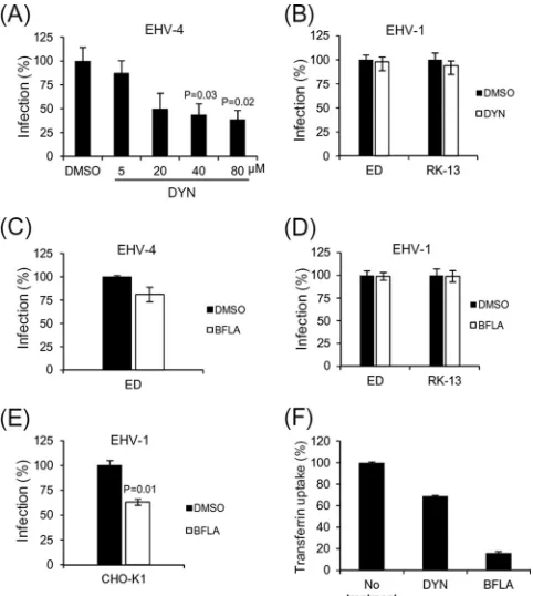
Equine epithelial (ED) cells were preincubated with chlor-promazine, which prevents the assembly of clathrin-coated pits through the inhibition of clathrin adaptor protein 2 assembly (40), or EIPA, a sodium-proton exchange inhibitor, as a specific inhibitor of macropinocytosis (41). Consistent with these activi-ties, chlorpromazine and EIPA caused a significant reduction in the uptake of transferrin, a marker of clathrin-dependent endocy-tosis, and dextran, a marker of macropinocyendocy-tosis, respectively (Fig. 1AandB). When tested in ED cells, neither drug inhibited the entry of either EHV-1 or EHV-4 (Fig. 1CandD), yet the percentage of EHV-4-infected cells was slightly reduced in the presence of EIPA. Although this reduction was not significant (P⬎0.05), it may reflect interference of the drug with another
endocytic pathway, as EIPA has been shown to have pleiotropic effects on other endocytic processes (42). In addition, incubation of CHO-K1 or Vero cells with chlorpromazine did not inhibit infection with either EHV-1 or EHV-4, respectively (data not shown). We concluded from these data that neither macropinocy-tosis nor clathrin-mediated endocymacropinocy-tosis is involved in the entry of either EHV-1 or EHV-4.
When ED cells were treated with nocodazole, a microtubule-disrupting drug (43), before infection for 30 min at 37°C, the number of infected cells was reduced significantly (P⬍0.05) by 40% in the case of EHV-1 and 80% in the case of EHV-4 (Fig. 1E). These results support a role for trafficking of both viruses along the microtubular network after entry. However, treatment with nocodazole does not allow us to distinguish between trafficking of naked nucleocapsids or enveloped viruses in vesicles within cells. Cellular tyrosine kinase activity is important for endocytosis, and internalization by caveolae depends on specific signaling events through tyrosine phosphorylation of Cav-1 at residue 14 (44,45). Previous reports have suggested that cellular tyrosine kinases play a role in alphaherpesvirus infection (10,26,27). Here, we tested the effect of a tyrosine kinase inhibitor, genistein, on virus infection. ED cells were incubated with increasing doses (10, 25, 50, and 100g/ml) of genistein for 1 h before infection with EHV-1 or EHV-4. In the case of EHV-4, the number of infected cells was significantly reduced in a dose-dependent manner (Fig. 1F). In contrast, genistein, even at a concentration of 100 g/ml, had no effect on EHV-1 infection of ED or RK13 but sig-nificantly (P ⬍ 0.05) reduced the EHV-1 infection rate of CHO-K1 cells (Fig. 1GandH). Taken together, the results of the inhibitor experiments suggested that an endocytic pathway and tyrosine kinase signaling play a role during the entry of EHV-4, but not EHV-1, into equine epithelial cells.
Role of dynamin II and endosomal acidification in EHV en-try.Dynamin, the high-molecular weight GTPase, is a key factor that controls fission of endocytic vesicles (46). Dynasore is a po-tent noncompetitive inhibitor of dynamin GTPase activity and blocks dynamin-dependent endocytosis in cells (47,48). Addition of dynasore to culture medium before infection resulted in a ⬎50% reduction of EHV-4 infection of ED cells at the highest concentration tested (Fig. 2A). In contrast, EHV-1 infection was not significantly reduced after the addition of dynasore to ED or RK13 cells (Fig. 2B). However, we could not test the role of dy-namin II in CHO-K1 cells, as dynasore proved to be toxic to these cells even at very low concentrations (seeFig. 6E).
Previous reports had suggested that EHV-1 entry into CHO-K1 cells requires acidic pH, whereas low-pH requirements for EHV-1 infection of ED cells are discussed controversially (26, 27). To further elucidate equine herpesvirus entry, cells were in-cubated with BFLA, an inhibitor of vacuolar ATPase, for 1 h be-fore infection. EHV-4 infection was reduced, albeit not signifi-cantly (P⬎0.05), in ED cells in the presence of the drug (Fig. 2C). Similarly, EHV-1 infection was not affected in ED or RK13 cells but was significantly (P⬍0.05) reduced in CHO-K1 cells (Fig. 2D andE).
To ensure that dynasore and BFLA indeed inhibited dynamin-mediated endocytosis and endosomal acidification, in our exper-imental setup, their effect on internalization of transferrin was determined. Flow cytometric analysis revealed that both dynasore and BFLA significantly blocked transferrin uptake in ED cells (Fig. 2F). Collectively, the data showed that dynamin II is involved
FIG 1EHV-1 and EHV-4 infection in the presence of different inhibitors. (A, B) Chlorpromazine (CHLO) and EIPA block transferrin and dextran uptake, respectively. ED cells (untreated or pretreated with 10 g/ml CHLO or 75M EIPA) were incubated with Alexa Fluor 647-labeled trans-ferrin (50g/ml) or FITC-labeled dextran (1 mg/ml), respectively. After 1 h, cells were washed and the uptake of transferrin or dextran was analyzed by flow cytometry. ED (C to G), RK13 (G), or CHO-K1 (H) cells were either mock treated or treated with CHLO and EIPA (C, D), nocodazole (Noc) (E), or genistein (F to H) and infected with either EHV-1 or EHV-4 (MOI⫽5) as indicated in Materials and Methods. At 8 to 12 h after infection, the percentage of infected cells was determined by flow cytom-etry. Error bars represent the mean⫾standard deviation of three indepen-dent experiments. The infection rate in the absence of inhibitors was set to 100%.
on November 7, 2019 by guest
http://jvi.asm.org/
[image:3.585.44.283.65.406.2]in EHV-4 entry into equine epithelial cells and that EHV-4 utilizes a nonacidic compartment for entry into ED cells.
Role of cholesterol in EHV entry.To test whether EHV entry requires cholesterol-rich lipid rafts, two cholesterol-depleting drugs, filipin and MCD, were used to inhibit lipid raft formation in the plasma membrane (49). Our data showed that filipin has no effect on EHV-4 or EHV-1 infection (Fig. 3AandB). Since filipin also failed to block cholera toxin B internalization in ED cells, we concluded that filipin was unable to sequester cholesterol in these cells (Fig. 3C). On the other hand, MCD significantly reduced the uptake of cholera toxin B in ED cells (Fig. 3C). Incubation of ED cells with MCD significantly reduced the infectivity of EHV-4 (P⬍0.05), but not EHV-1, particularly at higher concen-trations (Fig. 3BandD). We concluded from the data that choles-terol in the plasma membrane is required for EHV-4 but not EHV-1 entry into equine epithelial cells.
In summary, our drug inhibitor studies suggest that EHV-4 can enter equine epithelial cells at neutral pH through an endo-cytic pathway that is dependent on tyrosine kinase activity, dy-namin, and cholesterol. EHV-1, on the other hand, seems to enter ED cells by fusion with the plasma membrane at neutral pH.
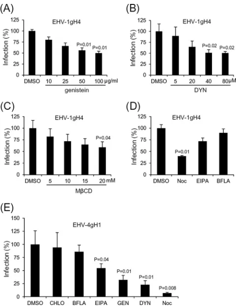
gH determines the entry route of EHV.Recently, we demon-strated that gD, but not gH, plays an important role in the cellular host range of EHV-1 and EHV-4, with integrins having no major role as entry receptors (20, 33). Following up on our previous observations, we further explored the role of gD and gH in routing the entry pathway of EHV-1 and EHV-4. On the basis of our inhibitor studies, we concluded that EHV-1gD4 and EHV-4gD1 follow the entry pathway of their parents. In other words, gD1 and gD4 do not alter the entry pathway of the respective viruses, be it by direct fusion at the plasma membrane or by endocytosis (data not shown). In contrast, EHV-1gH4 became sensitive to tyrosine kinase and dynamin II depletion and adopted characteristics of infection seen for EHV-4 (Fig. 4AandB). Incubation of cells with either genistein or dynasore reduced the number of infected cells by approximately 50% (P⬍0.05). The use of the cholesterol-depleting agent MCD also resulted in a significant (P⬍0.05) reduction of the infection rate (Fig. 4C). Similarly, nocodazole and EIPA reduced the number of cells infected by EHV-1gH4 by 60% and 25%, respectively (Fig. 4D). BFLA still did not have any effect on EHV-1gH4 infection, indicating that the virus uses a pH-neutral compartment for entry (Fig. 4D). On the other hand, EHV-4gH1 still entered equine epithelial cells by endocytosis and was indistinguishable from the parent virus (Fig. 4E).
Following up on these results, we infected equine dermal cells with EHV-1gHS440A, an EHV-1 mutant in which the␣41 integ-rin-binding motif SDI was mutated to a sequence found in the EHV-4 counterpart, ADI, and therefore can no longer bind to integrins (33). In contrast to parental EHV-1, EHV-1gHS440A in-fection in the presence of different inhibitors resulted in entry rates that were reduced by 45% in the presence of genistein,
dyna-FIG 2Effects of dynasore (DYN) and BFLA on EHV-1 and EHV-4 entry. (A) ED cells were infected with EHV-4 (MOI⫽5) in the presence of increasing doses of dynasore. (B) ED or RK13 cells were infected with EHV-1 (MOI⫽5) in the presence or absence of dynasore at a concentra-tion of 80M. (C to E) ED, RK13, or CHO-K1 cells were infected with either EHV-4 or EHV-1 in the presence or absence of BFLA at a concen-tration of 2M. At 8 to 12 h after infection, cells were detached and the percentage of infected cells was determined by flow cytometry. Infection rates in the absence of inhibitors was set to 100%. (F) ED cells were incu-bated with Alexa Fluor 647-labeled transferrin (50g/ml) (in the presence or absence of dynasore at 80M or BFLA at 2M). After 1 h, cells were washed and the uptake of transferrin was analyzed by flow cytometry. Error bars represent the mean⫾standard deviation of three independent exper-iments.
FIG 3EHV-4 entry into ED cells is cholesterol dependent. ED cells were incubated with either filipin (5g/ml) or MCD (20 mM) for 30 min before infection with EHV-4 (A) or EHV-1 (B). (C) ED cells were incu-bated with Alexa Fluor 647-labeled cholera toxin B (0.5g/ml) in the presence or absence of either filipin (5g/ml) or MCD (20 mM). After 30 min, cells were washed and the internalization of cholera toxin B was analyzed by flow cytometry. (D) ED cells were infected with EHV-4 in the presence of increasing doses of MCD. The percentage of infected cells was determined by flow cytometry. The percentage of infection in the absence of inhibitors was set to 100%. Error bars represent the mean⫾standard deviation of three independent experiments.
on November 7, 2019 by guest
http://jvi.asm.org/
[image:4.585.299.540.64.256.2] [image:4.585.43.284.66.335.2]sore, and MCD; by 15% in the presence of EIPA; and by 70% in the presence of nocodazole (Fig. 5AtoD).
From these experiments, we concluded that gH plays an im-portant role in routing EHV-1 into a specific entry pathway through a mechanism that is dependent on the interaction of gH with integrins expressed on the cell surface.
Routing the entry pathway of EHV-1 by cross-linking of
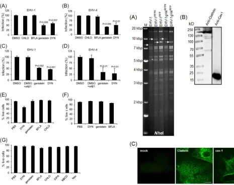
␣41 integrins.␣41 integrins are expressed on approximately 40% of ED cells (33). To further elucidate the role of integrins, we analyzed EHV-1 entry in the presence of different inhibitors after blocking virus interaction with cell surface integrins. Cells were incubated with an anti-␣41 integrin MAb for 1 h and with vari-ous drugs before infection with EHV-1. Entry of EHV-1, as well as EHV-4, was inhibited by genistein and dynasore, and the number of infected cells was significantly (P⬍0.05) reduced by approxi-mately 50% after infection (Fig. 6AandB). Incubation of the cells with␣47 or␣V5 integrin antibodies, as controls, had no effect on the infection rate of EHV-1 in the presence of genistein or dynasore (data not shown), whereas the entry of EHV-4 into equine epithelial cells remained sensitive to both drugs in the pres-ence of either antibody (data not shown). Furthermore, incuba-tion of EHV-1 and EHV-4 with soluble␣41 integrin before in-fection resulted in a reduction of infected cells when genistein or dynasore was added before infection (Fig. 6CandD).
Collectively, we concluded from the data that␣41 integrins play an important role during virus entry and acts as a routing factor that is able to change the entry pathway of the viruses. In addition, blocking the interaction between gH and integrins di-rects EHV-1 to the endocytic pathway. It seems likely that the physical interaction between gH1 and integrins may activate cel-lular signaling pathways that, in turn, allow virus fusion at the plasma membrane. Once this signaling pathway is blocked, virus entry is redirected to an endocytic pathway.
EHV-4 entry is caveolin dependent.Caveolae are cholesterol-and sphingolipid-rich smooth invaginations of the plasma mem-brane that play a major role in cellular uptake and traffic. Caveola/ lipid raft-dependent endocytosis has been shown to be sensitive to cholesterol depletion with drugs such as filipin and MCD (50), dependent on dynamin II (51), and inhibited by tyrosine kinase inhibitors such as genistein (52). To test the role of caveolin in EHV-4 entry, we investigated the colocalization of different vi-ruses with either Cav-1 or clathrin early after infection. First, two-step Red-mediated recombination was used to insert the gene for mRFP1 into the viral gene encoding the small capsid protein (VP26) in order to facilitate visualization of the virus (Fig. 7A). The viruses generated, EHV-1RFPand EHV-1gH4RFP, grew with kinetics that were virtually identical to those of the parental vi-ruses, and both viruses emitted red fluorescence after excitation. In contrast, EHV-4RFPand EHV-4gH1RFPshowed severe growth defects on equine cells. For that reason, we performed double staining of cells infected with parental EHV-4 using polyclonal antibodies against viral gD and either anti-Cav-1 or anti-clathrin antibodies as cellular markers. The specificity of the anti-Cav-1 and anti-clathrin-heavy chain antibodies in ED was examined by Western blotting and/or indirect immunofluorescence assays (Fig. 7BandC).
ED cells were infected with EHV-1RFPand EHV-1gH4RFP, and colocalization with either Cav-1 or clathrin was determined by
FIG 4Entry of EHV-1gH4 and EHV-4gH1 into ED cells. Cells were treated with different inhibitors, as indicated, before infection (MOI⫽5) with either EHV-1gH4 (A to D) or EHV-4gH1 (E) for 8 to 12 h. The percentage of infected cells was determined by flow cytometry. The percentage of infection in the absence of inhibitors was set to 100%. Error bars represent the mean⫾ stan-dard deviation of three independent experiments. CHLO, chlorpromazine; GEN, genistein; Noc, nocodazole.
FIG 5EHV-1gHS440Aentry into ED cells. Cells were pretreated with genistein
(A), dynasore (DYN) (B), MCD (C), EIPA, or nocodazole (Noc) (D) before infection with EHV-1gHS440A(MOI⫽5). At 8 to 12 h after infection,
mono-layers were detached and the percentage of infected cells was determined by flow cytometry. The percentage of infection in the absence of inhibitors was set to 100%. Error bars represent the mean⫾standard deviation of three inde-pendent experiments.
on November 7, 2019 by guest
http://jvi.asm.org/
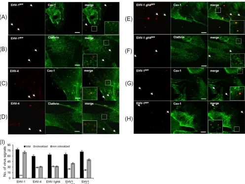
[image:5.585.291.541.63.262.2] [image:5.585.43.283.64.376.2]confocal microscopy with the respective antibodies. No signif-icant colocalization with either Cav-1 or clathrin was detected in the case of EHV-1RFP(Fig. 8AandB). In the case of EHV-4 and EHV-1gH4RFP, more than 50% of the virus particles were colocalizing with Cav-1 (Fig. 8CandE), whereas no colocaliza-tion above the background levels was detected with clathrin at the same time point (Fig. 8DandF). Twelve fields were ran-domly chosen, and approximately 60 signals were counted for each virus (Fig. 8I).
To further confirm the role of␣41 integrin in the routing of EHV-1 entry, cells were incubated with anti-␣41 integrin MAb for 1 h before infection with EHV-1RFP. In another experiment,
EHV-1RFPwas incubated with soluble␣41 integrin for 1 h before infection. Six fields were randomly selected, and more than 60 signals were counted (Fig. 8I). Approximately 39% and 31% of the virus signals were colocalizing with Cav-1, but not clathrin, after either blocking of the surface integrin with the anti-␣41 integrin MAb or incubation of the virus with soluble integrin, respectively (Fig. 8GandH).
To further confirm our results, we performed correlative fluo-rescence and transmission electron microscopy (TEM), where
FIG 6Effects of MAb P4C2 and soluble␣41 integrin on EHV-1 entry. ED cells were incubated with 20g/ml of anti-␣41 integrin MAb P4C2. After washing, cells were incubated with the indicated inhibitors before infection with EHV-1 (A) or EHV-4 (B) at an MOI of 5. (C, D) EHV-1 or EHV-4 was incubated with soluble␣41 integrin before infection of ED cells in the pres-ence of the indicated inhibitors. At 8 to 12 h after infection, the percentage of infected cells was determined by flow cytometry. The percentage of infection in the absence of inhibitors was set to 100%. Error bars represent the mean⫾ standard deviation of three independent experiments. (E to G) Toxicity assays for pharmacological inhibitors on different cells. Pharmacological inhibitor uptake in CHO-K1 (E), RK-13 (F), or ED (G) cells following 7 h of incubation with the indicated inhibitors. The number of live cells (no pharmacological inhibitor uptake) relative to the total cell population was determined after flow cytometric analysis and is given in percent. Error bars represent the means⫾ standard deviations of two independent experiments.
FIG 7Identification of mRFP1-labeled viruses. (A) Purified DNA from pa-rental EHV-1 or mRFP1-labeled viruses was digested with NheI. mRFP1 was inserted into VP26, which is located within a 7.6-kbp NheI fragment. This band disappeared and was replaced by a band of around 9.3 kbp because of the insertion of mRFP1Kanrin the case of EHV-1RFPKand EHV-1gH4RFPK. The
removal of the gene for Kanrsubtracted 1 kbp from the final constructs
(EHV-1RFPand EHV-1gH4RFP), and a fragment of around 8.3 kbp appeared.
Frag-ments in the mutants that appeared as a consequence of the insertion of the mRFP1 sequence are marked by arrows. (B) Detection of clathrin and Cav-1 expression by Western blot analysis. ED cell lysates were prepared, and pro-teins were separated by SDS–10% PAGE before transfer to a polyvinylidene difluoride membrane. Blots were incubated with anti-clathrin or anti-Cav-1 antibody (1/200 dilution), followed by anti-rabbit IgG peroxidase antibodies (1/10,000 dilution). (C) Indirect immunofluorescence detection of clathrin and Cav-1. ED cells were grown and fixed on gridded MatTek coverslips. Cells were subsequently stained with anti-clathrin or anti-Cav-1 antibodies, fol-lowed by Alexa Fluor 488-labeled goat anti-rabbit IgG (1:1,000). Cells were imaged with a Zeiss LSM 510 confocal microscope. Images were taken with a 63⫻oil immersion objective. The scale bar represents 5m.
on November 7, 2019 by guest
http://jvi.asm.org/
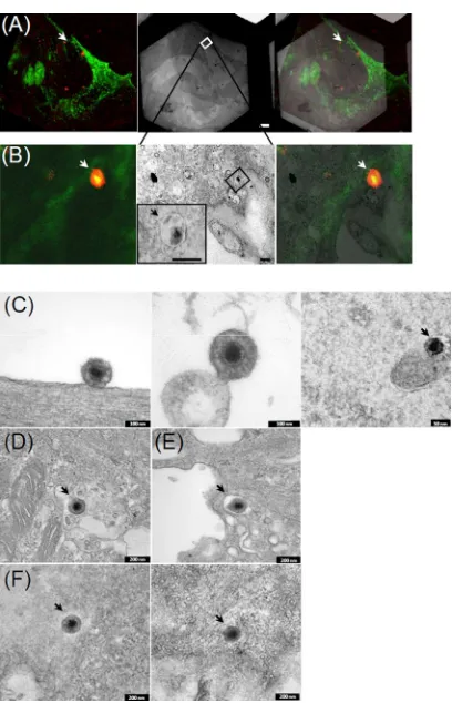
[image:6.585.43.508.61.429.2]identical areas were investigated by fluorescence and subsequent electron microscopy. EHV-1gH4RFP particles colocalized with Cav-1 (Fig. 8E) corresponded to a virus particle-containing vesi-cle, as imaged by TEM (Fig. 9AandB). Furthermore, our results showed that EHV-1 nucleocapsids were present in the cytosol (Fig. 9C, right panel), and virus attachment and fusion events at the plasma membrane were observed (Fig. 9C, left and middle panels). Enveloped EHV-1 was seen within vesicles only when cell surface integrins were blocked with anti-␣41 integrin MAb or when the virus was incubated with soluble␣41 integ-rin before infection (Fig. 9F). In contrast, enveloped EHV-4 and EHV-1gH4 were frequently found in vesicles in ED cells. The vesicles corresponded to the Cav-1-positive compartments observed by confocal microscopy. No fusion events with the plasma membrane were detected when viruses expressed gH4 (Fig. 9B,D, andE).
From the confocal and EM experiments, we concluded that EHV-1 infection of equine epithelial cells is not dependent on
either caveolin or clathrin, as entry occurred predominantly by fusion at the plasma membrane. Exchange of EHV-1 gH with that of EHV-4 or blocking of the interaction of gH1 with␣41 integ-rin, however, redirected the virus to an endocytic pathway, which was caveolin/raft dependent and therefore similar to that used by EHV-4.
DISCUSSION
For many viruses, factors that are responsible for routing the entry of viral particles, either by fusion at the plasma membrane or from within endosomal vesicles, are still unknown. In principle, fusion requires a timely coordinated interaction between viral and cellu-lar membranes. Recently, it was shown that entry routes of HSV are dictated by the cell, where integrins relocalize nectin 1 to lipid rafts independently of the virus (17, 18). Here, we show that EHV-1, an animal alphaherpesvirus with a tropism for epithelial and endothelial, as well as leukocytes, can enter cells by at least two distinct mechanisms; direct fusion at the plasma membrane and
FIG 8Colocalization of viral particles with caveolin during entry. ED cells were incubated with EHV-1RFP, EHV-4, or EHV-1gH4RFP(MOI⫽20) at 4°C for 2
h as indicated. The medium was replaced with preheated medium at 37°C, and cells were fixed at 5 min after the temperature shift. Cells were stained with anti-Cav-1 (green, A, C, E, G, H), anti-clathrin (green, B, D, F), and/or anti-EHV-4gD antibodies (red, C, D). (G) Cells were first incubated with anti-␣41 integrin MAb before infection with EHV-1RFP. (H) EHV-1RFPwas preincubated with soluble␣41 integrin before addition to cells. (I) Numbers of virus particles
colocalizing with caveolin signals after infection with various viruses and in the presence of antibodies or soluble integrins as determined in randomly selected fields of infected ED cells. The scale bars in panels A to H represent 5m.
on November 7, 2019 by guest
http://jvi.asm.org/
[image:7.585.44.541.65.438.2]caveolin/raft-dependent endocytosis, respectively. The decision of which of the two pathways will be taken is dependent mainly on viral gH and its ability to interact with␣41 integrins expressed on the surface of target cells (Table 2).
EHV-1 entry into different cells has been studied previously (26–28). However, no data are available on the details of EHV-4 entry. We initially evaluated the entry pathway of the two closely related viruses into epithelial (ED) cells, and our data showed that each virus can enter these cells by a different route. In the case of EHV-1, EM studies revealed tethering of viral particles to the plasma membrane. Prefusion states and fusion events with the plasma membrane could also be detected, while naked nucleocap-sids were observed just underneath the plasma membrane. Fur-thermore, virus entry appeared to be nonsensitive to any of the pharmacological inhibitors of endocytosis and viral particles did not colocalize with either Cav-1 or clathrin. The reduction of EHV-1 infection in the presence of the microtubule-depolymer-izing agent nocodazole seemed to be due to utilization of the mi-crotubular network for efficient travel to the nucleus postentry. Microtubule disruption cannot distinguish between virus entry via an endocytic route or by direct fusion at the plasma membrane (53). Our results are consistent with data of Frampton and co-workers, who showed that EHV-1 enters ED cells through direct fusion at the plasma membrane, and also with earlier results show-ing that EHV-1 can enter RK-13 cells through direct fusion at the plasma membrane and through low-pH-dependent endocytosis in CHO-K1 cells (26). However, our data contradict results claim-ing that yet another EHV-1 strain, Ab4p, can enter ED cells via energy- and pH-dependent endocytosis (27). It is important to note that the authors failed to find any colocalization of the virus with either caveolin or clathrin.
We were able to demonstrate that EHV-4 can enter ED cells using a pathway that is dependent on tyrosine kinase activity, dy-namin II, microtubule integrity, cholesterol, and caveolin 1 but does not require low pH or clathrin. EM studies further confirmed our conclusions and revealed the presence of enveloped viruses within noncoated vesicles in the cytoplasm. This clear difference between the entry pathways of the two closely related viruses di-rected us toward investigation of the viral and cellular factors that might control this process. EHV-1 (54,55) and EHV-4 (Azab et al., unpublished observations) enter ED cells by utilizing MHC-I molecules as receptors. Two of the glycoproteins (gD and gH) have integrin-binding motifs, RSD and SDI, respectively, that ex-ist in EHV-1 but not EHV-4 (20,33). We therefore investigated the route of entry of EHV-1 and EHV-4 by exchanging the respec-tive glycoproteins or by mutating the putarespec-tive integrin-binding motifs of gD1 and gH1 to be similar to their nonfunctional coun-terpart in EHV-4. While almost all of the recombinant viruses
FIG 9Correlative fluorescence and electron microscopy. (A, B) The location of an EHV-1gH4RFPparticle colocalized with a Cav-1-positive cellular
com-partment was visualized by confocal laser scanning microscopy and corre-sponds to a virus-containing vesicle imaged by TEM (left panel, fluorescence image; middle panel, electron micrograph; right panel, correlation of fluores-cence image with electron micrograph by alignment of cellular surface struc-tures). The particle of interest is indicated by white arrows (fluorescent signal) and within a vesicular intracellular compartment by a black arrow. Scale bars: 2m (A) and 200 nm (B). (C to F) EM analysis of virus entry into ED cells. The same fields of confocal microscopy were used for further analysis by TEM. EHV-1RFP(C), EHV-4 (D), EHV-1gH4RFP(E), or ED (F) cells were first
incubated with anti-␣41 integrin antibodies before infection with EHV-1RFP
(left panel), or EHV-1RFPwas incubated with soluble␣41 integrin before the
[image:8.585.62.266.62.379.2]infection of ED cells (right panel). Scale bar sizes are indicated.
TABLE 2Summary of the results obtained in this work
Inhibitor
Reduction of virus infectivitya
EHV-1 EHV-1gD4 EHV-1gH4 EHV-1S440A EHV-4 EHV-4gD1 EHV-4gH1 EHV-1 (anti-␣41) EHV-1 (sol␣41)
Chlorpromazine ⫺ ⫺ ⫺ ⫺ ⫺ ⫺ ⫺ ⫺ ⫺
BFLA ⫺ ⫺ ⫺ ⫺ ⫺ ⫺ ⫺ ⫺ ⫺
EIPA ⫺ ⫺ ⫹ ⫹ ⫹ ⫹ ⫹ NAb NA
Nocodazole ⫹⫹ ⫹⫹ ⫹⫹⫹ ⫹⫹⫹ ⫹⫹⫹ ⫹⫹⫹ ⫹⫹⫹ NA NA
Genistein ⫺ ⫺ ⫹⫹⫹ ⫹⫹⫹ ⫹⫹⫹ ⫹⫹⫹ ⫹⫹⫹ ⫹⫹⫹ ⫹⫹⫹
Dynasore ⫺ ⫺ ⫹⫹⫹ ⫹⫹⫹ ⫹⫹⫹ ⫹⫹⫹ ⫹⫹⫹ ⫹⫹⫹ ⫹⫹⫹
Filipin ⫺ ⫺ ⫺ ⫺ ⫺ ⫺ ⫺ NA NA
MCD ⫺ ⫺ ⫹⫹⫹ ⫹⫹⫹ ⫹⫹⫹ ⫹⫹⫹ ⫹⫹⫹ NA NA
a
Degrees of reduction:⫺, none;⫹, slight;⫹⫹, moderate;⫹⫹⫹, strong.
bNA, not applicable.
on November 7, 2019 by guest
http://jvi.asm.org/
[image:8.585.41.547.594.707.2]tested, EHV-1gD4, EHV-4gD1, and EHV-4gH1, followed the en-try pathway of their parents, only EHV-1gH4 changed its route of entry into ED cells: EHV-1gH4 enters ED cells through a pathway that is dependent on tyrosine kinase activity, dynamin II, micro-tubules, cholesterol, and neutral pH. Furthermore, we could de-tect significant colocalization of the virus with Cav-1, but not clathrin, and enveloped virus was detected by EM studies within endocytic vesicles.
Caveolin/raft-mediated entry has been demonstrated for en-veloped and nonenen-veloped viruses, including simian virus 40, polyomavirus, echovirus 1, amphotropic murine leukemia virus, and KSHV (39,50,56,57). Caveolin-dependent endocytosis was also determined as the route of entry of EHV-1 in equine brain endothelial cells (27). Caveolar endocytosis requires several fac-tors besides Cav-1, among them dynamin and tyrosine kinases, as well as lipid rafts. However, these factors are not exclusive for the caveolar pathway, as they are also integral for other endocytic pathways (57–60). Inhibitor studies with dynasore, genistein, and MCD revealed that dynamin, tyrosine kinase activity, and lipid rafts are involved in EHV-4 and EHV-1gH4 entry. However, we could not test the effect of either dominant negative dynamin II or Cav-1 plasmids because of the very low transfection efficiency that can be achieved in ED cells. The colocalization of the viruses with Cav-1 supports the notion that caveolin/raft-mediated endocyto-sis promotes entry of EHV-4 or EHV-1 mutants carrying gH4 into equine epithelial cells, although they have relatively big diameters
(150 to 200 nm). It is important to note that caveolae can inter-nalize large molecular complexes such as bacteria (61) and latex beads with diameters of 200 to 1,000 nm (62).
A crucial step in the entry of enveloped viruses via endocytosis is fusion of the viral envelope with cellular membranes to allow the release of nucleocapsids into the cytoplasm. This process can be triggered by low pH, which is known to induce conformational changes in several viral glycoproteins thereby promoting virus uncoating (43,63,64). Viral entry through caveolae is indepen-dent of acidic pH, as the pH of caveosomes is neutral (50,56,57). The effect of the BFLA used in our study suggested that entry of EHV-4, EHV-4gH1, and EHV-1gH4 into ED cells occurs at neu-tral pH and that infection by these viruses would therefore be pH independent. Endocytic entry of enveloped viruses through a pathway that is independent of low pH has also been described for other viruses, such as duck hepatitis B virus, EBV, and HSV-1 (65–67).
The amino acid motif LDV and the related motifs LDI and SDI have been shown to bind␣4 integrins with similar affinities and avidities (68,69). Also, our previous studies showed that␣41 integrins are expressed on the surface of ED cells (33). To deter-mine the role of integrins in defining the route of virus entry, ED cells were incubated with antibodies directed against␣41 and then infected with EHV-1 in the presence of different inhibitors. Our studies showed that, after blocking of␣41 integrins, EHV-1 entered cells through a caveolin/raft-mediated endocytosis.
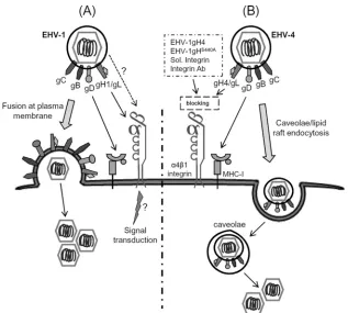
Fur-FIG 10Putative model of the route of entry of EHV-1 and EHV-4 into equine epithelial cells. Virions first attach to target cells via gC and/or gB, which binds to heparin sulfate- and chondroitin-containing cell surface proteoglycans. (A) In the case of wild type EHV-1, fusion at the plasma membrane starts with gD binding to its cognate receptor (MHC-I), followed by the activation of a gH/gL complex that can “prime” gB fusion activity. Yet, a strong interaction between gH1 and␣41 integrins, as well as at least one additional (unknown) viral factor, must occur before fusion can take place. (B) The interruption of this “fusion complex” prevents fusion with the plasma membrane; however, the virus is redirected to the endocytic pathway, which leads to the efficient release of
nucleocapsids into the cytoplasm. Sol., soluble; Ab, antibody.
on November 7, 2019 by guest
http://jvi.asm.org/
[image:9.585.134.451.63.348.2]thermore, incubation of EHV-1 with soluble␣41 integrin before infection also routed the virus to the same pathway. Interestingly, the same results were obtained after mutation of the SDI integrin-binding motif of EHV-1 gH to make it identical to that of EHV-4 gH (EHV-1S440A) (33). Confocal and EM studies showed signifi-cant colocalization of the virus with Cav-1 and the presence of enveloped viruses within endocytic vesicles. Our previous data showed that integrins have no measurable effect on the rate of infection of either EHV-1 or EHV-4 (20,33). We show here that integrins have a role in rerouting the EHV-1 entry pathway and that fusion at either the plasma membrane or the caveolar membrane are equally efficient in allowing virus entry and es-tablishment of infection. The salient outcome of these experi-ments was, therefore, that viral (gH) and cellular (␣41 integ-rins) components share the responsibility of routing the entry of EHV either to direct fusion at the plasma membrane or to a pH-independent caveolar endocytic pathway. Knowledge of these pathways may facilitate the development of novel strate-gies to prevent infection.
Interestingly, endowing EHV-4 with gH1 did not redirect the virus to fusion with the plasma membrane. Therefore, gH1 and its integrin-binding domain likely are not solely responsible for fu-sion from without. This finding supports, in our model, the hypothesis that fusion of the viral envelope with the plasma membrane can occur only if there is a strong interaction be-tween gH1 and␣41 integrins and at least one additional, so far unknown, viral factor. Once this interaction is disrupted, the virus cannot fuse with the plasma membrane any longer but is redirected to an endocytic pathway. On the other hand, EHV-1gH4 cannot fuse with the plasma membrane, as there is no interaction between gH4 and␣41 integrins (Fig. 10). The molecular mechanisms underlying the change of the virus en-try pathway are still unknown. A possible explanation is the loss of interaction between gH and␣41 integrins, which may result in perturbation of subsequent signaling events. The modulation of signaling events and its effect on the route of entry would explain results that we obtained in the presence or absence of gH-integrin interaction and are supported by other reports, which showed such cross talk to be important for the routing of viruses to specific compartments (35,39,70). Others have demonstrated that the activation of Ca2⫹signaling
path-ways is also associated with membrane fusion for some envel-oped viruses, including HIV-1, HSV, and HCMV (71–74). Fur-ther studies are in progress to address the role of various signal transductions during EHV entry.
ACKNOWLEDGMENTS
We thank Gabriele Drescher for technical assistance with the preparation of samples for electron microscopy. We thank Guanggang Ma for helping with Western blot analysis for detection of Cav-1.
This work was supported by a grant from the Alexander-von-Hum-boldt Foundation to W.A. and by unrestricted funds made available to N.O. by Freie Universität Berlin.
REFERENCES
1.Stein BS, Gowda SD, Lifson JD, Penhallow RC, Bensch KG, Engleman EG.1987. pH-independent HIV entry into CD4-positive T cells via virus envelope fusion to the plasma membrane. Cell49:659 – 668.
2.Matlin KS, Reggio H, Helenius A, Simons K.1981. Infectious entry pathway of influenza virus in a canine kidney cell line. J. Cell Biol.91:601– 613.
3.Mercer J, Schelhaas M, Helenius A.2010. Virus entry by endocytosis. Annu. Rev. Biochem.79:803– 833.
4.Rahn E, Petermann P, Hsu MJ, Rixon FJ, Knebel-Morsdorf D.2011. Entry pathways of herpes simplex virus type 1 into human keratinocytes are dynamin- and cholesterol-dependent. PLoS One 6:e25464. doi:10 .1371/journal.pone.0025464.
5.Schelhaas M, Shah B, Holzer M, Blattmann P, Kuhling L, Day PM, Schiller JT, Helenius A.2012. Entry of human papillomavirus type 16 by actin-dependent, clathrin- and lipid raft-independent endocytosis. PLoS Pathog.8:e1002657. doi:10.1371/journal.ppat.1002657.
6.Campadelli-Fiume G, Menotti L.2007. Entry of alphaherpesviruses into the cell, p 93–111.InArvin A, Campadelli-Fiume G, Mocarski E, Moore PS, Roizman B, Whitley R, Yamanishi K (ed), Human herpesviruses: bi-ology, therapy, and immunoprophylaxis. Cambridge University Press, Cambridge, United Kingdom.
7.Geraghty RJ, Krummenacher C, Cohen GH, Eisenberg RJ, Spear PG.
1998. Entry of alphaherpesviruses mediated by poliovirus receptor-related protein 1 and poliovirus receptor. Science280:1618 –1620.
8.Hutchinson L, Browne H, Wargent V, Davis-Poynter N, Primorac S, Goldsmith K, Minson AC, Johnson DC.1992. A novel herpes simplex virus glycoprotein, gL, forms a complex with glycoprotein H (gH) and affects normal folding and surface expression of gH. J. Virol.66:2240 – 2250.
9.Shukla D, Liu J, Blaiklock P, Shworak NW, Bai X, Esko JD, Cohen GH, Eisenberg RJ, Rosenberg RD, Spear PG.1999. A novel role for 3-O-sulfated heparan sulfate in herpes simplex virus 1 entry. Cell99: 13–22.
10. Nicola AV, Hou J, Major EO, Straus SE.2005. Herpes simplex virus type 1 enters human epidermal keratinocytes, but not neurons, via a pH-dependent endocytic pathway. J. Virol.79:7609 –7616.
11. Richart SM, Simpson SA, Krummenacher C, Whitbeck JC, Pizer LI, Cohen GH, Eisenberg RJ, Wilcox CL.2003. Entry of herpes simplex virus type 1 into primary sensory neurons in vitro is mediated by Nectin-1/ HveC. J. Virol.77:3307–3311.
12. Wittels M, Spear PG.1991. Penetration of cells by herpes simplex virus does not require a low pH-dependent endocytic pathway. Virus Res.18: 271–290.
13. Gianni T, Campadelli-Fiume G, Menotti L.2004. Entry of herpes sim-plex virus mediated by chimeric forms of nectin1 retargeted to endosomes or to lipid rafts occurs through acidic endosomes. J. Virol.78:12268 – 12276.
14. Milne RS, Nicola AV, Whitbeck JC, Eisenberg RJ, Cohen GH.2005. Glycoprotein D receptor-dependent, low-pH-independent endocytic en-try of herpes simplex virus type 1. J. Virol.79:6655– 6663.
15. Nicola AV, McEvoy AM, Straus SE.2003. Roles for endocytosis and low pH in herpes simplex virus entry into HeLa and Chinese hamster ovary cells. J. Virol.77:5324 –5332.
16. Clement C, Tiwari V, Scanlan PM, Valyi-Nagy T, Yue BY, Shukla D.
2006. A novel role for phagocytosis-like uptake in herpes simplex virus entry. J. Cell Biol.174:1009 –1021.
17. Gianni T, Gatta V, Campadelli-Fiume G.2010. {alpha}V{beta}3-integrin routes herpes simplex virus to an entry pathway dependent on cholesterol-rich lipid rafts and dynamin2. Proc. Natl. Acad. Sci. U. S. A.107:22260 – 22265.
18. Gianni T, Campadelli-Fiume G.2012.␣V3-integrin relocalizes nectin1 and routes herpes simplex virus to lipid rafts. J. Virol.86:2850 –2855. 19. Davison AJ, Eberle R, Ehlers B, Hayward GS, McGeoch DJ, Minson AC,
Pellett PE, Roizman B, Studdert MJ, Thiry E.2009. The order Herpes-virales. Arch. Virol.154:171–177.
20. Azab W, Osterrieder N.2012. Glycoproteins D of equine herpesvirus type 1 (EHV-1) and EHV-4 determine cellular tropism independently of integ-rins. J. Virol.86:2031–2044.
21. Osterrieder N, Van de Walle GR.2010. Pathogenic potential of equine alphaherpesviruses: the importance of the mononuclear cell compartment in disease outcome. Vet. Microbiol.143:21–28.
22. Patel JR, Heldens J.2005. Equine herpesviruses 1 1) and 4 (EHV-4)— epidemiology, disease and immunoprophylaxis: a brief review. Vet. J.
170:14 –23.
23. Csellner H, Walker C, Wellington JE, McLure LE, Love DN, Whalley JM.2000. EHV-1 glycoprotein D (EHV-1 gD) is required for virus entry and cell-cell fusion, and an EHV-1 gD deletion mutant induces a protec-tive immune response in mice. Arch. Virol.145:2371–2385.
24. Frampton AR, Jr, Goins WF, Cohen JB, von Einem J, Osterrieder N,
on November 7, 2019 by guest
http://jvi.asm.org/
O’Callaghan DJ, Glorioso JC.2005. Equine herpesvirus 1 utilizes a novel herpesvirus entry receptor. J. Virol.79:3169 –3173.
25. Neubauer A, Braun B, Brandmuller C, Kaaden OR, Osterrieder N.
1997. Analysis of the contributions of the equine herpesvirus 1 glycopro-tein gB homolog to virus entry and direct cell-to-cell spread. Virology
227:281–294.
26. Frampton AR, Jr, Stolz DB, Uchida H, Goins WF, Cohen JB, Glorioso JC.2007. Equine herpesvirus 1 enters cells by two different pathways, and infection requires the activation of the cellular kinase ROCK1. J. Virol.
81:10879 –10889.
27. Hasebe R, Sasaki M, Sawa H, Wada R, Umemura T, Kimura T.2009. Infectious entry of equine herpesvirus-1 into host cells through different endocytic pathways. Virology393:198 –209.
28. Van de Walle GR, Peters ST, VanderVen BC, O’Callaghan DJ, Oster-rieder N.2008. Equine herpesvirus 1 entry via endocytosis is facilitated by alphaV integrins and an RSD motif in glycoprotein D. J. Virol.82:11859 – 11868.
29. Clark EA, Brugge JS.1995. Integrins and signal transduction pathways: the road taken. Science268:233–239.
30. Chesnokova LS, Nishimura SL, Hutt-Fletcher LM. 2009. Fusion of epithelial cells by Epstein-Barr virus proteins is triggered by binding of viral glycoproteins gHgL to integrins alphavbeta6 or alphavbeta8. Proc. Natl. Acad. Sci. U. S. A.106:20464 –20469.
31. Wang D, Shenk T.2005. Human cytomegalovirus virion protein complex required for epithelial and endothelial cell tropism. Proc. Natl. Acad. Sci. U. S. A.102:18153–18158.
32. Akula SM, Pramod NP, Wang FZ, Chandran B. 2002. Integrin alpha3beta1 (CD 49c/29) is a cellular receptor for Kaposi’s sarcoma-associated herpesvirus (KSHV/HHV-8) entry into the target cells. Cell
108:407– 419.
33. Azab W, Zajic L, Osterrieder N.2012. The role of glycoprotein H of equine herpesviruses 1 and 4 (EHV-1 and EHV-4) in cellular host range and integrin binding. Vet. Res.43:61.
34. Dorner M, Zucol F, Alessi D, Haerle SK, Bossart W, Weber M, Byland R, Bernasconi M, Berger C, Tugizov S, Speck RF, Nadal D.2010. beta1 integrin expression increases susceptibility of memory B cells to Epstein-Barr virus infection. J. Virol.84:6667– 6677.
35. Krishnan HH, Sharma-Walia N, Streblow DN, Naranatt PP, Chandran B.2006. Focal adhesion kinase is critical for entry of Kaposi’s sarcoma-associated herpesvirus into target cells. J. Virol.80:1167–1180.
36. Rudolph J, O’Callaghan DJ, Osterrieder N. 2002. Cloning of the ge-nomes of equine herpesvirus type 1 (EHV-1) strains KyA and racL11 as bacterial artificial chromosomes (BAC). J. Vet. Med. B Infect. Dis. Vet. Public Health49:31–36.
37. Azab W, Kato K, Arii J, Tsujimura K, Yamane D, Tohya Y, Matsumura T, Akashi H.2009. Cloning of the genome of equine herpesvirus 4 strain TH20p as an infectious bacterial artificial chromosome. Arch. Virol.154: 833– 842.
38. Tischer BK, von Einem J, Kaufer B, Osterrieder N.2006. Two-step red-mediated recombination for versatile high-efficiency markerless DNA manipulation in Escherichia coli. Biotechniques40:191–197.
39. Kerur N, Veettil MV, Sharma-Walia N, Sadagopan S, Bottero V, Paul AG, Chandran B.2010. Characterization of entry and infection of mono-cytic THP-1 cells by Kaposi’s sarcoma associated herpesvirus (KSHV): role of heparan sulfate, DC-SIGN, integrins and signaling. Virology406: 103–116.
40. Wang LH, Rothberg KG, Anderson RG.1993. Mis-assembly of clathrin lattices on endosomes reveals a regulatory switch for coated pit formation. J. Cell Biol.123:1107–1117.
41. Harris C, Fliegel L.1999. Amiloride and the Na(⫹)/H(⫹) exchanger protein: mechanism and significance of inhibition of the Na(⫹)/H(⫹) exchanger (review). Int. J. Mol. Med.3:315–321.
42. Fretz M, Jin J, Conibere R, Penning NA, Al-Taei S, Storm G, Futaki S, Takeuchi T, Nakase I, Jones AT.2006. Effects of Na⫹/H⫹exchanger inhibitors on subcellular localisation of endocytic organelles and intracel-lular dynamics of protein transduction domains HIV-TAT peptide and octaarginine. J. Control. Release116:247–254.
43. Bayer N, Schober D, Prchla E, Murphy RF, Blaas D, Fuchs R.1998. Effect of bafilomycin A1 and nocodazole on endocytic transport in HeLa cells: implications for viral uncoating and infection. J. Virol.72:9645– 9655.
44. Aoki T, Nomura R, Fujimoto T. 1999. Tyrosine phosphorylation of caveolin-1 in the endothelium. Exp. Cell Res.253:629 – 636.
45. Simons K, Toomre D.2000. Lipid rafts and signal transduction. Nat. Rev. Mol. Cell Biol.1:31–39.
46. Doherty GJ, McMahon HT.2009. Mechanisms of endocytosis. Annu. Rev. Biochem.78:857–902.
47. Kirchhausen T, Macia E, Pelish HE.2008. Use of dynasore, the small molecule inhibitor of dynamin, in the regulation of endocytosis. Methods Enzymol.438:77–93.
48. Macia E, Ehrlich M, Massol R, Boucrot E, Brunner C, Kirchhausen T.
2006. Dynasore, a cell-permeable inhibitor of dynamin. Dev. Cell10:839 – 850.
49. Sieczkarski SB, Whittaker GR.2002. Dissecting virus entry via endocy-tosis. J. Gen. Virol.83:1535–1545.
50. Beer C, Andersen DS, Rojek A, Pedersen L.2005. Caveola-dependent endocytic entry of amphotropic murine leukemia virus. J. Virol.79: 10776 –10787.
51. Nabi IR, Le PU.2003. Caveolae/raft-dependent endocytosis. J. Cell Biol.
161:673– 677.
52. Pelkmans L, Puntener D, Helenius A.2002. Local actin polymerization and dynamin recruitment in SV40-induced internalization of caveolae. Science296:535–539.
53. Frampton AR, Jr, Uchida H, von Einem J, Goins WF, Grandi P, Cohen JB, Osterrieder N, Glorioso JC.2010. Equine herpesvirus type 1 (EHV-1) utilizes microtubules, dynein, and ROCK1 to productively infect cells. Vet. Microbiol.141:12–21.
54. Kurtz BM, Singletary LB, Kelly SD, and Frampton AR, Jr.2010.Equus caballusmajor histocompatibility complex class I is an entry receptor for equine herpesvirus type 1. J. Virol.84:9027–9034.
55. Sasaki M, Hasebe R, Makino Y, Suzuki T, Fukushi H, Okamoto M, Matsuda K, Taniyama H, Sawa H, Kimura T. 2011. Equine major histocompatibility complex class I molecules act as entry receptors that bind to equine herpesvirus-1 glycoprotein D. Genes Cells.16:343–357. 56. Pelkmans L, Kartenbeck J, Helenius A.2001. Caveolar endocytosis of simian virus 40 reveals a new two-step vesicular-transport pathway to the ER. Nat. Cell Biol.3:473– 483.
57. Pietiäinen V, Marjomaki V, Upla P, Pelkmans L, Helenius A, Hyypia T.
2004. Echovirus 1 endocytosis into caveosomes requires lipid rafts, dy-namin II, and signaling events. Mol. Biol. Cell15:4911– 4925.
58. Henley JR, Krueger EW, Oswald BJ, McNiven MA.1998. Dynamin-mediated internalization of caveolae. J. Cell Biol.141:85–99.
59. Pelkmans L, Helenius A.2002. Endocytosis via caveolae. Traffic3:311– 320.
60. Pelkmans L, Helenius A.2003. Insider information: what viruses tell us about endocytosis. Curr. Opin. Cell Biol.15:414 – 422.
61. Shin JS, Abraham SN.2001. Caveolae as portals of entry for microbes. Microbes Infect.3:755–761.
62. Rejman J, Oberle V, Zuhorn IS, Hoekstra D. 2004. Size-dependent internalization of particles via the pathways of clathrin- and caveolae-mediated endocytosis. Biochem. J.377:159 –169.
63. Ashok A, Atwood WJ.2003. Contrasting roles of endosomal pH and the cytoskeleton in infection of human glial cells by JC virus and simian virus 40. J. Virol.77:1347–1356.
64. Mellman I, Fuchs R, Helenius A.1986. Acidification of the endocytic and exocytic pathways. Annu. Rev. Biochem.55:663–700.
65. Köck J, Borst EM, Schlicht HJ.1996. Uptake of duck hepatitis B virus into hepatocytes occurs by endocytosis but does not require passage of the virus through an acidic intracellular compartment. J. Virol.70: 5827–5831.
66. Miller N, Hutt-Fletcher LM.1992. Epstein-Barr virus enters B cells and epithelial cells by different routes. J. Virol.66:3409 –3414.
67. Rigg RJ, Schaller H.1992. Duck hepatitis B virus infection of hepatocytes is not dependent on low pH. J. Virol.66:2829 –2836.
68. Graham KL, Fleming FE, Halasz P, Hewish MJ, Nagesha HS, Holmes IH, Takada Y, Coulson BS.2005. Rotaviruses interact with alpha4beta7 and alpha4beta1 integrins by binding the same integrin domains as natu-ral ligands. J. Gen. Virol.86:3397–3408.
69. Komoriya A, Green LJ, Mervic M, Yamada SS, Yamada KM, Humphries MJ.1991. The minimal essential sequence for a major cell type-specific adhesion site (CS1) within the alternatively spliced type III connecting segment domain of fibronectin is leucine-aspartic acid-valine. J. Biol. Chem.266:15075–15079.
70. Cheshenko N, Liu W, Satlin LM, Herold BC. 2005. Focal adhesion kinase plays a pivotal role in herpes simplex virus entry. J. Biol. Chem.
280:31116 –31125.
on November 7, 2019 by guest
http://jvi.asm.org/
71. Cheshenko N, Del Rosario B, Woda C, Marcellino D, Satlin LM, Herold BC.2003. Herpes simplex virus triggers activation of calcium-signaling pathways. J. Cell Biol.163:283–293.
72. Holm CK, Jensen SB, Jakobsen MR, Cheshenko N, Horan KA, Moeller HB, Gonzalez-Dosal R, Rasmussen SB, Christensen MH, Yarovinsky TO, Rixon FJ, Herold BC, Fitzgerald KA, Paludan SR.2012. Virus-cell fusion as a trigger of innate immunity dependent on the adaptor STING. Nat. Immunol.13:737–743.
73. Keay S, Baldwin BR, Smith MW, Wasserman SS, Goldman WF.1995. Increases in [Ca2⫹]i mediated by the 92.5-kDa putative cell membrane receptor for HCMV gp86. Am. J. Physiol.269:C11–21.
74. Liu QH, Williams DA, McManus C, Baribaud F, Doms RW, Schols D, De Clercq E, Kotlikoff MI, Collman RG, Freedman BD.2000. HIV-1 gp120 and chemokines activate ion channels in primary macrophages through CCR5 and CXCR4 stimulation. Proc. Natl. Acad. Sci. U. S. A.
97:4832– 4837.