Structural and Biochemical Analyses of Swine Major Histocompatibility
Complex Class I Complexes and Prediction of the Epitope Map of
Important Influenza A Virus Strains
Shuhua Fan,a,bYanan Wu,aSong Wang,aZhenbao Wang,aBo Jiang,aYanjie Liu,aRuiying Liang,aWenzhong Zhou,aNianzhi Zhang,a Chun Xiaa,c
Department of Microbiology and Immunology, College of Veterinary Medicine, China Agricultural University, Beijing, People’s Republic of Chinaa; College of Life Science and Agronomy, Zhoukou Normal University, Zhoukou, People’s Republic of Chinab; Key Laboratory of Animal Epidemiology and Zoonosis, Ministry of Agriculture, China Agricultural University, Beijing, Chinac
ABSTRACT
The lack of a peptide-swine leukocyte antigen class I (pSLA I) complex structure presents difficulties for the study of swine
cyto-toxic T lymphocyte (CTL) immunity and molecule vaccine development to eliminate important swine viral diseases, such as
in-fluenza A virus (IAV). Here, after cloning and comparing 28 SLA I allelic genes from Chinese Heishan pigs, pSLA-3*hs0202 was
crystalized and solved. SLA-3*hs0202 binding with s

2m and a KMNTQFTAV (hemagglutinin [HA]-KMN9) peptide from the
2009 pandemic swine H1N1 strain clearly displayed two distinct conformations with HA-KMN9 peptides in the structures,
which are believed to be beneficial to stimulate a broad spectrum of CTL immune responses. Notably, we found that different
HA-KMN9 conformations are caused, not only by the flexibility of the side chains of residues in the peptide-binding groove
(PBG), but also by the skewing of
␣
1 and
␣
2 helixes forming the PBG. In addition, alanine scanning and circular-dichroism
(CD) spectra confirmed that the B, D, and F pockets play critical biochemical roles in determining the peptide-binding motif of
SLA-3*hs0202. Based on biochemical parameters and comparisons to similar pockets in other known major histocompatibility
complex class I (MHC-I) structures, the fundamental motif for SLA-3*hs0202 was determined to be
X-(M/A/R)-(N/Q/R/F)-X-X-X-X-X-(V/I) by refolding
in vitro
and multiple mutant peptides. Finally, 28 SLA-3*hs0202-restricted epitope candidates were
identified from important IAV strains, and two of them have been found in humans as HLA-A*0201-specific IAV epitopes.
Struc-tural and biochemical illumination of pSLA-3*hs0202 can benefit vaccine development to control IAV in swine.
IMPORTANCE
We crystalized and solved the first SLA-3 structure, SLA-3*hs0202, and found that it could present the same IAV peptide with
two distinct conformations. Unlike previous findings showing that variable peptide conformations are caused only by the
flexi-bility of the side chains in the groove, the skewing of the
␣
1 and
␣
2 helixes is important in the different peptide conformations in
SLA-3*hs0202. We also determined the fundamental motif for SLA-3*hs0202 to be X-(M/A/R)-(N/Q/R/F)-X-X-X-X-X-(V/I)
based on a series of structural and biochemical analyses, and 28 SLA-3*hs0202-restricted epitope candidates were identified from
important IAV strains. We believe our structure and analyses of pSLA-3*hs0202 can benefit vaccine development to control IAV
in swine.
I
nfluenza A virus (IAV) has always been a major cause of
mor-bidity and mortality throughout history, and new emerging
IAVs have posed an increasing threat to human health in recent
years (
1–3
). IAV consists of eight single-stranded-RNA segments
encoding 12 proteins: the nucleoprotein (NP), three polymerase
proteins (PA, PB1, and PB2), two matrix proteins (M1 and M2),
two nonstructural proteins (NS1 and NS2), two surface
glycopro-teins, hemagglutinin (HA), neuraminidase (NA), and two newly
identified proteins (PB1-F2 and PB1-N40) (
4–6
). Reassortment of
the eight gene segments from different IAV strains is a common
cause of novel IV strains (
7
,
8
). Pigs are considered a “mixing
bowl” to produce new IAV strains and act as a primary host in
cross-species transmission of IAV because they have receptors that
bind to both avian and human IAV strains (
9–11
). This was
high-lighted by the emergence of a swine origin IAV in 2009, also called
2009 pandemic IAV (pH1N1), which caused a worldwide
epi-demic in the new century (
2
,
12
). Elimination of IAV in swine is
essential to control IAV in humans.
Currently, vaccination is the principal means to prevent IAV
infection. The available vaccines are strain specific and are used to
acquire neutralizing antibodies (
13
,
14
). However, gene
reassort-ment and rapid antigenic mutation make the vaccines ineffective
against newly emerged IAV strains. In addition to the
antibody-induced humoral immune response, cellular immunity is also
critical in defending against IAV infection (
15
), which has been
increasingly documented (
16–19
). Cytotoxic T lymphocytes
(CTLs) have been shown to play a significant role in the control of
Received23 January 2016 Accepted3 May 2016
Accepted manuscript posted online11 May 2016
CitationFan S, Wu Y, Wang S, Wang Z, Jiang B, Liu Y, Liang R, Zhou W, Zhang N,
Xia C. 2016. Structural and biochemical analyses of swine major histocompatibility complex class I complexes and prediction of the epitope map of important influenza A virus strains. J Virol 90:6625–6641.doi:10.1128/JVI.00119-16.
Editor:D. S. Lyles, Wake Forest School of Medicine
Address correspondence to Nianzhi Zhang, [email protected], or Chun Xia, [email protected].
Copyright © 2016, American Society for Microbiology. All Rights Reserved.
on November 7, 2019 by guest
http://jvi.asm.org/
primary IAV infection and to provide cross-protection against
different IAV strains in mice and humans (
20–24
). New vaccine
strategies are increasingly aimed at conserved CTL and B cell
epitopes for IAV to overcome seasonal variations in influenza
vi-rus antigens (
25–27
).
The major histocompatibility complex class I (MHC-I)
mole-cules can present viral peptides to specific T-cell receptors (TCRs),
resulting in the proliferation of CTLs and eventually clearance of
the virus from the host (
28
). Structural and biochemical studies
have revealed that the MHC-I heavy chain, epitope peptide, and

2-microglobulin (

2m) could form a ternary complex (peptide–
MHC-I complex [pMHC-I]) (
29
). Epitope peptides are fixed in
the peptide-binding groove (PBG) of the MHC-I heavy chain by
six pockets (A to F) (
30
). In human and mouse, MHC-I heavy
chains are encoded by thousands of different alleles from several
loci, which are highly polymorphic genes. Polymorphisms
deter-mine the distinct three-dimensional (3D) structure of the MHC-I
PBG (
31
). Each classical MHC-I molecule is able to bind
numer-ous CTL epitopes containing specific motifs, based on the
com-patibility of the pockets in the PBG. The crystal structure of
MHC-I and its peptide-binding motif is the basis to identify CTL
epitopes. Thus far, a number of the human and mouse pMHC-I
structures have been solved, and these structures have greatly
fa-cilitated the identification of MHC-I-restricted CTL epitopes
(
http://www.rcsb.org/pdb/home/home.do
). To date, more than
1,000 specific CTL epitopes for humans and mice have been
iden-tified and deposited in the Immune Epitope Database and
Analy-sis Resource (IEDB).
The swine MHC is also called swine leukocyte antigen (SLA),
and there are 3 expressed classical SLA I genes,
SLA-1
,
SLA-2
, and
SLA-3
. Currently, a total of 116 SLA I genes have been identified in
the
SLA-1
,
SLA-2
, and
SLA-3
loci and deposited in the Immune
Polymorphism Database (IPD [
http://www.ebi.ac.uk/ipd/index
.html
]). We solved the first SLA-1*0401 structure with a peptide
from IAV and Ebola virus and found that SLA-1*0401 has its own
characteristic peptide presentation; for example, Arg156 in the D
pocket has a “one-ballot veto” function in peptide binding (
32
).
Nevertheless, no pSLA I structures have been solved since then.
The only SLA-1*0401 structure is limited in identifying CTL
epitopes in swine IAV, especially in identifying the specific IAV
epitopes from
SLA-2
and
SLA-3
allelic genes.
Chinese Heishan pigs are wild boars that are believed to be
highly resistant to diseases (
33
,
34
). In this study, 28 SLA I allelic
genes were cloned from the swine, and their restricted IAV
pep-tides were predicted
in silico
(
http://www.cbs.dtu.dk/services/Net
MHCpan/
). The results suggested that the SLA-3*hs0202 allele
may bind more IAV peptides than other cloned alleles, and six
candidate IAV epitope peptides were then tested by
in vitro
refold-ing. To illuminate the structural basis, one peptide (KMNTQF
TAV) derived from the HA protein, termed HA-KMN9, in
com-plex with SLA-3*hs0202 and swine

2m (s

2m) was crystalized.
The crystal structure of SLA-3*hs0202-HA-KMN9-s

2m
(pSLA-3*hs0202) has a high resolution of 1.48 Å. Within one asymmetric
unit, two pSLA-3*hs0202 molecules (referred to below as M1 and
M2) with distinct overall structures were found. The differences
between the PBGs of M1 and M2 lead to the different peptide
conformations. Additionally, the peptide-binding motif of
SLA-3*hs0202 was assessed using mutant peptides and circular
dichr-oism (CD) spectral methods. Finally, 28 SLA-3*hs0202-restricted
epitope candidates were identified from important IAV strains.
The pSLA-3*hs0202 structures will contribute to new strategies in
vaccine development to control IAV in swine.
MATERIALS AND METHODS
Prediction and synthesis of epitope peptides.To identify the potential SLA I allele-specific peptides from pH1N1 IAV, two programs, accessed through the websites NetMHCpan2.8server (http://www.cbs.dtu.dk
/services/NetMHCpan-2.8/) and SYFPEITHI (http://www.syfpeithi.de
/bin/MHCServer.dll/EpitopePrediction.htm) (35,36), were used. The
re-sults ofin silicoprediction indicated that SLA-3*hs0202 might have more IAV epitopes than other cloned SLA I alleles. Six peptides with high pre-dicted scores for SLA-3*hs0202 were synthesized and purified by reverse-phase high-performance liquid chromatography (HPLC) (SciLight Biotechnology). The other 17 mutant peptides used to test the peptide-binding motif of SLA-3*hs0202 were also synthesized and purified in the same way.
Preparation of proteins.A gene fragment corresponding to the full-length SLA-3*hs0202 (1,105 bp) was amplified by reverse transcription (RT)-PCR with allele group-specific PCR primer pairs designed for SLA-3 alleles using total RNA extracts isolated from the kidneys of Heishan pigs from Changbai Mountain. The gene fragment was cloned into the pMD-18T vector (TaKaRa) and named pMD-0202, and the extracellular region of the SLA-3*hs0202 heavy-chain gene was amplified from the pMD-0202 vector by PCR, cloned into the pET-21a vector (Novagen) with 5=NdeI and 3=XhoI restriction sites, and transformed intoEscherichia colistrain BL21(DE3). The recombinant proteins were expressed as inclusion bodies and were purified as described previously (37). All the primer sequences will be made available on request.
Refolding of SLA-3*hs0202 with IAV peptides.To form a complex with each peptide, SLA-3*hs0202 and s2m were refolded using the grad-TABLE 1X-ray diffraction data processing and refinement statistics
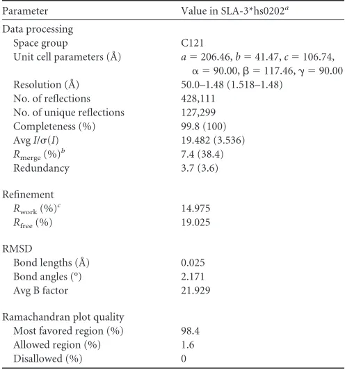
Parameter Value in SLA-3*hs0202a
Data processing
Space group C121
Unit cell parameters (Å) a⫽206.46,b⫽41.47,c⫽106.74,
␣ ⫽90.00, ⫽117.46,␥ ⫽90.00
Resolution (Å) 50.0–1.48 (1.518–1.48)
No. of reflections 428,111
No. of unique reflections 127,299
Completeness (%) 99.8 (100)
AvgI/(I) 19.482 (3.536)
Rmerge(%)
b 7.4 (38.4)
Redundancy 3.7 (3.6)
Refinement
Rwork(%)c 14.975
Rfree(%) 19.025
RMSD
Bond lengths (Å) 0.025
Bond angles (°) 2.171
Avg B factor 21.929
Ramachandran plot quality
Most favored region (%) 98.4
Allowed region (%) 1.6
Disallowed (%) 0
a
The numbers in parentheses indicate the highest-resolution shell.
bR
merge⫽ ⌺hkl⌺i|Ii(hkl)⫺ ⬍I(hkl)⬎|/⌺hkl⌺iIi(hkl), whereIi(hkl) is the observed
intensity and áI(hkl)ñ is the average intensity from multiple measurements.
cR⫽ ⌺
hkl||Fobs|⫺k|Fcalc||⌺hkl|Fobs|, whereRfreeis calculated for a randomly chosen
5% of reflections andRworkis calculated for the remaining 95% of reflections used for
structure refinement.
on November 7, 2019 by guest
http://jvi.asm.org/
[image:2.585.297.545.77.343.2]ual-dilution method, as previously described (38,39). As a negative con-trol, SLA-3*hs0202 and s2m were also refolded without peptide. After 48 h of incubation at 277 K, the soluble fraction of the complexes was con-centrated and then purified by size exclusion chromatography on a Su-perdex 200 16/60 column and by Resource-Q anion-exchange chroma-tography (GE Healthcare).
Crystallization and data collection.One peptide with the sequence KMNTQFTAV, derived from the HA protein of the 2009 pandemic H1N1 (A/Beijing/01/2009) strain (GenBank accession no.ACR54994.1), could form a stable complex with SLA-3*hs0202 followingin vitrorefolding and was selected for crystallization with the SLA-3*hs0202 heavy chain and s2m. The pSLA-3*hs0202 complexes were ultimately concentrated to 10 mg/ml in a buffer containing 20 mM Tris (pH 8.0) and 50 mM NaCl for crystallization. The sample was mixed with reservoir buffer at a 1:1 ratio and crystallized by the hanging-drop vapor diffusion technique at 291 K. A PEG/Ion kit (Hampton Research, Riverside, CA) was used to screen for optimal crystal growth conditions. After several days, crystals of SLA-3*hs0202 complexed with the HA-KMN9 peptide and s2m were ob-tained with PEG/Ion Screen solution 3 (20% [wt/vol] polyethylene glycol 3350, 0.2 M ammonium fluoride). Diffraction data were collected at a resolution of 1.48 Å using an in-house X-ray source (Rigaku Micro-Max007 desktop rotating anode X-ray generator with a Cu target operated at 40 kV and 30 mA) and an R-Axis IV⫹⫹imaging-plate detector at a wavelength of 1.5418 Å. The crystals were first soaked in reservoir solution containing 25% glycerol as a cryoprotectant and were then flash-cooled in a stream of gaseous nitrogen at 100 K (40). The collected intensities were indexed, integrated, corrected for absorption, scaled, and merged using the HKL2000 package (41).
Structure determination and refinement. The structures of the pSLA-3*hs0202 complex were solved by molecular replacement using the MOLREP program with SLA-1*0401 (Protein Data Bank [PDB] code
3QQ3) as a search model. Extensive model building was performed by
hand with COOT (42), and restrained refinement was performed using REFMAC5. Additional rounds of refinement were performed using the phenix refine program implemented in the PHENIX package (43) with isotropic atomic displacement parameter (ADP) refinement and bulk sol-vent modeling. The stereochemical quality of the final model was assessed with the PROCHECK program (44).
Circular-dichroism spectra.CD experiments for the pSLA-3*hs0202 complexes with mutant HA-KMN9 peptides were performed on a CD instrument (Chirascan; Applied Photophysics Ltd.) with a thermal con-troller. The far-UV CD spectra (180 to 260 nm) were collected at a protein concentration of 0.2 mg/ml in 20 mM Tris (pH 8.0) buffer, using a 1-mm-path-length cuvette with a 0.1-nm spectral resolution. The ellipticity at 218 nm was continuously recorded during heating. The water jacket cell containing the sample was heated at a linear rate of 1°C/min. Thermal-denaturation curves were determined by monitoring the CD value at 218 nm as the temperature was raised from 25 to 94°C at a rate of 1°C/min. The fractions of unfolded protein were calculated from the mean residue ellipticity () by the standard method. The unfolded fraction (%) is ex-pressed as ( ⫺ N)/(U⫺ N), whereNandUare the mean residue ellipticity values in the fully folded and fully unfolded states. The midpoint transition temperature (Tm) was determined by fitting data to the dena-turation curves using the Origin 8.0 program (Origin Lab) as described previously (45).
Biochemical assays and whole-genome screening of IAV strains.A total of 17 mutant HA-KMN9 peptides with single-residue substitutions at position 2 (P2), 3 (P3), or 9 (P9) was synthesized. Their binding affin-ities to SLA-3*hs0202 were tested byin vitrorefolding as described above. Finally, the candidate SLA-3*hs0202-restricted epitopes were screened throughout the whole genomes of four IAV strains, A/Beijing/01/2009 (H1N1), A/Texas/50/2012 (H3N2), A/chicken/Nigeria/SO494/2006 (H5N1), and A/Shanghai/4664T/2013 (H7N9), with the website NetMHCpan2.8server (35).
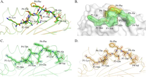
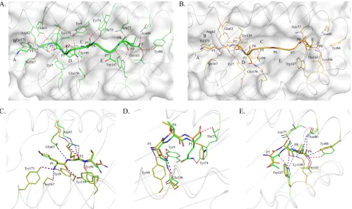
FIG 1Distinct structures of pSLA-3*hs0202 M1 and M2 in one asymmetric unit. (A) Superimposition of the overall structures of SLA-3*hs0202 M1 (green) and M2 (orange). They display a typical MHC-I structure but with obvious skewing in the␣1,␣2, and␣3 domains. (B) Superimposition of the HA-KMN9 peptides presented by M1 and M2, respectively. The maximum distance between the same atoms in the two peptides, which could reach 2.1 Å, is represented by the dashed red line.
on November 7, 2019 by guest
http://jvi.asm.org/
[image:3.585.79.506.65.337.2]Mutation of pSLA-3*hs0202 to pSLA-3*hs0202-Ala156.To investi-gate the function of Glu156 in SLA-3*hs0202, Glu156 was mutated to Ala156 by overlap PCR. The mutated gene, termed SLA-3*hs0202-Ala156, was inserted into the pET21a vector and expressed in BL21(DE3) cells. Recombinant SLA-3*hs0202-Ala156 was expressed in inclusion bodies and further purified, as described previously (38). SLA-3*hs0202-Ala156 was refolded with s2m and P3 mutant peptides of HA-KMN9 (P3 A, P3 Q, P3 R, P3 E, and P3 F). The complexes formed by refolding were further purified by anion-exchange chromatography as described above. In addition, the modeling structure of SLA-3*hs0202-Ala156 was built using SWISS-MODEL (http://swissmodel.expasy.org).
Accession numbers.The crystal structures have been deposited in the Protein Data Bank (http://www.pdb.org/pdb/home/home.do) with ac-cession number5H94; the sequence of SLA-3*hs0202 is available at the National Center for Biotechnology Information (NCBI) database under the accession numberKJ555032.
RESULTS
In silico
prediction and
in vitro
refolding of
SLA-3*hs0202-re-stricted IAV peptides.
Twenty-eight different SLA I genes were
cloned from Heishan pigs, and the restricted peptides from
pH1N1 HA and NP were predicted
in silico
(
http://www.cbs.dtu
.dk/services/NetMHCpan/
). The results suggested that
SLA-3*hs0202 could bind more IAV epitope peptides than other MHC
molecules. Then, six predicted epitope peptides from HA and NP
proteins were synthesized and tested for binding with
SLA-3*hs0202 by
in vitro
refolding. One peptide, HA-KMN9 (KMNT
QFTAV), could form a stable pMHC-I with SLA-3*hs0202 and
s

2m. Thus, the pSLA-3*hs0202 complex was further purified by
gel filtration and anion-exchange chromatography and used for
further experiments.
Distinct overall structures of two pSLA-3*hs0202 complexes.
SLA-3*hs0202 complexed with HA-KMN9 was crystallized in the
C121 space group with a high resolution of 1.48 Å (
Table 1
).
pSLA-3*hs0202 displayed a canonical pMHC-I structure, which
consisted of
␣
1,
␣
2, and
␣
3 domains of the heavy-chain and the
light-chain s

2m, and the peptide was located in the PBG formed
by the
␣
1 and
␣
2 domains (
Fig. 1A
). Within one asymmetric unit,
there are two SLA-3*hs0202 molecules, referred to below as M1
and M2, with a root mean square difference (RMSD) of 0.873 Å,
which is higher than the RMSD value of the two SLA-1*0401
(RMSD, 0.401; PDB code
3QQ3
) and BoLA-N*01801 (RMSD,
0.295; PDB code
3PWV
) molecules (
32
,
46
). The high RMSD
value indicated that the structures of M1 and M2 are not
coin-cident, and there should be some differences between them
(
Fig. 1B
).
The overall structural alignment of M1 and M2 showed that
their carbon backbones do not overlap. The skewing between M1
and M2 could be found in every domain of SLA-3*hs0202, and
these discordances not only occur in the flexible loops, but also
exist in the
␣
1 and
␣
2 helixes and the

-sheets of the
␣
3 domain
and s

2m (
Fig. 1A
). The
␣
1 and
␣
2 helixes were not perfectly
superimposed, even when only the peptide-binding domains were
compared. The two peptides presented by M1 and M2 were not
well aligned, and the distances between some atoms of the
pep-tides could reach 2.1 Å (
Fig. 1B
).
Different exposed areas of HA-KMN9 peptides presented by
pSLA-3*hs0202 M1 and M2.
The comparison showed that the
carbon backbones of the HA-KMN9 peptide in M1 and M2 are
not coincident (
Fig. 1B
). Further analysis showed that the
diver-FIG 2Distinct conformations and electronic densities of HA-KMN9 peptides found in pSLA-3*hs0202 M1 and M2. (A) Detailed comparison of HA-KMN9 peptides shown as a stick model (green, M1; orange, M2). The mismatch of the two peptides is obvious, especially at P6 Phe. (B) Different exposed areas of HA-KMN9 peptides. The exposed P4 Thr and P6 Phe of the M2-HA-KMN9 peptide are substantially higher than the two residues of the M1-HA-KMN9 peptide. (C and D) Electron density at the 1contour level of HA-KMN9 peptides shown in the pSLA-3*hs0202 M1 and M2 structures. The electron density maps are clear and indicate that the two distinct peptide conformations are reliable.on November 7, 2019 by guest
http://jvi.asm.org/
[image:4.585.46.543.67.328.2]gences occurred after P3 of the peptide (
Fig. 2A
). The P1 and P2
residues at the N termini of the two peptides are superimposed
perfectly, but the two P3 Asn residues show an obvious shift,
es-pecially in their side chains. The mismatch reached a peak at the
P6 residue, where the two Phe residues in M1 and M2 are the
maximum distance apart. After P5 Phe, the conformational
dif-ferences between the two peptides are diminished but still exist.
The P7 to P9 residues in M1 are located deeper in the PBG than
their counterparts in M2. Overall, the arch of the M2 peptide is
higher than that of the M1 peptide, but the length is shorter. The
side chains of P1, P4, P6, and P8 protruded from the PBG,
espe-cially P4 and P6, which are most likely recognized by specific
TCRs. The most striking divergence between the two peptides is in
the exposed region (
Fig. 2B
). The higher arch made the P4 and P6
residues of M2 more exposed than the two residues in the M1
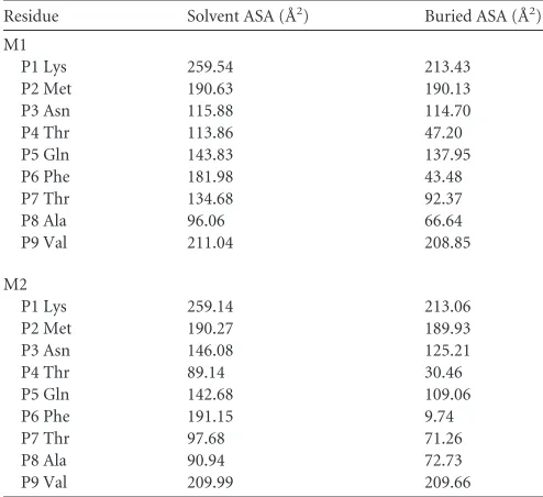
peptide. The exposed area (solvent-accessible surface area
[sol-vent ASA]) and buried area (buried ASA) of the M1 and M2
peptides are 1,447.5 and 1,114.8 Å, and 1,417.0 and 1,031.1 Å,
respectively, and detailed information about each residue is
provided in
Table 2
. The clear and complete electron density
maps of the two peptides indicate that these differences are
credible (
Fig. 2C
and
D
).
The phenomenon where peptides presented by the same
MHC-I molecule show different conformations has been found in
other known pMHC-I structures (
46
). Two nonapeptides
pre-sented by SLA-1*0401 showed similar M-like conformations, but
their main chains obviously diverge after the P3 residue, similar to
what we found in SLA-3*hs0202 (
Fig. 3A
). Using the same
pep-tide, BoLA-N*01801 also showed results similar to those for
SLA-3*hs0202. There are two BoLA-N*01801 molecules in one
asym-metric unit, and the peptide conformations are very different (
Fig.
3B
) (
46
). By comparing all the known pMHC-I structures, the M1
peptide conformation was found to be almost identical to that of
HLA-B*0801 (PDB code
1M15
) (
Fig. 3C
). The complex structure
of HLA-B*0801-TCR shows that the large side chain of P7 Tyr
interacts with the TCRs CDR1
␣
and CDR3

(
47
). In HA-KMN9
peptides of pSLA-3*hs0202, P6 Phe has a location and aromatic
ring similar to those of P7 Tyr of HLA-B*0801.
Different PBG conformations found in M1 and M2 and
var-ious interactions with the HA-KMN9 peptide.
The PBG
[image:5.585.39.286.87.313.2]confor-mations of M1 and M2 are shown in
Fig. 4
. The PBG of
pSLA-3*hs0202 consists of 6 pockets, similar to other known pMHC-I
structures (
48
). However, superimposition of the M1 and M2
PBGs showed that they have different conformations. The distinct
residues that directly interact with the HA-KMN9 peptide were
TABLE 2Accessible surface area (ASA) and buried surface area (BSA)calculations for HA-KMN9 peptides in M1 and M2 of pSLA-3*hs0202
Residue Solvent ASA (Å2) Buried ASA (Å2)
M1
P1 Lys 259.54 213.43
P2 Met 190.63 190.13
P3 Asn 115.88 114.70
P4 Thr 113.86 47.20
P5 Gln 143.83 137.95
P6 Phe 181.98 43.48
P7 Thr 134.68 92.37
P8 Ala 96.06 66.64
P9 Val 211.04 208.85
M2
P1 Lys 259.14 213.06
P2 Met 190.27 189.93
P3 Asn 146.08 125.21
P4 Thr 89.14 30.46
P5 Gln 142.68 109.06
P6 Phe 191.15 9.74
P7 Thr 97.68 71.26
P8 Ala 90.94 72.73
P9 Val 209.99 209.66
FIG 3Peptide presentation characteristics of pSlA-3*hs0202 compared to those of other pMHC molecules. (A and B) HA-KMN9 peptide conformations (thick sticks) compared to those of other peptides (thin sticks) with distinct conformations presented by the same MHC-I allele. (A) Comparison with peptides presented by SLA-1*0401 (PDB3QQ3, light pink; PDB3QQ4, cyan). (B) Comparison with the same peptide presented by two BoLA-A11 (N*01801) molecules in one asymmetric unit (PDB3PWU, pink and slate). (C) Peptide conformation similar to that of a peptide presented by HLA-B*0801 (PDB1M15, blue) to a specific TCR. Phe6 of the M1-HA-KMN9 peptide has a location and an aromatic ring similar to those of Tyr7 of the peptide presented by HLA-B*0801.
on November 7, 2019 by guest
http://jvi.asm.org/
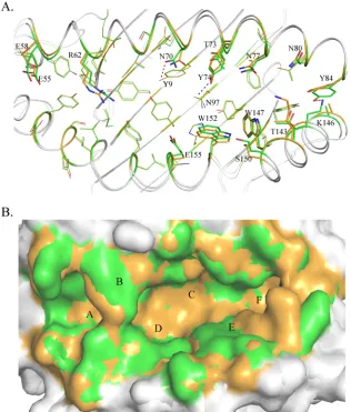
[image:5.585.43.545.477.673.2]selected and are shown as thick lines in the figure. The differences
in these residues are caused not only by shifts in their side chains,
but also by the mismatch of the
␣
1 and
␣
2 helixes. These distinct
residues are mainly located in the middle region and C-terminal
region of the PBG, where the helixes are highly divergent. Among
these residues, N70 and Y74 could form different hydrogen bonds
in M1 and M2. N70 forms a hydrogen bond only with Y9 in M1,
and Y74 could interact only with N97 in M2 (
Fig. 4A
).
Superim-position in a surface model (
Fig. 4B
) shows the side wall in the
middle of the overlapped PBGs (mainly in green) and the bottom
of the PBG (mainly in orange). This indicates that the PBG of M1
is narrower but deeper than the M2 PBG, which is consistent with
the finding that the
␣
1 and
␣
2 helixes in M1 are closer than in M2
(
Fig. 4B
).
The different PBG conformations of M1 and M2 lead to
vari-ous peptide interactions (
Fig. 5
). In M1, there are 19 hydrogen
bonds between the PBG and the HA-KMN9 peptide (
Fig. 5A
), but
in M2, the number is 16 hydrogen bonds (
Fig. 5B
). In the A and B
pockets of M1 and M2, the interactions with P1 Lys and P2 Met are
the same (
Fig. 5C
). The differences are mainly found in the middle
of the groove and in the C-terminal region. In M1, P3 Asn formed
two hydrogen bonds, with Glu156 and Tyr9, but in M2, P3 Asn
formed a hydrogen bond only with Glu156 (
Fig. 5D
). P5 Gln
formed two hydrogen bonds, with Thr73 and Tyr74, in M1 but did
not form any hydrogen bonds in M2 (
Fig. 5D
and
Table 3
). In the
F pockets, Trp147 could bind to P7 and P8 in M1 but could
interact only with P8 in M2. Lys146 formed only two hydrogen
bonds with P8 and P9 in M2 (
Fig. 5E
). In addition to the
hy-drogen bonds, the interactions formed by van der Waals forces
(VDWs) are also different in the two SLA-3*hs0202 H chains.
The total numbers of VDWs were 373 and 299 in M1 and M2,
respectively. Complete interaction information is provided in
Table 3
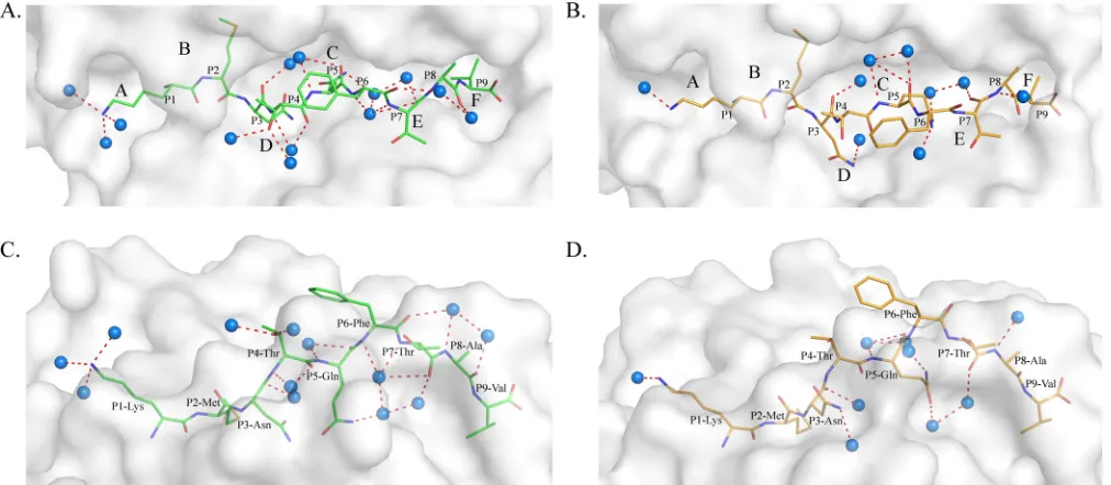
. Moreover, the water molecules involved in the
hydro-gen bond network between the PBG and the HA-KMN9
pep-tide are different in M1 and M2. In M1, 13 water molecules
form 17 hydrogen bonds with the HA-KMN9 peptide (
Fig. 6A
and
C
). In M2, only 9 water molecules and 10 direct bonds are
found (
Fig. 6B
and
D
). The diverse water molecules could fit
FIG 4Different conformations of HA-KMN9 peptides in the binding grooves of M1 and M2. (A) Superimposition of M1 and M2 PBGs in a stick model. The distinct residues that directly interact with the HA-KMN9 peptide are labeled and shown as thick sticks. They are mainly located in the middle and at the C terminus of the PBG, where the␣1 and␣2 helixes are mismatched. Two different hydrogen bonds formed by these residues are shown as dashed lines (M1, red; M2, blue). (B) Structural superimposition of M1 and M2 PBGs in a surface model. The six pockets (A, B, C, D, E, and F) are labeled in the PBGs. The M2 PBG is deeper and narrower than the M1 PBG, based on the fact that most of the bottom is yellow and the side wall is green.on November 7, 2019 by guest
http://jvi.asm.org/
[image:6.585.135.451.67.438.2]into the different PBG conformations of M1 and M2 to stabilize
the distinct peptide conformations (
49
).
Anchor residues determined by alanine scanning and CD
spectra.
Although the conformations of HA-KMN9 peptides in
M1 and M2 are different, the accommodation of residues by the 6
pockets is roughly the same. The major proportion of P1, P2, P3,
P5, and P9 in the HA-KMN9 peptide is accommodated by the A,
B, D, C, and F pockets, respectively (
Fig. 5
). To determine the
primary anchor residues, these five residues were tested by alanine
scanning (
Fig. 7
). The P1 Ala mutant peptide could efficiently
form a complex with SLA-3*hs0202, similar to the wild-type
HA-KMN9 peptide. As in the known pMHC-I structures, P1 Lys binds
the A pocket with its main chain (
Fig. 5C
) (
45
). Therefore,
al-though the binding between P1 and the A pocket is strong, the
substitution of the P1 residue cannot affect the peptide binding.
For the other four residues, interactions with the pockets largely
depend on their side chains. The variation of these residues may
affect the binding between HA-KMN9 and the PBG of
SLA-3*hs0202. The
in vitro
refolding results for these mutant peptides
showed that mutation of P2, P3, and P9 decreased the refolding
efficiency and stability of the complex (
Fig. 7A
and
B
).
Interest-ingly, although the M1 structure showed P5 Gln could form two
hydrogen bonds with pocket C, the refolding efficiency of P5 Ala
was not affected.
CD spectra were further used to test the stabilities of
pSLA-3*hs0202 complexes with the mutant peptides (
Fig. 7C
). The
T
mvalue of the wild-type HA-KMN9 peptide is 47.4°C, similar to the
T
mvalue of pSLA-1*0401, which we tested previously (47.1°C).
The results indicated that the binding between the HA-KMN9
peptide and SLA-3*hs0202 is quite stable. The
T
mvalue of the
SLA-3*hs0202 and P2 Ala mutant peptide complex
(pSLA-3*hs0202-
P2A) is 44.3°C, which is slightly lower than that of the
wild-type HA-KMN9 and SLA-3*hs0202, demonstrating that
the side chains of P2 residues contributed to the peptide binding.
The
T
mvalues of pSLA-3*hs0202-
P3Aand pSLA-3*hs0202-
P9Aare
40.0°C and 38.3°C, respectively, which are significantly decreased
compared to that of the wild-type HA-KMN9. The results showed
that the side chains of P3 and P9 play key roles in HA-KMN9
peptide binding and that these two residues are the primary
an-chor residues. The
T
mvalue of pSLA-3*hs0202-
P5Ais 48.6°C and is
higher than that of the wild-type complex. This suggested that the
interactions formed by the side chains of P5 residues are not
es-sential for the stability of pSLA-3*hs0202. Based on these results,
we determined that the P2, P3, and P9 residues are the primary
anchor residues of epitope peptides presented by SLA-3*hs0202.
The peptide-binding motif of SLA-3*hs0202.
The
peptide-binding motif of SLA-3*hs0202 is the set of acceptable
combina-tions of P2, P3, and P9 residues, because they are the primary
FIG 5Different peptide interactions found in pSLA-3*hs0202. (A and B) Hydrogen bonds found in pSLA-3*hs0202 M1 (A) and M2 (B) (both shown as dashed red lines). (C) Comparison of the interactions of P1 Lys and P2 Met in the A and B pockets (the N terminus of PBG), respectively. The numbers and positions of hydrogen bonds are the same in M1 and M2. (D) Comparison of the interactions of P3 Asn and P5 Gln in the D and C pockets (the middle of the PBG), respectively. P3 Glu forms two hydrogen bonds with Tyr99 in M1 (dashed red lines) but forms only one hydrogen bond with Tyr99 in M2 (dashed blue line). P5 Gln forms two hydrogen bonds with Thr73 and Tyr74 in M1 but does not form any hydrogen bonds in M2. (E) Comparison of the interactions of P7 Thr, P8 Ala, and P9 Val in the E and F pockets (the C terminus of PBG). Trp147 could form two hydrogen bonds with P7 and P8 in M1 (dashed red lines) but only one hydrogen bond with P8. Lys146 could form two hydrogen bonds in M2 (dashed blue lines) but could not form any hydrogen bonds in M1.on November 7, 2019 by guest
http://jvi.asm.org/
[image:7.585.42.545.66.366.2]anchor residues for the binding peptides. The B, D, and F pockets
accommodated the side chains of P2, P3, and P9, respectively, so
they are very critical to determine the peptide-binding motif of
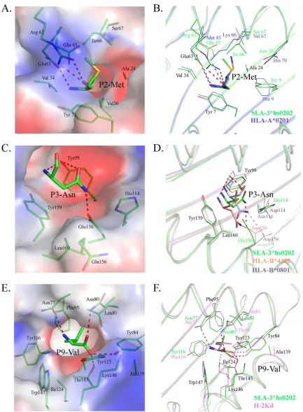
SLA-3*hs0202. The B pocket consists of Tyr7, Ala24, Val25,
Gly26, Val34, Glu45, Arg62, Glu63, Ile66, and Ser67, and its
po-larity is shown in
Fig. 8A
. The long side chain of Arg62 stretches to
the
␣
2 helix and seals the top of the B pocket. There are three
charged residues (Glu45, Arg62, and Glu63) in the B pocket, and
two of them form hydrogen bonds with the main-chain atoms of
P2 M. The side chain of P2 M stretches to the
␣
1 helix and is
inserted into the B pocket, and it is surrounded by uncharged
amino acids, except for Glu45. The mutant HA-KMN9 peptides
with charged P2 E or P2 R were used to test the influence of Glu45,
and the refolding results showed that the binding of P2 R is better
than that of P2 E, but both of them are not as efficient as the
[image:8.585.41.539.77.529.2]wild-type HA-KMN9 peptide (
Fig. 9A
). P2 M as a primary anchor
residue is rare in the other known pMHC-I structures, and only
HLA-A*0201 has been found to present peptides with P2 M,
sim-ilar to SLA-3*hs0202 (
50
). Although the residues in the B pockets
of SLA-3*hs0202 and HLA-A*0201 are not same, they bind P2 M
in very similar manners (
Fig. 8B
). The B pocket of HLA-A*0201
could accommodate uncharged residues, such as M, L, V, I, T, and
F. We used the P2 V/L/I/T/F mutant HA-KMN9 peptides to verify
whether the binding motif of the SLA-3*hs0202 B pocket is the
same as that of the HLA-A*0201 B pocket. The refolding results
showed that SLA-3*hs0202 could not bind these mutant
pep-tides as efficiently as the wild-type KMN peptide (
Fig. 9A
and
B
). Only the P2 A/R mutant peptides could form stable
com-plexes that survived the strong ionic buffer during anion
ex-change (
Fig. 7B
and
9B
). Our results confirmed that P2 M/A/R
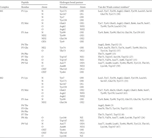
TABLE 3Hydrogen bonds and van der Waals interactions between HA-KMN9 peptides and peptide-binding domains of M1 and M2 moleculesComplex
Peptide Hydrogen bond partner
Van der Waals contact residuesa
Residue Atom Residue Atom
M1 P1 Lys N Tyr171 OH Leu5, Tyr7, Tyr59, Arg62, Glu63, Tyr59, Leu163, Ser167,
Glu170, Tyr171 (59)
N Ser167 OG
N Tyr7 OH
O Tyr159 OH
P2 Met N Glu63 OE1 Tyr7, Tyr9, Glu45, Arg62, Glu63, Ile66, Asn70, Ser67,
Tyr99, Tyr59, Leu163 (74)
O Arg62 NH1
O Arg62 NH2
P3 Asn N Tyr99 OH Tyr9, Ile66, Tyr99, His114, Glu156, Tyr159 (65)
ND2 Tyr99 OE1
ND2 Glu156 OE2
OD1 Tyr9 OH
P4 Thr Arg62, Ile66, Trp152 (5)
P5 Gln NE2 Tyr74 OH Tyr9, Asn70, Thr73, Tyr74, Asn97, Tyr99, His114,
Tyr116, Trp152 (37)
O Thr73 OG1
P6 Phe Thr73, Asp69 (13)
P7 Thr O Trp147 NE1 Thr73, Trp147, Ser150, Trp152 (27)
P8 Ala O Trp147 NE1 Thr73, Val76, Asn77, As80, Trp147 (15)
P9 Val N Asn77 OD1 Asn77, Asn80, Leu81, Tyr84, Phe95, Tyr123, Thr143,
Lys146, Trp147 (59)
O Asn80 ND2
OXT Thr143 OG1
OXT Tyr84 OH
M2 P1 Lys N Tyr7 OH Leu5, Tyr7, Tyr59, Arg62, Glu63, Tyr159, Leu163,
Ser167, Glu170, Tyr171 (53)
N Tyr171 OH
N Ser167 OG
O Tyr159 OH
P2 Met N Glu63 OE2 Tyr7, Tyr9, Ala24, Glu45, Arg62, Glu63, Ile66, Ser67,
Tyr99, Tyr159, Leu163 (63)
O Arg62 NH1
O Arg62 NH2
P3 Asn N Tyr99 OH Tyr9, Ile66, Tyr99, Trp152, Gln155, Glu156, Tyr159 (48)
ND2 Glu156 OE2
P4 Thr Arg62, Ile66 (3)
P5 Gln Asn70, Thr73, Tyr116, Trp152 (18)
P6 Phe
P7 Thr Thr73, Trp147, Trp152 (8)
P8 Ala O Lys146 NZ Thr73, Val76, Asn77, As80, Lys146, Trp147 (24)
O Trp147 NE1
P9 Val N Asn77 OD1 Asn77, Asn80, Leu81, Tyr84, Phe95, Tyr123, Thr143,
Lys146, Trp147 (67)
O Asn80 ND2
OXT Tyr84 OH
OXT Thr143 OG1
O Lys146 NZ
a
The numbers in parentheses are the amounts of van der Waals force.
on November 7, 2019 by guest
http://jvi.asm.org/
is suitable for binding the B pocket of SLA-3*0202 and that P2
M is the better choice.
The D pocket is composed of Tyr99, His114, Gln155, Glu156,
Tyr159, and Leu160 and showed obvious negatively charged
po-larity (
Fig. 8C
). Glu156 could form a hydrogen bond with the side
chain of P3 N, and this interaction is critical to anchor the peptide
because the P3 A mutant attenuates the peptide-binding stability.
Tyr99 could form two hydrogen bonds with the P3 residue in M1
and is important in stabilizing the peptide; however, it does not
restrict the scope of the binding peptide because it contacts the
main chain of the P3 residue. HLA-B*0801 and HLA-B*4402 have
D pockets similar to those of SLA-3*hs0202 (
51
,
52
). Their D
pockets can accommodate P3 N and bind it in a manner similar to
that of SLA-3*hs0202 (
Fig. 8D
). These three D pockets have
sim-ilar compositions, including a negatively charged residue in the
same position (156); therefore, they should have similar
prefer-ences for P3 residues. The D pockets of B*0801 and
HLA-B*4402 could accommodate R, N, and F. The P3 Q/R/F/E mutant
HA-KMN9 peptides were used to test the binding motif of the D
pocket of SLA-3*hs0202 (
Fig. 9C
and
D
). The refolding results
showed that the P3 Q mutant peptide could bind to SLA-3*hs0202
as well as the wild-type HA-KMN9 peptide. The P3 R mutant
peptide could also form a stable complex with SLA-3*hs0202, but
its refolding efficiency was lower than that of the wild-type and P3
Q mutant peptides. The refolding efficiency of the P3 F mutant
peptide was better than that of the P3 R mutant peptide, but the
complex formed by the P3 F peptide was not stable and was mostly
dissociated during anion exchange. As expected, refolding of the
P3 E mutant peptide was the least efficient among these four
mu-tant peptides. These results suggested P3 N and Q are the best
choices, and P3 R and F were also acceptable for the D pocket of
SLA-3*hs0202.
Our previous work proved that the charged Arg156 of
SLA-1*0401 is vital to the peptide binding of the D pocket. Here,
Glu156 is also a charged residue in the D pocket of SLA-3*hs0202.
To clarify the role of Glu156, a mutated heavy chain,
SLA-3*hs0202-Ala156 was co-refolded with the HA-KMN9 and the P3
mutant peptides. The refolding results showed that the binding
properties of HA-KMN9 and P3 Q mutant peptides are not much
affected by Ala156 mutation, and P3 A, F, and E peptides bind
with SLA-3*hs0202-Ala156 better than with SLA-3*hs0202. Only
the P3 R peptide could not bind with SLA-3*hs0202-Ala156 as
well as the wild-type heavy chain (
Fig. 10A
). By comparison with
SLA-3*hs0202, the modeling structure of SLA-3*hs0202-Ala156
was shown to have a bigger and more hydrophobic D pocket (
Fig.
10B
and
C
). This change made the D pocket of
SLA-3*hs0202-Ala156 prefer the hydrophobic P3 residues, such as P3 A and P3 F,
and have weaker resistance to P3 E. Therefore, these three peptides
can bind with the mutant heavy chain better than with the
wild-type chain. The mutant D pocket cannot form the salt bridge with
P3 R, so it cannot bind well with the P3 R peptide. The results
indicate that Glu156 is a vital residue for the peptide motif of
SLA-3*hs0202.
The F pocket is composed of the conserved residues Tyr84,
Tyr123, Thr143, Lys146, and Trp147, as well as the less conserved
residues Asn77, Asn80, Leu81, Phe95, Ser97, Tyr116, Ile124, and
Ala139. It showed strong hydrophobic properties (
Fig. 8E
). The
nonpolar side chain of P9-V stretched into the F pocket and
formed many VDWs with the surrounding residues on the
bot-tom and wall of the pocket (
Table 3
). Four hydrogen bonds are
formed by the main chain of P9 V and the residues at the top of the
F pocket. The F pocket of H-2Kd could accommodate P9 V in a
similar manner (
Fig. 8F
) and could also bind other P9 residues,
such as I, L, and F (
53
). Therefore, mutant HA-KMN9 peptides
with P9 I/L/F were used to test the binding of the SLA-3*hs0202 F
pocket (
Fig. 9E
and
F
). The refolding efficiencies of P9 mutant
peptides were lower the those of the wild-type HA-KMN9 peptide,
and only P9 I could form a stable complex that could survive in the
FIG 6Different water molecules found in pSLA-3*hs0202 M1 and M2 structures. (A and C) Water molecules of pSLA-3*hs0202 M1 in a top view (A) and a side view (C); 13 water molecules form 17 hydrogen bonds with the HA-KMN9 peptide. (B and D) Water molecules of SLA-3*hs0202 M2 in a top view (B) and a side view (D); 9 water molecules in M2 form 10 direct bonds. The blue balls represent water molecules.on November 7, 2019 by guest
http://jvi.asm.org/
[image:9.585.41.547.67.288.2]anion-exchange buffer. These results suggest P9 V is the most
suitable anchor residue for the F pocket of SLA-3*hs0202, and P9
I is another possible choice. Therefore, the 9-mer peptide-binding
motif of SLA-3*hs0202 was preliminarily determined to be X-(M/
A/R)-(N/Q/R/F)-X-X-X-X-X-(V/I).
Biochemical data predicted in H1N1, H3N2, H7N9, and
H5N1 strains.
Based on the preliminary motif described above,
we screened the whole protein sequences of four current
epi-demic IAV strains: A/Beijing/01/2009 (H1N1), A/Texas/50/2012
(H3N2), A/chicken/Nigeria/SO494/2006 (H5N1), and A/Shanghai/
4664T/2013 (H7N9) (
54–57
). There were 28 total 9-mer epitope
peptides that were predicted to fit the motif. Six of them were
completely conserved in the H1N1, H3N2, H5N1, and H7N9
strains, and five have amino acid substitutions among different
strains (
Fig. 11A
). Two conserved peptides, RMQFSSLTV
(PB2-RMQ9) and RRNYFTAEV (PA-RRN), were selected to test their
affinities to SLA-3*hs0202 by
in vitro
refolding, and the results
confirmed that both of them could form stable complexes with
SLA-3*hs0202 (
Fig. 11B
and
C
). Both HA-KMN9 and PB2-RMQ9
were considered HLA-A*0201-restricted epitopes, but only
PB2-RMQ9 could stimulate an effective CTL response after injection
(
58
). None of the other peptides we predicted here have been
deposited in the IEDB.
DISCUSSION
Distinct conformations of peptides presented by the same MHC
class I molecule, and even the same peptide, have been found in
mammalian pMHC-I structures (
32
,
46
,
59
). The pMHC-I
struc-tures of swine, cattle, and rhesus macaques show that the flexible
PBG adopts various conformations to accommodate the peptides,
leading to the distinct peptide conformations (
32
,
46
,
59
). It is
believed that the various peptide conformations presented by the
same MHC-I could elicit a broad range of CTL responses (
60
).
The distinct peptide conformations are mainly caused by the
flex-ible side chains of the pockets and do not involve the
␣
-helixes and

-sheets that form the PBG. Our data showed that the HA-KMN9
peptide displays different conformations in M1 and M2.
How-ever, the distinct HA-KMN9 peptide conformations are not only
FIG 7Anchor residues of the HA-KMN9 peptide verified by alanine scanning and CD spectra. (A) Gel filtration chromatograms of the refolded products of the HA-KMN9 peptide and its Ala mutants. Peak 1, peak 2, and peak 3 represent the aggregated H chain, the correctly refolded SLA-3*hs0202 complex (44 kDa), and the redundant s2m, respectively. The refolding efficiencies are represented by the relevant concentration ratios and heights of peak 2 formed by complexes with mutant peptides. A high peak 2 indicates high efficiency of the peptide to help the MHC renature. However, a low or no peak 2 indicates that the peptide cannot stabilize the complex. (B) The stabilization of refolded complexes with mutant peptides was further tested by anion exchange. Under the anion-exchange conditions, complexes with P1 A and P5 A mutant peptides can be eluted at NaCl concentrations of 14 to 16% as well as the complex with the wild-type peptide. The peak of the complex with the P2 A mutant peptide is lower than the three high peaks and indicates less stability. With peptides P3 A and P9 A, the refolded complex proteins dissociated at NaCl concentrations of 14 to 16%, implying very poor stability. (C) The thermostabilities of SLA-3*hs0202 with five peptides (wild-type HA-KMN9 peptide, P2 A, P3 A, P5 A, and P9 A) were tested by CD spectroscopy. The temperature was increased by 1°C/min. The curves for the unfolded fractions were determined by monitoring the CD value at 218 nm (45).on November 7, 2019 by guest
http://jvi.asm.org/
[image:10.585.111.474.60.392.2]FIG 8Compositions of the B, D, and F pockets of SLA-3*hs0202 with other solved pMHC-I structures. The residues comprising the pockets and the residues of bound peptides accommodated by the pockets are shown as stick models. The hydrogen bonds between the peptides and the pockets are shown as dashed red lines. (A) The electrostatic potential of pocket B with the P2 residue (red, negative; blue, positive; gray, neutral) is shown, with the HA-KMN9 peptide in green. (B) B pocket alignment between SLA-3*hs0202 and HLA-A*0201 (PDB3HPJ), which both accommodate P2 Met. (C) Electrostatic potential of pocket D with the P3 residue. (D) D pocket alignment between SLA-3*hs0202 with HLA-B*4402 (PDB3DX6) and HLA-B*0801 (PDB4QRT). They all bind P3 Asn in similar manners. (E) Electrostatic potential of pocket F with the P9 residue. (F) F pocket alignment between SLA-3*hs0202 and H-2Kd (PDB2FWO), which both accommodate P9 Val or P9 Ile.
on November 7, 2019 by guest
http://jvi.asm.org/
[image:11.585.76.509.63.652.2]FIG 9Determination of the peptide-binding motif of SLA-3*hs0202 byin vitrorefolding with mutant peptides. (A) Gel filtration chromatograms of the wild-type HA-KMN9 peptide and its P2 mutants (P2 R, P2 E, P2 F, P2 T, P2 I, P2 L, and P2 V). The P2 R (magenta) peptide is more efficient than other mutant peptides in helping MHC to renature, but its efficiency is lower than that of the wild-type HA-KMN9 peptide. (B) Results from further stabilization assays of the refolded complexes with P2 mutants by anion exchange. Consistent with the results shown inFig. 8A, only partial complexes of P2 R can be eluted at normal NaCl concentrations of 14 to 16%, implying less stability. (C) Gel filtration chromatograms of the HA-KMN9 peptide and its P3 mutants (P3 Q, P3 F, P3 R, and P3 E). P3 Q (red) efficiently helps the MHC to renature as well as the wild-type peptide, and the P3 F (blue) peptide has the second highest peak. (D) Results from further stabilization assays of the refolded complexes with P3 mutant peptides by anion exchange. The complex with P3 Q could be eluted normally at NaCl concentrations of 14 to 16%, but very little of the complex with P3 F survived. (E) Gel filtration chromatograms of the wild-type peptide and its P9 mutants (P9 I, P9 L, and P9 F). The P9 I (magenta) and P9 F (blue) peptides can help the MHC renature. (F) Results from further stabilization assays of the P9 mutants by anion exchange. Only a small amount of P9 I could be eluted normally at NaCl concentrations of 14 to 16%.
on November 7, 2019 by guest
http://jvi.asm.org/
[image:12.585.43.540.64.591.2]caused by the side chains in the PBG, but also by the skewing of the
␣
1 and
␣
2 helixes. SLA-3*hs0202 M1 and M2 are in one
asym-metric unit, but the differences in their overall structures are
ob-vious. The RMSD between M1 and M2 is 0.873 Å, which is higher
than the RMSD of the SLA-1*0401 (0.401 Å) and BoLA-N*01801
(0.267 Å) molecules, which are also within one asymmetric unit
(
32
,
46
). As described in detail in Results above, these
discor-dances not only occur in the flexible loops but are also present in
the
␣
1 and
␣
2 helixes and the

-sheets of the
␣
3 domain and s

2m
(
Fig. 1A
), which has not been described in the structures of
SLA-1*0401 and other molecules. The major differences in the
HA-KMN9 peptides occur at the mismatches of the
␣
-helixes of M1
and M2. This finding indicates that, besides the side chains of the
PBG, the flexibility of the swine MHC class I carbon backbone
might expand the peptide conformation and facilitate the
activa-tion of an increased TCR repertoire.
Pockets B, D, and F accommodate the side chains of the P2, P3,
and P9 residues, respectively, and determine the peptide-binding
motif of SLA-3*hs0202. In addition to the canonical B and F
pock-ets of HLA molecules, the D pocket of SLA-3*hs0202 also plays a
vital role in binding peptides. This is similar to SLA-1*0401, in
which the D pocket is the major limitation to peptide binding
(
32
). In the D pocket, residue 156 interacting with the P3 peptide
residue is critical to determine the motif in both SLA-3*hs0202
and SLA-1*0401. The peptide-binding motif determined by the B,
D, and F pockets may be common among SLA I alleles.
The conformations of the SLA-3*hs0202 pockets are unique,
but similar pockets were found in other solved pMHC-I
struc-tures, such as the B pocket of A*0201, the D pockets of
HLA-B*4401 and HLA-B*0801, and the F pocket of H2-Kd (
50–53
).
These pockets could accommodate the same residues of the
HA-KMN9 peptide in similar manners. Therefore, we tested the
pep-tide-binding motif of SLA-3*hs0202 based on the known binding
residues. Indeed, the strategy we used to determine the motif of
SLA-3*hs0202 is same one used to classify the HLA I supertypes
and to predict allele-specific epitopes
in silico
. The
in vitro
refold-ing results showed that some candidate residues are acceptable to
SLA-3*hs0202, at least in mutant HA-KMN9 peptides, and the
9-mer peptide-binding motif of SLA-3*hs0202 was determined
to be X-(M/A/R)-(N/Q/R/F)-X-X-X-X-X-(V/I). The structural
pocket alignment showed accuracy in determining the
peptide-binding motif of a new MHC-I allele.
Based on our analysis and previous studies, the
peptide-bind-ing motif of SLA-3*hs0202 partly overlaps that of the most
popu-lar HLA allele, HLA-A*0201, which prefers L or M in position 2
and I or V at the C terminus (
50
). Previous studies demonstrated
that HA-KMN9 and PB2-RMQ9 peptides derived from other
sub-types of IAV are HLA-A*0201-restricted epitope peptides (
58
).
The sequences of the HA-KMN9 peptide derived from the PR8
and Qinghai IAV strains are KMNIQFTAV and KMNTQFEAV,
with a substitution of P4/P8 residues in the HA-KMN9 peptide
from H1N1, respectively (
19
,
58
). However, the anchor residues
FIG 10Comparative analysis of D pockets between SLA-3*hs0202 and SLA-3*hs0202-Ala156 mutants. (A) Anion-exchange chromatography results for HA-KMN9 and its P3 N mutant peptides co-refolding with SLA-3*hs0202 and SLA-3*hs0202-Ala156. (B) D pocket of pSLA-3*hs0202 in the electrostatic potential surface model (red, negative; blue, positive; gray, neutral). Glu156 is shown in red as a stick model. (C) 3D model of the pSLA-3*hs0202-Ala156 D pocket built by the SWISS-MODEL program. Ala156 is shown in red as a stick model. Compared with that of SLA-3*hs0202, the D pocket of SLA-3*hs0202-Ala156 became bigger and more hydrophobic without the negative side chain of Glu156.on November 7, 2019 by guest
http://jvi.asm.org/
[image:13.585.49.545.66.373.2]are conserved in all the HA-KMN9 peptides. Both HA-KMN9
peptides from the PR8 and Qinghai strains cannot elicit significant
CTL responses in HLA-A*0201 transgenic mice (
19
,
58
). This may
be caused by the intermediate affinity between the HA-KMN9
peptide and HLA-A*0201, which was tested by both quantitative
inhibition assays and
in vitro
refolding, because the low affinity
could result in impaired pMHC stability and loss of
immunoge-nicity. However, in our experiment, the HA-KMN9 peptide
showed high binding affinity for SLA-3*hs0202, and its
immuno-genicity to SLA-3*hs0202 pigs may be different from that of
HLA-A*0201 individuals. The sequence of the PB2-RMQ9 peptide from
the PR8 strain is RMQFSSFTV, with a substitution at the P7
resi-due (F/L) compared to the pH1N1 peptide we used. The PR8
PB2-RMQ9 peptide has a high affinity for HLA-A*0201 and could
elicit CTL responses after the injection of the peptide (
58
).
Nota-bly, the HA-KMN9 peptide is part of a B cell epitope located at
HA391 to HA410 and has been documented in previous studies
(
61–63
). The PB2-RMQ9 peptide we used has the same anchor
residues and high affinity for SLA-3*hs0202. The substitution
lo-cated in the region recognized by TCR may affect its
immunoge-nicity, but L also has a long side chain that forms the “featured”
conformation. Other peptides we identified here have not been
described previously, and they are conserved in different subtypes
of IAV, especially the peptides from the PB1, PB2, PA, NP, and M1
proteins.
In brief, the first structure of the SLA-3 allele determined here
should help increase our understanding of antigen presentation in
swine, and the “epitope map” for SLA-3*hs0202 can benefit the
development of CTL vaccines against IAV diseases.
ACKNOWLEDGMENTS
This work was supported by the 863 Project of the China Ministry of Science and Technology (grant no. 2013AA102503), the State Key Pro-gram of the National Natural Science Foundation of China (grant no. FIG 11Distribution of SLA-3*hs0202-restricted epitope peptides in important IAV strains. (A) Genome-wide scanning for SLA-3*hs0202-restricted peptides in four IAV strains, A/Beijing/01/2009 (H1N1), A/Texas/50/2012 (H3N2), A/chicken/Nigeria/SO494/2006 (H5N1), and A/Shanghai/4664T/2013 (H7N9). Twenty-eight nonapeptides derived from the PB1, PB2, PA, HA, NA, NP, M1, and NS1 proteins were selected based on the motif X-(M/A/R)-(N/Q/R/F)-X-X-X-X-X-(V/I). (B) Gel filtration chromatograms of peptides RMQFSSLTV and RRNYFTAEV. (C) Further stabilization assays of the two peptides by anion exchange. The results confirmed that both could form stable complexes with SLA-3*hs0202.
on November 7, 2019 by guest
http://jvi.asm.org/
[image:14.585.40.543.67.457.2]31201887), the 973 Project of the China Ministry of Science and Technol-ogy (grant no. 2013CB835302), and the Youth Teacher Scientific Research and Innovation Program of Zhoukou Normal University (grant no. ZKNUB115208).
We thank the Shanghai Synchrotron Radiation Facility (SSRF), Shanghai, People’s Republic of China, for diffraction data on the crystals.
We declare no conflict of interest.
FUNDING INFORMATION
This work, including the efforts of Chun Xia, was funded by The 863 Proj-ect of the China Ministry of Science and Technology (2013AA102503). This work, including the efforts of Nianzhi Zhang, was funded by Na-tional Science Foundation of China (31201887). This work, including the efforts of Chun Xia, was funded by The 973 Project of the China Ministry of Science and Technology (2013C835302). This work, including the ef-forts of Shuhua Fan, was funded by The Youth Teacher Scientifc Research and Innovation Program of Zhoukou Normal University (ZK-NUB115208).
The funders had no role in study design, data collection and interpreta-tion, or the decision to submit the work for publication.
REFERENCES
1.Ducatez MF, Hause B, Stigger-Rosser E, Darnell D, Corzo C, Juleen K, Simonson R, Brockwell-Staats C, Rubrum A, Wang D, Webb A, Crumpton JC, Lowe J, Gramer M, Webby RJ.2011. Multiple reassort-ment between pandemic (H1N1) 2009 and endemic influenza viruses in pigs, United States. Emerg Infect Dis17:1624 –1629.http://dx.doi.org/10
.3201/1709.110338.
2.Naffakh N, van der Werf S.2009. April 2009: an outbreak of swine-origin influenza A(H1N1) virus with evidence for human-to-human transmis-sion. Microbes Infect11:725–728.http://dx.doi.org/10.1016/j.micinf .2009.05.002.
3.Nelson MI, Stratton J, Killian ML, Janas-Martindale A, Vincent AL. 2015. Continual reintroduction of human pandemic H1N1 influenza A viruses into swine in the United States, 2009 to 2014. J Virol89:6218 – 6226.http://dx.doi.org/10.1128/JVI.00459-15.
4.Chen W, Calvo PA, Malide D, Gibbs J, Schubert U, Bacik I, Basta S, O’Neill R, Schickli J, Palese P, Henklein P, Bennink JR, Yewdell JW. 2001. A novel influenza A virus mitochondrial protein that induces cell death. Nat Med7:1306 –1312.http://dx.doi.org/10.1038/nm1201-1306. 5.Kingsford C, Nagarajan N, Salzberg SL.2009. 2009 Swine-origin
influ-enza A (H1N1) resembles previous influinflu-enza isolates. PLoS One4:e6402.
http://dx.doi.org/10.1371/journal.pone.0006402.
6.Wise HM, Foeglein A, Sun J, Dalton RM, Patel S, Howard W, Anderson EC, Barclay WS, Digard P.2009. A complicated message: identification of a novel PB1-related protein translated from influenza A virus segment 2 mRNA. J Virol83:8021– 8031.http://dx.doi.org/10.1128/JVI.00826-09. 7.Stincarelli M, Arvia R, De Marco MA, Clausi V, Corcioli F, Cotti C, Delogu M, Donatelli I, Azzi A, Giannecchini S. 2013. Reassortment ability of the 2009 pandemic H1N1 influenza virus with circulating hu-man and avian influenza viruses: public health risk implications. Virus Res 175:151–154.http://dx.doi.org/10.1016/j.virusres.2013.04.012. 8.Tao H, Steel J, Lowen AC.2014. Intrahost dynamics of influenza virus
reassortment. J Virol 88:7485–7492. http://dx.doi.org/10.1128/JVI .00715-14.
9.Claas EC, Kawaoka Y, de Jong JC, Masurel N, Webster RG. 1994. Infection of children with avian-human reassortant influenza virus from pigs in Europe. Virology204:453– 457.http://dx.doi.org/10.1006/viro .1994.1553.
10. Ito T, Couceiro JN, Kelm S, Baum LG, Krauss S, Castrucci MR, Donatelli I, Kida H, Paulson JC, Webster RG, Kawaoka Y. 1998. Molecular basis for the generation in pigs of influenza A viruses with pandemic potential. J Virol72:7367–7373.
11. Kundin WD.1970. Hong Kong A-2 influenza virus infection among swine during a human epidemic in Taiwan. Nature228:857.
12. Smith GJ, Vijaykrishna D, Bahl J, Lycett SJ, Worobey M, Pybus OG, Ma SK, Cheung CL, Raghwani J, Bhatt S, Peiris JS, Guan Y, Rambaut A. 2009. Origins and evolutionary genomics of the 2009 swine-origin H1N1 influenza A epidemic. Nature459:1122–1125.http://dx.doi.org/10.1038 /nature08182.
13. Wang TT, Tan GS, Hai R, Pica N, Ngai L, Ekiert DC, Wilson IA, Garcia-Sastre A, Moran TM, Palese P.2010. Vaccination with a synthetic peptide from the influenza virus hemagglutinin provides protection against distinct viral subtypes. Proc Natl Acad Sci U S A107:18979 –18984.
http://dx.doi.org/10.1073/pnas.1013387107.
14. Yusuf M, Konc J, Sy Bing C, Trykowska Konc J, Ahmad Khairudin NB, Janezic D, Wahab HA.2013. Structurally conserved binding sites of hemagglutinin as targets for influenza drug and vaccine development. J Chem Infect Model53:2423–2436.http://dx.doi.org/10.1021/ci400421e. 15. Sun Y, Shi Y, Zhang W, Li Q, Liu D, Vavricka C, Yan J, Gao GF.2010.
In silico characterization of the functional and structural modules of the hemagglutinin protein from the swine-origin influenza virus A (H1N1)-2009. Sci China Life Sci53:633– 642.http://dx.doi.org/10.1007/s11427 -010-4010-8.
16. Bui HH, Peters B, Assarsson E, Mbawuike I, Sette A.2007. Ab and T cell epitopes of influenza A virus, knowledge and opportunities. Proc Natl Acad Sci U S A104:246 –251.http://dx.doi.org/10.1073/pnas.0609330104. 17. McMichael AJ, Gotch FM, Noble GR, Beare PA.1983. Cytotoxic T-cell immunity to influenza. N Engl J Med309:13–17.http://dx.doi.org/10
.1056/NEJM198307073090103.
18. Rimmelzwaan GF, Fouchier RA, Osterhaus AD.2007. Influenza virus-specific cytotoxic T lymphocytes: a correlate of protection and a basis for vaccine development. Curr Opin Biotechnol18:529 –536.http://dx.doi .org/10.1016/j.copbio.2007.11.002.
19. Sun Y, Liu J, Yang M, Gao F, Zhou J, Kitamura Y, Gao B, Tien P, Shu Y, Iwamoto A, Chen Z, Gao GF. 2010. Identification and structural definition of H5-specific CTL epitopes restricted by HLA-A*0201 derived from the H5N1 subtype of influenza A viruses. J Gen Virol91:919 –930.
http://dx.doi.org/10.1099/vir.0.016766-0.
20. Bender BS, Croghan T, Zhang L, Small PA, Jr.1992. Transgenic mice lacking class I major histocompatibility complex-restricted T cells have delayed viral clearance and increased mortality after influenza virus chal-lenge. J Exp Med175:1143–1145.http://dx.doi.org/10.1084/jem.175.4 .1143.
21. Langley WA, Bradley KC, Li ZN, Talekar GR, Galloway SE, Steinhauer DA.2010. The effects of preexisting immunity to influenza on responses to influenza vectors in mice. Vaccine28:6305– 6313.http://dx.doi.org/10 .1016/j.vaccine.2010.06.112.
22. Liu J, Wu B, Zhang S, Tan S, Sun Y, Chen Z, Qin Y, Sun M, Shi G, Wu Y, Liu N, Ning K, Ma Y, Gao B, Yan J, Zhu F, Wang H, Gao GF.2013. Conserved epitopes dominate cross-CD8⫹T-cell responses against influ-enza A H1N1 virus among Asian populations. Eur J Immunol43:2055– 2069.http://dx.doi.org/10.1002/eji.201343417.
23. Sridhar S, Begom S, Bermingham A, Ziegler T, Roberts KL, Barclay WS, Openshaw P, Lalvani A.2012. Predominance of heterosubtypic IFN-gamma-only-secreting effector memory T cells in pandemic H1N1 naive adults. Eur J Immunol 42:2913–2924. http://dx.doi.org/10.1002/eji
.201242504.
24. Yap KL, Ada GL.1978. Cytotoxic T cells in the lungs of mice infected with an influenza A virus. Scand J Immunol7:73– 80.http://dx.doi.org/10.1111 /j.1365-3083.1978.tb00428.x.
25. Choo JA, Liu J, Toh X, Grotenbreg GM, Ren EC.2014. The immu-nodominant influenza A virus M158-66 cytotoxic T lymphocyte epitope exhibits degenerate class I major histocompatibility complex restriction in humans. J Virol88:10613–10623.http://dx.doi.org/10
.1128/JVI.00855-14.
26. Tan PT, Khan AM, August JT.2011. August. Highly conserved influenza A sequences as T cell epitopes-based vaccine targets to address the viral variability. Hum Vaccin7:402– 409.
27. Wu KW, Chien CY, Li SW, King CC, Chang CH.2012. Highly conserved influenza A virus epitope sequences as candidates of H3N2 flu vaccine targets. Genomics100:102–109.http://dx.doi.org/10.1016/j.ygeno.2012.06.003. 28. MacDonald IK, Harkiolaki M, Hunt L, Connelley T, Carroll AV, MacHugh
ND, Graham SP, Jones EY, Morrison WI, Flower DR, Ellis SA.2010. MHC class I bound to an immunodominant Theileria parva epitope demonstrates unconventional presentation to T cell receptors. PLoS Pathog6:e1001149.
http://dx.doi.org/10.1371/journal.ppat.1001149.
29. Bjorkman PJ, Saper MA, Samraoui B, Bennett WS, Strominger JL, Wiley DC.1987. Structure of the human class I histocompatibility anti-gen, HLA-A2 Nature329:506 –512.
30. Madden DR.1995. The three-dimensional structure of peptide-MHC complexes. Annu Rev Immunol13:587– 622.http://dx.doi.org/10.1146 /annurev.iy.13.040195.003103.
on November 7, 2019 by guest
http://jvi.asm.org/
31. Reimann J, Miller RG.1983. Polymorphism and MHC gene function. Dev Comp Immunol 7:403– 412.http://dx.doi.org/10.1016/0145-305X (83)90025-3.
32. Zhang N, Qi J, Feng S, Gao F, Liu J, Pan X, Chen R, Li Q, Chen Z, Li X, Xia C, Gao GF.2011. Crystal structure of swine major histocompati-bility complex class I SLA-1 0401 and identification of 2009 pandemic swine-origin influenza A H1N1 virus cytotoxic T lymphocyte epitope pep-tides. J Virol85:11709 –11724.http://dx.doi.org/10.1128/JVI.05040-11. 33. Groenen MA, Archibald AL, Uenishi H, Tuggle CK, Takeuchi Y,
Roth-schild MF, Rogel-Gaillard C, Park C, Milan D, Megens HJ, Li S, Larkin DM, Kim H, Frantz LA, Caccamo M, Ahn H, Aken BL, Anselmo A, Anthon C, Auvil L, Badaoui B, Beattie CW, Bendixen C, Berman D, Blecha F, Blomberg J, Bolund L, Bosse M, Botti S, Bujie Z, Bystrom M, Capitanu B, Carvalho-Silva D, Chardon P, Chen C, Cheng R, Choi SH, Chow W, Clark RC, Clee C, Crooijmans RP, Dawson HD, Dehais P, De Sapio F, Dibbits B, Drou N, Du ZQ, Eversole K, Fadista J, Fairley S, Faraut T, Faulkner GJ, Fowler KE, Fredholm M, Fritz E, Gilbert JG, Giuffra E, Gorodkin J, Griffin DK, Harrow JL, Hayward A, Howe K, Hu ZL, Humphray SJ, Hunt T, Hornshoj H, Jeon JT, Jern P, Jones M, Jurka J, Kanamori H, Kapetanovic R, Kim J, Kim JH, Kim KW, Kim TH, Larson G, Lee K, Lee KT, Leggett R, Lewin HA, Li Y, Liu W, Loveland JE, Lu Y, Lunney JK, Ma J, Madsen O, Mann K, Matthews L, McLaren S, Morozumi T, Murtaugh MP, Narayan J, Nguyen DT, Ni P, Oh SJ, Onteru S, Panitz F, Park EW, Park HS, Pascal G, Paudel Y, Perez-Enciso M, Ramirez-Gonzalez R, Reecy JM, Rodriguez-Zas S, Rohrer GA, Rund L, Sang Y, Schachtschneider K, Schraiber JG, Schwartz J, Scobie L, Scott C, Searle S, Servin B, Southey BR, Sperber G, Stadler P, Sweedler JV, Tafer H, Thomsen B, Wali R, Wang J, Wang J, White S, Xu X, Yerle M, Zhang G, Zhang J, Zhang J, Zhao S, Rogers J, Churcher C, Schook LB.2012. Analyses of pig genomes provide insight into porcine demography and evolution. Nature 491:393–398. http://dx.doi.org/10 .1038/nature11622.
34. Li M, Tian S, Jin L, Zhou G, Li Y, Zhang Y, Wang T, Yeung CK, Chen L, Ma J, Zhang J, Jiang A, Li J, Zhou C, Zhang J, Liu Y, Sun X, Zhao H, Niu Z, Lou P, Xian L, Shen X, Liu S, Zhang S, Zhang M, Zhu L, Shuai S, Bai L, Tang G, Liu H, Jiang Y, Mai M, Xiao J, Wang X, Zhou Q, Wang Z, Stothard P, Xue M, Gao X, Luo Z, Gu Y, Zhu H, Hu X, Zhao Y, Plastow GS, Wang J, Jiang Z, Li K, Li N, Li X, Li R.2013. Genomic analyses identify distinct patterns of selection in domesticated pigs and Tibetan wild boars. Nat Genet45:1431–1438.http://dx.doi.org /10.1038/ng.2811.
35. Hoof I, Peters B, Sidney J, Pedersen LE, Sette A, Lund O, Buus S, Nielsen M.2009. NetMHCpan, a method for MHC class I binding pre-diction beyond humans. Immunogenetics61:1–13.http://dx.doi.org/10
.1007/s00251-008-0341-z.
36. Rammensee H, Bachmann J, Emmerich NP, Bachor OA, Stevanovic S. 1999. SYFPEITHI: database for MHC ligands and peptide motifs. Immu-nogenetics50:213–219.http://dx.doi.org/10.1007/s002510050595. 37. Chen W, Gao F, Chu F, Zhang J, Gao GF, Xia C.2010. Crystal structure
of a bony fish beta2-microglobulin: insights into the evolutionary origin of immunoglobulin superfamily constant molecules. J Biol Chem285: 22505–22512.http://dx.doi.org/10.1074/jbc.M109.095000.
38. Chu F, Lou Z, Gao B, Bell JI, Rao Z, Gao GF.2005. Complex assembly, crystallization and preliminary X-ray crystallographic studies of rhesus macaque MHC Mamu-A*01 complexed with an immunodominant SIV-Gag nonapeptide. Acta Crystallogr Sect F Struct Biol Cryst Commun61: 614 – 616.http://dx.doi.org/10.1107/S1744309105016453.
39. Zhou M, Xu Y, Lou Z, Cole DK, Li X, Liu Y, Tien P, Rao Z, Gao GF. 2004. Complex assembly, crystallization and preliminary X-ray crystallo-graphic studies of MHC H-2Kd complexed with an HBV-core nonapep-tide. Acta Crystallogr D Biol Crystallogr60:1473–1475.http://dx.doi.org
/10.1107/S0907444904013587.
40. Harp JM, Timm DE, Bunick GJ.1998. Macromolecular crystal anneal-ing: overcoming increased mosaicity associated with cryocrystallography. Acta Crystallogr D Biol Crystallogr54:622– 628.http://dx.doi.org/10.1107
/S0907444997019008.
41. Jensen LH.1997. Refinement and reliability of macromolecular models based on X-ray diffraction data. Methods Enzymol277:353–366.http://dx
.doi.org/10.1016/S0076-6879(97)77020-4.
42. Emsley P, Cowtan K.2004. Coot: model-building tools for molecular graphics. Acta Crystallogr D Biol Crystallogr60:2126 –2132.http://dx.doi
.org/10.1107/S0907444904019158.
43. Adams PD, Grosse-Kunstleve RW, Hung LW, Ioerger TR, McCoy AJ,
Moriarty NW, Read RJ, Sacchettini JC, Sauter NK, Terwilliger TC. 2002. PHENIX: building new software for automated crystallographic structure determination. Acta Crystallogr D Biol Crystallogr58:1948 – 1954.http://dx.doi.org/10.1107/S0907444902016657.
44. Laskowski RA, Moss DS, Thornton JM.1993. Main-chain bond lengths and bond angles in protein structures. J Mol Biol231:1049 –1067.http:
//dx.doi.org/10.1006/jmbi.1993.1351.
45. Tobita T, Oda M, Morii H, Kuroda M, Yoshino A, Azuma T, Kozono H.2003. A role for the P1 anchor residue in the thermal stability of MHC class II mole