Dynamic Interaction of Stress Granules, DDX3X, and IKK-
␣
Mediates
Multiple Functions in Hepatitis C Virus Infection
Véronique Pène, Qisheng Li, Catherine Sodroski, Ching-Sheng Hsu, T. Jake Liang
Liver Diseases Branch, National Institute of Diabetes and Digestive and Kidney Diseases, National Institutes of Health, Bethesda, Maryland, USA
ABSTRACT
The ubiquitous ATP-dependent RNA helicase DDX3X is involved in many cellular functions, including innate immunity, and is
a pivotal host factor for hepatitis C virus (HCV) infection. Recently, we showed that DDX3X specifically recognizes the HCV 3
=
untranslated region (UTR), leading to the activation of IKK-␣
and a cascade of lipogenic signaling to facilitate lipid droplet
bio-genesis and viral assembly (Q. Li, V. Pene, S. Krishnamurthy, H. Cha, and T. J. Liang, Nat Med 19:722–729, 2013,
http://dx.doi
.org/10.1038/nm.3190
). The interaction of DDX3X with HCV core protein seems to be dispensable for its proviral role. In this
study, through systematic imaging and biochemical and virologic approaches, we identified a dynamic association between
DDX3X and various cellular compartments and viral elements mediating multiple functions of DDX3X in productive HCV
in-fection. Upon HCV infection, the HCV 3
=
UTR interacts with DDX3X and IKK-␣, which redistribute to speckle-like cytoplasmic
structures shown to be stress granules (SGs). As viral proteins accumulate in infected cells, DDX3X granules together with
SG-associated proteins redistribute and colocalize with HCV core protein around lipid droplets (LDs). IKK-
␣
, however, does not
relocate to the LD but translocates to the nucleus. In HCV-infected cells, various HCV nonstructural proteins also interact or
colocalize with DDX3X in close proximity to SGs and LDs, consistent with the tight juxtaposition of the replication complex and
the assembly site at the surface of LDs. Short interfering RNA (siRNA)-mediated silencing of DDX3X and multiple SG
compo-nents markedly inhibits HCV infection. Our data suggest that DDX3X initiates a multifaceted cellular program involving
dy-namic associations with HCV RNA and proteins, IKK-␣, SG, and LD surfaces for its crucial role in the HCV life cycle.
IMPORTANCE
DDX3X is a proviral host factor for HCV infection. Recently, we showed that DDX3X binds to the HCV 3
=
UTR, activating IKK-␣
and cellular lipogenesis to facilitate viral assembly (Q. Li et al., Nat Med 19:722–729, 2013,
http://dx.doi.org/10.1038/nm.3190
).
Here, we report associations of DDX3X with various cellular compartments and viral elements that mediate its multiple
func-tions in the HCV life cycle. Upon infection, the HCV 3
=
UTR redistributes DDX3X and IKK-␣
to speckle-like cytoplasmic
struc-tures shown to be SGs. Subsequently, interactions between DDX3X, SG, and HCV proteins facilitate the translocation of
DDX3X-SG complexes to the LD surface. HCV nonstructural proteins are shown to colocalize with DDX3X in close proximity to
SGs and LDs, consistent with the tight juxtaposition of the HCV replication complex and assembly site at the LD surface. Our
data demonstrate that DDX3X initiates a multifaceted cellular program involving dynamic associations with HCV elements,
IKK-␣, SGs, and LDs for its critical role in HCV infection.
D
espite recent advances in therapeutics, hepatitis C virus
(HCV) infection is still a leading cause of chronic liver
dis-ease. Approximately 80% of HCV infections become chronic,
with many cases necessitating antiviral treatment. Such a high
persistence rate is unusual for a human pathogen and can be
at-tributed to various viral immune evasion strategies (
1
,
2
). Chronic
hepatitis C patients have a high risk of developing hepatic
steato-sis, liver cirrhosteato-sis, and hepatocellular carcinoma. While current
therapeutic regimens are improving, a protective HCV vaccine
still is unavailable (
3
,
4
).
HCV has a single positive-strand RNA genome of about 9.6 kb
consisting of two untranslated regions (UTRs) at the 5=
and 3=
termini, respectively, that are needed for translation and
replica-tion of viral RNA. Between the 5=
and 3=UTRs, a single open
read-ing frame encodread-ing a large polyprotein is processed further into
both structural (core protein, E1, and E2) and nonstructural (P7,
NS2, NS3/4A, NS4B, NS5A, and NS5B) proteins. The viral
ge-nome replicates at the so-called replication complex (RC), an
en-doplasmic reticulum (ER) membrane-associated replicase
struc-ture engulfing viral nonstructural proteins. The core protein
forms the viral nucleocapsid (
5
), oligomerizes, binds to HCV RNA
through its N-terminal domain, and associates with lipid droplets
(LDs) and the ER through its C-terminal hydrophobic domain.
The association between HCV core protein and the LD is essential
for production of infectious viral particles (
6
,
7
). Core protein also
recruits viral RCs to LD-associated membranes (
6
) and induces
Received3 November 2014Accepted25 February 2015
Accepted manuscript posted online4 March 2015
CitationPène V, Li Q, Sodroski C, Hsu C-S, Liang TJ. 2015. Dynamic interaction of stress granule, DDX3X, and IKK-␣mediates multiple functions in hepatitis C virus infection. J Virol 89:5462–5477.doi:10.1128/JVI.03197-14.
Editor:J.-H. J. Ou
Address correspondence to Qisheng Li, [email protected], or T. Jake Liang, [email protected].
V.P. and Q.L. contributed equally to this work.
Supplemental material for this article may be found athttp://dx.doi.org/10.1128 /JVI.03197-14.
Copyright © 2015, American Society for Microbiology. All Rights Reserved.
doi:10.1128/JVI.03197-14
on November 7, 2019 by guest
http://jvi.asm.org/
the accumulation of LDs in hepatocytes to facilitate viral assembly
(
8
,
9
).
One strategy for viral escape from the host immune system is to
hijack cellular proteins involved in antiviral immunity. DEAD
(Asp-Glu-Ala-Asp) box helicase 3, X-linked (DDX3X), is a
ubiq-uitous, multifunctional ATP-dependent RNA helicase and an
RNA-dependent ATPase that is involved in a variety of cellular
processes related to RNA processing, such as transcription,
mRNA splicing, export and translation, RNA decay, and ribosome
biogenesis (
10
). The precise mechanisms for these functions are
not well understood. DDX3X also has been shown to be involved
in the cellular stress response and stress granule (SG) assembly
independent of its RNA helicase activity (
11
). SG contains
trans-lation-initiation components and specific RNA-binding proteins.
The SG is one of the two best-characterized RNA granules, the
other being the processing bodies (P bodies) that encompass the
mRNA decay machinery (
12
). DDX3X also has been proposed to
act as a viral RNA sensor, signaling intermediate, and
transcrip-tional coactivator (
13
,
14
). Interestingly, several recent studies
have reported that despite being involved in the induction of
IFN-

mediated by RIG-I-like helicases (
15
,
16
), DDX3X is
nec-essary for the replication of several human-pathogenic viruses that
impose major global health threats, including HIV, hepatitis B
virus, and poxviruses (
17
,
18
). DDX3X also appears to be a strong
proviral host factor for HCV (
19
,
20
). Recently, we showed that
the highly structured HCV 3=UTR interacts with DDX3X and then
activates I-kappa-B kinase-alpha (IKK-
␣
) to induce cellular
lipo-genesis and LD biolipo-genesis through a novel innate pathway
inde-pendent of NF-
B (
21
). In addition, HCV core protein binds to
and redistributes DDX3X to the viral assembly sites around LDs
(
22
,
23
); however, this interaction is dispensable not only for HCV
replication but also for HCV-triggered activation of the DDX3X–
IKK-
␣
pathway (
21
).
In this study, we aim to dissect the precise role of DDX3X in the
HCV life cycle and HCV-triggered cellular stress responses
through confocal microscopic, biochemical, and virologic
ap-proaches. We demonstrate that DDX3X dynamically interacts
with various cellular compartments, including SGs and LDs,
IKK-
␣
, and viral elements, conferring multiple functions in
pro-ductive HCV infection.
MATERIALS AND METHODS
Cell culture.The human hepatoma cell line Huh-7.5.1 was provided by Francis Chisari (The Scripps Research Institute, La Jolla, CA). Cells were maintained at 37°C with 5% CO2in Dulbecco’s modified Eagle’s medium (DMEM; Life Technologies) supplemented with 10% fetal bovine serum (FBS; Omega Scientific). The HCV genotype 2a JFH-1 strain was propa-gated and viral titer was determined as previously described (24,25). HCV infection was performed at a multiplicity of infection (MOI) of 0.5. Cul-ture supernatants and cell lysates were collected at various time points postinfection.
Antibodies.Anti-HCV core protein monoclonal antibody was produced from the␣-core 6G7 hybridoma cells provided by Harry Greenberg and Xiaosong He (Stanford University, Palo Alto, CA). Monoclonal antibody 9E10 (␣-NS5A) was a gift of Charles Rice (The Rockefeller University, New York, NY). The following antibodies were obtained commercially: mouse (C7-50)␣-HCV core protein (MA1-080; Thermo Scientific), mouse (8 G-2) ␣-HCV NS3 (ab65407; Abcam), rabbit␣-HCV NS5B (ab65410; Abcam), rabbit ␣-DDX3X antibody (A300-474A, Bethyl Laboratories), purified mouse␣-human IKK-␣(clone B78-1; BD Pharmingen), rabbit␣-IKK-␣ (ab4111; Abcam), mouse ␣-IKK-␣ (ab54626; Abcam), mouse ␣-G3BP (ab59533; Abcam), rabbit ␣-G3BP (ab56574; Abcam), mouse ␣-PABP
(sc32318; Santa Cruz Biotechnology), rabbit␣-PABP (ab21060; Abcam), mouse␣-catalase (ab88650; Abcam), rabbit␣-catalase (ab1877; Abcam), rabbit␣-LC3B (2775; Cell Signaling Technology), rabbit␣-DDX6 (ab40684; Abcam), mouse ␣-EDC4 (sc-8418; Santa Cruz Biotechnology), rabbit ␣-EDC4 (ab72408; Abcam),␣-hemagglutinin (HA) rabbit monoclonal an-tibody (C29F4; Cell Signaling Technology), mouse␣-Flag (F1804; Sigma), monoclonal␣--tubulin (TUB 2.1; Sigma), mouse IgG2b,(clone MPC-11; BD Biosciences), mouse IgG1 monoclonal isotype (ab184443; Abcam), nor-mal rabbit IgG (2729; Cell Signaling Technology), Alexa Fluor 488 goat ␣-mouse IgG (A11001; Life Technologies), Alexa Fluor 488 goat␣-rabbit IgG (A11008; Life Technologies), Alexa Fluor 568 goat␣-mouse IgG (A11004; Life Technologies), Alexa Fluor 568 goat␣-rabbit IgG (A11011; Life Technol-ogies), Alexa Fluor 647 goat␣-mouse IgG (A21235; Life Technologies), Alexa Fluor 647 goat␣-rabbit IgG (A21244; Life Technologies), goat␣-mouse IgG (A2554; Sigma), and goat␣-rabbit IgG (A0545; Sigma).
Plasmid transfection.HA-IKK-␣plasmid was a gift of Ebrahim Zandi (University of Southern California, Los Angeles, CA). pFLAG-CMV-NS3 and pFLAG-CMV-NS5B, both derived from HCV JFH-1 strain, were gen-erated by amplification of the respective viral genes, which then were subcloned into p3xFLAG-CMV-7.1 expression vector (Sigma). DNA transfections were performed using FuGENE 6 transfection reagent (Roche) according to the manufacturer’s instructions.
In vitrotranscription and RNA transfection.The pUC/JFH-1 plas-mid was provided by Takaji Wakita (NIID, Tokyo, Japan). The plasplas-mids pFK-J6/C3 (JC1) and pFK-Con1/C3 were gifts of Ralf Bartenschlager (University of Heidelberg, Germany). The plasmid carrying the HCV 3=UTR was generated as previously described (21). pBlueScript KS(⫹) enhanced green fluorescent protein (EGFP) plasmid was generated by cloning the EGFP gene into the pKS(⫹) vector between AvrII and NotI sites. EGFP was amplified from pEGFP-N1 plasmid using primers 5=-CCC CTA GGG ATG GTG AGC AAG GGC GAG-3=and 3=-GAG GCG GCC GCT TGT ACA GCT CGT CCA TGC-5=. pHCV-CLX-CMV wild-type (WT) plasmid was provided by Michael Niepmann (Justus-Liebig-University-Giessen, Giessen, Germany). R-Luc/JFH-1-replicon was pro-vided by Taka Kato (NIID, Tokyo, Japan). Plasmids were linearized by enzyme digestion and purified by phenol-chloroform-isoamyl alcohol ex-traction.In vitrotranscription was performed by using the MEGAscript T7 kit (Ambion) according to the manufacturer’s protocol. RNA was purified by use of TRIzol RNA isolation reagent (Life Technologies), and their quality and quantity were evaluated by electrophoresis and Nano-Drop spectrophotometry (Thermo Scientific). RNA transfection was per-formed using DMRIE-C reagent (Life Technologies) according to the manufacturer’s instructions.
siRNA transfection.SMARTpool short interfering RNA (siRNA) (Dharmacon) was transfected by Oligofectamine (Life Technologies) into Huh-7.5.1 cells at a 50 nM final concentration, using a reverse transfection protocol as previously described (26). Unless otherwise indicated, further treatments or assays typically were performed 72 h after siRNA transfec-tion, when gene-silencing efficiency reached maximal levels.
HCV core protein staining.Huh-7.5.1 cells were treated with a nontar-geting (NT) control or DDX3X SMARTpool siRNA at a concentration of 50 nM for 72 h and then infected with the HCV JFH-1 strain. Cells were fixed 48 h after infection, immunostained, and imaged for HCV core protein expres-sion. Core protein staining was performed as previously described (21).
Quantification of viral RNA.Total RNA was isolated from whole-cell lysate using the RNeasy minikit (Qiagen) or from culture medium with a QIAamp viral RNA minikit (Qiagen). Intracellular and extracellular copy numbers of HCV RNA were determined by quantitative PCR with the probe, primers, and parameters described previously (27). The relative amount of HCV RNA was normalized to the internal-control human 18S rRNA (Applied Biosystems).
HCV internal ribosome entry site (IRES)-mediated translation as-say.Huh-7.5.1 cells were transfected with the indicated siRNAs for 3 days and then transiently transfected with pHCV-CLX-CMV RNA harboring a firefly luciferase reporter gene. After 24 h, cell lysates were obtained and
on November 7, 2019 by guest
http://jvi.asm.org/
firefly luciferase activity was measured with the Firefly luciferase assay system (Promega) using a POLARstar Omega multidetection microplate reader (BMG Labtech).
HCV subgenomic replicon assay.Huh-7.5.1 cells were treated with the indicated siRNAs for 3 days and subsequently were transfected with JFH-1-RLuc subgenomic replicon RNA. Cell lysates were collected at 48 h posttransfection, andRenillaluciferase activity was determined using the
Renillaluciferase assay system (Promega).
Immunofluorescence and confocal microscopy.Cells grown on Lab-Tek II borosilicate 4-well chamber coverslips (Nunc) were fixed with 4% paraformaldehyde, permeabilized in 0.1% Triton X-100, and incubated with blocking solution in phosphate-buffered saline (PBS) containing 3% bovine serum albumin (BSA) and 10% normal goat serum (Vector Lab-oratories). Cells then were labeled with appropriate primary antibodies diluted in PBS with 1% BSA and subsequently incubated with Alexa Fluor 488, 568, or 647 secondary antibody (Life Technologies) in PBS with 1% BSA. Nuclei were counterstained with Hoechst 33342 (Life Technologies) at 1:5,000 in PBS. LDs were stained with BODIPY 493/503 (Life Technol-ogies) at 1g/ml for 1 h in PBS with 1% BSA. Mitochondria were labeled with MitoTracker red CMXRos (Life Technologies) at 100 nM for 30 min at 37°C. Each step was followed by three washes with PBS. Confocal laser-scanning microscopy analysis was performed with an Axio Observer.Z1 microscope equipped with a Zeiss LSM 5 Live DuoScan system under a 1.4-numeric-aperture oil-immersion 63⫻objective lens (Carl Zeiss). Im-ages were acquired using ZEN 2009 software (Carl Zeiss). Dual or triple color images were acquired by consecutive scanning with only one laser line active per scan to avoid cross-excitation. Pearson’s correlation coef-ficient (CR), which demonstrates the intensity of colocalization, was de-termined using ZEN software on selected areas. The CR can range from ⫺1 to 1. A CR of⫺1 indicates a perfect negative linear relationship be-tween two signals (exclusion), a CR of 0 indicates the absence of any relationship between two signals, and a CR of 1 indicates a perfect colo-calization between two signals.
Immunoprecipitation.Cells were lysed with lysis buffer (1% NP-40, 150 mM NaCl, 50 mM Tris, pH 8.0) supplemented with complete pro-tease inhibitor cocktail (Roche) for 20 min on ice and then subjected to centrifugation at 14,000 rpm for 20 min. Lysates were immunoprecipi-tated with the appropriate antibodies at 4°C overnight. The immune com-plexes were incubated with protein A-agarose beads (KPL) for 1 h, washed five times with wash buffer (0.5% NP-40, 150 mM NaCl, 50 mM Tris, pH 8.0), and eluted in Laemmli buffer for SDS-PAGE.
Immunoblotting.After denaturation at 95°C for 5 min, proteins were separated in a NuPAGE 4 to 12% Bis-Tris gel (Life Technologies) in NuPAGE morpholineethanesulfonic acid (MES) or morpholinepropane-sulfonic acid (MOPS) SDS running buffer (Life Technologies). Gels were electroblotted onto a nitrocellulose membrane (Life Technologies) in NuPAGE transfer buffer (Life Technologies). Membranes were probed overnight with appropriate primary antibodies in blocking buffer (2% blocking agent [GE Healthcare] in 0.05% Tris-buffered saline [TBS]-Tween) and washed five times for 5 min with 0.05% TBS-Tween, followed by incubation with horseradish peroxidase-labeled secondary antibody (Sigma) for 1 h. After five washing steps with 0.05% TBS-Tween and an additional washing step in TBS, immunoreactive proteins were detected using ECL Advance (GE Healthcare).
Gene expression assay.Total cellular RNA was extracted using the RNeasy minikit (Qiagen). cDNA was synthesized using the first-strand cDNA synthesis kit (Roche). The mRNA expression levels of target genes were quantified by quantitative PCR using gene-specific primers and probes (IDT) and TaqMan gene expression master mix (Applied Biosys-tems) on an ABI 7500 real-time PCR system. Relative transcript levels were calculated using the⌬⌬CTmethod (CTindicates threshold cycle), with 18S rRNA as the normalizing control gene.
Statistical analysis.The results are presented as the means⫾standard deviations (SD). The two-tailed unpaired Student’sttest was used for
statistical analysis. The level of significance is denoted in each figure (*,
P⬍0.05; **,P⬍0.01). RESULTS
HCV 3
=
UTR RNA induces DDX3X and IKK-
␣
redistribution to
SGs.
We recently showed that upon HCV infection, HCV RNA,
through its 3=UTR complexes with DDX3X, in turn recruits and
activates IKK-
␣
to enhance viral assembly (
21
). DDX3X also has
been shown to redistribute to the surface of LDs in HCV-infected
cells (
23
). To further characterize DDX3X-HCV interactions, we
first transfected Huh-7.5.1 cells with
in vitro
-transcribed HCV
3=UTR RNA and performed immunofluorescence and confocal
imaging analysis at 24 h posttransfection. DDX3X and IKK-
␣
formed large granular cytoplasmic structures (1 to 2.5
m in
di-ameter) that did not colocalize with LDs (
Fig. 1A
). These
DDX3X-IKK-
␣
granules stimulated by HCV 3=UTR RNA also did not
co-localize with mitochondria (MitoTracker was used as a marker),
peroxisomes (catalase as a marker), or autophagosomes (LC3B as
a marker) (see Fig. S1A to D in the supplemental material).
In-triguingly, they colocalized exclusively with GTPase-activating
(SH3 domain) binding protein 1 (G3BP1), a bona fide SG marker
(
Fig. 1B
to
E
). DDX3X has been shown to specifically bind to RNA
containing a complex secondary structure and function as a
pat-tern recognition receptor in innate immunity (
14
,
21
). As such, we
tested poly(I·C), a synthetic viral mimetic that serves as a
patho-gen-associated molecule pattern (PAMP) and potentially can be
recognized by DDX3X. In the poly(I·C)-treated cells, we observed
that DDX3X and IKK-
␣
also similarly redistributed to the SGs
(
Fig. 1B
to
E
).
HCV infection triggers DDX3X and IKK-␣
redistribution to
SGs and P bodies.
We next examined DDX3X–IKK-
␣
localization
in Huh-7.5.1 cells infected with the JFH-1 strain of HCVcc (HCV
grown in cell culture). Confocal imaging analyses demonstrated
that HCV infection induced large DDX3X–IKK-
␣
granules like
the ones described above (1 to 2.5
m in diameter) that did not
colocalize with LDs, mitochondria, peroxisomes, or
autophago-somes (
Fig. 2A
; also see Fig. S1A and C to F, focus on the cells in
selected blue areas) but colocalized with SGs (G3BP1 and PABP1
as markers) (
Fig. 2B
,
C
, and
E
; also see Fig. S2A, focus on the cells
in selected blue areas). Interestingly, DDX3X, but not IKK-
␣
, also
exhibited numerous smaller (
⬍
1
m in diameter) and round
structures that surrounded the LDs (
Fig. 2A
,
E
, and
F
; also see Fig.
S1E and F, focus on the cells in selected orange areas). These
DDX3X structures predominantly colocalized with HCV core
protein, whereas the large, irregular-shaped DDX3X granules did
not (
Fig. 2D
to
F
). Upon HCV infection, DDX3X–IKK-
␣
granules
also colocalized with DDX6 and EDC4 (see Fig. S2B and C),
mark-ers of P bodies that are closely linked to SGs in stressed cells (
28
).
The physical interaction of DDX3X and G3BP1 also was shown
by coimmunoprecipitation assay (see Fig. S2D). Together,
these results demonstrate that two forms of DDX3X granular
structures are generated by stimulation with HCV infection in
hepatocytes: (i) large DDX3X–IKK-
␣
–viral RNA–SG
com-plexes induced by HCV 3=UTR and (ii) smaller DDX3X-core
protein complexes around LDs induced by HCV core protein
as previously described (
22
,
23
). The differential phenotypes
manifested by HCV 3=UTR RNA transfection or HCVcc
infec-tion in triggering DDX3X and IKK-
␣
redistribution suggests
that a sequential and dynamic relocalization of these proteins
from early to late infection exists.
Pène et al.
on November 7, 2019 by guest
http://jvi.asm.org/
Dynamic cellular distribution of DDX3X in HCV infection.
To determine if the various patterns of DDX3X distribution were
related to HCV infection kinetics, we performed a time course
infection study (
Fig. 3
; also see Fig. S3 and S4 in the supplemental
material). No DDX3X granular structures could be detected at the
time of HCV inoculation, but they appeared as early as 30 min
postinfection and colocalized exclusively with G3BP1 and PABP1
granules but not with LDs at all early time points (30 min to 8 h)
(
Fig. 3
; also see Fig. S4). These DDX3X granules were not triggered
by inoculation of cells with culture supernatant from naive cells or
with heat-inactivated JFH-1 HCVcc (see Fig. S5). They may be
induced by HCV 3=UTR RNA that is present in newly infected
cells either just after inoculation or as the virus spreads and infects
naive neighboring cells.
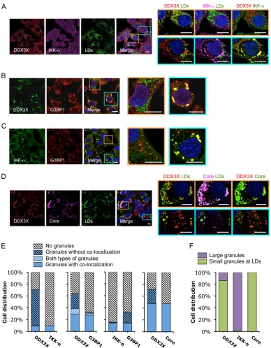
FIG 1HCV 3=UTR RNA induces DDX3X–IKK-␣colocalization in stress granules (SGs). Huh-7.5.1 cells were transfected with EGFP control (Ctrl) or HCV 3=UTR RNA for 24 h. Various cellular proteins and compartments were stained and subsequently examined by confocal microscopy. DDX3X (A and B) and G3BP1 (a SG marker) (D) were detected with specific primary rabbit antibodies, and IKK-␣(A and D) and G3BP1 (B) were detected with specific primary mouse antibodies, followed by the secondary antibody Alexa Fluor 488 (green), 568 (red), or 647 (magenta). Lipid droplets (LDs) were stained with BODIPY 493-503 (green). Magnifications of selected areas are shown on the right. Scale bars, 10m. (A) DDX3X–IKK-␣granules and LDs. Colors in the far right picture were digitally changed for better visualization of IKK-␣(green instead of magenta) and DDX3X (red). (B) DDX3X and SGs. (C) Quantification of DDX3X structures in cells under various conditions. Confocal microscopic images (6 to 12 randomly selected fields from at least three independent experiments performed for panel B, at least 90 cells under each condition) were counted. Results shown are percentages of cells without DDX3X granules (light gray striped bars) or those with DDX3X granular structures either colocalized with SGs (blue bars) or not colocalized (blue striped bars). (D) IKK-␣and SGs. (E) Quantification of IKK-␣ structures in cells that underwent various treatments. Confocal microscopic images (5 to 10 randomly selected fields from at least three independent experiments performed for panel D, at least 50 cells under each condition) were counted. Results shown are percentages of cells without IKK-␣granules (light gray striped bars) or those with IKK-␣granules either colocalized with SGs (blue bars) or not colocalized (blue striped bars).
on November 7, 2019 by guest
http://jvi.asm.org/
[image:4.585.102.488.66.513.2]FIG 2HCV infection induces formation of two subsets of DDX3X granular structures with distinct subcellular localizations. Huh-7.5.1 cells were infected with JFH-1 HCVcc for 2 days. Various cellular proteins and organelles then were detected by immunofluorescence and confocal microscopy. Scale bars, 10m. (A to D) Magnifications of two independent selected areas are shown on the right: small DDX3X-HCV core protein granules at the LD surface (orange area) and large DDX3X–IKK-␣granules (blue area). (A) DDX3X with IKK-␣or LDs. (B) DDX3X with SGs. (C) IKK-␣with SGs. (D) DDX3X, HCV core protein, and LDs. (A and D) Colors in images on the far right were changed for better visualization of IKK-␣(A) or HCV core protein (D), shown in green instead of magenta. (E) Quantification of cell distribution of colocalizing granular structures positive for various combinations of proteins. Confocal microscopic images from each comparison group (10 to 25 randomly selected fields from three independent experiments performed for panels A to D, at least 120 cells per group) were counted. The comparison groups are DDX3X with IKK-␣(far left), DDX3X with G3BP1 (second from the left), IKK-␣with G3BP1 (second from the right), and DDX3X with HCV core protein (far right). Results shown are percentages of cells without any granular structures (light gray striped bars) or those with granular structures either colocalized with each other (blue bars) or not colocalized (blue striped bars). Blue-white striped bars represent percentages of cells with both colocalized and noncolocalized granules. (F) Quantification of cell distribution containing large granules (purple bars) or small granules that colocalize with LDs (green bars) stained positive for DDX3X, IKK-␣, or HCV core protein. At least 40 cells for each protein were counted, and percentages of cells with each pattern of granular structure are shown.
Pène et al.
on November 7, 2019 by guest
http://jvi.asm.org/
[image:5.585.98.486.63.561.2]At 24 to 72 h postinfection, when HCV core protein is present
in sufficient levels (see Fig. S3A in the supplemental material), the
second population of DDX3X structures became evident. These
small and numerous DDX3X-positive structures colocalized with
core protein around LDs (see Fig. S3). The formation of these
structures suggests that DDX3X redistributes with core protein to
the LDs when HCV protein expression is robust late during
infec-tion. Intriguingly, some DDX3X-core protein-containing
struc-tures at the LD surface were stained positive for SG-associated
proteins 48 h postinfection (
Fig. 3
and
4
; also see Fig. S3). All of the
observed structures can coexist in the same population of cells but
rarely in the same cell, demonstrating the spreading and different
stages of infection among cells (
Fig. 3
and
4
; also see Fig. S3). HCV
3=UTR RNA transfection, in contrast, only stimulated large
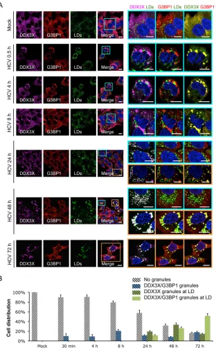
FIG 3Dynamic shift of DDX3X subcellular localization during HCV infection. (A) Huh-7.5.1 cells were mock infected or infected with JFH-1 HCVcc for 0.5 to 72 h. DDX3X, G3BP1, and LDs were stained, and their subcellular localizations and associations were examined. Magnifications of selected areas are shown on the right. Colors on the far right were modified for better visualization of DDX3X, shown in green instead of magenta. Scale bars, 10m. (B) Quantification of DDX3X colocalization with SGs and/or LDs. Five to 10 confocal microscopic pictures from at least three independent experiments with a total of at least 30 cells were analyzed for colocalization signals. Percentages of cells without granular structures (light gray striped bars) or presenting DDX3X granules that are colocalized with SGs only (blue stripped bars), LDs only (green striped bars), or both SGs and LDs (green bars) are shown.
on November 7, 2019 by guest
http://jvi.asm.org/
[image:6.585.139.451.65.566.2]DDX3X-SG structures (
Fig. 4
), suggesting that viral protein
ex-pression is required for redistribution of DDX3X and G3BP1
granules to LD surfaces.
Functions of DDX3X and SGs in HCV infection.
We
previ-ously demonstrated a role of DDX3X in facilitating HCV assembly
through triggering an IKK-
␣
-dependent proviral effect upon
as-sociation of DDX3X with HCV 3=UTR RNA (
21
). DDX3X also
has been shown to be important for HCV replication (
19
,
20
). We
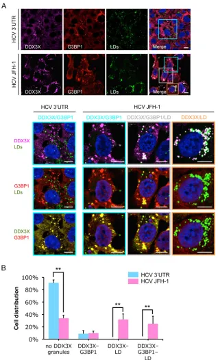
FIG 4Different patterns of DDX3X granular structures coexist in a population of HCV-infected cells. Huh-7.5.1 cells were transfected with HCV 3=UTR RNA for 24 h or infected with JFH-1 HCVcc for 48 h. G3BP1, DDX3X, and LD then were detected by staining, and their associations were evaluated. (A) DDX3X colocalization with SGs or LDs. Magnifications of selected areas are shown in the lower panels. Colors in the lowest panel were modified for better visualization of DDX3X (green instead of magenta) and G3BP1 (red). Scale bars, 10m. (B) Quantification of DDX3X granules associated with SGs or LDs in HCV 3=UTR-transfected or HCV-infected cells. Confocal microscopy images were analyzed visually. Cells were classified into four groups according to the presence or absence of DDX3X granules and their association with SGs or LDs. Percentages of cells in the four groups are shown. Five to 10 randomly selected fields with a total of at least 60 cells were counted. Error bars represent⫾SD from triplicate experiments. **,P⬍0.01.
Pène et al.
on November 7, 2019 by guest
http://jvi.asm.org/
[image:7.585.134.452.64.590.2]silenced DDX3X in Huh-7.5.1 cells using siRNA and performed
various HCV life cycle assays (
29
): quantification of intracellular
and extracellular HCV RNA or immunostaining of HCV core
pro-tein. Silencing of DDX3X led to a significant inhibition of
intra-cellular as well as extraintra-cellular HCV RNA levels and reduced the
number of core protein-positive cells after 2 days of infection (see
Fig. S6A to C in the supplemental material). The role of DDX3X in
the early steps of HCV infection was studied by transfecting
DDX3X-silenced Huh-7.5.1 cells with a firefly luciferase-encoded
construct driven by the HCV IRES (see Fig. S6D) or a subgenomic
JFH-1 replicon encoding
Renilla
luciferase (see Fig. S6E). One or 2
days after transfection, the firefly or
Renilla
luciferase activity in
cells was determined as a marker of HCV IRES-mediated
transla-tion or RNA replicatransla-tion, respectively. Depletransla-tion of DDX3X in cells
significantly inhibited HCV IRES-mediated translation and viral
replication (see Fig. S6D and E). Since HCV RNA triggers the
interaction of SGs and DDX3X as shown above, we then knocked
down three major SG-nucleating proteins: G3BP1,
T-cell-re-stricted intracellular antigen 1 (TIA1), and its paralog, TIA1
cyto-toxic granule-associated RNA binding protein-like 1 (TIAL1).
Depletion of these genes by siRNA in hepatocytes resulted in
sig-nificant reduction of both intracellular and extracellular HCV
RNA levels (see Fig. S6F and G). Moreover, G3BP1 silencing also
led to a reduction in the number of core protein-positive cells and
a 2-fold inhibition of HCV replication (see Fig. S6H and I). These
results demonstrate an important role of SGs in productive HCV
infection.
To further address the interactions of SGs with DDX3X and
IKK-
␣
in HCV infection, we depleted various SG-associated
pro-teins by siRNAs. Silencing of G3BP1 or PKR (protein kinase R),
but not IKK-
␣
or PABP1 expression, abrogated the formation of
HCV-induced SGs detected with a specific anti-G3BP1 antibody
(
Fig. 5
; also see Fig. S7 in the supplemental material). The eIF2
␣
kinase PKR activation is a prerequisite of SG formation upon
cel-lular stress involving the presence of double-stranded RNA (such
as the HCV replication intermediate) (
30
). Intriguingly, in cells
deprived of DDX3X, no SG was induced either (
Fig. 5B
),
indicat-ing that DDX3X expression is necessary for HCV-triggered SG
formation. Moreover, the association of DDX3X with the large SG
was observed in infected cells despite the silencing of IKK-
␣
ex-pression (see Fig. S7A), consistent with our previous finding that
DDX3X functions upstream of IKK-
␣
in this signaling cascade
(
21
). Cells treated with PKR siRNA were unable to aggregate
IKK-
␣
upon HCV infection (see Fig. S7B), and in
siG3BP1-trans-fected cells, neither IKK-
␣
nor PAPB1 granules were detected (
Fig.
5C
; also see Fig. S7C). However, DDX3X staining in complex with
HCV core protein on the surface of LDs still was detected in these
cells (
Fig. 5A
; also see Fig. S7A). These observations are consistent
with the notion that colocalization of DDX3X and HCV core
pro-tein on LDs is independent of the SG–DDX3X–IKK-
␣
-mediated
signaling pathway (
21
,
22
). These results also suggest the
impor-tance of SGs in the recruitment of IKK-
␣
by DDX3X upon HCV
infection.
Interaction of DDX3X and IKK-␣
with HCV nonstructural
proteins.
To explore the potential role of various HCV proteins in
the dynamics of DDX3X-SG redistribution, we studied the effects
of various HCV expression constructs in Huh-7.5.1 cells: a
sub-genomic replicon of JFH-1 encoding only the nonstructural
pro-teins, the complete genome of JFH-1, and two chimeras of JFH-1,
Con1/C3 and Jc1, which expressed the structural proteins of either
Con1 (genotype 1b) or J6 (genotype 2a), respectively (
Fig. 6
and
7
). At 2 days posttransfection, cells were examined by
immuno-staining and confocal microscopy. When transfected with a
con-trol RNA, most cells displayed weak and diffuse DDX3X staining
(
Fig. 6A
). About 3% of cells acquired DDX3X granules (
Fig. 6B
),
which could be attributed to spontaneous activation of DDX3X
during the electroporation process for RNA transfection. Upon
transfection of any of the four abovementioned HCV constructs,
the vast majority of the cells produced HCV proteins (NS5A or
NS3) at detectable levels, and most of these viral
protein-express-ing cells displayed DDX3X granules (
Fig. 6A
and
B
). In cells
har-boring the subgenomic replicon of JFH-1, DDX3X-containing
granules colocalized extensively with NS5A in the ER-derived
membranous web, presumably at the RC, but colocalized only a
little with LDs (
Fig. 6A
and
C
). In contrast, upon transfection of
full-length HCV genome (JFH-1, Con1/C3, or Jc1), DDX3X
co-localized predominantly with NS5A at the surface of LDs (
Fig. 6A
and
C
). Similar patterns of NS5A or LD association were observed
for SGs with G3BP1 staining (
Fig. 7
). These data suggest that HCV
nonstructural proteins mediate the localization of DDX3X-SG
complex to the membranous web on the ER. The data also
con-firm the role of HCV core protein in mediating DDX3X-SG
redis-tribution to LDs.
We previously demonstrated that the HCV 3=UTR mediates
the formation of a DDX3X–IKK-
␣
complex, leading to IKK-
␣
activation and nuclear translocation for downstream
transcrip-tional functions (
21
). To further investigate the dynamic process
of IKK-
␣
localization and association with DDX3X-SGs during
HCV infection, we performed an HCV infection time course
ex-periment (
Fig. 8
). As early as 30 min postinfection, IKK-
␣
gran-ules started to appear and colocalized with DDX3X and SGs but
not with LDs at any time point (
Fig. 8
). At 24 h postinfection, a
smaller population of DDX3X colocalizing with LDs was observed
in a subset of cells as described in the legend to
Fig. 3
; however,
IKK-
␣
was not detected in these structures (
Fig. 8A
). Moreover, in
cells that exhibited DDX3X and LD colocalization, the granular
cytoplasmic structures of IKK-
␣
no longer existed (
Fig. 8A
).
Lastly, HCV infection or transfection of plasmid constructs
expressing individual HCV proteins showed that both
endoge-nously and exogeendoge-nously expressed IKK-
␣
colocalized with NS3
and NS5B but not with core protein, as demonstrated by
immu-nofluorescence and coimmunoprecipitation assays (
Fig. 9A
to
D
).
NS3 protein also was coimmunoprecipitated with DDX3X and
IKK-
␣
in cells transfected with subgenomic or full-length HCV
RNA (
Fig. 9E
). Therefore, HCV nonstructural proteins seem to be
important for the association among IKK-
␣
, DDX3X, and SGs.
Unlike DDX3X, IKK-
␣
does not interact with HCV core protein
for subsequent trafficking to the LD surface. Instead, IKK-
␣
from
the initial SG-associated complex is phosphorylated and
subse-quently translocated to the nucleus, as suggested by our previous
study (
21
).
DISCUSSION
We and other groups have previously demonstrated that DDX3X
interplays with multiple cellular compartments and HCV
ele-ments and exerts an important role in HCV replication and
as-sembly (
19–21
,
31
). The mechanisms underlying these
DDX3X-mediated effects remain elusive; in particular, how DDX3X is
involved in multiple concurrent cellular machineries that are
ex-ploited by HCV for efficient infection is largely unknown. In this
on November 7, 2019 by guest
http://jvi.asm.org/
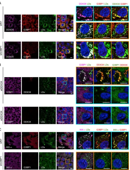
FIG 5Effect of G3BP1 or DDX3X silencing on SG formation and DDX3X–IKK-␣localization. Huh-7.5.1 cells were treated with nontargeting control (siNT) or G3BP1 or DDX3X siRNA for 3 days and then infected with JFH-1 HCVcc for 2 days. LDs and various indicated proteins then were stained and analyzed for their subcellular localizations and associations. Magnifications of the selected area are shown on the right. In the majority of siG3BP1- or siDDX3X-treated cells, G3BP1 or DDX3X was efficiently depleted. A few cells still showed G3BP1 or DDX3X expression; therefore, SG-DDX3X-LD colocalization still exists in these cells (A, lower, and B, upper). (A) Small DDX3X granules are associated with LDs in G3BP1-silenced cells. (B) DDX3X depletion abrogated SG formation. (C) Lack of IKK-␣granules in G3BP1-silenced cells. Colors of pictures on the far right were modified for better visualization of DDX3X (A), G3BP1 (B), or IKK-␣(C), shown in green instead of magenta. Scale bars, 10m.
Pène et al.
on November 7, 2019 by guest
http://jvi.asm.org/
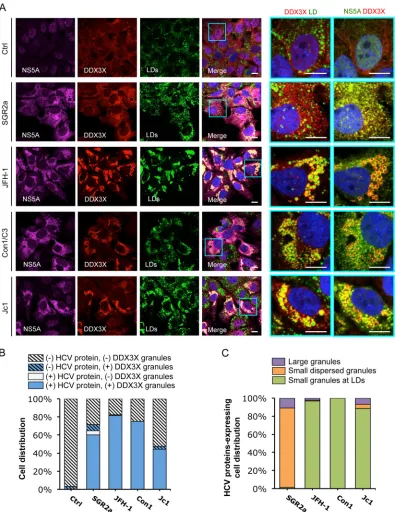
[image:9.585.86.505.63.609.2]FIG 6HCV NS5A protein colocalizes with DDX3X and LDs. Huh-7.5.1 cells were transfected within vitro-transcribed EGFP control RNA (Ctrl), subgenomic replicon RNA of HCV genotype 2a (SGR2a), genomic-length JFH-1, Con1/C3, or Jc1 chimeric RNA for 48 h. DDX3X, HCV NS5A, and LDs were stained and characterized for their localizations and associations. (A) Confocal microscopic analyses of DDX3X, NS5A, and LD staining in cells treated with various HCV RNAs. Magnifications of selected areas are shown on the right. Colors of pictures on the far right were digitally changed for better visualization of NS5A (green instead of magenta) and DDX3X (red). Scale bars, 10m. (B) Quantification of cell distribution for DDX3X granules in HCV protein-negative or -positive cells. Confocal microscopic images (6 to 12 per condition from at least three independent experiments with at least 80 cells in total) were analyzed for the presence or absence of DDX3X granules in HCV protein-positive or -negative cells, as determined by detection of NS5A (A), NS3 (not shown), or core protein (not shown). Results shown are percentages of cells that are HCV protein negative and either with (blue striped bars) or without (light gray striped bars) DDX3X granules and cells that are HCV protein positive and either with (blue bars) or without (light gray bars) DDX3X granules. (C) Characterization and quantification of DDX3X-positive structures in HCV protein-positive cells. HCV protein-positive cells with DDX3X granules (blue bars from panel B) were further classified into three groups: cells with a few large DDX3X granules not associated with LDs (purple bars), cells with DDX3X granules dispersed throughout the ER (orange bars, probably representing HCV replicative intermediates), and cells with small and numerous DDX3X granules localized to LDs (green bars). Percentages of cells in each group are shown (a total of at least 40 cells).
on November 7, 2019 by guest
http://jvi.asm.org/
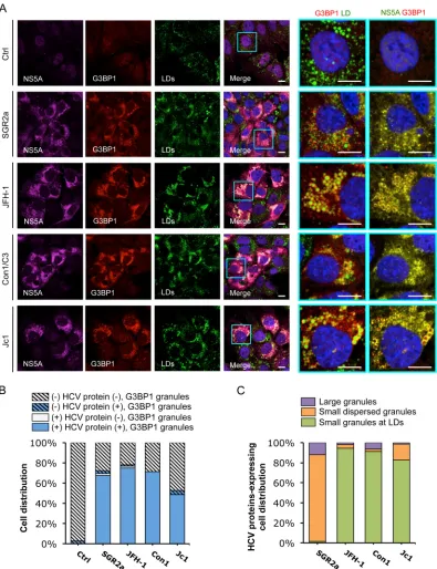
[image:10.585.95.490.65.577.2]FIG 7Colocalization of HCV NS5A protein with SGs.In vitro-transcribed control RNA (Ctrl), SGR2a, JFH-1, Con1/C3, or Jc1 chimera RNA-transfected Huh-7.5.1 cells (as described forFig. 6) were stained for HCV NS5A, SGs, and LDs. (A) Confocal microscopic imaging of G3BP1 (for SGs), NS5A, and LDs. Magnifications of selected areas are shown on the right. Colors in pictures on the far right were digitally changed for better visualization of NS5A (green instead of magenta) and G3BP1 (red). Scale bars, 10m. (B) Quantification of SGs in HCV protein-negative or -positive cells. Six to 12 randomly selected microscopic images from at least three independent experiments with a total of at least 80 cells were analyzed for the presence or absence of SGs in HCV protein-positive or -negative cells. Results shown are percentages of cells that are HCV protein negative and either with (blue striped bars) or without (light gray striped bars) SGs and cells that are HCV protein positive and either with (blue bars) or without (light gray bars) SGs. (C) Characterization of SGs in HCV protein-positive cells. This population of cells, which also presents SGs (blue bars in panel B), were further classified into three groups: cells with a few large SGs not associated with LDs (purple bars), cells with G3BP1 granules dispersed throughout the ER (orange bars), and cells with small and numerous SGs localized to LDs (green bars). Percentages of HCV-positive cells (in a total of at least 40 cells) in each group are shown.
Pène et al.
on November 7, 2019 by guest
http://jvi.asm.org/
[image:11.585.95.490.65.579.2]FIG 8Associations between IKK-␣, DDX3X, SGs, and LDs upon HCV infection. Huh-7.5.1 cells were mock infected or infected with JFH-1 HCVcc and then stained for IKK-␣, DDX3X, SGs, and LDs at various time points. (A) Association of IKK-␣with DDX3X. (B) Association of IKK-␣with SGs. Magnifications of selected areas are shown on the right. Colors in pictures on the far right were digitally changed for better visualization of IKK-␣, shown in green instead of magenta. Scale bars, 10m.
on November 7, 2019 by guest
http://jvi.asm.org/
[image:12.585.116.471.64.688.2]study, applying systematic imaging and virologic approaches, we
examined the dynamic subcellular localization and association of
DDX3X with various cellular components and viral elements and
explored the intrinsic functions of DDX3X in the HCV life cycle
and HCV-mediated cellular responses.
Once entering host cells, HCV releases its genomic RNA, the
3=UTR of which interacts with DDX3X and initiates a cascade of
signaling processes, including PKR-mediated stress granule
for-mation, DDX3X and IKK-
␣
association, and recruitment of
DDX3X–IKK-
␣
complex to the SGs (
Fig. 10
). We recently
dem-onstrated that IKK-
␣
, a major I-
B kinase for activation of the
NF-
B pathway, exerts a proviral role in HCV assembly through
the induction of host cell lipogenic gene expression and lipid
droplet formation. This transcriptional role of IKK-
␣
is
indepen-dent of NF-
B but is subsequent to HCV 3=UTR-DDX3X
inter-action (
21
). DDX3X, bound to and activated by the HCV 3=UTR,
interacts with and recruits IKK-
␣
to the SGs, leading to its
phos-phorylation and nuclear translocation through an unidentified
mechanism. Here, we show that DDX3X–IKK-
␣
association and
activation take place at the stress granules but not in other
subcel-lular organelles, such as the autophagosome, mitochondria,
per-oxisome, or lipid droplets, that have been suggested as the sites for
HCV replication and production or implicated in the antiviral
cellular responses that restrain HCV infection.
Stress granules are cytoplasmic macromolecular structures of
stalled translation initiation complexes that are induced in
re-sponse to various stress conditions, including viral infection (
32
).
The formation of SGs is driven by aggregation of several key RNA
binding proteins, such as TIA-1, TIA-1-related protein (TIAR),
and G3BP1 (
33
). Cells depleted of either G3BP1, TIA-1, or TIAR
subsequently are deficient in SG formation (
34–36
). The primary
role of SGs in translation suppression and RNA decay suggests this
cellular event impacts the viral life cycle and can be manipulated
by viruses for their own survival. Indeed, SG formation is a
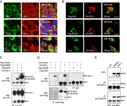
hall-FIG 9Interaction of IKK-␣with HCV nonstructural proteins. (A and B) Colocalization of IKK-␣with HCV NS3 and NS5B proteins. Huh-7.5.1 cells were infected with JFH-1 HCVcc for 48 h (A) or were transfected with various plasmids encoding HCV proteins for 24 h (B). The subcellular localization of IKK-␣and its association with HCV proteins were detected and analyzed by immunofluorescence and confocal microscopy. Scale bars, 20m. Pearson’s correlation coefficient (CR) was determined on the selected areas (in white boxes). (C and D) Cells were transfected with plasmids encoding various Flag-tagged HCV proteins and/or HA-IKK-␣for 24 h. Immunoprecipitation (IP) with anti-HA, anti-Flag, or IgG control antibody was performed, followed by Western blotting (WB) using anti-Flag or anti-HA antibody. (E) Cells were transfected within vitro-transcribed EGFP control RNA (Ctrl), SGR2a, or JFH-1 RNA for 48 h. Immunoprecipitation on whole-cell lysate (WCL) with anti-IKK-␣or anti-DDX3X antibody was conducted, followed by Western blotting using anti-NS3 antibody. HC, IgG heavy chain.
Pène et al.
on November 7, 2019 by guest
http://jvi.asm.org/
[image:13.585.76.512.67.430.2]mark of infection for many RNA viruses, albeit with a range of
phenotypes. Generally, it appears to be inhibited during infection
by most viruses, including influenza virus, HIV-1, rotavirus, and
two important members of the
Flaviviridae
family, West Nile virus
(WNV) and dengue virus (
37
). Since SGs potentially are antiviral
by sequestering and altering cell components that are pivotal for
viral replication, the antagonization of SG formation by these
vi-ruses may be a viral strategy to subvert the cellular stress responses
that restrict infections (
37
). Several other viruses, such as
reovi-ruses (
38
), respiratory syncytial virus (RSV) (
39
), and
coronavi-ruses (
40
,
41
), in contrast, induce and coopt SG responses as part
of their replication cycle.
HCV, a hepatotropic RNA virus, also has been shown to
ma-nipulate SG formation and interact with SG components for
sur-vival and efficient propagation. During HCV infection, G3BP1 is
associated with the viral NS5B protein and the negative-strand
viral RNA; thus, it constitutes part of the HCV replication
com-plexes (RCs) (
42
). HCV also recruits other components of SGs to
the viral RCs, and these SG markers play an important role in
HCV genome amplification (
43
). Using live-cell imaging
technol-ogy, two recent studies showed that the assembly and disassembly
of SGs is a highly dynamic (oscillating) process in HCV-infected
cells (
30
,
44
). In addition, HCV-triggered SG formation is
associ-ated with delayed cell division, stalled translation, and prolonged
cell survival, suggesting that HCV exploits this important stress
response to establish persistent infection in hepatocytes (
30
).
Apart from the effect on HCV RNA replication, the SG proteins
have been shown to be required for either HCV assembly (G3BP1
and TIA-1) or secretion (TIAR) (
31
).
Here, we show that SGs, stimulated by HCV 3=UTR RNA in
infected cells, provide a platform for DDX3X and IKK-
␣
associa-tion and activaassocia-tion (
Fig. 10
). The SG-associated DDX3X–IKK-
␣
complex binds to multiple HCV nonstructural proteins, which,
together with viral genomic RNA, establish the HCV replicase
machinery on the membranous web derived from the ER
mem-brane. Late in HCV infection, DDX3X and SGs redistribute from
the ER to LDs (
Fig. 10
). In the absence of core protein expression
(for example, in cells with subgenomic replicon RNA
transfec-tion), the DDX3X-SG complex does not translocate to the LDs but
is retained in the yet-to-be fully established HCV RNA machinery
within the ER-derived membranous web. IKK-
␣
does not relocate
to the LDs either but instead translocates to the nucleus upon
phosphorylation for a role in transcriptionally regulating cellular
lipogenesis (
Fig. 10
) (
21
).
The relocation of SGs to LDs at the late step of viral infection
also has been reported by Ariumi et al. They have shown that HCV
hijacks the SGs as well as P-body contents around LDs for
en-hancement of viral production (
43
). However, how the SG
com-ponents are redistributed to the LDs and their contingent roles in
modulating HCV assembly still are unclear. DDX3X has been
shown to interact with HCV core protein, but this interaction is
dispensable for productive HCV infection (
22
); hence, it is not
likely to mediate SG relocalization, as the relocated, LD-associated
SG proteins ultimately are required for HCV assembly (
31
). In
addition, the major SG component G3BP1 does not colocalize
with an ectopically expressed core protein, which mainly localizes
to LDs, suggesting that other cellular or viral factors (such as
non-structural proteins) contribute to the redistribution of SGs to the
LDs in association with HCV core protein (
45
).
Among the early initiation factors present in SGs is the
poly(A)-binding protein 1 (PABP1). PABP1 stimulates initiation
factor recruitment to the mRNA, leading to mRNA circularization
and efficient translation (
14
). In a previous study, PABP1 was
shown to directly bind to DDX3X, and downregulation of
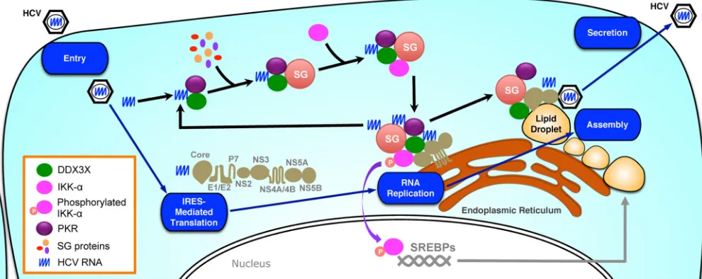
FIG 10Proposed model for dynamic localization and association of DDX3X and IKK-␣with various cellular compartments during HCV life cycle. Using an HCV life cycle map as the background, the dynamic interactions of DDX3X, IKK-␣, SG, LD, and HCV elements are shown. HCV infection, through the 3=UTR RNA, stimulates PKR- and DDX3X-dependent SG formation, which then recruits IKK-␣, leading to IKK-␣activation at the SGs. The SG-associated DDX3X– IKK-␣complex also interacts with HCV nonstructural proteins, which, together with viral genome RNA, constitute the HCV replicase machinery on the ER-derived membranous web. The formation of functional complexes harboring the HCV 3=UTR, DDX3X, IKK-␣, and SGs is further amplified by viral replication by providing more HCV genomic RNA. Late in HCV infection, DDX3X and SGs, via interaction with the core protein, redistribute from the ER to the surface of LDs where assembly occurs. In contrast, the transcription factor IKK-␣does not relocate to the LDs but translocates to the nucleus for a role in activating cellular lipogenesis and LD biogenesis.
on November 7, 2019 by guest
http://jvi.asm.org/
[image:14.585.44.539.71.268.2]DDX3X in cells interfered with SG assembly (
11
). This is
consis-tent with our finding that DDX3X knockdown significantly
im-paired HCV-induced SG formation (
Fig. 5B
). We also showed
that silencing of G3BP1 completely abolished the formation of
SGs as well as the formation of DDX3X–IKK-
␣
granules (
Fig. 5A
and
C
). These data further confirmed the important role of G3BP1
in SG formation as previously described (
35
) and implicated an
intrinsic role of SG in DDX3X–IKK-
␣
activation for the
down-stream role in facilitating HCV assembly. In addition, depletion of
either DDX3X or the SG component G3BP1 or TIA paralogs
sig-nificantly reduced viral RNA levels in cells, demonstrating the
involvement of DDX3X-SG complexes in HCV replication. This
effect was further supported by the presence of HCV
nonstruc-tural proteins in the DDX3X-SG complex in the context of either
full-length or subgenomic HCV replicon transfection. Moreover,
G3BP1 directly binds to HCV NS5B protein and associates with
viral RNA (
42
), implying that SGs participate in the formation of
the HCV RC that controls viral RNA replication.
DDX3X previously has been shown to (i) function as an
eIF4E-inhibitory protein to specifically repress cap-dependent
transla-tion by trapping eIF4E in a translatransla-tionally inactive complex (
46
);
(ii) preferentially promote the translation initiation of structured
5=UTR regions (
47
,
48
); (iii) associate with the cytoplasmic
mul-tisubunit translation initiation factor eIF3 (
49
), which is involved
in HCV translation, by specifically binding to the IRES in the HCV
5=UTR (
50
,
51
); and (iv) interact with HCV core protein to inhibit
translation of capped but not uncapped RNA (
52
). As all of these
effects of DDX3X can be attributed to SG functions, we
extrapo-late that HCV may hijack DDX3X-SG complexes to reguextrapo-late the
expression of viral and cellular proteins for its own advantage.
It is known that LDs are essential for the assembly of infectious
HCV particles, and that HCV core protein plays a key role in this
process. The core protein promotes the accumulation of LDs to
facilitate virus assembly. It has been proposed that HCV core
pro-tein recruits nonstructural propro-teins, mainly NS5A, viral RNA, and
the RC- to LD-associated membranes (
6
), and that NS5A plays a
dual role in HCV replication and assembly and meanwhile acts as
a switch between these two processes (
53
,
54
). In addition to
NS5A, other nonstructural proteins, such as NS2, NS3, NS4A, and
NS4B, were shown to be critical for HCV assembly (
44
,
55–57
).
Moreover, HCV core protein can interact with NS5B and
modu-late its RNA-dependent RNA polymerase activity (
58
). The
com-plex and dynamic interactions of HCV components with DDX3X
and SG proteins in LD-associated membranes would allow the
tight control of each step of the HCV life cycle as well as potential
inhibition of innate antiviral responses.
ACKNOWLEDGMENTS
We thank Helen Cha, Keng-Hsin Lan, and Zongyi Hu for technical assis-tance. We also thank all members of the Liver Diseases Branch, NIDDK, for discussion and support.
This work was supported by the Intramural Research Program of the National Institute of Diabetes and Digestive and Kidney Diseases (NIDDK), U.S. National Institutes of Health (NIH).
REFERENCES
1.Heim MH.2013. Innate immunity and HCV. J Hepatol58:564 –574.http: //dx.doi.org/10.1016/j.jhep.2012.10.005.
2.Thimme R, Binder M, Bartenschlager R.2012. Failure of innate and adaptive immune responses in controlling hepatitis C virus infection. FEMS Microbiol Rev36:663– 683.http://dx.doi.org/10.1111/j.1574-6976 .2011.00319.x.
3.Liang TJ. 2013. Current progress in development of hepatitis C virus vaccines. Nat Med19:869 – 878.http://dx.doi.org/10.1038/nm.3183. 4.Liang TJ, Ghany MG.2013. Current and future therapies for hepatitis C
virus infection. N Engl J Med368:1907–1917.http://dx.doi.org/10.1056 /NEJMra1213651.
5.McLauchlan J.2000. Properties of the hepatitis C virus core protein: a structural protein that modulates cellular processes. J Viral Hepatitis7:2– 14.http://dx.doi.org/10.1046/j.1365-2893.2000.00201.x.
6.Miyanari Y, Atsuzawa K, Usuda N, Watashi K, Hishiki T, Zayas M, Bartenschlager R, Wakita T, Hijikata M, Shimotohno K.2007. The lipid droplet is an important organelle for hepatitis C virus production. Nat Cell Biol9:1089 –1097.http://dx.doi.org/10.1038/ncb1631.
7.Rouillé Y, Helle F, Delgrange D, Roingeard P, Voisset C, Blanchard E, Belouzard S, McKeating J, Patel AH, Maertens G, Wakita T, Wychowski C, Dubuisson J.2006. Subcellular localization of hepatitis C virus structural proteins in a cell culture system that efficiently replicates the virus. J Virol80:2832–2841.http://dx.doi.org/10.1128/JVI.80.6.2832 -2841.2006.
8.Abid K, Pazienza V, de Gottardi A, Rubbia-Brandt L, Conne B, Pugnale P, Rossi C, Mangia A, Negro F.2005. An in vitro model of hepatitis C virus genotype 3a-associated triglycerides accumulation. J Hepatol42:
744 –751.http://dx.doi.org/10.1016/j.jhep.2004.12.034.
9.Boulant S, Douglas MW, Moody L, Budkowska A, Targett-Adams P, McLauchlan J.2008. Hepatitis C virus core protein induces lipid droplet redistribution in a microtubule- and dynein-dependent manner. Traffic
9:1268 –1282.http://dx.doi.org/10.1111/j.1600-0854.2008.00767.x. 10. Rocak S, Linder P.2004. DEAD-box proteins: the driving forces behind
RNA metabolism. Nat Rev Mol Cell Biol5:232–241.http://dx.doi.org/10 .1038/nrm1335.
11. Shih JW, Wang WT, Tsai TY, Kuo CY, Li HK, Wu Lee YH. 2012. Critical roles of RNA helicase DDX3 and its interactions with eIF4E/ PABP1 in stress granule assembly and stress response. Biochem J441:119 – 129.http://dx.doi.org/10.1042/BJ20110739.
12. Adjibade P, Mazroui R.2014. Control of mRNA turnover: Implication of cytoplasmic RNA granules. Semin Cell Dev Biol34:15–23.http://dx.doi .org/10.1016/j.semcdb.2014.05.013.
13. Schroder M.2010. Human DEAD-box protein 3 has multiple functions in gene regulation and cell cycle control and is a prime target for viral manipu-lation. Biochemical Pharmacol79:297–306.http://dx.doi.org/10.1016/j.bcp .2009.08.032.
14. Shih JW, Lee YH.2014. Human DExD/H RNA helicases: Emerging roles in stress survival regulation. Clin Chim Acta436C:45–58.http://dx.doi .org/10.1016/j.cca.2014.05.003.
15. Oshiumi H, Sakai K, Matsumoto M, Seya T.2010. DEAD/H BOX 3 (DDX3) helicase binds the RIG-I adaptor IPS-1 to up-regulate IFN-beta-inducing potential. Eur J Immunol40:940 –948.http://dx.doi.org/10.1002 /eji.200940203.
16. Schroder M, Baran M, Bowie AG.2008. Viral targeting of DEAD box protein 3 reveals its role in TBK1/IKKepsilon-mediated IRF activation. EMBO J27:2147–2157.http://dx.doi.org/10.1038/emboj.2008.143. 17. Li C, Ge LL, Li PP, Wang Y, Dai JJ, Sun MX, Huang L, Shen ZQ, Hu
XC, Ishag H, Mao X.2014. Cellular DDX3 regulates Japanese encepha-litis virus replication by interacting with viral un-translated regions. Vi-rology449:70 – 81.http://dx.doi.org/10.1016/j.virol.2013.11.008. 18. Schroder M.2011. Viruses and the human DEAD-box helicase DDX3:
inhibition or exploitation? Biochem Soc Trans39:679 – 683.http://dx.doi .org/10.1042/BST0390679.
19. Ariumi Y, Kuroki M, Abe K, Dansako H, Ikeda M, Wakita T, Kato N.
2007. DDX3 DEAD-box RNA helicase is required for hepatitis C virus RNA replication. J Virol81:13922–13926.http://dx.doi.org/10.1128/JVI .01517-07.
20. Randall G, Panis M, Cooper JD, Tellinghuisen TL, Sukhodolets KE, Pfeffer S, Landthaler M, Landgraf P, Kan S, Lindenbach BD, Chien M, Weir DB, Russo JJ, Ju J, Brownstein MJ, Sheridan R, Sander C, Zavolan M, Tuschl T, Rice CM.2007. Cellular cofactors affecting hepatitis C virus infection and replication. Proc Natl Acad Sci U S A104:12884 –12889. http://dx.doi.org/10.1073/pnas.0704894104.
21. Li Q, Pene V, Krishnamurthy S, Cha H, Liang TJ.2013. Hepatitis C virus infection activates an innate pathway involving IKK-alpha in lipogenesis and viral assembly. Nat Med19:722–729.http://dx.doi.org/10.1038/nm .3190.
22. Angus AG, Dalrymple D, Boulant S, McGivern DR, Clayton RF, Scott MJ, Adair R, Graham S, Owsianka AM, Targett-Adams P, Li K, Wakita Pène et al.
on November 7, 2019 by guest
http://jvi.asm.org/
T, McLauchlan J, Lemon SM, Patel AH.2010. Requirement of cellular DDX3 for hepatitis C virus replication is unrelated to its interaction with the viral core protein. J Gen Virol91:122–132.http://dx.doi.org/10.1099 /vir.0.015909-0.
23. Owsianka AM, Patel AH.1999. Hepatitis C virus core protein interacts with a human DEAD box protein DDX3. Virology257:330 –340.http://dx .doi.org/10.1006/viro.1999.9659.
24. Kato T, Matsumura T, Heller T, Saito S, Sapp RK, Murthy K, Wakita T, Liang TJ.2007. Production of infectious hepatitis C virus of various genotypes in cell cultures. J Virol 81:4405– 4411. http://dx.doi.org/10 .1128/JVI.02334-06.
25. Wakita T, Pietschmann T, Kato T, Date T, Miyamoto M, Zhao Z, Murthy K, Habermann A, Kräusslich HG, Mizokami M, Bartenschlager R, Liang TJ.2005. Production of infectious hepatitis C virus in tissue culture from a cloned viral genome. Nat Med11:791–796.http://dx.doi .org/10.1038/nm1268.
26. Li Q, Brass AL, Ng A, Hu Z, Xavier RJ, Liang TJ, Elledge SJ.2009. A genome-wide genetic screen for host factors required for hepatitis C virus propagation. Proc Natl Acad Sci U S A106:16410 –16415.http://dx.doi .org/10.1073/pnas.0907439106.
27. Zhang YY, Zhang BH, Ishii K, Liang TJ.2010. Novel function of CD81 in controlling hepatitis C virus replication. J Virol84:3396 –3407.http: //dx.doi.org/10.1128/JVI.02391-09.
28. Jonas S, Izaurralde E.2013. The role of disordered protein regions in the assembly of decapping complexes and RNP granules. Genes Dev27:2628 – 2641.http://dx.doi.org/10.1101/gad.227843.113.
29. Li Q, Zhang YY, Chiu S, Hu Z, Lan KH, Cha H, Sodroski C, Zhang F, Hsu CS, Thomas E, Liang TJ.2014. Integrative functional genomics of hepatitis C virus infection identifies host dependencies in complete viral replication cycle. PLoS Pathog 10:e1004163.http://dx.doi.org/10.1371 /journal.ppat.1004163.
30. Ruggieri A, Dazert E, Metz P, Hofmann S, Bergeest JP, Mazur J, Bankhead P, Hiet MS, Kallis S, Alvisi G, Samuel CE, Lohmann V, Kaderali L, Rohr K, Frese M, Stoecklin G, Bartenschlager R. 2012. Dynamic oscillation of translation and stress granule formation mark the cellular response to virus infection. Cell Host Microbe12:71– 85.http://dx .doi.org/10.1016/j.chom.2012.05.013.
31. Garaigorta U, Heim MH, Boyd B, Wieland S, Chisari FV.2012. Hep-atitis C virus (HCV) induces formation of stress granules whose proteins regulate HCV RNA replication and virus assembly and egress. J Virol
86:11043–11056.http://dx.doi.org/10.1128/JVI.07101-11.
32. Beckham CJ, Parker R.2008. P bodies, stress granules, and viral life cycles. Cell Host Microbe3:206 –212.http://dx.doi.org/10.1016/j.chom .2008.03.004.
33. Anderson P, Kedersha N.2008. Stress granules: the Tao of RNA triage. Trends Biochem Sci33:141–150.http://dx.doi.org/10.1016/j.tibs.2007.12 .003.
34. Gilks N, Kedersha N, Ayodele M, Shen L, Stoecklin G, Dember LM, Anderson P.2004. Stress granule assembly is mediated by prion-like ag-gregation of TIA-1. Mol Biol Cell15:5383–5398.http://dx.doi.org/10 .1091/mbc.E04-08-0715.
35. Tourriere H, Chebli K, Zekri L, Courselaud B, Blanchard JM, Bertrand E, Tazi J.2003. The RasGAP-associated endoribonuclease G3BP assem-bles stress granules. J Cell Biol160:823– 831.http://dx.doi.org/10.1083/jcb .200212128.
36. White JP, Cardenas AM, Marissen WE, Lloyd RE.2007. Inhibition of cytoplasmic mRNA stress granule formation by a viral proteinase. Cell Host Microbe2:295–305.http://dx.doi.org/10.1016/j.chom.2007.08.006. 37. White JP, Lloyd RE.2012. Regulation of stress granules in virus systems. Trends Microbiol20:175–183.http://dx.doi.org/10.1016/j.tim.2012.02.001. 38. Smith JA, Schmechel SC, Raghavan A, Abelson M, Reilly C, Katze MG,
Kaufman RJ, Bohjanen PR, Schiff LA.2006. Reovirus induces and ben-efits from an integrated cellular stress response. J Virol80:2019 –2033. http://dx.doi.org/10.1128/JVI.80.4.2019-2033.2006.
39. Lindquist ME, Lifland AW, Utley TJ, Santangelo PJ, Crowe JE, Jr.2010. Respiratory syncytial virus induces host RNA stress granules to facilitate viral replication. J Virol84:12274 –12284.http://dx.doi.org/10.1128/JVI .00260-10.
40. Raaben M, Groot Koerkamp MJ, Rottier PJ, de Haan CA.2007. Mouse hepatitis coronavirus replication induces host translational shutoff and mRNA decay, with concomitant formation of stress granules and
process-ing bodies. Cell Microbiol9:2218 –2229.http://dx.doi.org/10.1111/j.1462 -5822.2007.00951.x.
41. Sola I, Galan C, Mateos-Gomez PA, Palacio L, Zuniga S, Cruz JL, Almazan F, Enjuanes L.2011. The polypyrimidine tract-binding protein affects coronavirus RNA accumulation levels and relocalizes viral RNAs to novel cytoplasmic domains different from replication-transcription sites. J Virol85:5136 –5149.http://dx.doi.org/10.1128/JVI.00195-11. 42. Yi Z, Pan T, Wu X, Song W, Wang S, Xu Y, Rice CM, Macdonald MR,
Yuan Z.2011. Hepatitis C virus coopts Ras-GTPase-activating protein-binding protein 1 for its genome replication. J Virol85:6996 –7004.http: //dx.doi.org/10.1128/JVI.00013-11.
43. Ariumi Y, Kuroki M, Kushima Y, Osugi K, Hijikata M, Maki M, Ikeda M, Kato N.2011. Hepatitis C virus hijacks P-body and stress granule components around lipid droplets. J Virol85:6882– 6892.http://dx.doi .org/10.1128/JVI.02418-10.
44. Jones DM, Patel AH, Targett-Adams P, McLauchlan J.2009. The hep-atitis C virus NS4B protein can trans-complement viral RNA replication and modulates production of infectious virus. J Virol83:2163–2177.http: //dx.doi.org/10.1128/JVI.01885-08.
45. Pager CT, Schutz S, Abraham TM, Luo G, Sarnow P.2013. Modulation of hepatitis C virus RNA abundance and virus release by dispersion of processing bodies and enrichment of stress granules. Virology435:472– 484.http://dx.doi.org/10.1016/j.virol.2012.10.027.
46. Shih JW, Tsai TY, Chao CH, Wu Lee YH. 2008. Candidate tumor suppressor DDX3 RNA helicase specifically represses cap-dependent translation by acting as an eIF4E inhibitory protein. Oncogene27:700 – 714.http://dx.doi.org/10.1038/sj.onc.1210687.
47. Geissler R, Golbik RP, Behrens SE.2012. The DEAD-box helicase DDX3 supports the assembly of functional 80S ribosomes. Nucleic Acids Res
40:4998 –5011.http://dx.doi.org/10.1093/nar/gks070.
48. Lai MC, Lee YH, Tarn WY.2008. The DEAD-box RNA helicase DDX3 associates with export messenger ribonucleoproteins as well as tip-associated protein and participates in translational control. Mol Biol Cell
19:3847–3858.http://dx.doi.org/10.1091/mbc.E07-12-1264.
49. Lee CS, Dias AP, Jedrychowski M, Patel AH, Hsu JL, Reed R.2008. Human DDX3 functions in translation and interacts with the translation initiation factor eIF3. Nucleic Acids Res36:4708 – 4718.http://dx.doi.org /10.1093/nar/gkn454.
50. Hellen CU, Pestova TV.1999. Translation of hepatitis C virus RNA. J Viral Hepatitis6:79 – 87.http://dx.doi.org/10.1046/j.1365-2893.1999.00150.x. 51. Sun C, Querol-Audi J, Mortimer SA, Arias-Palomo E, Doudna JA,
Nogales E, Cate JH.2013. Two RNA-binding motifs in eIF3 direct HCV IRES-dependent translation. Nucleic Acids Res41:7512–7521.http://dx .doi.org/10.1093/nar/gkt510.
52. Mamiya N, Worman HJ.1999. Hepatitis C virus core protein binds to a DEAD box RNA helicase. J Biol Chem274:15751–15756.http://dx.doi.org /10.1074/jbc.274.22.15751.
53. Gawlik K, Baugh J, Chatterji U, Lim PJ, Bobardt MD, Gallay PA.2014. HCV core residues critical for infectivity also are involved in core-NS5A complex formation. PLoS One 9:e88866. http://dx.doi.org/10.1371 /journal.pone.0088866.
54. Hughes M, Griffin S, Harris M.2009. Domain III of NS5A contributes to both RNA replication and assembly of hepatitis C virus particles. J Gen Virol90:1329 –1334.http://dx.doi.org/10.1099/vir.0.009332-0. 55. Mousseau G, Kota S, Takahashi V, Frick DN, Strosberg AD. 2011.
Dimerization-driven interaction of hepatitis C virus core protein with NS3 helicase. J Gen Virol 92:101–111. http://dx.doi.org/10.1099/vir.0 .023325-0.
56. Phan T, Kohlway A, Dimberu P, Pyle AM, Lindenbach BD.2011. The acidic domain of hepatitis C virus NS4A contributes to RNA replication and virus particle assembly. J Virol85:1193–1204.http://dx.doi.org/10 .1128/JVI.01889-10.
57. Popescu CI, Callens N, Trinel D, Roingeard P, Mor