EVALUATION OF WOUND HEALING EFFECT OF AQUEOUS STEM
EXTRACT OF Zanthoxylum rhetsa (Roxb.) DC.
ON WISTAR RATS
A Dissertation Submitted to
THE TAMIL NADU Dr. M. G. R. MEDICAL UNIVERSITY
CHENNAI-600 032
In partial fulfillment of the requirement for the award of the Degree of
MASTER OF PHARMACY
IN
PHARMACOLOGY
OCTOBER
-
2017
DEPARTMENT OF PHARMACOLOGY
KMCH COLLEGE OF PHARMACY
KOVAI ESTATE, KALAPPATTI ROAD,
EVALUATION OF WOUND HEALING EFFECT OF AQUEOUS
STEM EXTRACT OF Zanthoxylum rhetsa (Roxb.) DC.
ON WISTAR RATS
A Dissertation submitted to
THE TAMIL NADU Dr. M .G .R. MEDICAL UNIVERSITY
CHENNAI-600 032
In partial fulfillment of the requirement for the award of the Degree of
MASTER OF PHARMACY
IN
PHARMACOLOGY
OCTOBER
-
2017
Submitted by
Reg. No. 261525809
DEPARTMENT OF PHARMACOLOGY
KMCH COLLEGE OF PHARMACY
KOVAI ESTATE, KALAPPATTI ROAD,
Prof. Dr. A. Rajasekaran, M. Pharm., Ph.D., Principal,
KMCH College of Pharmacy, Kovai Estate, Kalapatti Road, Coimbatore - 641 048.
Tamil Nadu
CERTIFICATE
This is to certify that the dissertation work entitled “EVALUATION OF WOUND HEALING EFFECT OF AQUEOUS STEM EXTRACT OF
Zanthoxylum
rhetsa
(Roxb.)DC. ON WISTAR RATS
” was carried out by Reg. No. 261525809. The work mentioned in the dissertation was carried out at the Department of Pharmacology, KMCH College of Pharmacy, Coimbatore, Tamil Nadu, for thepartial fulfillment for the degree of Master of Pharmacy during the academic year
2016-2017 and is forwarded to the Tamil Nadu Dr. M. G. R. Medical University,
Chennai.
Date: Prof. Dr. A. Rajasekaran, M. Pharm., Ph.D.,
Place: Coimbatore Principal
GUIDE
Dept. of Pharmacology, KMCH College of Pharmacy, Kovai Estate, Kalapatti Road,
Coimbatore - 641 048.
Tamil Nadu
CERTIFICATE
This is to certify that the dissertation work entitled “EVALUATION OF WOUND HEALING EFFECT OF AQUEOUS STEM EXTRACT OF
Zanthoxylum rhetsa
(Roxb.)DC. ON WISTAR RATS
” is a bonafide work carried out by Reg. No. 261525809. The work mentioned in the dissertation was carried out at the Department of Pharmacology, KMCH College of Pharmacy,Coimbatore, Tamil Nadu, under my supervision and guidance during the academic
year 2016-2017.
This research work either in part or full does not constitute any of any thesis /
dissertation.
DECLARATION
I do here by declare that to the best of my knowledge and belief ,the dissertation
work entitled “EVALUATION OF WOUND HEALING EFFECT OF AQUEOUS STEM EXTRACT OF
Zanthoxylum rhetsa
(Roxb.)DC. ON WISTAR
RATS
” submitted to the Tamil Nadu Dr. M.G.R. Medical university , Chennai, in the partial fulfillment for the Degree of Master of Pharmacy in Pharmacology, was carried out at Department of Pharmacology, KMCH College of Pharmacy,Coimbatore, during the academic year 2016-2017.
Date:
EVALUATION CERTIFICATE
This is to certify that the work embodied in the thesis entitled “EVALUATION OF WOUND HEALING EFFECT OF AQUEOUS STEM EXTRACT OF
Zanthoxylum rhetsa
(Roxb.)DC. ON WISTAR RATS
” submitted by Reg No: 261525809 to the Tamil Nadu Dr. M.G.R. Medical university, Chennai, in the partial fulfillment for the Degree of Master of Pharmacy in Pharmacology, is a bonafide research work carried out by the candidate during the academic year2016-2017 at KMCH College of Pharmacy, Coimbatore, Tamil Nadu and the same was
evaluated by us.
Examination Center: KMCH College of Pharmacy, Coimbatore
Date:
Place: Coimbatore
Internal Examiner External Examiner
EVALUATION OF WOUND HEALING EFFECT OF AQUEOUS
STEM EXTRACT OF Zanthoxylum rhetsa (Roxb.) DC.
ON WISTAR RATS
A Dissertation submitted to
THE TAMIL NADU Dr. M. G. R. MEDICAL UNIVERSITY
CHENNAI-600 032
In partial fulfillment of the requirement for the award of the Degree of
MASTER OF PHARMACY
IN
PHARMACOLOGY
OCTOBER
-
2017
DEPARTMENT OF PHARMACOLOGY
KMCH COLLEGE OF PHARMACY
KOVAI ESTATE, KALAPPATTI ROAD,
EVALUATION OF WOUND HEALING EFFECT OF AQUEOUS
STEM EXTRACT OF Zanthoxylum rhetsa (Roxb.) DC.
ON WISTAR RATS
A Dissertation submitted to
THE TAMIL NADU Dr. M. G. R. MEDICAL UNIVERSITY CHENNAI-600 032
In partial fulfillment of the requirement for the award of the Degree of
MASTER OF PHARMACY
IN
PHARMACOLOGY
OCTOBER-2017
Submitted by
S. Parthiban
(Reg. No. 261525809)
Under the Guidance of
Mrs. A. Abarnadevika, M.Pharm.,
Asst. Professor, Department of Pharmacology
DEPARTMENT OF PHARMACOLOGY KMCH COLLEGE OF PHARMACY, KOVAI ESTATE, KALAPATTI ROAD,
Prof. Dr. A. Rajasekaran, M. Pharm., Ph.D., Principal,
KMCH College of Pharmacy, Kovai Estate, Kalapatti Road, Coimbatore - 641 048.
Tamil Nadu
CERTIFICATE
This is to certify that the dissertation work entitled “EVALUATION OF WOUND HEALING EFFECT OF AQUEOUS STEM EXTRACT OF
Zanthoxylum
rhetsa
(Roxb.) DC. ON WISTAR RATS” was carried out by
S. Parthiban (Reg. No. 261525809). The work mentioned in the dissertation was carried out at the Department of Pharmacology, KMCH College of Pharmacy, Coimbatore, Tamil Nadu,under the guidance of Mrs. A. Abarnadevika, M. Pharm., for the partial fulfillment for the degree of Master of Pharmacy during the academic year 2016-2017 and is
forwarded to the Tamil Nadu Dr. M. G. R. Medical University, Chennai.
Date: Prof. Dr. A. Rajasekaran, M. Pharm., Ph.D.,
Mrs. A. Abarnadevika, M. Pharm., Asst. Professor, Dept. of Pharmacology, KMCH College of Pharmacy,
Kovai Estate, Kalapatti Road, Coimbatore - 641 048. Tamil Nadu
CERTIFICATE
This is to certify that the dissertation work entitled “EVALUATION OF WOUND HEALING EFFECT OF AQUEOUS STEM EXTRACT OF
Zanthoxylum
rhetsa
(Roxb.) DC. ON WISTAR RATS”
is a bonafide work carried out by S. Parthiban (Reg. No. 261525809). The work mentioned in the dissertation was carried out at the Department of Pharmacology, KMCH College of Pharmacy,Coimbatore, Tamil Nadu, under my supervision and guidance during the academic
year 2016-2017.
This research work either in part or full does not constitute any of any thesis /
dissertation.
DECLARATION
I do here by declare that to the best of my knowledge and belief ,the dissertation work
entitled “EVALUATION OF WOUND HEALING EFFECT OF AQUEOUS STEM EXTRACT OF
Zanthoxylum rhetsa
(Roxb.)DC. ON WISTAR RATS”
submitted to the Tamil Nadu Dr. M. G. R. Medical university , Chennai, in the partialfulfillment for the Degree of Master of Pharmacy in Pharmacology, was carried out at Department of Pharmacology, KMCH College of Pharmacy, Coimbatore under the
guidance of Mrs. A. Abarnadevika, M. Pharm., during the academic year 2016-2017.
Date: S. Parthiban
EVALUATION CERTIFICATE
This is to certify that the work embodied in the thesis entitled
“EVALUATION OF WOUND HEALING EFFECT OF AQUEOUS STEM EXTRACT OF
Zanthoxylum rhetsa
(Roxb.)DC. ON WISTAR
RATS” submitted by
S. Parthiban to the Tamil Nadu Dr. M.G.R. Medical university, Chennai, in the partial fulfillment for the Degree of Master of Pharmacy in Pharmacology, is a bonafide research work carried out by the candidate during the academic year 2016-2017 at KMCH College of Pharmacy,Coimbatore, Tamil Nadu and the same was evaluated by us.
Examination Center: KMCH College of Pharmacy, Coimbatore
Date:
Place: Coimbatore
Internal Examiner External Examiner
ACKNOWLEDGEMENT
By the blessings of Lord, encouragement of parents, continuous guidance and support of my esteemed teachers and timely help of my friends and peers, who orchestrated this work. I take an opportunity to thank them for making me to achieve the desired goal.
Many thanks to Almighty God, for it He who began this work in me and carried it to completion. It is he who has blessed me with the people whose names I feel privileged to mention here.
I would like to express my whole hearted gratitude to my father, mother and grandmother, without whose blessings, love, and inspiration, this endeavor would not have been completed.
I take this opportunity to express my sincere gratitude and indebtedness and heartfelt thanks to my esteemed research guide, Mrs. A. Abarnadevika, M.Pharm, Asst.Professor, Department of Pharmacology, who by his constant evaluation made sure that I stayed focused on my work and with his continued encouragement motivated me to learn more.
I would like to thank Dr. K. T. Manisenthil Kumar, HOD and Professor, Department of Pharmacology, for suggesting and guiding me throughout. I will forever be grateful for his invaluable ideas and support. His patience has always encouraged me in learning many things in this work and in my life too.
It is my privilege to thank Dr. A. Rajasekaran, Principal, KMCH College of Pharmacy, Coimbatore, who has provided excellent facilities to do research in this institution.
I express my deep gratitude to Dr. G. Ariharasivakumar, M.pharm.,PhD. Professor, Dr. K.S.G. Arulkumaran M.Pharm., PhD., Dr. N. Adhirajan, M.Pharm.,Ph.D., Dr. C. Sundaramoorthy, M.Pharm Ph.D., Dr. Arivukkarasu, M.Pharm., Ph.D., Dr. M Senthil Kumar, M.Pharm.,PhD., Mr. Ramasamy, Mr. J. Saravanan, Mrs.J.Vennila for their sensible help and suggestions.
It was a pleasure to share Master studies with wonderful people. I am greatly indebted to all my loving friends T.Boopathi, Anusree, Anu Sebastain, Anna Mathew, Neethu Dhevasia.
I owe my heartfelt thanks to my greatful friend M.Thanga kokila
This project would not be a resplendent one without the timely help and continuous support by ever loving friends S. Vinod, G. Sangeetha. R. Soundaria, Kirthika, and Alga antony.
I am thankful to all my Seniors Manimaran, Kanchana, Sree kala, Jobson, Jibi Mathew.
I express my special thanks to my brother Sanjai.
I am greatful thanks to as my brothers Sanish Devan, Gopala satheesh kumar, Jeevanantham, and also Heartful thanks to my loving brothers Arul kumar, Gowtham, Aswin, Deepak, Akash, Saravanan, Dinesh, Anguraj, Ganesh, Dhayanithi, Vignesh, Nazeeb Rahman, Guruvijayan.
I am thankful to all my Encouraging juniors Shyamla Lolita, Abinaya, Srividhya, Kanu priya, Treesa,
I am greatful to Mrs. Dhanalakshmi for helping in animal maintenance during the study.
Thanks is a small word for the constant love and encouragement given to me by my beloved father Mr. S.P. Singaravelu, my mother Mrs. S. Ganthimathi, my uncle Mr. M. Thiru moorthi, my sister S.T. Sathana, and all my family members who not only supported me but also for their prayers and inspiration during the course of the study.
Thanks to all those for whom I am unable to name individually, but still remember with appreciation.
Above all I dedicate myself before the unfailing presence of God Almighty throughout the way my success.
S. Parthiban
ABBREVIATIONS
ABBREVIATIONS FULL FORM
AQZR Aqueous stem Extract of Zanthoxylum rhetsa
ABTS 2,2'-azinobis-(3-ethylbenzothiazoline-6-sulfonic acid)
DMSO Dimethyl Sulfoxide
DPPH 1, 1-diphenyl-2-picrylhydrazyl
GAE Gallic acid Equivalent
QE Quercetin Equivalent
WHO World Health Organization
NO Nitric Oxide
ABS Absorption
MTCC Microbial Type Culture Collection
µg Micro gram
Fig Figure
ºC Degree centigrade
mm Milli meter
[image:17.595.102.526.149.651.2]LIST OF FIGURES
FIGURE NO PARTICULARS PAGE NO
1 Zanthoxylum rhetsa (Roxb.) DC 18
2 Estimation of total phenolic content of AQZR 40
3 Estimation of total flavonoid content of AQZR 41
4 DPPH radical scavenging activity of Quercetin 42
5 DPPH radical scavenging activity of AQZR 43
6 ABTS radical scavenging activity of Quercetin 44
7 ABTS radical scavenging activity of AQZR 45
8 NO radical scavenging activity of Quercetin 46
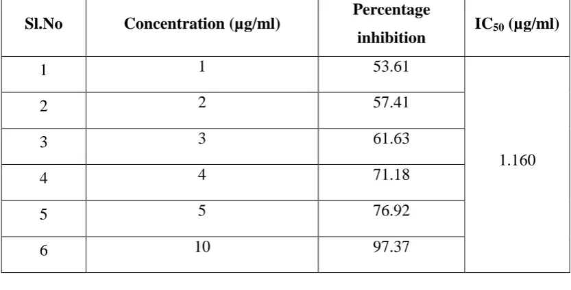
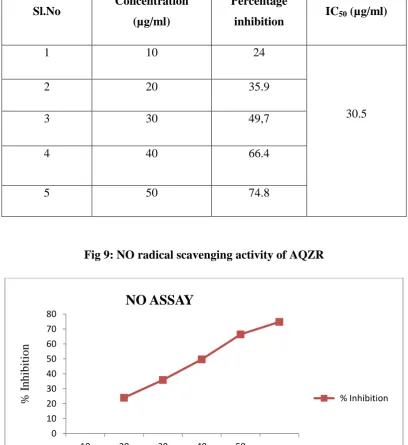
9 NO radical scavenging activity of AQZR 47
10 Zone of inhibition for Gram +ve organisms 48
11 Zone of inhibition for Gram –ve organisms 49
12 Antifungal acitivity of AQZR 49
13 MIC of AQZR 50
14 MIC of AQZR 51
15 Percentage wound contraction on excision wound model 52
16 Effect of AQZR on % Wound contraction in excision wound model 53
17 Effect of AQZR on Hydroxy proline level in excision wound model 54
18 Effect of AQZR on Hexosamine level in excision wound model 55
19 Effect of AQZR on Uronic acid level in excision wound model 56
20 Control 57
21 Simple base ointment 57
22 Standard 58
[image:18.612.69.550.73.714.2]LIST OF TABLES
TABLENO PARTICULARS PAGENO
1 Comparison between Acute and Chronic Inflammation 4
2 Bacterial strain used for the study with NCIM 30
3 Fungal strain used for the study with MTCC 31
4 Experimental design for excision wound model 34
5 Phytochemical Analysis of Zanthoxylum rhetsa(Roxb) DC 39
6 Estimation of total phenolic content of AQZR 40
7 Estimation of total flavonoid Content of AQZR 41
8 Percentage inhibition & IC50 values of DPPH radical by Quercetin 42
9 Percentage inhibition & IC50 values of DPPH radical by AQZR 43
10 Percentage inhibition & IC50 values of ABTS radical by Quercetin 44
11 Percentage inhibition & IC50 values of ABTS radical by AQZR 45
12 Percentage inhibition & IC50 values of NO radical by Quercetin 46
13 Percentage inhibition and IC50 values of NO radical by AQZR 47
14 Zone of inhibition for Gram +ve organisms 48
15 Zone of inhibition for Gram –ve organisms 48
16 Antifungal activity of AQZR 49
17 MIC values of AQZR 50
18 MIC values of AQZR 51
19 Percentage wound contraction in excision wound model 53
20 Hydroxyproline levels in Excision wound models 54
21 Hexosamine levels in Excision wound models 55
TABLE OF CONTENTS
SL NO: CONTENTS PAGE NO:
1 INTRODUCTION 1
2 PLAN OF WORK 14
3 REVIEW OF LITERATURE 15
4 PLANT PROFILE 18
5 METHODOLOGY 20
6 RESULTS 39
7 DISCUSSION 59
8 CONCLUSION 64
Discussion
ABSTRACT
The present investigation has been undertaken to study the wound healing properties of aqueous extract of Zanthoxylum rhetsa. The plant Zanthoxylum
rhetsa has a long history in herbal medicine in many countries. Experiments
were conducted following standard procedures. The extracts were evaluated for their in vitro antioxidant, antimicrobial and total phenol and flavonoid content. The AQZR ointment were administered topically, for evaluating the wound healing potential in excision wound model for twenty one days. Povidone iodine ointment was used as a standard for wound healing in excision wound model. Extract treated group showed in vitro antioxidant, antimicrobial properties compared with standard and control. AQZR exhibited similar in vivo wound healing activity that of the standard but with lesser magnitude. The result may be attributed to the phytoconstituents such as flavonoids and phenolics present in it which may be due to their individual or cumulative effect that enhanced wound healing and provided scientific evidence to the ethnomedicinal futures of Zanthoxylum rhetsa. These findings could justify the inclusion of this plant in the management of wound healing.
Introduction
Depatment of Pharmacology, KMCH College of Pharmacy 1
1. INTRODUCTION
1.1 HERBAL MEDICINES
[1][2][3][4]Herbal medicines which formed the basis of health care throughout the world since the earliest days of mankind are still widely used, and have considerable importance in international trade. Recognition of their clinical, pharmaceutical and economic value is still growing, although this varies broadly between countries.
Medicinal plants are important for pharmacological research and drug development, not only when plant constituents are used directly as remedial agents, but also as starting materials for the synthesis of drugs or as models for pharmacologically active compounds. Regulation of development and exportation is therefore essential, together with international cooperation and coordination for their conservation so as to ensure their availability for the future.
The United Nations Convention on Biological Diversity states that the conservation and sustainable use of biological diversity is of critical importance for meeting the food, health and other needs of the growing world population, for which purpose access to and sharing of both genetic resources and technologies are essential.
Legislative controls in respect of medicinal plants have not evolved around a structured control model. There are different ways in which countries define medicinal plants or herbs or products derived from them, and countries have adopted various approaches to licensing, dispensing, manufacturing and trading to ensure their safety, quality and efficacy.
Despite the use of herbal medicines over many centuries, only a relatively small number of plant species has been studied for possible medical applicat ions. Safety and efficacy data are available for an even smaller number of plants, their extracts and active ingredients and preparations containing them.
Introduction
Depatment of Pharmacology, KMCH College of Pharmacy 2
total global population remains dependent on traditional medicines for their primary healthcare. Herbs are occupying a comeback and an Herbal Renaissance is blooming across the world. They have been evidently prized for their medicinal, flavoring and aromatic qualities for centuries, yet for a while they were over shadow by synthetic products of modern civilization. Folk medicine is generally defined as traditional medicine that is practiced by non-professional healers or embodied in local custom or lore, generally involving the uses of natural and especially herbal remedies. The World Health Organization (WHO) defines traditional medicine as “the health practices, approaches, knowledge and believes incorporating plant, animal and mineral-based medicines, spiritual therapies, manual techniques and exercise, applied singularly or in combination to treat, diagnose and prevent illness or maintain well -being.” Treatments are specific to a particular ailment and may include the power of rare, dancing, sweet baths, massage, medicinal herbs, hot and cold foods or other means not practiced in modern medicine.
Once having realized their sources and adverse effects, people are going to nature with hopes of safety and security. The rich treasure of herbal drugs is forming a boon for our society. Plant derived compounds, apart from their nutritive values, could serve as important therapeutic weapons to fight various human and animal diseases, thereby making them indispensable in traditional medicine for treating a number of diseases. Plant drugs, popularly known as herbal medicines have since been unabatedly used to that various diseases. The major challenge is to protect traditional knowledge and will prove to be a beneficial asset to our human surrounding. For all the ailments herbal formulations are proved to be effective without any side effects commonly seen with allopathic drugs.
1.2 INFLAMMATION
[5][6]Introduction
Depatment of Pharmacology, KMCH College of Pharmacy 3
The classical signs of acute inflammation are pain, heat, redness, swelling, and loss of function. Inflammation is a generic response, and therefore it is considered as a mechanism of innate immunity, as compared to adaptive immunity, which is specific for each pathogen.
Inflammation is tightly regulated by the body. Too little inflammation could lead to progressive tissue destruction by the harmful stimulus (e.g. bacteria) and compromise the survival of the organism. In contrast, chronic inflammation may lead to a host of diseases, such hay fever, periodontitis, atherosclerosis, rheumatoid arthritis, and even cancer (e.g., gallbladder carcinoma). Inflammation is therefore normally closely regulated by the body.
Inflammation can be classified as either acute or chronic.
Acute inflammation is the initial response of the body to harmful stimuli and is achieved by the increased movement of plasma and leukocytes (especially granulocytes) from the blood into the injured tissues. Prolonged inflammation, known as chronic inflammation, leads to a progressive shift in the type of cells present at the site of inflammation and is characterized by simultaneous destruction and healing of the tissue from the inflammatory process.
Introduction
Depatment of Pharmacology, KMCH College of Pharmacy 4
Table:1 Comparison Between Acute and Chronic Inflammation [7]
Factors Acute Inflammation Chronic Inflammation
Causative agent Bacterial pathogens, injured tissues
Persistent acute
inflammation due to non-degradable pathogens, viral infection, persistent foreign bodies, or autoimmune reactions.
Major cells involved
Neutrophils (primarily), basophils (inflammatory response), and eosinophil’s (response to helminth worms and parasites),
Mononuclear cells
(monocytes, macrophages, lymphocytes, plasma cells), fibroblasts
Primary mediators
Vasoactive amines,
Eicosanoids.
IFN-γ and other cytokines, growth factors, reactive oxygen species, hydrolytic enzymes
Onset Immediate Delayed
Duration Few days Up to many months, or
years
Outcomes
Resolution,
chronic inflammation
Introduction
Depatment of Pharmacology, KMCH College of Pharmacy 5
Acute inflammation is a short-term process, usually appearing within a few minutes or hours and begins to cease upon the removal of the injurious stimulus. It is characterized by five cardinal signs: An acronym that may be used to remember the key symptoms is "PRISH" for Pain, Redness, Immobility (loss of function), Swelling and Heat.
The traditional names for signs of inflammation come from Latin:[8][9][10]
Dolor (pain)
Calor (heat)
Rubor (redness)
Tumor (swelling)
Functiolaesa (loss of function)
The first four (classical signs) were described by Celsus (ca. 30 BC–38 AD), while loss of function was added later by Galeneven though the attribution is disputed and the origination of the fifth sign has also been ascribed to Thomas Sydenham and Virchow.
Redness and heat are due to increased blood flow at body core temperature to the inflamed site; swelling is caused by accumulation of fluid; pain is due to the release of chemicals such as bradykinin and histamine that stimulate nerve endings. Loss of function has multiple causes.
Acute inflammation of the lung (pneumonia) does not cause pain unless the inflammation involves the parietal pleura, which does have pain.
Inflammatory disorders [11]
Inflammatory abnormalities are a large group of disorders that underlie a vast variety of human diseases.
Examples of disorders associated with inflammation include: Acne vulgaris
Asthma
Introduction
Depatment of Pharmacology, KMCH College of Pharmacy 6
Auto inflammatory diseases Celiac disease
Chronic prostatitis Glomerulonephritis Hypersensitivities
Inflammatory bowel diseases Reperfusion injury
Rheumatoid arthritis Sarcoidosis
Transplant rejection Vasculitis
Interstitial cystitis
1.3 WOUND HEALING ACTIVITY
[12]Wounds are inescapable events in life. Wounds may arise due to physical, chemical or microbial agents. Wound healing involves a complex series of interactions between different cell types, cytokine mediators, and the extracellular matrix. The phases of normal wound healing include hemostasis, inflammation, proliferation and remodeling. Each phase of wound healing is distinct, although the wound healing process is continuous, with each phase overlapping the next. Because successful wound healing requires adequate blood and nutrients to be supplied to the site of damaged tissue.
CLASSIFICATION OF WOUNDS
Wounds are classified as open wounds and closed wounds on the basis of underlying cause of wound creation and as acute and chronic wounds on the basis of physiology of wound healing
(a) Open Wound: [13]
Though the open wound blood escapes the body and bleeding is clearly visible.
Introduction
Depatment of Pharmacology, KMCH College of Pharmacy 7
Incised Wounds:
It is an injury with no tissue loss and minimal tissue damage. It is caused by a sharp object such as knife. Bleeding in such cases can be profuse, so immediate action should be taken.
Abrasions or superficial Wounds:
It is caused by sliding fall on to a rough surface. During abrasion the topmost layer of the skin i.e. epidermis is scraped off that exposes nerve ending resulting in a painful injury. Blood loss similar to a burn can result from serious abrasions.
Laceration wound or tears Wounds:
This is a nonsurgical injury in conjunction with some type of trauma, resulting in tissue injury and damage.
Puncture Wounds:
They are caused by some object puncturing the skin, such as needle or nail. Chances of injection in them are common because dirt can enter into the depth of wound.
Gunshot Wounds:
They are caused by a bullet or similar driving into or through the body. Penetration Wounds:
Penetration wounds are caused by an object such as knife entering and coming out from the skin.
(b) Closed Wounds:
In closed wounds blood escapes the circulating system but remain in the body. It includes contusion or bruises, heamatomas or blood tumor, crush injury etc.
Contusions or bruises:
Bruises are caused by a blunt force trauma that damage tissue under the skin.
Hematomas or blood tumor:
Introduction
Depatment of Pharmacology, KMCH College of Pharmacy 8
Crush injury:
Crush injury is caused when great or extreme amount of force is applied on the skin over long period of time.
Acute Wounds:
An acute wound is a tissue injury that normally proceeds through an orderly and timely reparative process that results in sustained restoration of anatomic and functional integrity. Acute wounds are usually caused by cuts or surgical incisions and complete the wound healing process within the expected time frame.
Chronic wounds:
C hronic wounds are wounds that have failed to progress through the normal stages of healing and therefore enter a state of pathologic inflammation chronic wounds either require a prolonged time to or recur frequently. Local infection, hypoxia, trauma, foreign bodies and systemic problems such as diabetes mellitus, malnutrition, immunodeficiency or medications are the most frequent causes of chronic wound.
FACTORS AFFECTING WOUND HEALING[14] Improper diet.
Infection at the wound site.
Insufficient oxygen supply and tissue perfusion to the wound area. Drugs.
Elderly age.
Diabetes and other diseases conditions.
Wound healing is normal biological process in the human body. Many factors can adversely affect this process and lead to improper and impaired wound healing.
Improper Diet:
Introduction
Depatment of Pharmacology, KMCH College of Pharmacy 9
Infection at the wound site:
Wound infection is probably the most common reason of impaired wound healing by
Streptococcus aereus, Streptococcus pyrogens, Escherichia coli, and Pseudomonas
aeruginosa.
Insufficient Oxygen Supply and Tissue Perfusion to the Wound area:
Adequate blood supply and tissue perfusion is extremely important for wound healing. Excessive pain, cold and anxiety can cause local vasoconstriction and increased healing time. Smoking and use of tobacco decreased tissue perfusion and oxygen tension in wound.
Drugs:
Many drugs are known to impair wound healing. Chemotherapeutic drugs are used in cancer are the largest group well known to delay wound repair. Systemic glucocorticoids interfere normal healing process by reducing collagen synthesis and fibroblast proliferation.
Elderly Age:
Elderly age is found to be associated with delay wound healing. It is reported that the fibroblast growth and activity diminishes and collagen production, wound contraction is slow in older individuals
Diabetes and Other Diseases:
Introduction
Depatment of Pharmacology, KMCH College of Pharmacy 10
PHASES INVOLVED IN WOUND HEALING: The Inflammatory phase
Fibroblastic phase Epithelialization phase Proliferative phase Contraction phase Remodeling phase
THE INFLAMMATORY PHASE: [15]
The inflammatory phase starts immediately after the injury that usually last between 24 and 48hrs and may persist for up to 2 weeks in some cases the inflammatory phase launches the haemostatic mechanisms to immediately stop blood loss from the wound site. Clinically recognizable cardinal sign of inflammation, rubor, calor, tumor, dolor and functionless appear as the consequence. This phase is characterized by vasoconstriction and platelet aggregation to induce blood clotting and subsequently vasodilatation and phagocytosis to produce inflammation at the wound site.
FIBROBLASTIC PHASE: [16]
The second phase of wound healing is the fibroblastic phase that lasts upto 2 days to 3 weeks after the inflammatory phase. This phase comprises of three steps viz. granulation, contraction and epithelialization in the granulation step fibroblasts form a bed of collagen and new capillaries are produced. Fibroblast produces a variety of substances essential for wound repair including glycosaminoglycans and collagen. Under the step of contraction wound edges pull together to reduces the defects in the third step epithelial tissues are formed over the wound site.
EPETHELIALIZATION PHASE: [17]
Introduction
Depatment of Pharmacology, KMCH College of Pharmacy 11
cells. Epithelial cell migrating across wound usually along the basal lamina or fibrin deposits, this phenomenon is called contact guidance and is an important factor in epithelial migration. Epithelial migration is followed by increased mitosis of epithelium. Recent evidence suggests that a water soluble heat labile substance called chalcone which is secreted at the wound site is responsible for regulation for mitosis.
PROLIFERATIVE PHASE: [18]
Proliferative phase (2 days to 3 weeks) includes:
Granulation stage: Fibroblasts lay bed of collagen fills defect and produces new capillaries.
Contraction stage: Wound edges pull together to reduce defect.
Epithelialization stage: Crosses moist surface cell travel about 3cm from
point of origin in all directions.
CONTRACTION PHASE: [19]
Introduction
Depatment of Pharmacology, KMCH College of Pharmacy 12
level of transforming growth factor, the most important instigator of wound contraction, was significantly higher in the granulation tissue of pony wounds compared with horse wounds.
REMODELING PHASE: [20]
This phase last for 3 weeks to 2 years. New collagen is formed in this phase. Tissue tensile strength is increased due to intermolecular cross-linking of collagen via vitamin-C dependent hydroxylation. The scar flattens and scar tissues become 80% as a strong as the original.
The wound healing activities of plants have since been explored in folklore. Many ayurvedic herbal plants have a very important role in the process of wound healing. Plants are more potent healers because they promote the repair mechanisms in the natural way. Extensive research has been carried out in the area of wound healing management through medicinal plants. Herbal medicines in the wound management involve disinfection, debridement and providing a moist environment to encourage the establishment of the suitable environment for natural healing process.
ACTIVITIES LEADING TO WOUND HEALING (a) Anti-inflammatory Activity:
The acute inflammatory response during the early stages of injury generates factors that are essential for tissue growth and repair. When prolonged, however, chronic inflammation can to be detrimental, preventing wound remodeling and matrix synthesis, leading to delay in wound closure and an increase in wound pain. Thus, it is possible that an anti-inflammatory effect could facilitate wound healing and improve patient comfort, although traditional texts and animal studies indicate that extracts exert an anti-inflammatory effect.
(b) Antioxidant Effect:
Introduction
Depatment of Pharmacology, KMCH College of Pharmacy 13
(c) Antimicrobial Activity:
Wound healing can also be delayed when microoganisms are present in large enough numbers. Therefore, reducing the bacterial load of a wound may be necessary to facilitate wound healing as well as to reduce local inflammation and tissue destruction. An ideal agent for the prevention and control of wound infection would therefore be one that directly destroys the pathogens while also stimulate immune activity.
(d) Analgesic Activity:
Plan of Work
Dept. of Pharmacology, KMCH College of Pharmacy 14
2. PLAN OF WORK
1. Review of Literature.
2. Selection Collection and Authentication of Plant Material.
3. Extraction of plant material stem with Water.
4. Preliminary Phytochemical analysis
5. Quantification of total Phenol and Flavonoid content.
6. Invitro antioxidant study
DPPH radical scavenging assay.
ABTS radical cation assay.
Nitric oxide scavenging assay.
7. In vitro antimicrobial study. Antibacterial.
Antifungal.
8. Pharmacological study.
9. Histopathological study.
Literature Review
Dept. of Pharmacology, KMCH College of Pharmacy 15
3. LITERATURE REVIEW
1. M.T.Rahman, et al., Antinociceptive and Antidiarrheal activity. The methanolic extract of the
Z
anthoxylum rhesta (Roxb.) DC. Stem bark. Significantly reduced the abdominal contraction induced by acetic acid and the diarrheal episodes induced by castor oil in mice. [21]2.
Md.Khrul Islam, et al., Anti-nociceptive and Antioxidant activity. Different parts of the medicinal plantZ
anthoxyllum budrunga wall seeds (ZBSE) to evaluate its antinociceptive and antioxidant potential. [22]3.
T.G.Nagaraja et al., In-vitro Antibacterial properties. Invitro screening of antibacterial properties ofZ
.rhesta (Roxb.) DC. of bark & stem showed antibacterial properties. [23]4.
T.G.Nagaraja et al., In-vitro evaluation of Anti-fungal properties. Invitro screening of anti-fungal properties of root, stem & bark ofZ
anthoxylum rhesta (Roxb.) DC. possess good fungicidal properties against alternaria alternate, aspergillus niger, fusarrium oxysporum, cladosporum sp., macrophoma phaseolina and thrichodermaviridi. [24]
5.
Aparna Saraf et al., Chemical profile studied. To establish the chemical fingerprint of various secondary metabolites ofZ
anthoxylum rhesta (Roxb.) DC. A medicinally important plant. HPTLC profiles of various individual secondary metabolites were done and profiles developed for authentication. This study was useful in differentiating the species from the adulterant and also act as biomarker for this plant in the pharmaceutical industry. [25]6.
Anwarul Islam abu sayeed et al., Brain shrimp lethality bioassay. The cytotoxic activity of free terpenes isolated from the bark ofZ
anthoxylum budrunga wall used in folk medicine in Bangladesh, was evaluated by brine shrimp lethality bioassay. [26]7.
Suresh lalitharani et al., G.C-MS analysis. The investigation deals with GC-MSanalysis of ethanolic extract of the spine of the
Z
anthoxylum rhesta (Roxb.) DC. plant fifteen compound were identified. [27]Literature Review
Dept. of Pharmacology, KMCH College of Pharmacy 16
plant. Pharmacognostic studies include microscopic, physicochemical constants, fluorescence analysis and preliminary phytochemical evaluations. [28]
9.
Temin payum et al., Antioxidant potencial .The methanolic extract of shoot were evaluated for total phenolic content (folin-ciocalteu’s method), total flavonoids content (colorimetric method) and antioxidant potential (DPPH and ABTS). [29] 10. R.Rajashri Naik, et al., GC-MS Analysis and biological evaluation of Essential oilof Zanthoxylum rhetsa(Roxb.) DC pericarp. The present study reports the chemical
composition, antioxidant, antimicrobial, antidiarrheal as well as the spasmolytic activity of Zanthoxylum rhetsa(Roxb)DC essential oil, fractionated oil and its principal constituent (Terpinen-4-ol) The constituents of essential oil were characterized by GC-FID and GC-MS. The pharmacological and biological activities of oil, its fraction and principal constituent were carried out in-vivo and in-vitro. The study suggests that the oil and its main active constituent (Terpinen-4-ol) of the studied plant would have high potential in the treatment of stress and gastro intestinal diseases. [30]
11. Rajashrinaik, et al., GC-FID Analysis of fatty acids and biological Activity of Zanthoxylum rhetsa ( Roxb.) DC seed oil. The fatty acid content and composition of
fixed oil from Zanthoxylum rhetsa seeds was determined. The seeds were found to contain about -19.5% of crude foxed oil on dry weight basis. Fatty acids were converted in to methyl esters and analyzed GC-FID. Ten fatty acids were identified using GC-FID. The major monosaturated and saturated fatty acids were oleic acids and palmitic acids respectively, whereas the alpha linolenic acid and linolenic acid were poly unsaturated fatty acid. Fixed oil exhibited significant free radical scavenging activity which was measured using DPPH, and is also known to inhibit the gastrointestinal motility significantly. [31]
12. A Ramesha Rao, et al., Chemoprotective influence of Zanthoxylum sps. On hepatic carcinogen metabolizing and antioxidant enzymes and skin papillomagenesis in murine model. In the present study, the putative of pericarp of dried fruit
Zanthoxylum (Rutaceae Family), a common spice additive in Indians west coast
Literature Review
Dept. of Pharmacology, KMCH College of Pharmacy 17
Cumulative, the findings strongly suggest cancer chemo preventive potential of
Zanthoxylum sps. [32]
13. S.K.Nazrul islam
et al.,Constituents and cytotoxicity of
Zanthoxylum rhetsa stem bark. Xanthyletin and sesamin have been isolated from petroleum ether extract of the stem bark of Zanthoxylum rhetsa. The petroleum ether extract showed cytotoxicity on brine shrimp nauplii. [33]14. Vijitr Udeye et al., Characteriaztion of the Essential oil and Fatty oil from Makhwaen fruit (Zanthoxylum rhetsa (Roxb.) DC). [34]
15. Khozirah shaari et al., Bioactive constituents of Zanthoxylum rhetsa. Bark and its Cytotoxicity Potential against B16-F10 Melanoma cancer and Normal Human Dermal Fibroblast (HDF) Cell lines. This plant has been predominantly used by Indian tribes for the treatment of many infirmities like diabetes, inflammation, rheumatism, toothache and diarrhea. The presence of bioactive lignans and alkaloids were suggested to be responsible for the cytotoxicity property of Z.rhetsa bark against B16-F10 cells. [35]
Plant profile
Dept. of Pharmacology, KMCH College of Pharmacy 18
4. PLANT PROFILE
[37][38][39]Figure:1 Zanthoxylum rhetsa (Roxb.) DC.
INTRODUCTION:
General Information of Zanthoxylum rhetsa(Roxb.) DC.
A genus of herbs belongs to the family Rutaceae well distributed in India, Bangladesh, Bhutan, China etc.
Fifteen species are found naturalized in India and Bangladesh. 1. Zanthoxylum rhetsa(Roxb.)DC.
2. Zanthoxylum nitidum 3. Zanthoxylum chalybeum 4. Zanthoxylum americanum. 5. Zanthoxylum oxyphyllum 6. Zanthoxylum armatum 7. Zanthoxylum piperitum 8. Zanthoxylum simulans
9. Zanthoxylum bungeanum max 10. Zanthoxylum sehinifolium 11. Zanthoxylum nitidumRoxb
12. Zanthoxylum armatum DC(or) alatumRoxb 13. Zanthoxylum limonella
Plant profile
Dept. of Pharmacology, KMCH College of Pharmacy 19
LATIN BINOMIAL & VERNACULAR NAMES:
Source : Zanthoxylum rhetsa (Roxb.) DC.
Synonym : Budrunga
Family : Rutaceae
VERNACULAR NAMES
:General Name - Kasabang(llk),kayatena(tag) English Name - Indian prickly ash-tree.
Botanical Name - Zanthoxylumrhetsa (Roxb.) DC. Tamil Name - Veerasangi
Sanskrit - Lakhuvalka(la), Asvaghra.
Malayalam - Mulilam, Mulliyllam, Karimurikka.
Kannada - Aramadala,Arampala,Jammina
Hindi Name - Badrang.
SCIENTIFIC CLASSIFICATION:
Kingdom - Plantae
Phylum - Tracheophyta
Division - Eukaryota
Class - Spematopsida
Order - Sapindales
Suborder - Rutineae
(Unranked) - Angiosperms, Eudicots, Rosids
Family - Rutaceae
Subfamily - Rutoideae
Tribe - Zanthoxyleae
Genus - Zanthoxylum
Methodology
Dept. of Pharmacology, KMCH College of Pharmacy 20
5. METHODOLOGY
5.1. PLANT COLLECTION AND AUTHENTICATION
The stem part of the plant Zanthoxylum rhetsa (Roxb) DC was collected from Palakkad District of Kerala and authenticated from the Plant Anatomy Research Center, Tambaram, Chennai, Tamil Nadu. The authentication certificate number is PARC/2016/3324. Soon after collection, the stems were cleaned and shade dried. After drying, these stems were crushed to a coarse powder, stored in air tight plastic container for further use.
5.2. EXTRACTION OF THE PLANT MATERIAL [40]
The coarsely powdered stem (200gm) were taken in a round bottom flask and with water for 48hours at room temperature. After extraction the extracts were evaporated or concentrated by using rotary evaporator and dried at room temperature. The obtained crude extracts were weighed and stored at 40C for the further analysis.
5.3. QUALITATIVE PHYTOCHEMICAL ANALYSIS OF AQZR [41][42][43][44] 5.3.1. Preparation of test sample
A small quantity of the extract was dissolved in 5ml of distilled water and filtered. The filtrate was tested to detect the presence of various phytochemical constituents in the sample.
5.3.2. TEST FOR CARBOHYDRATES
Molisch’s test
Methodology
Dept. of Pharmacology, KMCH College of Pharmacy 21 Fehling’s test
1ml Fehling’s-A (copper sulphate in distilled water) was added to 1ml of Fehling’s-B (potassium tartarate and sodium hydroxide in distilled water) solution, boiled for one minute. To this added 1ml of filtrate and heated gently. Formation of brick red precipitate indicates the presence of reducing sugars.
Benedict’s test
Few ml of filtrate was mixed with equal volume of Benedict’s reagent (alkaline solution containing cupric citrate complex) and heated in boiling water bath for 5min. Formation of reddish brown precipitate infers the presence of reducing sugars.
5.3.3. TEST FOR ALKALOIDS
Small amount of extract mixed with few ml of dilute hydrochloric acid. Shaken well and filtered. Following tests were performed with the obtained filtrate.
Dragendorff’s test
A few drops of Dragendorff’s reagent (potassium bismuth iodide solution) was added to 2-3ml of filtrate. Orange red precipitate indicates the presence of alkaloids.
Mayer’s test
A few drops of Mayer’s reagent (potassium mercuric iodide solution) was added to 2-3ml of filtrate. Cream (dull white) precipitate was formed.
Wagner’s test
A few drops of Wagner’s reagent (solution of iodine in potassium iodide) was added to 2-3ml of filtrate. Reddish brown precipitate was obtained.
Hager’s test
A few drops of Hager’s reagent (Picric acid) was added to 2-3ml of filtrate. Yellow precipitate was obtained.
5.3.4. TEST FOR TRITERPENOID
Libermann-Burchard test
Methodology
Dept. of Pharmacology, KMCH College of Pharmacy 22 Salkowski test
A small quantity of the extract was treated with chloroform and few drops of concentrated sulphuric acid and allowed to stand for few minutes. Yellow colour at the lower layer indicates the presence of triterpenoids.
5.3.5. TEST FOR GLYCOSIDES
Legal’s test
1ml of pyridine and 1ml of sodium nitroprusside was added to 1ml of extract. Pink to red colour indicates the presence of glycosides.
Keller-Killiani test
Glacial acetic acid was added to 2ml extract, followed by the addition of trace quantity of ferric chloride and 2 to 3drops of concentrated sulphuric acid. Reddish brown colour appears at the junction of two liquid indicates the presence of cardiac glycosides.
Baljet test:
2ml of extract was added to sodium picrate solution. Yellow to orange colour formation indicates the presence of glycosides.
5.3.6. TEST FOR STEROIDS AND STEROLS
Liebermann- Burchard reaction
2ml of extract was mixed with chloroform. To that mixture added 1-2ml of acetic anhydride and 2drops of concentrated sulphuric acid along the sides of the test tube. The solution becomes red, then blue and finally bluish green colour.
Salkowski reaction
2ml of extract was mixed with 2ml chloroform and 2ml concentrated sulphuric acid. Shaken well. Chloroform layer appears red and acid layer shows greenish yellow fluorescence.
5.3.7. TEST FOR PHENOLS
Ferric chloride test
Methodology
Dept. of Pharmacology, KMCH College of Pharmacy 23 Lead acetate test
Diluted 1ml of alcoholic solution of extract with 5ml distilled water and to this added few drops of 1% aqueous solution of lead acetate. Formation of yellow colour precipitate indicates the presence of phenols.
5.3.8. TEST FOR TANNINS
Lead acetate test
A few drop of lead acetate was added to 5ml of aqueous extract. Formation of yellow or red colour precipitate indicates the presence of tannins.
5.3.9. TEST FOR SAPONINS
Foam Test:
1ml of test sample was diluted with 20ml of distilled water and shaken it in a graduated cylinder for 3minutes. Foam of 1cm after 10min indicates the presence of saponins.
Froth test:
5ml of test sample was added to sodium bicarbonate solution. After vigorous shaking the mixture, kept it for 3minutes. A honey comb like froth formation indicates the presence of saponins.
5.3.10. TEST FOR FLAVONOIDS
Alkaline reagent test
A few drop of sodium hydroxide solution was added to the extract. Formation of an intense yellow colour, which turns to colourless on addition of few drops of dilute hydrochloric acid, indicates the presence of flavonoids.
Shinodas test [Magnesium hydrochloride reduction test]
Methodology
Dept. of Pharmacology, KMCH College of Pharmacy 24
5.3.11 TEST FOR PROTEINS AND AMINO ACIDS
Biuret test
3ml of test solution was added to 4% sodium hydroxide and few drops of 1% copper sulphate solution. Formation of violet colour indicates the presence of proteins.
Ninhydrin test
A mixture of 3ml test solution and 3drops of 5% Ninhydrin solution was heated in a boiling water bath for 10min. Formation of purple or bluish colour indicates the presence of free amino acids.
5.4. QUANTIFICATION OF TOTAL PHENOLICS AND FLAVONOIDS [45][46] 5.4.1. ESTIMATION OF TOTAL PHENOLICS
Reagents
Folin-Ciocalteu’s reagent Gallic acid (1mg/ml) 20% sodium carbonate Preparation of standard
Standard solution of was prepared by adding 10mg of accurately weighed Gallic acid in 10ml of distilled water.
Preparation of sample
10mg of the accurately weighed AQZR extract were separately dissolved in 10ml ethanol and used for the estimation.
.Procedure
Methodology
Dept. of Pharmacology, KMCH College of Pharmacy 25
5.4.2. ESTIMATION OF TOTAL FLAVONOIDS Reagents
Ethanol
10% Aluminium chloride 1M Potassium acetate
Preparation of standard
Standard solution of was prepared by adding 10mg of accurately weighed Quercetin in 10ml of ethanol.
Preparation of sample
10mg of the accurately weighed AQZR extracts were separately dissolved in 10ml ethanol and used for the estimation.
Procedure
The total flavonoid content of the AQZR was determined by using Aluminium chloride colorimetric method. To an aliquot of 100µl of extract (1mg/ml) or standard solutions of Quercetin (10, 20, 40, 60, 80, 100μg/ml) methanol was added separately to make up the solution upto 2ml. The resulting mixture was treated with 0.1ml of 10% aluminium chloride, 0.1ml of 1M potassium acetate and 2.8ml of distilled water. Shaken well and incubated at room temperature for 30minutes. The absorbance was measured at 415nm against blank, where a solution of 2ml ethanol, 0.1ml potassium acetate, 2.8ml distilled water and 0.1ml of aluminium chloride serve as blank solution. The total flavonoid content was determined from the standard Quercetin calibration curve. And it was expressed as milligrams of Quercetin equivalents (QE) per gram of extract.
5.5. INVITRO ANTIOXIDANT STUDY OF AQZR 5.5.1. DPPH Free Radical Scavenging Assay [47] Principle
Methodology
Dept. of Pharmacology, KMCH College of Pharmacy 26
reduced to the DPPHH and as consequence the absorbance’s decreased from the DPPH. DPPH radical is a stable radical by virtue of the delocalization of the spare electron over the molecule as a whole, so that the molecules do not dimerize, as would be the case with most other free radicals. The delocalization also gives rise to the deep violet colour, characterized by an absorption band in ethanol/methanol solution centred at about 520nm Radical to the DPPH-H form, results in decolorization (yellow colour) with respect to the number of electrons captured. More the decolorization more is the reducing ability. This test has been the most accepted model for evaluating the free radical scavenging activity of any new drug. When a solution of DPPH is mixed with that of a substance that can donate a hydrogen atom, then this gives rise to the reduced form (Diphenyl picryl hydrazine; non radical) with the loss of this violet colour (although there would be expected to be a residual pale yellow colour from the picryl group still present).
Procedure
The method adopted here is Blois method. Where by using the stable DPPH radical, the antioxidant capacity of the extract was measured in terms of hydrogen donating or radical scavenging ability. 1ml of 0.3mM solution of DPPH in ethanol was added to various concentrations of sample (10, 20, 40, 60, 80, 100 μg/ml) and the reference compound (5, 10, 15, 20, 25 and 30 μg/ml), shaken vigorously, and left to stand in the dark at room temperature. After 30 min absorbance was measured at 517nm against a blank. Quercetin was used as reference compound. A control reaction was also carried out without the test sample. All the tests were performed in triplicate in order to get the mean values. The percentage of inhibition was calculated by comparing the absorbance values of the control and test samples. Free radical scavenging activity was expressed as percentage inhibition (I%) and calculated using the following equation:
Percentage inhibition (I%) = (Abs control- Abs sample /Abs control) X 100
Different sample concentrations were used in order to obtain calibration curves and to calculate the IC50 values. (IC50 - concentration required to obtain a 50% radical
Methodology
Dept. of Pharmacology, KMCH College of Pharmacy 27
5.5.2. ABTS Assay [48][49] Principle
A method for the screening of antioxidant activity is reported as a decolorization assay applicable to both lipophilic and hydrophilic antioxidants, including flavonoids, hydroxyl cinnamates, carotenoids, and plasma antioxidants. The pre-formed radical monocation of 2,2'-azinobis-(3-ethylbenzothiazoline-6-sulfonic acid) (ABTS*+) is generated by oxidation of ABTS with potassium persulfate and is reduced in the presence of such hydrogen-donating antioxidants present in the sample. The influences of both the concentration of antioxidant and duration of reaction on the inhibition of the radical cation absorption are taken into account when determining the antioxidant activity. Chemistry involves the direct generation of the ABTS radical monocation with no involvement of an intermediary radical. It is a decolorization assay; thus the radical cation is pre-formed prior to addition of antioxidant test systems, rather than the generation of the radical taking place continually in the presence of the antioxidant. An antioxidant with an ability to donate a hydrogen atom will quench the stable free radical and inhibits the absorption of the radical cation which has characteristic long-wavelength absorption spectrum showing maxima at 660,734, and 820nm.The relatively stable ABTS radical has a green colour and is quantified spectrometrically at 734nm. It is applicable to both aqueous and lipophilic systems.
Procedure
ABTS radical scavenging activity of the extract was measured by Rice-Evans method. ABTS was dissolved in water to a 7mM concentration. ABTS radical cation (ABTS+) was produced by reacting ABTS stock solution with 2.45mM potassium
Methodology
Dept. of Pharmacology, KMCH College of Pharmacy 28
the mean values. The percentage inhibition of ABTS+ by the sample was calculated
according to the formula:
Percentage inhibition (I %) = (Abs control- Abs sample /Abs control) X 100
Different sample concentrations were used in order to obtain calibration curves and to calculate the EC50 values. (EC50 - concentration required to obtain a 50% radical
scavenging activity).
5.5.3. Nitric Oxide Assay [50][51] Principle :
Nitric oxide (NO) and reactive nitrogen species (RNS) are free radicals are derived from the interaction of NO with oxygen or reactive oxygen species. Nitric oxide is classified as a free radical because of its unpaired electron and displays important reactively with certain types of proteins and other free radical such as superoxide. NO is synthesized by three isoforms of the enzymatic nitric oxide synthase (NOS), endothelial NOS, neuronal NOS, and inducible NOS (iNOS). Nitric oxide (NO) is generated from amino acid L-arginine by the enzymes in the vascular endothelial cells, certain neuronal cells, and phagocytes. Low concentration of NO are sufficient in most cases to affect the physiological function of the radical. NO is a diffusible free radical that plays many roles as an effectors molecule in diverse biological systems including neuronal messenger, vasodilation and antimicrobial and antitumor activities. Chronic exposure to nitric oxide radical is associated with various carcinomas and inflammatory conditions including juvenile diabetes, multiple sclerosis, arthritis and ulcerative colitis. The toxicity of NO increases greatly when it react with the superoxide radical, forming the highly reactive peroxy nitrite anion (ONOO- ). Nitric oxide has been shown to be directly scavenged by flavonoids.
Procedure:
Methodology
Dept. of Pharmacology, KMCH College of Pharmacy 29
for 150 min. The sample was run as above but the blank was replaced with the same amount of water. After the incubation period, 2ml of the above incubated solution was added to 2ml of Griess reagent and incubated at room temperature for a period of 30mins. The absorbance of the chromophore formed was read at 540nm.
Percentage inhibition (I%) = (Abs control- Abs sample /Abs control) X 100
5.6 ANTI-MICROBIAL ACTIVITY OF AQZR [52] 5.6.1. Antibacterial study of AQZR
Preparation of inoculums
The inoculums for the experiment were prepared in fresh nutrient broth from the preserved slant culture. The turbidity of the culture can be adjusted by the addition of broth or sterile saline (if it is excessive) or by further incubation to get the required turbidity, and the newly prepared inoculums were standardized by adjusting the turbidity of the culture to that of McFarland standards.
Preparation of sterile swabs
Cotton wool swab on wooden applicator or plastics were prepared and sterilized by autoclaving or by dry heat (only for the wooden swabs). It was sterilized by packing the swabs in culture tubes, papers or tins etc.
Sterilization of forceps
Forceps can be sterilized by dipping in alcohol and burning off the alcohol.
Experiment
Methodology
Dept. of Pharmacology, KMCH College of Pharmacy 30
petri dish is divided into 2 parts. First compartment were loaded with sample disc as AQZR (100µg) and second compartment with standard Ciprofloxacin disc (10µg) with the help of sterile forceps. After that petri dishes are placed in the refrigerator at 4º C or at room temperature for 1hour for diffusion. Incubate at 37ºC for 24hours. Observe the zone of inhibition produced by different samples. Measure it using a scale and record the average of two diameters of each zone of inhibition.
Table 2: Bacterial strain used for the study with NCIM
Sl no Organism Strain NCIM 1
Gram+ve bacteria
Bacillus subtilis 2063
2 Staphylococcus aureus 2079
3
Gram–ve bacteria
E-coli 2065
4 Pseudomonas auregenosa 2200
5.6.2. Anti fungal activity of AQZR Procedure
Preparation of inoculums
The inoculums for this particular experiment were prepared in fresh sabouraud dextrose broth from preserved slant culture. The turbidity of the culture can be adjusted by the addition of broth or sterile saline (if it is excessive) or by further incubation to get the required turbidity, and the newly prepared inoculums were standardized by adjusting the turbidity of the culture to that of McFarland standards.
Preparation of sterile swabs
Methodology
Dept. of Pharmacology, KMCH College of Pharmacy 31
Sterilization of forceps
Forceps can be sterilized by dipping in alcohol and burning off the alcohol.
Experiment
The standardized inoculums is inoculated in the sterilized plates prepared earlier (aseptically) by dipping a sterile in the inoculums removing the excess of inoculums by passing and rotating the swab firmly against the side of the culture tube above the level of the liquid and finally streaking the swab all over the surface of the medium 3times rotating the plate through an angle of 60º after each application. Finally pass the swab round the edge of the agar surface. Leave the inoculums to dry at room temperature with the lid closed. The sterile discs are soaked overnight in sample solutions, AQZR. Each Petri dish is divided into 2 parts. First compartment were loaded with sample disc as AQZR (100µg) and second compartment with standard Fluconazole disc (10µg) with the help of sterile forceps. After that petri dishes are placed in the refrigerator at 4ºC or at room temperature for 1hour for diffusion. Incubate at 37ºC for 24hours. Observe the zone of inhibition produced by different samples. Measure it using a scale and record the average of two diameters of each zone of inhibition.
Table 3: Fungal strain used for the study with MTCC
Si no Fungi strains used MTCC
1 Aspergillus niger 1344
Methodology
Dept. of Pharmacology, KMCH College of Pharmacy 32
5.6.3. DETERMINATION OF MINIMUM INHIBITORY CONCENTRATION OF AQZR [53][54]
The minimum inhibitory concentration (MIC) is the lowest concentration of a chemical that prevents the visible growth of the organism.ie, the lowest concentration at which it has bacteriostatic activity.
5.6.3.1. Preparation of test drug:
The selected test drugs were prepared in DMSO at a concentration 2000µg/ml
5.6.3.2. Preparation of inoculums:
Bacillus subtilis, Staphylococcus aureus, E-coli, Pseudomonas auregenosa, Aspergillus
niger, and Monascus purpureus were the strains of organisms selected for the study.
Overnight culture are grown at 370C Kirby- Bauer procedure and diluted to Muller Hinton Broth and Sabouraud Dextrose Broth for bacterial and fungal strains respectively. This overnight culture was diluted to 10-2.
Inoculation
1. The sterile tubes were labeled 1-8 and 8th tube was taken as control. 2. 1ml of Muller Hinton Broth was transferred to all tube.
3. 1ml of drug solution was added to 1st tube and mixed well.
4. From the 1st tube transfer 1ml of solution to the 2nd tube and was repeated up to 7th tube.
5. From the 7th tube 1ml of solution was pipette out and discarded. 6. 0.01ml of culture was added to all the test tubes.
7. All the tubes were incubated at 370C for 18-24hrs. 8. After incubation observe the turbidity by visually
Methodology
Dept. of Pharmacology, KMCH College of Pharmacy 33
5.7. .PHARMACOLOGICAL EVALUATION OF AQZR 5.7.1. ACUTE TOXICITY STUDY [55]
Based on previously conducted study the dose was selected.
5.7.2. ANIMALS AND MANAGEMENT
Female Wistar rats of 6-8 weeks old and 160-180g body weight were offered by KMCH College of Pharmacy, Coimbatore. All rats were kept at room temperature and allowed to accommodate in standard co