JOURNAL OFVIROLOGY, Feb.1992, p. 804-815 Vol. 66, No. 2 0022-538X/92/020804-12$02.00/0
Copyright © 1992, American SocietyforMicrobiology
Simian Virus 40 T-Antigen DNA Helicase Is a Hexamer Which
Forms a
Binary Complex
during Bidirectional Unwinding
from the Viral Origin of DNA Replication
RAINER WESSEL, JOHANNES SCHWEIZER,t AND HANS STAHL*
Fakultatfur Biologie, Universitat Konstanz, D-7750 Konstanz, Germany
Received 9September 1991/Accepted 28 October 1991
The roleof simian virus 40(SV40) largetumorantigen (Tantigen)as aDNA helicaseatthereplication fork was studied. We found that a T-antigen hexamer complex acts during the unidirectional unwinding of appropriate DNA substrates and is localized directly in the center of the fork, contacting the adjacent double strandaswellastheemerging singlestrands. When bidirectional DNAunwinding, initiated at the viral origin of DNA replication, was analyzed, a largerT-antigen complexthat is simultaneously active atboth branch
points ofan unwinding bubble was observed. The size and shape of this helicase complex imply that the T-antigen dodecamercomplex, assembled attheorigin and active in the localizedmelting of duplex DNA, is subsequently also usedtocontinue DNA unwindingbidirectionally. Then, however,the dodecamer complex
does notsplit into two hexamer subunits that track along theDNA; rather, the DNA is threaded through the intactcomplex, with theconcomitant extrusion ofsingle-strandedloops.
The DNAofprokaryotic and eukaryotic cells as well as the DNA ofmany viruses is semiconservatively replicated following an apparently common molecular biological con-cept. According to this concept, DNA synthesis is
per-formedbidirectionally in a semidiscontinuous fashion
start-ing from defined points within thegenome, conventionally called
origins
of DNA replication. The double-stranded DNA(dsDNA)attheorigin is melted byaninitiatorprotein, and this primaryreplication
bubble isextended at its forksby a helicase, followed by the synthesis of daughter DNA
(forreviews, see references 27 and28).
In studies of the role of replicative helicases in the pathway of DNA replication, the simian virus 40 (SV40) largetumorantigen(Tantigen)has proventobeveryuseful. Viral DNA replication, which takes place in the nuclei of
monkey andhuman cells, requires Tantigen in addition to numerouscellularfactors(30, 50;for reviews,seereferences 15and48). Tantigenspecifically recognizes the pentanucle-otide 5'-GAGGC-3', present in four copies within the 64bp
ofthe SV40 core origin ofDNA replication (11, 14, 52). In the presence of
Mg2+
and ATP (Mg-ATP), it forms a double-hexamercomplex,coveringthecomplete coreorigin plus 12bp ineachdirection (10, 31). This complexpartiallyuntwists the bound DNA region without the need for ATP
hydrolysis(6, 37). From the meltedorigin region, T antigen cancontinue DNA
unwinding
bidirectionallyin the presenceof a single-stranded-DNA (ssDNA)-binding protein (SSB) and a topoisomerase, with hydrolysis of ATP being now
essential (16, 35, 39).
From theSV40replication studies and also from
biochem-icalanalyses, it is clear that T antigen is a DNA helicase that
catalyzesboth the localized melting of duplex DNA, with a
highbut not exclusive specificity for the SV40 origin (40), and also the processive unwinding of long stretches of dsDNA (46, 60). The two activities can be biochemically
differentiated by theirdifferent response to some
T-antigen-specific monoclonal antibodies and by their template
re-*Correspondingauthor.
tPresentaddress: Institut Pasteur,75015Paris,France.
quirements (40). ForDNA melting, aminimumlengthof 50 to 60 bp ofthe dsDNA substrate (50to 60 bp)is essential,
most probably reflecting the size of the active T-antigen complex. Incontrast, DNA
unwinding
of very short dsDNAregions (less than 20 bp) can be
efficiently
started by Tantigen if the substrate contains a
single-stranded
3'exten-sion of5 to 10nucleotides as anentry site
(49,
60).
Confirming the role as the viral replication fork helicase (47,59),DNAunwinding byTantigenproceedsataboutthe samerate as the rateofSV40DNA
replication
(60). More-over, in analogy to theprokaryotic
systems, Tantigen
hasalso been found to associate with the DNA
polymerase
cx-primase
complex (17, 20, 44),though the 3'-to-5' direction-ality oftheT-antigen helicasemovementdiffers fromthatof itsprokaryotic
counterparts and would place it on thetemplate fortheleading strandatthe
replication
fork(oppo-site the position of the polymerase a-primase complex catalyzing SV40
lagging-strand
DNAsynthesis [22, 29, 60]). While these data demonstrate aT-antigen interaction withthe
replication
machinery, more recent findings place it at the same time at the nuclear matrix, indicating that SV40DNAreplication proceedsatthisstructure (43).Therefore,a more detailed analysis ofhow T antigen combines its
bio-chemnical
and structural functionsduring viralDNA replica-tion is essential.Asmentionedabove, localDNAdenaturationat theSV40
origin apparentlyis initiated bytheassembly ofaT-antigen
dodecamercomplex. However, the size of the active
heli-case complex essential for the extension of this initial replicationbubble is unknown, as is therelationship, ifany,
between this helicase complex and the initial T-antigen
melting activity.We show that the basic DNA helicase of T
antigen is a hexamer that is active during unidirectional unwinding, started from single strand/double strand (ss/ds)
junctionsofpartiallydouble-stranded DNA. Footprinting of the T-antigen helicase bound at an unwinding fork and
electron microscopic analysis reveal that the hexamer
com-plex is localized directlyin the centerof the fork, contacting the double strand as well as the emerging single strands.
However, duringbidirectional DNA unwinding,started from
804
on November 10, 2019 by guest
http://jvi.asm.org/
ROLE OF T ANTIGEN AS A REPLICATIVE DNA HELICASE 805
the viral origin of DNA replication, a
bilobed
T-antigen helicase complex was observed operatingsimultaneouslyatboth forks of an unwinding bubble, with the ssDNA looped
out between. Thus, the double-hexamer complex formingat
the viral replication origin becomes active as a processive helicase with two reactive centers subsequentto itsinitiation function. Since the helicase complex does not track along
the DNA but rather the DNA is translocated in relation to
the complex, our results provide direct supportfor a model
of SV40 DNA replication in which the DNA is threaded through a fixed replication center.
MATERIALS ANDMETHODS
Reagents and enzymes. Nucleotides, restriction
enzymes,
endonucleaseP1,exonuclease III, T4 polynucleotidekinase,
and Klenow enzyme were purchased from Boehringer
Mannheim, Escherichia coli SSB was from Pharmacia,
Se-quenase (version 2.0) was from U.S. Biochemical, and A exonuclease was from Bethesda Research Laboratories.
Radioactive nucleotides were from Amersham.
Glutaralde-hyde was from TAAB, formamide was from Fluka, and Bio-Gel A5m was from Bio-Rad. All other chemicals were purchased from Sigma. T antigenwas isolatedby the immu-noaffinity procedure (46), usingextractsfromhybrid adeno-virus-SV40-infected HeLa cells (33). The T-antigen-specific monoclonal antibody PAb 101 was prepared as described previously (47).
DNA constructs used as DNA helicase substrates. A
par-tially double-stranded DNA containing a preformed fork
(used in the experiments shown inFig. 4 and 5) wasprepared as follows. One strand, calledhere the 3' strand and
repre-senting the fast-migrating strand ofthe denatured HindlIl-PvuII fragment (322 bases, HindIII site at the 5' end) of
plasmid pSVC4 (39) was preparedbyelutionafterseparation
of the restriction fragment on adenaturing agarose gel. A 70-mer oligonucleotidewithbases 31to 70complementaryto bases 93 to 132 ofthe3' strand wasannealedand elongated up to the end withSequenase(usedunderconditions recom-mended by the supplier);thisoligonucleotidewasdesignated
the 5' strand ofthissubstrate. As can be also seen fromthe schematic drawing in Fig. 4A, the substrate consists of a 132-bp double-stranded part carrying one blunt end at one side and two noncomplementarysingle strands of 190 bases
(3' strand)and 30 bases (5' strand)atthe other. The 5'strand
was radioactively labeled either at the 5' end with
[_y-32P]dATP by T4 polynucleotide kinase prior toannealing or at the 3' end by replacingdATPwith [a-32P]dATP
during
the elongation reaction with the Sequenase. The 3' strand was labeledat its 5' end also by using the T4polynucleotide kinase and [y-32P]dATP(38).
A partially double-strandedM13 DNA(usedin the
exper-iments shown in Fig. 1) was created by
annealing
a30-nucleotide primer complementary to nucleotides 6510 to
6539 ofM13mp7 positivestrandfollowed
by
elongation
withKlenow enzyme in the presenceof
[a-32P]dATP
andnonra-dioactive dCTP by 8 nucleotides (for details, see reference 46).
A A exonuclease-digested linearized pUC8 DNA
(used
intheexperiments shown in Fig. 2 and3) was
prepared
using
1.5 ,ug ofScaI-cleavedpUC8 DNAincubated for10swith30
U ofexonuclease as described
previously (41).
Theproce-dure nearly quantitatively generated linear dsDNA
mole-cules with single-stranded 3' ends of a mean
length
of approximately 200 bases, as revealed byquantitative
elec-tron microscopy (EM) (for details, see reference41).
The large
fragment
ofAvaII-restrictedpSVMO1
DNA(used for
Fig.
6 and 7) (49)carrying
the SV40origin
of replication 1.5 and 1.1 kbp from the ends was used in theorigin-specific
DNAunwinding experiments.
Nuclease protection experiments. 32P-labeled DNA with a
preformed fork (2.5to 5 ng;see
above)
wasincubatedunder standard helicase reaction conditions butin the presenceof2 mM
adenylyl(y,3-methylene)-diphosphonate
(AMP-PCP)
instead ofATPwith indicatedamounts ofT
antigen
for 10 min at 37°C. Then 2.3 ,ugM13mp7
ssDNAwasadded,
andincubation continued for additional 10min. Nuclease
diges-tion was started
by
additionof105 to 135 Uofexonuclease III andincubation for10minat37°C
orby
addition of5to10 ,ug of endonuclease P1 and incubation for 15 minat37°C.
Exonuclease III was used at
limiting
concentrations to preventnonspecific
degradation
ofthesingle-stranded
part ofthe substrate. Reactionswerestopped
with 3 volumesof 50mMEDTA-400
mM sodiumacetate-0.5%
sodium dode-cyl sulfate(SDS).
Afterphenol-chloroform
treatment andethanol
precipitation,
theDNAwasdriedanddissolved in10,ul of 47% formamide-10 mM EDTA. The heat-denatured DNAwas then
analyzed
on a6%DNAsequencing
gel,
andthe G+A
Maxam-Gilbert-sequenced (32) 32P-5'-end
labeled 3' strand was runalongside.
DNA helicase
experiments.
Standard DNAhelicase assays were performedessentially
as describedpreviously (46).
Briefly,
2.5 to5 ng of32P-labeled
DNAwas incubated with 50ngto 1,ug ofTantigen
in 20 mMTris-HCI
(pH
7.5)-4
mMMgCl2-2
mMATPat37°C
fortheindicatedtimes. Reactionproducts were
analyzed
by
SDS-polyacrylamide
gel
electro-phoresis
andautoradiography
(40);
forquantitative
analysis,
thebandsfromthe
gel
wereslicedoutandcountedinaliquid
scintillation counterasdescribed elsewhere
(60).
For
EM,
helicase assays contained 20 mM triethanol-amine-HCI(pH
7.5)
instead ofTris-HCI,
lowerconcentra-tions
(2
mM)
ofMgCl2
(to
avoidaggregation
ofTantigen
that wasobservedathigher
concentrations of freeMg2+;
unpub-lished
observations),
80 mM NaCl(to
inhibitnonspecific
binding
ofTantigen),
and 500 ngofE.coli SSB(to
stabilize unwoundsingle
strands).
Unwinding
reactionswerestopped
by
theaddition of0.1volume of1%glutaraldehyde
in 20 mMtriethanolamine-HCI
(pH
7.5)
and incubationfor 15 min at37°C.
Unlessindicated,
freeprotein
andglutaraldehyde
wereremoved before use of the
samples
for EMby
spin gel
filtration
using
Bio-Gel
ASm in 20 mMtriethanolamine-HCl
(pH
7.5)
asdescribedpreviously
(58).
EM. Fixed
samples
werespread
by
using
benzylalkyldi-methylammonium
chloride(BAC) (56)
andprocessed
forshadowing
as describedpreviously
(58).
Rotary
shadowing
wasdone withtungsten atan
angle
of8°
andcontrolled witha quartz
crystal.
The thickness of thetungsten
layer
wasadjusted
to5 nmby
thefrequency
change
oftheoscillating
quartzcrystal
(500
Hz;
seemanual BB 800 048BD,
Balzers,
Liechtenstein),
and diameters of shadowedT-antigen
heli-case
complexes
were corrected for this value. Fornegative
staining,
samples
weredirectly
adsorbed for 1 min to air-glowdischarge-treated
(15
s, 7Pa,
110V,
20 to 40mA)
carbon-coated
grids
and stained with 5%uranyl
acetate for 15 to20 s. Excessuranyl
acetate was removedby blotting,
and the
grids
were air dried.Micrographs
were taken at aHitachi
H-7000,
and tilt seriesweredone withaPhilips
400-Ttransmission electron
microscope.
Negatively
stained to-bacco mosaic virus was used as externallength
standard.Length
measurements ofpSVMO1
DNA for determinationof the SV40
origin
position
were done on a CRPdigitizer
(Cybernetical
Research&Production,
Konstanz,
Germany),
VOL. 66, 1992
on November 10, 2019 by guest
http://jvi.asm.org/
J.
VIROL.
806 WESSEL ET AL.using magnified (100,000-fold)positives. Size measurements
ofT-antigen complexes were performed with a measuring lens using magnified (500,000-fold) positives. Diameters of individual complexes were calculated from four measure-ments, rotating the positive image clockwise after each measurement by an angle of 45°. The mass of T-antigen complexes was calculated from the mean diameter
(deter-mined from 50 complexes) of the negatively stained com-plexes, using the formula formassdetermination of a spher-ical protein body: Mr = d3 -rw* p NA!6, where Mr is the
molecular weight, d is the mean diameter ofthe T-antigen complexes, pis thespecific density of proteins(1.33g cm-3 [25]),and NA is Avogardo'snumber(6.022 x 10²³ molecules permol).
Sucrose gradient centrifugation. Gradients (4.5 ml)
con-taining5to20%sucrosein 10 mMTris-HCI (pH 7.5)-0.1mM ATP-0.1 mMMgCl2-1mMdithiothreitol-10% glycinewere centrifuged in an SW55 rotor(Beckman) at 39,000 rpm for 19.5 h at0°C and elutedin250-pulfractions.Samplesusedfor the T-antigen helicase assay and for the EM analysis were taken fromthe sucrose gradientfractions immediately after
centrifugation and subsequently used for the respective
assays. Forcalibration, gradientswith marker proteins
(he-moglobin, 4.3S, 65 kDa; aldolase, 7.4S, 158 kDa;catalase,
11.3S, 233 kDa ferritin, 17S, 440 kDa; and thyroglobulin,
19S, 669 kDa) wereruninparallelunderidentical conditions.
RESULTS
Formation of helicase-active hexamers in the absence of DNA. Incubation of T antigen at 37°C in the presence of
Mg-ATPbutin the absenceofDNAresults intheformation of T-antigen oligomers with hexamers as the most specific complexes (31). Using a partially double-stranded DNA substrate, we found theT-antigen helicase still active after preincubation for 1 h under these conditions. At the same
time, a sedimentation analysisdisclosed ashift ofthe
T-an-tigenprotein (data not shown; 46) and ofthe DNA helicase
activityfromabout6S to 17S (Fig. 1C).
Whenanalyzed by EM after negative staining with uranyl acetate, controlTantigen (not preincubated with Mg-ATP)
from the enzymatically active 6S peak (fraction 15 in Fig. 1C) appeared as 6- to 8-nm particles as would be expected
for monomeric to dimeric forms (Fig. 1B). In contrast,
micrographs ofthe 17S complexes manifested about 90% of Tantigen in regularglobular structures (Fig. 1A), with the restofthe complexes showing a smaller and heterogeneous
size (not shown). The diameter of the main class of 17S
complexeswas 10.8 ±0.7 nm(Table 1). A calculation of the Mr ofa globular protein complex with this experimentally
determined diameter (25; see also Materials and Methods)
givesa meanvalue of 528,000. This is 6.4 times larger than the Mr ofaT-antigen monomer (82,000) and implies that the helicase activity is associated with a hexameric form of T
antigen, though the technique used here may not be
suffi-ciently accurate to definitively distinguish between pen-tamericto heptameric forms of T antigen (see Discussion).
In the presence of ATP, free hexamers were stable at
37°C, whereas incubation of T antigen in the absence of Mg-ATPat37°C for 1 h did not lead to detectable
oligomer-ization and even destroyed preformed hexamers (data not
shown). Moreover, incubation on ice in the presence of Mg-ATP did also not support the assembly of hexamer
complexes,indicating that T antigen is converted on cooling
into a state unable to form hexamers (data not shown; 31).
Since ofthe various possible interactions, only hydrophobic
A
B
C Thyroglobulin Ferretin Catalase Aidolase Hemoglobln
10
-Co
-0
1 5 1~~~10 15 2
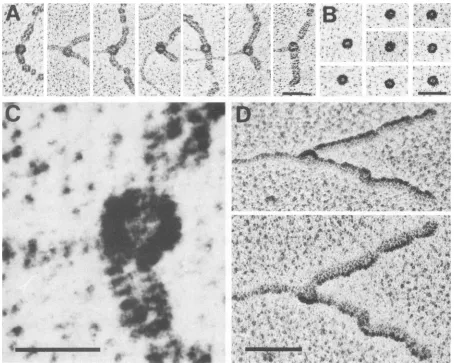
fraction number top FIG. 1. DNA helicaseactivity inT-antigen hexamercomplexes assembled in the absence ofDNA. (A) EM analysis ofT-antigen complexes taken from fraction 8 of the sucrosegradientshown in panel C, whichwasloadedwith Tantigenpreincubated at37°Cin thepresenceof Mg-ATP but in the absenceof DNA.Sampleswere
fixedwithglutaraldehyde,directly adsorbedtocarbon-coatedgrids, andnegatively stained (see Materials andMethods). Thecomplexes shown represent about 90% of the Tantigen. (B) EManalysis by negative staining of fraction 15 ofthe sucrose gradient shown in panel Cand loadedwithcontrolTantigen(withoutpreincubation). Theshown complexes represent more than90% of the Tantigen. Bar, 50nm.(C)A60-,ugsample ofTantigen withoutpreincubation (circles)orofTantigen incubated for1 hat 37°C inatotal volume of100,ul in the presence of1 mMMg-ATP under otherwise standard helicase reaction conditions but in the absence of DNA (squares) wasseparated by sucrose gradientcentrifugation (see Materials and Methods). Sedimentation wasfrom righttoleft. Aliquots (10
p1u)
of collected fractions (250pd)
weretested forhelicase activity under standardconditions, using5ngof partiallydouble-stranded M13mp7 DNA as asubstrate. Helicase reaction productswere analyzed by gel electrophoresis and quantitated as described in Materials and Methods. The helicase activitywasexpressed as arbitrary units (1 U represents the unwinding of 0.2 fmol of partially double-stranded M13mp7 DNA).interactions are impaired at low temperature (36), it is
conceivable that they may have an important role in
main-taining the hexameric structure of T antigen. Moreover,
hydrophobic interactions could also explain the observed salt stability of theT-antigen helicase complex (60; unpub-lishedobservations).
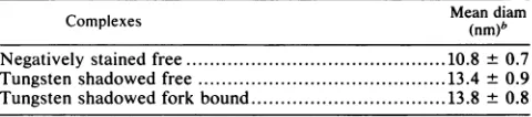
TABLE 1. Dimensions ofT-antigenhelicase complexesa
Complexes Mean(nm)bdiam
Negativelystained free... 10.8 + 0.7
Tungsten shadowedfree... 13.4 + 0.9
Tungsten shadowed fork bound... 13.8 + 0.8
"Freecomplexeswere fromtheexperiment shownin Fig.1A;fork-bound complexes werefrom theexperiment shown inFig.2A.
bDeterminedasdescribedinMaterialsandMethods and calculatedfrom 50 individual complexesofeach type.
on November 10, 2019 by guest
http://jvi.asm.org/
[image:3.612.311.551.636.689.2]VOL.66,1992~~~~ROLE OF T ANTIGEN AS A REPLICATIVE DNA HELICASE 807
I,
*
1)~~~~~~~~~~i
FIG. 2. EMdemonstration of fork-boundT-antigenhelicasecomplexesinvolved inprogressiveunidirectional DNAunwinding.(A)Linear
pUC8DNA(0.1 p.g),containing3'-extendedsingle-strandedendsofca.200bases, wasincubated with 0.2 gxgof Tantigenfor 15minunder
helicasereactionconditions, andreactionproducts were processedfor EManalysis(see Materials andMethods). Electronmicrographsof
progressively unwoundDNAmolecules are shown. A circularcomplex withhigh contrastis seen at the end(most probably atthe ss/ds
junction)of the first molecule andatthebranchpointofunwindingforks. Note thatssDNA,coated with E. coliSSB,is condensed about 2.7
timesrelativetoduplex DNA,thusaccountingfor thediscrepancyin thelengthsofprogressivelyunwound DNAs(27). (B)Anunwinding
reactionsampleidenticaltothat used forpanelAwasincubated with PAb 101(500ng)(23)afterglutaraldehydefixation andspin gelfiltration
for 15 min on ice. Sodium chloride (200 mM)was added;after fixation, a second spin gel filtration wasperformed toremove nonbound
antibodies. Because of the presence of boundantibodies, thecomplexesatthe branchpoints nowappearlargerandmoreamorphous than
thosewithout antibodies(showninpanel A). Bar,200nm.
DetectionofanactiveT-antigenhelicasecomplexin unwind-ingforksbyEM. PreformedT-antigen hexamers may disso-ciate after isolation, and resulting substructures could give
risetothe helicaseactivity detected in the 17Speak ofFig.
1. Therefore, we also analyzed individual T-antigen
com-plexes actually involved in DNA unwinding by EM. A
linearized plasmid DNA of 2.7 kb was used as a helicase substrate. The DNA contained small 3'-extended
single-strandedregions at each end, created byashort incubation with theX exonuclease, that servedashighly efficient entry
sites of the T-antigen helicase for unidirectional unwinding
(60; for experimental details, see reference 41). After
incu-bation of the DNA with Tantigen for 15minunderhelicase conditions in the presence of E.coliSSB,sampleswerefixed
immediately with glutaraldehyde added directly to the
un-windingreactions and spreadfor EM analysis. As has been
reported before (16, 40), progressively unwound DNA
mol-ecules with twofree single strands produced by the
T-anti-gen helicase activity were obtained (Fig. 2). The denatured DNA is covered by the SSB and thus exceeds in diameter
and contrastthe nondenatured partof the DNA molecules.
Since the 3' single-stranded endsof the substrate served as
highly efficient entry sites, T antigen apparently always
started from the ends of the DNA molecules; unwinding
bubblesresultingfrom internalstarts(40)werenotobserved.
TheBACspreadingtechniquewasusedincombination with
quartz crystal-controlled rotary tungsten shadowing,
result-ing in a good resolution of the different proteins in a
nucleoprotein complex(58).
A protein knob witha higher contrast than the SSB was
seen in the center ofmost unwindingforks (90%) and was
believedtobe Tantigen, theonlyproteinin additiontoSSB
present in the reaction sample (Fig. 2A). This conclusion
was confirmed by staining of the protein-DNA complexes
with a T-antigen-specific monoclonal antibody (PAb 101
[23]). Under these conditions, the -protein knob appeared
moreamorphousandlargerthan in thepreparationswithout
antibodies(Fig.2B). Astheunwindingwascarriedoutunder
stringentreaction conditions (80 mM salt), most DNA
mol-ecules were free of nonspecifically bound T antigen (49).
Fromthepanelof thenucleoproteinstructures showninFig.
2A,itappearsthat thehelicasecomplexfirstbindstotheend
A,
t t
B
VOL. 66,1992
on November 10, 2019 by guest
http://jvi.asm.org/
[image:4.612.76.554.70.402.2]808 WESSEL ET AL.J.Vo.
A 0.
'LI
.
4,-'
"1 .. *0
44. '0
4'
/ b
WV%
dEW.
Af
41
'I yW
I4
A'
44
0
4,.
141 .~~~~~~~~~~~~~~~~~~~~~~~~~~~~~~~~~~~~~~~4
[image:5.612.78.532.66.429.2]
-4~~~~~~~~~~~~~~~~~~~~~~~~~~~~~~~~~~-.~~~~~~,1'344~~~~~~~~~~~~~~~~~~Al
FIG. 3. Shapeof fork-boundT-antigenhelicasecomplexes.(A)Electronmicrographsof the centralpart of DNAunwindingforks with the
T-antigen helicase bound toadjacent ssDNAanddsDNAregions. DNAhelicase substrate, helicaseconditions, and EMprocedureswere
identicaltothose usedfor Fig. 2. Note thecircular palespot in thecenter of eachT-antigencomplex, whichlikely reflects thesphericor hemispheric shapeoftheT-antigencomplexshadowed ata smallangle. Bar,50nm.(B) T-antigen complexesfrom the 17Sfraction ofFig.
1 butspread bythe BAC techniqueand rotary shadowed with tungstenexactly asdone forpanelAfor visualization ofT-antigenhelicase complexesboundtounwindingforks.Bar, 50nm.(C)Highermagnificationof the firstunwindingfork shown inpanelA. Bar,20nm.(D)An
unwindingfork(thesecondoneshown inpanelA)photographedunderdifferent tiltangles(upper, +600;lower, -40').Notethehemispheric
appearanceof thefork-boundT-antigen complex withaheight dependingonthe tiltangle. Bar, 50nm.
of the DNAmolecules, most
probably
atthe ss/dsjunction,
andthen
processively
invades the double-stranded section. Fork-bound Tantigen complexes
remained stable after treat-ment ofunwinding
intermediates with salt (500 mM) orEDTA(1 mM) before fixation with
glutaraldehyde
but werenot observed when
unwinding
reactions were run in the absence ofMg-ATP (data not shown).The
T-antigen
helicase wasalways
bounddirectly
to the center of the fork,apparently interacting
with all three strands. Athigh magnification (Fig.
3A and C), helicasecomplexes
from differentunwinding
intermediatesalways
were identical and
appeared
as a circular dark spot with apale
center, whichlikely
reflects themicrographic
appear-anceof
hemispheric
orspheric complexes
afterrotaryshad-owing
atasmallangle.
Thisassumption
wasconfirmedby
atilt series of electron
micrographs,
whichgives
more infor-mation about the three-dimensionalshape
(Fig.
3D).Com-plexes
delineatedatanangleof-40'or +600 nowappeared
asdistinctcaps with aheight resembling
theangular profile.
Sizemeasurementsof fork-bound
T-antigen complexes
(Ta-ble 1)revealed diameters
ranging
from 12.7to 14.9nm, withan average value of 13.8 ± 0.8 nm. This size is identical to that of the 17S hexameric
complexes
from the sucrosegradient
ofFig.
1prepared by
the BACspreading
method(Fig.
3B) and shows that the fork-boundT-antigen
com-plexes
are also hexamers(Table 1).Footprint
of theT-antigen
DNA helicase bound at anunwinding
fork.In theunwinding
intermediatesofFig.
2and3, helicase
complexes appeared always
bounddirectly
tothe center of the fork,apparently interacting
with all three strands. Tomaptheposition
of theT-antigen
helicase within the fork moreprecisely,
we triedfootprinting experiments.
We used a
partially
double-stranded DNA of knownse-quence as a helicase substrate that consisted of a
132-bp
duplex
region
withtwononcomplementary single
strands of 190 and 30 bases at one side(Fig.
4A). Thepredicted
structureof theDNAconstructwith this
preformed
unwind-ing
fork was confirmedby
proving
theaccessibility
of the J. VIROL.'P.
-I.mm -.
A
'Mik.,
.s. 4M"f',A
V MM
on November 10, 2019 by guest
http://jvi.asm.org/
ROLE OF T ANTIGEN AS A REPLICATIVE DNA HELICASE 809
A
B
/vg
N
3'
r-
--_
r _ - tl I
c~~~~~~~~~
g
JI
X
\R
/I C T
AG +
T T
.0 T TT
' A
A G
T G
C iG
T iA
A 'A
T IT
T
'TG
-T
'-GA
A--T T--A
n G--C
N2
C--GT
T--A
3' 5' i 2 3 4 5
"IF 0 VI ON
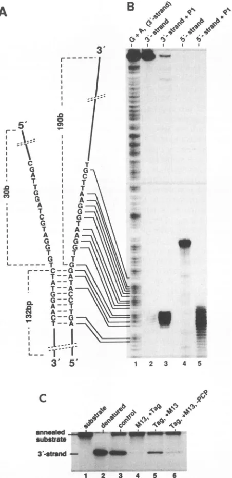
FIG. 4. Construction and function of a T-antigen DNA helicase substrate containing a preformed unwinding fork. (A) Schematic drawing of the DNA helicase substrate used in the nuclease protec-tionexperiments. Individual strands are referred to as the 3 strand and 5' strand according to their 3' and 5' single-stranded ends, respectively. The lengths of the double-stranded part and of the single-stranded ends as well as the DNA sequence in the vicinity of the fork are indicated.Single-stranded regionsoftheDNAconstruct were analyzed by theirsensitivity tonucleaseP1(B). The DNAwas labeled either at the 5' end of the 3' strand(lanes 1to3) or atthe3' end of the 5' strand (lanes 4 and 5) and subjected either to a Maxam-Gilbert G+A reaction (lane 1) or a nuclease P1 (10
pu.g)
digestion (lanes 3 and 5). Reaction products were analyzed by autoradiography after separation on a 6%DNA sequencing gel in comparison with the untreated 3' strand (lane 2) and 5' strand (lane 4). (For experimental details, see Materials and Methods.) (C) Analysis by polyacrylamide gel electrophoresis and autoradiogra-phy ofunwinding of the substrate DNA shown in panel B by T antigen. Binding of T antigen (Tag; 0.3,ug)to the substrateDNA(2.5 ng; labeled at the 5' end of the 3' strand; specific activity, 700
overhanging single strands by the single-strand-specific
nu-clease P1. After digestion, the radioactively labeled sub-strate was examined on a sequencing gel next to a G+A
sequencingladder,showingthat nucleaseP1 removed
essen-tially all of the overhanging single strands (Fig. 4A, lane 3
and 5). Moreover, using denatured substrate DNA as a control, we obtained completely digested DNA under the sameconditions(data notshown). Thedouble-strandedpart
of the DNA construct was established by its sensitivity to
double-strand-specific 3' exonucleaseIII (see below). The DNAsubstratewasefficientlyunwound by T antigen under standard DNA helicase conditions (Fig. 4B, lane 3). ssDNA (M13DNA) was aneffective inhibitorof the unwind-ing reactionbecause itcompetedwith thesubstrate DNA for T-antigen binding (lane 4) (60). However,
prieincubation
of the substrate DNA with T antigen in the presence of thenonhydrolyzable ATP analogAMP-PCPallowed the forma-tion ofaT-antigen preinitiation complex, which, after addi-tion ofATP,performedthereactioneven inthepresenceof M13 ssDNA(lane 5). Thus, stalled T-antigen helicase
com-plexes were bound to the preformed unwinding forks, a
reactionthat was stronglystimulatedby AMP-PCP. Infact, 1 mM ATP-PCP did not inhibit the T-antigen ATPase or DNA helicase activities during the subsequent unwinding
step after addition of2 mM ATP but was slightly
stimula-tory, most probably by promotion of suitable complex formation(data notshown;7).
The exactposition ofTantigenatthepreformed
unwind-ing fork wasinvestigated by DNase protectionexperiments (Fig.
5).
The interaction ofTantigen with each of the twosingle strands was analyzed by protection against
degrada-tion by nucleaseP1 (Fig. 5A andB). Afterpreincubation of
the DNA substrate (labeled at either strand) with Tantigen
and Mg-AMP-PCP under helicase reaction conditions, P1 digestionwasperformedin thepresenceofan excess
of
M13 DNA to remove nonspecifically bound T antigen. Theex-periments performed in the absence of T antigen again
showed that digestion ceasedonboth strands near thess/ds
border(lanes 2 and 3 in Fig. 5Aand lane 1 inFig. 5B).Only
in the presenceofTantigen,
P1
digestion ofboth overhang-ing soverhang-ingle strands resulted in additional sharp bands thatcouldbemapped 16 to 17nucleotidesin front of the forkon the
5'-extended
single strand(Fig.5A,
lanes 5 and6)and 9to 10 nucleotides on the 3'-extended single strand (Fig.SB,
lanes 2 to4).Nuclease
protection
was stimulatedby,
thoughnotabsolutelydependenton,
Mg-AMP-PCP
(compare lanes 4 and 5 in Fig.SA),
which is consistent withsimilar
obser-vations concerning the ATP-independent formation of aT-antigen unwinding complex atthe SV40
origin
(31). Thepercentage of the
protected
DNA molecules was low butsimilartothatof molecules unwoundin thepresence ofM13 ssDNA after
preincubation
with PCP(Fig. 4B),
indicating
that the
protection
was really due to fork boundT-antigen
helicasecomplexes.
Bq/ng), was allowed only during preincubation under helicase reactionconditions but in the presence of AMP-PCP(2mM)instead ofATPat37°Cfor15 min.Afterpreincubation,excess amounts(2.3
,ug) ofM13 ssDNAand4mM ATPwereadded,andtheincubation was continued for additional 30 min (lane 5). Reactions were
performedinthe absence ofM13 DNA(control; lane3),withM13 DNApresentduringthepreincubation step(lane4),in theabsence ofAMP-PCPduring preincubation,orwith AMP-PCP added after preincubation(lane6).Analysisof thenative(lane1)anddenatured (lane2) substrateDNAisalsoshown.
VOL.66, 1992
on November 10, 2019 by guest
http://jvi.asm.org/
[image:6.612.66.292.68.534.2]810 WESSEL ET AL.
A
C
31
B
G
T A
0
A |w
_ G: v_
e45 -23I
70
II k60
11 r ML
a
A. T
I
50
1 1
A
I I
A
I I I
C',40
D
5'
16 b3'
ss/ds-junction
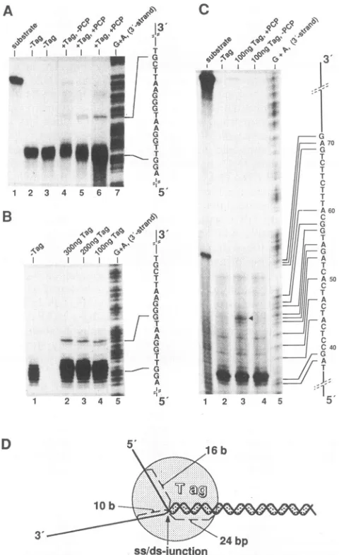
FIG. 5. Footprint of the T-antigen helicase complex bound at a preformed unwinding fork. (A)P1protection of the 5' strand of the DNAsubstrate shown inFig.4A.DNA (2.5 ng; see Fig. 1A)labeled atthe 3' end of the 5' strand (lane 1) waspreincubated without (lanes 2 and3,showing identical experiments) or with (lanes4-6)Tantigen (Tag;200ng)under standard helicase reaction conditions but in the presence of2 mM AMP-PCP instead of ATP for 15 min at 37°C (lanes 2, 3, 5, and 6) or with no nucleotide (lane 4). After a further incubation period of 15 min in the presence of 2.3
p.g
ofM13 ssDNA, endonucleaseP1digestion and analysis of the resulting products on a6%polyacrylamide sequencing gel were done as described in the legend toFig. 4A and Materialsand Methods. The experiment in lane 6 isshown as an overexposure (twofold) of the autoradiogram. AMaxam-Gilbert G+A sequencing reaction of the complementary5'-end-labeled 3' strand was analyzed in parallel as a size marker (lane 7). Positions of the ss/ds junction (first single-stranded
nucle-otide)
andofthe band induced by T-antigen protection are indicated andarealigned with thesequence of the 3' strand. (B)P1protection of the3'strandlabeled at the 5' end (see Materials and Methods). Experimental conditions were the same as for panel A. Lanes: 1 to 4, no T antigen or 0.3, 0.2, and 0.1 ,ug of Tantigen,
respectively, addedduringthepreincubation step in the presence of AMP-PCP; 5, Maxam-Gilbert G+A sequencing reaction of the 3' strand, with alignment of the positions of the ss/ds junction and of the band protected by T antigen to its DNA sequence. (C) Exonuclease III protection of the 5' strand. The substrate DNA (lane 1) was labeled atthe5'end ofthe5'strand (theintermediate band represents some labeled nonannealed 70-meroligonucleotide used for theconstruc-To
analyze
thebinding
oftheT-antigen
helicase to thedouble-stranded
portion
of thesubstrate,
weused exonucle-ase III(Fig.
SC).Theresults,
obtained in the absence of Tantigen,
showed a smearof DNAending
atadefined bandcorresponding
toastop
of theexonuclease 10bp
in front of the ds/ss transition(lane 2).
With bound Tantigen,
oneadditionalbandbecame
visible,
mapping
at24bp
behindthe fork(compare
lanes2, 3,
and4).
Thisprobably
indicatesthe extent ofprotection
within the double-strandedportion
by
thefork-bound
T-antigen
helicasecomplex.
Withincreasing
amounts of
protein,
additional clusters of bandsappeared,
indicating protection
oflonger
stretches of the double-strandedportion
(data
notshown),
mostprobably
due toenhanced
binding
of Tantigen
to dsDNA in the presence of ATPaspreviously reported (12, 40).
InFig. SD,
theprotec-tion data are summarized in a model
showing
the dimen-sional characteristics of theT-antigen
helicaseplaced
slightly asymmetrically
overthefork.T-antigen
complexes involvedinbidirectionalDNAunwind-ing. Inthe presence of
Mg-ATP,
Tantigen
assemblesattheoriginas adouble-hexamer
complex
mostprobably
resulting
intheuntwisting
oftwo tothree turns of DNA(for
areview,
seereference
5).
Aftermelting
of theorigin region,
thisinitialT-antigen complex
maysplit
into two helicase-active hex-amers, eachmigrating
totheopposite
directionduringbidi-rectional DNA unwinding. To test this model,
T-antigen
complexes,
involved in the unwinding of plasmid DNAcontaining
the SV40origin (large fragment
ofAvaIl-re-stricted
pSVMO1),
werespread
by
the BAC method andanalyzed
by
EM.Figure6shows thedifferenttypes ofnucleoprotein struc-tures found after incubation of the DNA with T
antigen
under stringent helicase reaction conditions that allowed
only
origin-specific unwinding (Fig. 7A) (49).. First, we observed bilobedT-antigen complexes (Fig. 6B)
bound at theorigin
that were also obtained after incubation in the presence ofa nonhydrolyzable ATP analog but not in the absence of ATP(data not shown). These origincomplexes
appeared identical to those described by others (10) and most probably represent the double hexamers
apparently
involved in the initialmelting of origin DNA
(31).
About5%of theDNA molecules scoredwerefoundtobe
unwinding intermediatesoftwoapparently different confor-mations. One type, representingabout 75% ofthe
unwinding
intermediates(forastatistical analysis, seeFig.7B),showed
a clear unwinding bubble with a protein knob within the center ofeach fork and SSB covering ssDNA. The
protein
complex inthe centerofthe forkswas determinedto have
the same dimensions described above for the T-antigen
helicase
(Fig.
2 and 3) and could also beimmunologically
stained by PAb 101 (data not shown). The other type,
representingabout25%ofunwindingintermediates,consists
tion of thesubstrate DNA; see Materials andMethods). Preincuba-tion ofthe DNA with T antigen, exonuclease III digestion, and analysis of the resultingproducts were done as described in Mate-rials and Methods and forpanel A.Preincubations were performed withoutTantigen (lane 2) or with 0.1 ,ug of T antigen in the presence (lane 3; the triangle marks the bandinduced by T-antigen protection) orabsence(lane 4) of AMP-PCP.AMaxam-Gilbert G+A sequenc-ingreaction of the 5'-end-labeled 3' strand was run in parallel asa sizemarker; the related DNA sequence is shown (lane 5). (D) Model showing the localization of the helicase complex at the unwinding fork. Numbersindicate thelengths (in bases or base pairs) ofDNA strands protected by Tantigen.
J. VIROL.
i
on November 10, 2019 by guest
http://jvi.asm.org/
[image:7.612.59.299.68.461.2]at
44 r~~~~~~~~~~~
-
.. 'C,,'
9'i
t5r ,'-
.5
4~ ~ ~ ~ 4
dIsDNA
Tag* r-.
46
SdQ,vNjssDNA
,'
,.: G t' 4 ',I t
J'
rz
'...
dDAts'r;,.*
;-
; ,
s'#
~ 49 P4
-B- :.-.-
- : .
>.-.
-:.>>;.) :--:.
s
;t*-+J
4g98-v
_e=9
;;s^so
., .. .. .' , ... t;
,,;z se
@ _ * *
.. .ew
,t,,
.-.X. -,fX,..,,,,'t.. e;..'..,-.>',..
*''s-{.{.
;,',11s 8j#,Rt^
_.,.@tt6,x,v
,et.
C.~~
K
ft~~~~~~~~~~~~~~~~~~~~~~~~
,' .i
..
|s
-'*
ti.6
0.
tP
.
,*ri.
;-,.0..6Y..
4..S.
E..;.. .. ... ... .. s. .. ... .. ..
44t..,U..
[image:8.612.71.557.38.567.2]* . - ... . vts ...: s :;',-:,
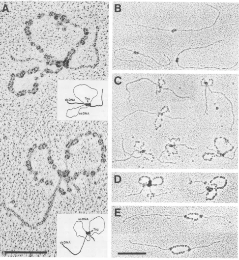
FIG. 6. Electron micrographs ofunwindingintermediates started from the SV40originof DNAreplication. (A)Thelarge fragmentof Avall-restricted pSVMO1DNA(0.1 pLg),containingtheoriginofreplication1.1 and 1.5kbp fromtheends,wasincubated with Tantigen (1
,ug)underorigin-specifichelicase reaction conditions(see Materials andMethods) for15min,spread by the BAC method, and processed for EMasdescribed forFig.2. Shownarehigh magnificationsofonetypeofunwindingintermediatestructure, containing single-stranded loops (rabbit ears). Bar, 100nm. Inserts show model drawings deduced from themicrographs. Thebilobed protein complexin thecenterof the unwinding intermediates, connectingboth forks andresultingintheloopingoutof the DNAsingle strands,has beenidentifiedasTantigen (Tag)(see panel D). (B) Micrographsfrom the reaction mixture used forpanel A, showing bilobed T-antigen complexesboundattheorigin position. (C) Micrographsof the type (rabbit ears-containing structures) shown inpanel A, representingdifferent stages of theunwinding
process. Thetypeofcomplexesshown comprisesabout 25% ofunwindingintermediates obtained(forstatisticalanalysis,see Fig.7). (D)
Micrographs representing unwindingintermediates obtainedafterapreincubationof Tantigenwiththe monoclonalantibodyPAb 101(0.7 ,ug) for15minonice beforetheonsetof the helicase reactionbythe addition of thelargepSVMO1Avallfragment.Helicase reaction conditions
and processingofunwinding intermediateswere otherwiseexactlyasdescribed forpanelA. The rabbitears-containingstructures shown
comprise about 75% ofunwindingintermediates obtained under these conditions. Note thelarge sizeandamorphous shape(duetobound
antibodies)oftheprotein complex connectingboth forks of each molecule. (E) Micrographsof the helicasereactionmixture usedinpanel A butincubatedin thepresence of 10 mM EDTA at00C for 5 minpriortofixation with glutaraldehyde. The main speciesofunwinding intermediates obtained underthisconditions arefully expanded unwindingbubbles whicharosebybidirectionalunwindingstarted fromthe
origin (seealsoFig. 7A). Note the individualT-antigen complexesboundatthe branchpointsofboth forks of each molecule.Bar,200nm.
811
C .1
.. ti.
on November 10, 2019 by guest
http://jvi.asm.org/
812 WESSEL ET AL.
A
B
theoretical
1000- ri~~~~~~~~~o-position
0 ~~~~~~~~~~molecule
0 5 10 15 20 25 30 35 40 number
reaction conditions structureofunwindingIntermediates bubbles rabbit ears standard reaction conditions 75% 25%
EDTAbefore fixation 96% 4%
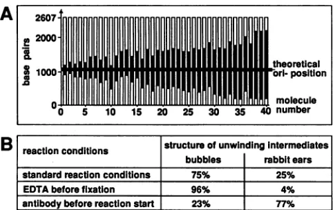
[image:9.612.58.299.67.219.2]antibodybeforereaction start 23% 77% FIG. 7. Statisticalanalysisofunwindingintermediatesobtained with substrate DNA containing the SV40 replication origin. (A) T-antigen-catalyzedbidirectional DNAunwindingfromtheoriginof
replication.A0.1-.g sampleofthelargeAvallfragment (2,607 bp)
ofthepSVMO1 plasmidthatcontained theoriginofreplication 1.1 and 1.5kbpfrom the endswasused. Helicase reaction conditions and EManalysiswereexactlyasdescribed forFig.6. Nodifferences for theorigin specificity were observed in the different protocols
used. Unwindingintermediateswerephotographed,andthelengths
of the dsDNA arms were measured on a digitizing board. The shorterarmwastaken tobe theoneclosest tothereplicationorigin
and wasplacedat thebottom ofthefigure.White columnsrepresent
dsDNAregions;blackcolumnsrepresentunwound ssDNAregions
coatedbySSB. Thethick horizontal linerepresentsthe theoretical
origin position. The linearized 4,360-bp plasmid pBR322 was in-cludedas aninternallength standard;thestandard deviation of the
lengthmeasurementswas3.5%.(B) Frequencyof differenttypesof
unwinding intermediates. The percentage ofunwinding intermedi-ates with expanded unwinding bubbles or rabbit ears-containing structures, obtained under different protocols, is indicated.
Stan-dardreaction conditionswere asdescribed forFig.6A toC;addition
of EDTAbeforefixationwas asdescribed forFig.6E;antibodywas added beforethe start of the reactionas described forFig. 6D.
of two loops of ssDNA (again covered with SSB) which appear to emerge from, and return to, a bilobed protein
complex, sitting on the otherwise entirely double-stranded
DNA (Fig. 6A and C). Thecentral protein knobs appeared
similar in size and shape to the bilobed T antigen complex
obtained at the origin before theinitiation ofunwinding. In
fact,it couldbeimmunologicallystainedafterfixation witha
T-antigen-specific monoclonal antibody (see below). There-fore,in thiskindofunwinding intermediates,thetwo branch points ofeach single-stranded bubble are held together by
the internal T-antigen complex. Individual unwinding
com-plexeshadsingle-strandedloopsof differentlengths ("rabbit ears")representingdifferentstages ofunwinding.Whenthe unwindingreactionwas stoppedwithEDTAbeforefixation, unwinding intermediates with single-stranded loops were found to be drastically reduced (4% of controls), and the
fraction of unwinding intermediates with completely
un-folded bubbleswere accordingly increased (Fig. 7B).
Thepercentageofunwindingintermediateswith therabbit
ears structures was significantly increased when T antigen
was preincubated with the monoclonal antibody PAb 101
before theunwindingreaction(Fig. 6D). Inthiscase, 77%of unwinding intermediatescontained rabbit ears. The
T-anti-gen knobs holding together the branch points were
some-what larger and amorphous because of bound antibodies (Fig. 6D). Inaddition,when Tantigenwaspreincubated with PAb 101, the percentage of unwinding intermediates also
was considerably
higher (at
leastfivefold;
data notshown),
suggestinga moreefficient formation of helicase-active
T-an-tigen complexes. Similar datawere obtained previously
by
biochemical unwinding experiments using an SV40
origin-containing DNA fragment as a substrate but also with
non-origin-containing DNA substrates (40). Because of its bivalent nature, onemolecule ofthe PAb 101 antibodymay interact with both hexamers of a T-antigen initiation com-plex and cross-link them, especially since PAb 101 was added prior to the formation of helicase complexes. This
possibility was confirmed in aprevious study (40) in which Fabfragments ofPAb 101 were shownto lose their stimu-lating activity.
DISCUSSION
With the exception of T antigen, all known helicases involved in bidirectional DNA replication are unable tostart DNA unwinding within an intact dsDNA substrate.
They
assemble on a premelted DNA region at the
replication
origin created by a corresponding initiatorprotein. In con-trast, extensive biochemical analyses have shown that T
antigen catalyzes both the melting ofduplex DNA, with a
highbutnot exclusivespecificityfor the SV40origin(6,
37,
39), and the processive unwinding of long stretches of dsDNA asatrue DNA helicase (22, 60).
In thisstudy, we first characterized the composition of the basicT-antigen DNA helicase. After preincubation at 37°C
in the presence ofMg-ATP but in the absence ofDNA, T antigen assembled into stable complexes that could be separated as a discrete species sedimenting at 17S in a sucrosegradient. Sedimentation behavior and size determi-nation by EM, using the method of negative staining, iden-tifiedthe 17S complexes as T-antigen multimers, most
prob-ably hexamers. Our conclusions were strongly confirmedby
scanning transmission EM, which allowed more precise size measurements(31). By this method, T antigen has also been shown underthe same conditions to form free hexamersthat were, however, not reported to possess DNA helicase
activity, in contrast to the results presented here. Free T-antigen hexamers proved to be stable in the presence of
ATP, arguing against the formal possibility that preformed hexamerscould disassemble andsubsequentlyreassemble at DNA forks. Moreover, preformed complexes were also active in an origin-dependent unwinding assay, indicating
that they can also assemble into functional dodecamer complexes (unpublished observations).
Actively unwinding T-antigen helicase complexes were found byEM to havealmostthe samesize asthe
(helicase-active)17Scomplexes (Table1). Thesmalldifference,if any, may result from the DNAcontained withinthe fork-bound complexes. The EM analysis of unwinding intermediates therefore confirms that the hexamer complex indeed resem-blesthehelicase-active T-antigen form. Actually unwinding helicase complexes were bound directly in the center of
unwindingforks, which was further confined by footprinting
experiments. The data clearly demonstrate that the T-anti-gen helicase complex interacts with all three strands of a
fork, protecting about 10 and 16 bases of theprotruding 3' and 5' single strands, respectively, and about 24 bp of the
double-stranded portion ahead ofthe fork. Even though T
antigeninteracts with alongerstretch of the single-stranded 5' strand, this does not necessarily contradict the earlier biochemicalfindings that the interaction with the 3' strand
probablydetermines thedirectionalityof helicase movement
(22, 60). Ourdata arealsoconsistentwith the concept that T J. VIROL.
on November 10, 2019 by guest
http://jvi.asm.org/
ROLE OF T ANTIGEN AS A REPLICATIVE DNA HELICASE 813
antigen, specifically interacting with the 3' strand, simulta-neously forms close contacts with the DNA polymerase-primase complex at the 5' strand of the replication fork. The protected DNA within the fork most likely also reflects dimensions of the T-antigen helicase complex, although we cannot exclude the possibility that the nucleases used stopped some nucleotides in front of the helicase complex as a result of steric hindrance. We note, however, that T antigen was found in this study to protect a length of the 3'-extended single strand similar to that required for an efficient start of the T-antigen helicase at partially double-stranded DNA (5 to 10 nucleotides [60]). The size of the helicase complex deduced from the nuclease protection data and from EM analysis amounts to approximately half of that determined for the dodecamer complex formed at the origin and covering about 84 bp (12, 31). Taken together, our data strongly indicate that a hexameric complex represents the basic T-antigen helicase. Interestingly, two helicases of E.
coli, the DnaB and Rho proteins, are also active as hexamers (4, 8, 19), and it may be speculated that this property has been conserved for functional reasons during evolution.
At
37°C
and in the presence of ATP, T antigen assembles at the SV40 origin as a double-hexamer complex which is assumed to perform local DNA melting (31). Interestingly, this double-hexamer complex is shown here to bind simul-taneously to both forks of unwinding bubbles that were formed during bidirectional DNA unwinding started from the origin. As the most plausible explanation for the generation of these structures, we propose that the double-hexamer complex, assembled at the SV40 origin and used for the initial melting, subsequently becomes also active in bidirec-tional DNA unwinding. Since unwinding is performed at two unwinding centers within the bilobed complex, the basic helicase again is a T-antigen hexamer, as shown for unidi-rectional unwinding with partially double-stranded sub-strates. The correct orientation within the bilobed complex with regard to the unwinding direction of the helicase subunits may be ensured by the 27-bp inverted repeat at the origin during the T-antigen assembly process.Under standard reaction conditions, approximately 75%
of the unwinding intermediates showed a clear unfolded unwinding bubble with a T-antigen hexamer in the center of each fork. We do not know whether this structure resembles real unwinding intermediates resulting from potential split-ting of the bilobed T-antigen complex at later stages in the unwinding reaction or whether it is an artifact of the prepa-ration. PAb 101, bound to T antigen before the onset of the unwinding reaction, apparently stimulates the formation of or stabilizes binary helicase complexes, since in the pres-ence of the antibodies, the rabbit ears-containing structures are the major unwinding intermediates. Bound antibodies may simulate in vivo conditions in which the T-antigen unwinding complex seems to be bound to structural ele-ments (nuclear matrix), possibly resulting in a stabilization and also stimulation of the replication complex(see below). EDTA treatment seems to disrupt the double-hexamer complex, whereas single T-antigen hexamers, bound tothe center of unwinding forks, appear to remain stablybound to
DNA under these conditions. Therefore, different
mecha-nisms must be involved in the formation of single T-antigen hexamers and the association of dodecamer complexes at
the origin. Whereas hydrophobic interactions seem to be
involved in the formation of hexamer complexes (see
Re-sults), magnesium-mediated protein-protein interactions of T-antigen molecules have been reported to induce DNA loops, and it seems that similar interactions are also
impor-tant during bidirectional DNA unwinding (41). However, EDTA may also affect some other metal ion such aszinc that may be bound by the zinc finger region of T antigen (30a).
DNA unwinding starting from an origin of replication and continued bidirectionally by one and the same helicase complex acting simultaneously at both forks provides direct support for previously proposed models of DNA replication in which the DNA moves while the replication machine remains at a fixed site. Moreover, the DNA helicase could function as an organizer of the replicationmachine andat the same time could fix it to the nuclear scaffold, possibly arranged in replication centers with multiple replication forks as has been suggested recently for viral and cellular DNA (13, 24, 34, 54). Accordingly, the (transient) binding of replication origins, as well as replicating DNA, to structural elements has been described (18, 21, 26, 55; for areview, see reference 53), and during alytic infection, a subfraction of T antigen is bound to the nuclear matrix and has been sug-gested to be actively involved in SV40DNAreplication (42, 43, 57). In this context, it is also interesting to note the frequent colocalization of detected replication origins with matrix attachment sites (1-3, 9).
Finally, the simultaneous binding ofthehelicasecomplex
to both branch points of a replication bubblewould guaran-tee a synchronous progression of related forks. Indeed, T antigen has been reported to be associated with replicating
intermediate SV40 chromatin particularly distinguished by synchronously moving forks (45, 51). The association of forks in a topologically closed replicating DNA molecule would also prevent an accumulation of torsional strain in replicated sections. An extensive catenation of daughter
strands could thus be avoided.
ACKNOWLEDGMENTS
Wethank R. Knippers for continuing support and helpful discus-sions, K. Mendgenfor making available the Hitachi H-7000, andH. Muller for helpwith the tilt series on the Philips 400 T transmission electron microscope. We also thank W. Ruyechan for critical reading of the manuscript and T. Kapitza for excellent technical assistance.
This work was supported by the Deutsche Forschungsgemein-schaft.
REFERENCES
1. Amati, B., and S. M. Gasser. 1988. Chromosomal ARS and CEN elements bind specifically to the yeast nuclear scaffold. cell 54:967-978.
2. Amati, B., and S. M. Gasser. 1990. Drosophila scaffold-attached regionsbind nuclear scaffolds and can functionasARSelements in both budding and fission yeasts. Mol. cell. Biol. 10:5442-5454.
3. Amati, B., L. Pick, T.Laroche,and S. M.Gasser. 1990. Nuclear scaffold attachment stimulates, but is not essential for ARS activityin Saccharomyces cerevisiae: analysisofDrosophila ftz SAR. EMBOJ. 9:4007-4016.
4. Arai, K.-I.,S.-I. Yasuda, and A. Kornberg. 1981. Mechanisms of dnaB protein action. J. Biol. Chem. 256:5247-5252. 5. Borowiec, J. A., F. B.Dean, P. A.Bullock,andJ.Hurwitz. 1990.
Bindingandunwinding-how T antigenengages the SV40origin of DNA replication. Cell60:181-184.
6. Borowiec, J. A., and J. Hurwitz. 1988. Localized melting and structural changesin the SV40 originofreplication inducedby T antigen. EMBO J. 7:3149-3158.
7. Bradly, M. K. 1990. Activation of ATPase activity of simian virus 40 large Tantigen by thecovalent affinity analogofATP, fluorosulfonylbenzoyl 5'-adenosine. J. Virol. 64:4939-4947. 8. Brennan,C. A.,A. J.Dombroski,and T. Platt. 1987.
Transcrip-tion termination factor rho is an RNA-DNA helicase. Cell 48:945-952.
VOL.66, 1992
on November 10, 2019 by guest
http://jvi.asm.org/
814 WESSEL ET AL.
9. Brun, C., Q. Dang, and R. Miassod. 1990. Studies of an
800-kilobase DNA stretch of the
Drosophila
X chromosome:comapping
ofa subclass ofscaffold-attached regions withse-quences able to
replicate
autonomously
in Saccharomycescerevisiae. Mol. Cell. Biol. 10:5455-5465.
10. Dean, F. B., M. Dodson, H. Echols, and J. Hurwitz. 1987.
ATP-dependent
formation ofaspecialized nucleoprotein
struc-tureby
simian virus 40(SV40)
large
tumorantigen
at theSV40
replication origin.
Proc. Natl. Acad. Sci. USA 84:8981-8985.11. Deb,S.,A. L.DeLucia,C. P.Baur,A.Koff,and P.Tegtmeyer.
1986. Domain structure of the simian virus 40 core
origin
ofreplication.
Mol. Cell. Biol. 6:1663-1670.12. Deb,S. P.,and P.Tegtmeyer. 1987. ATP enhances the
binding
of simian virus40
large
Tantigen
totheorigin
ofreplication.
J. Virol. 61:3649-3654.13. DeBruyn Kops, A., and D. M.Knipe.1988. Formation ofDNA
replication
structures inherpes
virus-infected cellsrequires
aviral DNAbinding protein. Cell 55:857-868.
14.
Delucia,
A.L.,B.A.Lewton,R.Tjian,
and P.Tegtmeyer.
1983.Topography of simian virus 40 A
protein-DNA
complexes:arrangementof
pentanucleotide
interaction sitesattheorigin
ofreplication.
J. Virol. 46:143-150.15. DePamphilis, M. L., and M. K. Bradley. 1986.
Replication
of SV40 andpolyoma
virus chromosomes, p. 99-246. In N. P. Salzman (ed.), ThePapovaviridae,
vol. 1. PlenumPublishing
Corp.,
NewYork.16. Dodson, M., F.B.Dean,P. Bullock,H.echols,andJ.Hurwitz. 1987.
Unwinding
of duplex DNA from the SV40 origin ofreplication
byTantigen. Science 238:964-967.17. Dornreiter, I.,A.Hoss,A.Arthur,andE. Fanning.1990. SV40 T
antigen
bindsdirectly
to thelarge
subunit ofpurified
DNApolymerase alpha.
EMBO J.9:3329-3336.18. Farache, G.,S. V. Razin,J.Rzeszowska-Wolny,J.Moreau, F. RecillasTarga,andK.Scherrer.1990.
Mapping
of structural and matrix attachment sites in thea-globin
gene domain of avianerythroblasts
anderythrocytes.
Mol. Cell. Biol. 10:5349-5358. 19.Finger,
L. R., and J. P. Richardson. 1982. Stabilization of thehexameric form of Escherichia coli protein rho under ATP
hydrolysis
conditions. J. Mol. Biol. 156:203-219.20. Gannon,J.V.,and D. P. Lane.1987.
p53
and DNApolymerasea compete for
binding
to SV40 T antigen. Nature (London) 329:456-458.21. Gayama, S.,T. Kataoko,M. Wachi,G.Tamura,and K.Nagai. 1990. Periodic formation of the oriC complex of Escherichia coli. EMBO J. 9:3761-3765.
22. Goetz,G.S., F.B.Dean,J.Hurwitz,andS. W. Matson. 1988. The
unwinding
ofduplex regionsinDNAbythe simian virus40large
tumorantigen-associated
DNA helicaseactivity. J. Biol. Chem. 263:383-392.23.
Gurney,
E.G.,R.0.Harrison,andJ.Fenno. 1980.Monoclonal antibodiesagainst
simian virus 40 T antigens: evidence for distinct subclasses oflarge
Tantigenandfor similarities among nonviralTantigens.J. Virol. 34:752-763.24. Harper, F.,Y.Florentin,and E.Puvion.1985.LargeT antigen-rich viral DNAreplicationloci inSV40-infectedmonkey kidney cells.
Exp.
Cell Res. 161:434-444.25. Hough,P. V. C., I. A. Mastrangelo,J. S. Wall, J. F.Hainfeld, M.
Sawadogo,
and R. G. Roeder. 1987. The gene-specific initiation factor USF(upstream stimulatory factor)boundatthe adenovirus type2majorlate promoter: mass and three-dimen-sional structure.Proc. Natl.Acad. Sci. USA 84:4826-4830. 26. Jones,C., and R. T.Su. 1987. Association of viralandplasmidDNA withnuclearmatrixduring productiveinfection. Biochim.
Biophys.
Acta910:52-62.27. Kornberg, A. 1982. Supplement to DNA replication. W. H. FreemanCo.,San Francisco.
28. Kornberg,A. 1988. DNAreplication. J. Biol. Chem.263:1-4. 29. Lee, S.-H., T. Eki, and J. Hurwitz. 199. Synthesis ofDNA
containing
the simian virus 40 origin of replication by the combined action of DNA polymerases a and B. Proc. Natl. Acad. Sci. USA 86:7361-7365.30. Li,J.J.,and T.J.Kelly.1985.Simian virus40 DNAreplication
in vitro: specificity of initiation and evidence for bidirectional replication. Mol. Cell. Biol. 5:1238-1246.
30a.Loeber, G., P. Parsons,and P.Tegtmeyer.1989. The zincfinger regionof simian virus 40largeTantigen.J. Virol.63:94-100. 31. Mastrangelo,I.A.,P. V. C.Hough,J. S.Wall,M.Dodson,F. B.
Dean, andJ.Hurwitz. 1989.ATP-dependentassemblyof double hexamers ofSV40Tantigenatthe viraloriginof DNA replica-tion. Nature(London)338:658-662.
32. Maxam, A. M., and W. Gilbert. 1977. A new method for sequencingDNA. Proc. Natl. Acad. Sci. USA74:560-564. 33. Mohr, I.J., B.Stillman, and Y. Gluzman. 1987. Regulationof
SV40 DNAreplication by phosphorylation ofTantigen.EMBO J. 6:153-160.
34. Nakayasu, H., and R. Berezney. 1989. Mapping replicational sites in theeucaryotic cell nucleus.J. Cell Biol. 108:1-11. 35. Parsons, R., M. E. A. Anderson, and P.Tegtmeyer. 1990. Three
domains in the simian virus 40 core origin orchestrate the binding, melting and DNA helicase activity of T antigen. J. Virol. 64:509-518.
36. Privalov, P. L., and S. J. Gill. 1988. Stabilityofproteinstructure and hydrophobic interaction. J. Adv. Protein Chem. 39:191-324.
37. Roberts, M. 1989. Simian virus 40(SV40) largetumorantigen causes stepwise changesinSV40 origin structureduring initia-tion of DNA replicainitia-tion. Proc. Natl. Acad. Sci. USA 86:3939-3943.
38. Sambrook, J., E. F. Fritsch,and T. Maniatis. 1989. Molecular cloning, 2nd ed. Cold Spring Harbor Laboratory Press, Cold Spring Harbor, N.Y.
39. Scheffner, M., R. Wessel, and H. Stahl. 1989. SV40 T antigen catalysed duplex DNA unwinding. Curr. Top. Microbiol. Im-munol. 144:37-45.
40. Scheffner, M., R. Wessel, and H. Stahl. 1989. Sequence inde-pendent duplex DNA opening reaction catalysed by SV40 large Tantigen. Nucleic Acids Res. 17:93-106.
41. Schiedner, G., R. Wessel, M. Scheffner, and H. Stahl. 1990. Renaturation and DNAlooping promoted by the SV40 largeT antigen. EMBO J. 9:2937-2943.
42. Schirmbeck, R., and W. Deppert. 1987. Specific interactionof simian virus 40 large T antigen with cellular chromatin and nuclear matrixduring thecourseof infection.J.Virol. 61:3561-3569.
43. Schirmbeck,R.,and W.Deppert. 1991.Structural topographyof simian virus 40 DNAreplication. J. Virol. 65:2578-2588. 44. Smale, S. T., and R. Tjian. 1986. T-antigen-DNA polymerase
alpha complex implicated in simian virus40 DNAreplication. Mol. Cell. Biol. 6:4077-4087.
45. Stahl, H.,M.Bauer,andR.Knippers. 1983. The simian-virus-40 large-tumor antigen in replicating viral chromatin. Asalt
resis-tantprotein-DNAinteraction.Eur. J. Biochem. 134:55-61. 46. Stahl, H., P. Droge, and R. Knippers. 1986. DNA helicase
activity of SV40 largetumorantigen. EMBOJ. 5:1939-1944. 47. Stahl, H., P. Droge, H. Zentgraf, and R. Knippers. 1985. A
large-tumor-antigen-specificmonoclonalantibody inhibits DNA replication of simian virus 40 minichromosomes inan in vitro elongationsystem. J.Virol. 54:473-482.
48. Stahl, H., and R. Knippers. 1987. The simian virus 40 large tumorantigen. Biochim. Biophys. Acta 910:1-10.
49. Stahl, H.,M.Scheffner,M.Wiekowski, and R. Knippers. 1988. DNA unwinding function of the SV40 large tumor antigen. CancerCells 6:105-112.
50. Stillman,B.W.,andY.Gluzman. 1985. Replication and super-coilingof simianvirus40 DNAincell extracts from human cells. Mol.Cell. Biol. 5:2051-2060.
51. Tack, L. C., and G. N. Proctor. 1987. Two major replicating simian virus40chromosome classes.J. Biol. Chem. 262:6339-6349.
52. Tjian, R. 1978. The binding site on SV40 DNA for a T antigen-related protein. Cell13:165-179.
53. VanderVelden,H. M. W., and F. Wanka. 1987. The nuclear matrix-its role in the spatial organization and replication of eukaryoticNA. Mol. Biol. Rep. 12:69-77.
54. Voelkerding, K., and D. F. Klessig. 1986. Identification of two J. VIROL.
on November 10, 2019 by guest
http://jvi.asm.org/
ROLE OF T ANTIGEN AS A REPLICATIVE DNA HELICASE 815 nuclear subclasses of the adenovirus type 5-encoded
DNA-binding protein.J. Virol. 60:353-362.
55. Vogelstein, B., D. M. Pardoll, and D. S. Coffey. 1980. Super-coiled loops and eucaryoticDNAreplication. Cell 22:79-85. 56. Vollenweider,H., J.Sogo, and T. Koller. 1975. A routine method
forprotein-freespreadingofdouble- andsingle-stranded nucleic acid molecules. Proc. NatI.Acad. Sci. USA72:83-87. 57. Watson, J. B., and J. D. Gralla.1987.Simian virus40associates
withnuclearsuperstructures atearlytimes of infection.J.Virol.
61:748-754.
58. Wessel,R.,H.Muller, and H. Hoffmann-Berling.1990.Electron microscopy ofDNAhelicase I complexes in theact of strand separation. Eur. J. Biochem. 189:277-285.
59. Wiekowski, M., P. Droge, and H. Stahl. 1987. Monoclonal antibodiesasprobes forafunction oflarge T antigenduring the
elongationprocessofsimian virus 40 DNAreplication. J. Virol.
61:411-418.
60. Wiekowski, M., M. W. Schwarz, and H. Stahl. 1988. Simian virus40largeTantigen DNA helicase.J. Biol. Chem.
263:436-442.
VOL.66, 1992