Formation and purification of tailored liposomes for
1
drug delivery using a module-based micro
continuous-2
flow system
3
4
Nikolay Dimov
1,+, Elisabeth Kastner
2,+, Maryam Hussain
3, Yvonne Perrie
3, and Nicolas
5
Szita
1,*6
7
1Department of Biochemical Engineering, University College London, London, WC1H 0AH, UK
8
2Aston Pharmacy School, School of Life and Health Sciences, Aston University, Birmingham, B4 7ET, UK
9
3Strathclyde Institute of Pharmacy and Biomedical Sciences, University of Strathclyde, Glasgow, G4 0RE,
10
Scotland
11
12
13
+these authors contributed equally to this work
14
15
ABSTRACT
16
17
Liposomes are lipid based bilayer vesicles that can encapsulate, deliver and release low-soluble drugs and small
18
molecules to a specific target site in the body. They are currently exploited in several nanomedicine formulations.
19
However, their development and application is still limited by expensive and time-consuming process development and
20
production methods. Therefore, to exploit these systems more effectively and support the rapid translation of new
21
liposomal nanomedicines from bench to bedside, new cost-effective and scalable production methods are needed. We
22
present a continuous process flow system for the preparation, modification and purification of liposomes which offers
lab-23
on-chip scale production. The system was evaluated for a range of small vesicles (below 300 nm) varying in lipid
24
composition, size and charge; it offers effective and rapid nanomedicine purification with high lipid recovery (>98%)
25
combined with effective removal of non-entrapped drug (propofol >95% reduction of non-entrapped drug present) or
26
protein (ovalbumin >90% reduction of OVA present) and organic solvent (ethanol >95% reduction) in less than 4
27
minutes. The key advantages of using this bench-top, rapid, process development tool are the flexible operating
28
conditions, interchangeable membranes and scalable high-throughput yields, thereby offering simultaneous
29
manufacturing and purification of nanoparticles with tailored surface attributes.
30
31
Introduction
32
33
Liposomes are a well-established formulation strategy to improve drug delivery and enhance therapeutic
34
outcomes for a range of drugs, such as pharmaceuticals, biopharmaceuticals, and vaccines. Due to their
35
bilayer vesicle structure, which is akin to natural cells, liposomes are able to incorporate drugs both within
36
their aqueous core and their lipidic bilayers. Through such means, the pharmacokinetics of a drug can be
37
controlled and dictated by the liposomal delivery system rather than the drug attributes. This has allowed the
38
development of a range of clinically approved liposome-based medicines including DOXIL/Caelyx®
39
(doxorubicin), AmBisome® (amphotericin B) and Daunoxome® (daunorubicin), which when combined have
40
an annual market revenue of approximately $100 million. However, despite these advantages, their wider
41
application is limited by their complex and costly production requirements. Currently, manufacturing methods
42
include the use of solvent injection, reverse-phase evaporation and emulsification methods1. Such methods
43
have the disadvantage of involving multi-step processes, often adopt large amounts of organic solvents and
44
are limited to batch-release processes. Furthermore, a crucial attribute to an effective liposomal drug system
45
is the vesicle size range, which can be controlled the production method, e.g. sonication (20-40 nm)2,
46
extrusion (70 - 415 nm)3 or high-pressure homogenisation (20 - 140 nm)4; and, more recently, microfluidic
47
mixing (20 - 80 nm)5, 6 or flow focusing (50 - 150 nm)7. Upon administration, the pharmacokinetic profile and
48
fate of liposomes is dictated by their size and therefore controlling particle size and polydispersity (PDI) is a
49
key issue in their manufacturing and a key parameter in the product specifications. To produce liposomes in
50
a controlled size range, downsizing through extrusion or homogenisation is often adopted. This adds further
51
steps to the manufacturing process and exposes the liposomes and drug constituents to harsh and
52
potentially detrimental processing conditions. To address these issues, and allow the wider adoption of
53
liposomal systems to improve health-care, new methods in liposome manufacture are therefore required.
54
55
Microfluidic devices operate with small volumes, offer exquisite control over the fluid flow8, 9, and make
56
formation of liposomes with uniform size distribution1, 6, 11. Typically, liposome formation occurs at the
58
interface of an aqueous and a solvent phase, containing lipid molecules12, and microfluidic devices are well
59
suited to establish and finely control such interfaces. In hydrodynamic flow focusing (HFF) devices, for
60
example, where the solvent phase is microinjected in between two co-flows of aqueous buffer, liposomes
61
with well-controlled size distributions can be assembled 6, 13. Furthermore, by changing the ratio between the
62
flow rates of the aqueous buffer and organic phase, the concentration of lipid molecules in the organic
63
phase, or by adapting the channel geometry, the size of the liposome can be finely prescribed 14, 15. Scaling
64
up of HFF devices, however, is difficult which constrains the amount of liposomes that can be produced per
65
unit time16. In contrast, devices based on chaotic advection micromixing11, 17 are more suitable for high
66
throughput production of liposomes. Lipid nanoparticles with sizes between 20 and 50 nm were reported
67
using a staggered herringbone mixer (SMH) by varying triglyceride ratios5. More recently, Kastner et al. used
68
the same SMH to prepare liposomes encapsulating propofol18, a poorly water-soluble drug. These works
69
demonstrate the potential application of microfluidics for the rapid, reproducible and size-controlled formation
70
of drug-loaded liposomes.
71
72
Purification remains a significant hurdle in the development of liposomal products. Irrespective of which
73
production method is adopted, non-entrapped contaminant molecules, small molecule drugs or proteins must
74
be removed from the final liposome product. Separation is typically achieved by filtration19, 20 or
ultra-75
centrifugation, which can be challenging for the large-scale purification. Other possible routes for removal of
76
non-encapsulated material include dialysis, gel-permeation chromatography, ion-exchange chromatography,
77
and size exclusion chromatography. However, these processes are time-intensive and can furthermore
78
diminish product yield by column equilibration, which dilutes the final liposomal product, even with size
79
exclusion chromatography21.
80
81
To address these issues of post assembly refinement, here we investigate a ‘lab-on-chip’ module-based
82
microfluidic manufacturing and purification system for the production of liposomes. In contrast to previously
83
reported on-chip devices by other groups and by us5, 18, 22, 23, where lengthy dialysis procedures for removal
84
of non-entrapped drug and solvent residues were required, we present a novel continuous microfluidic
85
liposome production and purification process train which generates purified liposomal products in less than 4
86
minutes. Furthermore, the purification step is based on a tangential flow-filtration device with an easily
87
exchangeable membrane, allowing therefore the purification of a broad variety of liposome formulations. This
88
robust process train facilitates the identification of prospective formulations, optimal operating conditions and
89
scale-up parameters, whilst significantly reducing the time required for developing versatile adjuvant and
90
drug delivering systems.
91
92
93
Results
94
95
In order to obtain a continuous microfluidic liposome production and purification process train, we first
96
characterised a tangential flow filtration (TFF) device24 and its purification capabilities for a variety of
97
liposome formulations using lipids as outlined in Table 1.
98
99
On-chip purification of liposomes for determination of the range of operational transmembrane
100
pressures
101
102
To identify the operational backpressure which yields the maximal recovery, we introduced in to the TFF
103
device a commonly used formulation of neutral liposomes (PC:Chol; 1:1 molar ratio; Table 1). By varying the
104
flow rates, and by implementing capillaries with different inner diameters and lengths downstream of the TFF
105
device, we investigated backpressures from 7 to 80 psi (Table 2). To assess the liposome retention on the
106
retentate side of the membrane (the volume of the liquid, which does not pass through the membrane), we
107
collected samples from both the retentate and permeate (the volume which passes through the membrane),
108
and measured the liposomal size and polydispersity index (PDI) at each of the backpressure conditions. The
109
results (Fig. 1) show that there was no significant change in the size (approximately 115 nm) and PDI (0.15)
110
of the liposome suspension in the retentate across the pressure range tested. However, at backpressures of
111
75 psi and 80 psi, particles were detected in the permeate as confirmed by qualitative image-based
112
nanoparticle tracking analysis NTA (Fig. 1). To confirm membrane integrity and exclude membrane damage
113
as a possible cause for the liposome transferred into permeate, a leak-test applying pressures higher than 80
114
psi was run and confirmed the membrane was intact. This suggests that at pressures above 75 psi, the TFF
115
device was unable to retain liposomes and these were being pushed across the membrane into the retentate
116
The effect of filtration on particle characteristics of cationic and anionic liposomes
118
119
After proving that neutral PC:Chol liposomes were retained, the application of the TFF to purify cationic
120
(DDA:TDB; 8:1 molar ratio; Table 1) and anionic liposomes (DPPC:Chol:DPPG; 4:4:1 molar ratio; Table 1)
121
was assessed (Fig. 2). The TFF device was first challenged with a batch-formulated cationic liposomal
122
adjuvant (DDA:TDB) in three diafiltration cycles with buffer replenishment after each cycle, compensating for
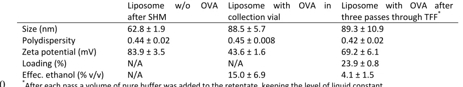
123
the volume of liquid passing into permeate. At a flow rate of 1 mL min-1, the backpressure was 49 psi
124
(capillary length 50 mm, and I.D. 100 µm, Table 2), yielding a calculated flow rate of water through the
125
cellulose membrane, Qtransmemb, of 0.25 mL·min-1 (based on a linear extrapolation from supplier data; 16 mL
126
min-1 cm-2 for 14.5 psi).
127
128
With the cationic liposomes, the particle concentration of liposomes was 4.1 x 10⁹ P/mL, which reduced to
129
3.8 x 10⁹ P/mL at the end of the third cycle (Fig. 2A) confirming the yield from the filtration process was 93%
130
for the cationic liposomes. With these systems the liposomal size (approximately 300 nm) and cationic
131
nature (approximately 60 mV) were not notably influenced by the filtration process (Fig. 2A). Furthermore,
132
NTA analysis (Fig. 2B) showed that there were no cationic liposomes detected in the permeate.
133
134
Similar results were demonstrated with anionic liposomes; filtration of batch-formulated anionic liposomes
135
(DPPC:Chol:DPPG) using three diafiltration cycles produced no notable changes in terms of vesicle size
136
(approximately 120 nm), PDI (0.14 to 0.15), ZP (-55 mV) and particle concentration (4.4 to 4.6 x 10¹⁰ P/mL)
137
(Fig. 2). As with the neutral and cationic liposomes, NTA analysis in each diafiltration cycle verified that no
138
liposomes were present in the permeate (Fig. 2B).
139
140
Purification of non-incorporated drugs from liposome formulations
141
142
Having successfully demonstrated the capability of the TFF to retain a wide range of different liposome
143
systems, we then focused on investigating the efficiency of the TFF to purify the liposomal nanomedicines
144
and remove non-incorporated drug (RC membrane, 10 kDa cutoff). To study drug removal, propofol (1
145
mg.mL-1) was added to a suspension of negatively charged liposomes (DPPC:Chol:DPPG) in aqueous
146
solution containing 20% (v/v) ethanol residual solvent levels found after liposome production by microfluidics
147
prior to purification. Propofol, was employed as it has previously been studied as a low-solubility drug
148
solubilised within liposomes. The TFF was shown to effectively purify the liposomes by removing both the
149
solvent and non-incorporated drug with 90 % of the non-incorporated propofol being removed in the first
150
diafiltration cycle, with a further removal of 80% in the second diafiltration run, and a further 60% after the
151
third diafiltration cycle (Fig. 2C). Thus, after three cycles only 1% of the ‘free’ non-incorporated drug
152
remained within the formulation (Fig. 2C). Simultaneously, the TFF system removed the ethanol which had
153
been used for the liposome formulation (Fig. 2C). Ethanol was reduced by approximately 50% in the first
154
diafiltration cycle, and after three diafiltration cycles the residual ethanol concentration was 3% (v/v) (Fig.
155
2C).
156
157
Purification of ‘free’ protein from liposome formulations
158
159
To investigate the removal of non-entrapped protein from liposome formulations, both cationic (DDA:TDB)
160
and anionic liposomes (DPPC:Chol:DPPG) were considered given that electrostatic interactions between
161
cationic liposomes and anionic proteins is exploited in the loading of antigens to liposomal adjuvant systems.
162
Therefore both liposome systems were mixed with ovalbumin (OVA; 100 μg mL-1). At a flow rate for the
163
retentate of 2.5 mL min-1, the backpressure was 62 psi (capillary length 25 mm, and I.D. 100 µm, Table 2),
164
yielding a calculated flow rate of water through the PES membrane (MWCO 300 kDa), Qtransmemb, of 1.69
165
mL·min-1 (based on a linear extrapolation from supplier data; 58 mL min-1 cm-2 for 10 psi). Thus, from 2.5 mL
166
initial sample only 0.81 mL remain in the retentate fraction, and 1.69 mL pass through the membrane. The
167
theoretical volume of permeate accounted for 67% of the initial liquid.
168
169
Given the anionic nature of OVA at the pH range used, electrostatic interactions with the cationic but not the
170
anionic liposomes occurred. Indeed, anionic liposomes maintained a similar size (approximately 120 nm)
171
after the addition of OVA (Fig. 3A); however, for the cationic liposomes, the electrostatic interactions with the
172
anionic OVA resulted in aggregation and in an increased vesicle size from around 220 nm to 300 nm, and a
173
drop in their cationic nature from 59.8 ± 1.9 mV to 17.5 ± 1.4 mV (Fig. 3B).
174
175
After the addition of protein, both systems were subjected to three diafiltration cycles. The size and PDI of
176
of the diafiltration cycles and particle recovery was 87% and 96%, respectively (Fig. 3). Protein (OVA) and
178
residual ethanol was removed into the permeate stream over the three diafiltration cycles, with a final
179
removal of 70% of the free protein and 95% of the solvent with the anionic liposomes (Fig. 3C). Similar
180
results were achieved with the cationic liposome systems (Fig. 3D); by using TFF purification, ethanol
181
residues were reduced to 4% (v/v) of the starting value and 75% protein was removed (Fig. 3B) after three
182
diafiltration cycles. The reduced levels of protein removed from the cationic system were a result of protein
183
loading onto the surface of the liposomes due to electrostatic interactions (but not related to the filtration
184
process in the TFF).
185
186
Micro continuous-flow system for production, modification and purification of liposomes
187
188
Having established the efficacy of the TFF purification system, the next stage was to develop a continuous
189
manufacturing and purification process for liposomes. To achieve this, a staggered herringbone micromixer
190
(SHM) was employed to generate the liposomal systems and directly feed the TFF device with the
191
liposomes. To optimise the throughput for each of the two devices separately, and to independently control
192
the flow rates, an intermediate collection vial was used (Fig. 4). Furthermore, the intermediate collection vial
193
allowed purification of the liposomes in diafiltration mode. After each diafiltration cycle, fresh buffer was
194
added manually into the intermediate vial to compensate for the volume passing through the membrane into
195
the permeate. Continuous production (Fig. 4) was demonstrated by using 1) neutral liposomes (PC:Chol, 4:1
196
molar ratio) with propofol and 2) cationic liposomes (DOPE:DOTAP, 1:1 molar ratio; Table 1) loaded with
197
surface-complexed protein. Lipid recovery after 4 diafiltration cycles remained at 100%, matching the initial
198
amount of lipids present prior to TFF (Fig. 5A). Without buffer replenishment, the system performed
199
concentration cycles for formulations (Fig. 5B). Within four cycles, the concentration of lipids measured in the
200
retentate doubled (Fig. 5B). This was due to a 50% reduction in volume as the overall quantity of lipids in the
201
retentate remained constant, matching the lipid content after the SHM (but before the TFF).
202
203
Continuous manufacture and purification of liposomes with bilayer loaded drug
204
205
This system was then applied for the continuous manufacture and purification of liposomes incorporating
206
propofol (Fig. 4B). Liposome (PC:Chol) production and drug encapsulation were performed in a staggered
207
herringbone mixer (SHM), operated with a volumetric flow rate of 2 mL min-1 and a 1:3 solvent:aqueous
208
buffer ratio. The resulting liposomes were 50 nm in size with a PDI of 0.3 (Table 3) in line with previously
209
reported studies25. Using the continuous manufacturing set up (with three diafilitrations), liposomes were
210
therefore both manufactured and purified. This continuous system was able to produce a purified liposome
211
product incorporating 51 mol% propofol (in line with previously reported drug loading achieved using a 2 step
212
manufacture and purification process based on dialysis18), with clinically acceptable ethanol levels (3% (v/v);
213
Table 3). Furthermore, liposomes manufactured and purified in this continuous systems retained their
214
physico-chemical attributes and were not significantly different in size, nor PDI from those not subjected to
215
TFF purification (Table 3). Examples of electron microscopy images of liposomes are shown in
216
Supplementary Figures S1 and S2.
217
218
To compare the characteristics and drug loading of PC:Chol liposomes loaded with propofol, the same
219
formulation was prepared using hand-held extrusion (10 passes through a 400 nm, 200 nm, 100 nm and final
220
50 nm pore size filters). Whilst this method of liposome manufacture was not the main focus of this study and
221
could be further optimized, again these liposomes were effectively purified to remove free drug using TFF
222
(with drug loading of 3.6 ± 0.38 mol %; data not shown) and the liposome size and PDI remained unchanged
223
by TFF purification (107.9 ± 14.1 nm and 109.9 ± 19.0 nm with PDI values of 0.17 ± 0.10 and 0.34 ± 0.06 pre
224
and post TFF purification respectively, results not shown).
225
226
Continuous manufacture and purification of cationic liposomes with adsorbed protein
227
228
The lab-on-chip micro continuous-flow system was next challenged with the production of cationic
229
(DOPE:DOTAP) liposomes, which were modified with added ovalbumin in the intermediate connection vial
230
and finally subjected to purification (Fig. 4C). Lipids were included in the ethanol stream and liposome
231
formation was performed using the SHM, which operated at 2 mL min-1 and a 1:3 solvent:aqueous buffer
232
ratio. The outflowing liposome solvent mixture was collected in the intermediate collection vial after 1 minute
233
of SHM operation, and analysed (size, PDI, ZP). The resulting liposomes had a size of 62.8 ± 1.9 nm, PDI of
234
0.4 ± 0.02 and ZP of 84 ± 3.5 mV prior to addition of OVA. Then ovalbumin was added to the intermediate
235
vial, resulting in vesicles with a larger size (88.5 ± 5.7 nm), unaltered polydispersity (0.45 ± 0.01), and
236
the anionic protein. After manufacture and purification on the system, liposomes were unaltered in size (89.3
238
± 10.9 nm; PDI 0.42 ± 0.02) and had an increased ZP (69.2 ± 6.1 mV) (Table 4), presumably through the
239
purification and removal of 74% ‘free’ protein from the system. Residual solvent levels were also reduced to
240
clinically acceptable levels (4 %; Table 4). Examples of electron microscopy images of liposomes are shown
241
in Supplementary Figure S1.
242
243
Discussion
244
245
We successfully investigated the region of operation for the Tangential Flow Filtration (TFF) device with
246
various liposome formulations and confirmed the upper limit of operational pressure for the presented
247
purification system to be 75 psi. A pressure range between 5 and 80 psi is a common backpressure
248
implemented in industrial filtrations26 which is virtually identical to our TFF. During pressure tests, the
249
membrane remained intact throughout, and therefore it can be considered that the measured backpressure
250
equaled the transmembrane pressure inside the TFF. This transmembrane pressure could be adjusted
251
accordingly using the data available in Table 2; alternatively, it could be calculated from Hagen Poiseuille’s
252
equation. Based on these findings we determined the optimal operational transmembrane pressure of 62 psi,
253
which corresponded to a maximum flow rate of 2.5 mL min-1 through a restrictive capillary with an internal
254
diameter of 100 µm and a length of 25 mm. At this flow rate, our sample (2 mL) took less than a minute (~48
255
s) to run through the system, which shows a distinct advantage over the current methods that require lengthy
256
bench-top, post-synthetic dialysis22. At high shear rates, drug release from liposomes can be a problem.
257
However, the calculated average shear rate at the maximum flow rate of 2.5 mL min-1 inside the retentate
258
channel is approximately 590 s-1 (Supplementary Information S3). This value is lower than previously
259
reported shear rates27 of 800 s-1 for which no influence on the permeability or integrity of the liposome
260
membranes was found. Furthermore, the flow rates matched those previously applied for liposome
261
manufacturing using a device with a SHM5, 25. Thus, we proved that a SHM can be coupled directly with the
262
TFF, and that we could generate and purify liposomes in a continuous mode without any losses into the
263
permeate. Overall our results show that our filtration system can be implemented for multistage purification of
264
a broad range of liposomal products.
265
266
Backpressures of 75 psi and higher, however, led to losses of liposomes through the intact cellulose
267
membrane into the permeate. One possible explanation could be that of particle extrusion across the
268
membrane at these high pressures. It is well known that liposomes can undergo extrusion through cylindrical
269
pores in membranes. Industrial scale extrusion tends to use higher lipid concentration than in our current
270
study and adopts higher pressures ranging between 100-700 psi. However, extrusion of liposomes is system
271
dependent; polycarbonate filters are used at pressures less than 100 psi, and low lipid concentrations
272
require lower pressure28. Therefore, to avoid extrusion, a backpressure of 75 psi was adopted as the critical
273
cut off value. Membrane characteristics also play an important role for liposome recovery as they influence
274
the flux from the retentate to the permeate. The calculated transmembrane flow was 0.32 mL min-1 (or 12.8%
275
of the total flow rate, TFR) for a hydrophilic membrane with a pore size of 0.22 µm, at a backpressure of 62
276
psi and nominal flow rate of 2.50 mL min-1 (retentate). In contrast, for the same backpressure and same
277
nominal flow rate, a membrane with a 0.45 µm pore size resulted in a transmembrane flux of 1.69 mL min-1,
278
corresponding to 67.6% of the retentate inflow. Furthermore, the presented setup demonstrates that a range
279
of capillaries with varying inner diameter and length can be applied to control the backpressure and the
280
dilution or concentration rates of the system, allowing to tailor resulting flow rates and to adjust throughputs.
281
282
Having established optimal operational conditions of our TFF device, its purification capacity for the removal
283
of non-incorporated hydrophobic drug (propofol) and residual ethanol was studied. Over three diafiltration
284
cycles, the quantity and quality of liposomes were preserved after purification for the anionic vesicles. For the
285
cationic liposomes, there was a small increase in polydispersity, but no significant increase in liposome size
286
(Fig. 2A). The propofol content decreases much faster in comparison to the ethanol (Fig. 2B), with the
287
hydrophilic membrane (0.22 µm pore size). This could potentially be due to capillary action that channels the
288
separation of lipophilic propofol29 (Log Kow=3.79) through the membrane. The ethanol content was the critical
289
factor, which determined the required number of diafiltration cycles. After three diafiltration cycles, only 1% of
290
non-incorporated propofol and 3% residual ethanol remained within the liposomal suspension with no
291
changes in liposome physico-chemical attributes or concentration (Fig. 2) demonstrating the ability of this
292
system to provide liposomes purified to a level as would be expected for a therapeutic product.
293
294
In terms of removal of non-associated protein from liposomes, as might be required for liposomal adjuvants
295
or biological therapeutics, protein removal is challenging because high concentrations of protein can lead to
296
electrostatic repulsion forces. Similar to propofol removal, the dilution by replenishing with fresh buffer, and
298
subsequent filtration facilitates the reduction in concentration of free protein in the retentate. Purification
299
therefore occurs as a result of two cumulative effects: one from the separation at the membrane and the
300
other from the dilution of the retentate. As demonstrated, separation can be controlled by adjusting flow rates
301
and restrictive capillary sizes; also by varying the amount of liquid that is replenished after each diafiltration
302
cycle. In our results, the volume amounted to the volume of the permeate, thus maintaining constant the
303
amount of liquid circulating in lab-on-chip purification system.
304
305
The tolerated levels of free protein depend on the requirements implied by the target application of the
306
liposomes and the number of diafiltration cycles can be adjusted accordingly to match those criteria for
307
purity. A particular focus in the delivery of protein antigens is the use of cationic liposomes, with electrostatic
308
attractive forces dominating and often leading to a surface-adsorption reaching close to 100% depending on
309
protein concentrations used30 and purification can further be complicated by the cross-linking and/or
310
aggregation of cationic liposomes (DDA:TDB) with protein. We have demonstrated the capability of the
311
filtration device to separate non-adsorbed ovalbumin (OVA) from a cationic liposome formulation and
312
residual solvent with high liposome adjuvants recovery (87%) (Fig. 3). This presents compelling evidence
313
that our micro continuous-flow purification device, i.e. TFF device, is capable of providing an effective
post-314
production purification step, with the option to recycle purified protein for subsequent applications.
315
316
The liposome process flow system presented here (Fig. 4) facilitates the complete removal of the free drug,
317
which was previously only achievable by time intensive, bench-top dialysis18, 22. The encapsulation and
318
solubilisation of drug with low aqueous solubility in the bilayer of liposomes has been investigated previously
319
using a microfluidics based system18. In that study the assembly of PC:Chol liposomes was performed using
320
a SHM, and the method was established as a robust, reproducible approach for preparing size-controlled
321
liposomes as solubilising agents. The same SHM is implemented in this herein reported system to
322
investigate the effects of continuous processing on drug encapsulation by measuring amounts of drug
323
encapsulated in the liposome bilayer. Very importantly, the amount of encapsulated drug and physical
324
characteristics (size, PDI) show that continuous processing and the pressures applied in the TFF have no
325
adverse effect on liposome integrity (Table 3). The presented assembly utilizes the methanol solubilisation
326
as the initial step of liposome production. However, it is possible to replace time-intensive production and
327
dialysis (hours) with a micro continuous-flow system (minute-long process) manufacturing and purification to
328
rapidly remove residual solvent. Among the main merits of using the continuous microfluidic process flow are
329
the mild conditions during the assembly of the liposomes and the replacement of long ultracentrifugation
330
steps for protein removal31. It can be concluded that the performance of the process flow system
331
demonstrated (Fig. 4) for liposomes is consistent with: (i) the results from the stand-alone SHM in terms of
332
particle characteristic; (ii) the results from the stand alone TFF in terms of purification.
333
334
Conclusions
335
336
We have successfully demonstrated for the first time the feasibility for on-chip purification of liposomal
337
batches for process development. Liposome manufacture, drug loading and removal of contaminants (such
338
as un-entrapped drug or protein as well as solvent residues) were performed in a continuous mode using two
339
microfluidic devices, allowing for manufacturing, purification and concentration of liposomal drug products.
340
These devices were successfully challenged with a range of liposomes, varying in lipid composition, surface
341
potential, size and concentration. The results demonstrate the ability of the on-chip filtration unit to be
342
tailored to a broad diversity of lipid-based nanoparticles by varying the operational parameters. The
343
microfluidic devices allow for an efficient and quick investigation of several lipid or drug candidates, and meet
344
high throughput requirements of early stage development processes. The continuous process may permit
345
determination of liposomal characteristics (e.g. size, surface potential, particle number) and encapsulation
346
efficiencies of a wide variety of drug molecules, allowing for future integration of process analytical
347
technologies (PAT) to further aid reproducibility. Furthermore, the setup is of considerable interest for
cost-348
intensive drugs or protein encapsulation development, as the process requires micro volumes. The
349
microfluidic device developed herein can cope with a variety of proteins developed by the biopharmaceutical
350
industry. The device has the flexibility of integrating different types of membranes to cater for a variety of
351
uses; also has the option of scalability through parallelization of the mixer chips and TFF membranes, and
352
thereby can be easily translated to industrial setting32.
353
354
Methods
355
Chemicals
357
358
Egg Phosphatidylcholine (PC), CAS: 8002-43-5, 1,2-Dipalmitoyl-sn-glycero-3-phospho-rac-(1-glycerol)
359
sodium salt (DPPG), CAS: 67232-81-9, 1,2-Dipalmitoyl-sn-glycero-3-phosphocholine (DPPC), CAS: 63-89-8
360
and Cholesterol (Chol), CAS: 57-88-5 were obtained from Sigma-Aldrich Company Ltd. (Poole, UK).
1,2-361
dioleoyl-sn-glycero-3-phsphoethanolamine (DOPE), CAS: 4004-05-1,
1,2-dioleoyl-3-trimethylammonium-362
propane (DOTAP), CAS: 144189-73-1, dimethyldioctadecylammonium bromide (DDA), CAS: 3700-67-2 and
363
trehalose 6,6-dibehenate (TDB), CAS: 66758-35-8 were purchased from Avanti Polar Lipids, Inc., (Alabaster,
364
AL), purity >99% (Table I). Ethanol, CAS: 64-17-5, and methanol, CAS: 67-56-1, were obtained from Fisher
365
Scientific (Loughborough, UK). TRIS Ultra Pure, CAS: 77-86-1, was obtained from ICN Biomedicals, Inc.,
366
(Aurora, Ohio). Propofol (2,6-Bis(isopropyl)phenol), CAS: 2078-54-8 and ovalbumin (chicken egg), CAS:
367
9006-59-1 were obtained from Sigma-Aldrich Company Ltd., (Poole, UK). Ultrafiltration regenerated cellulose
368
membranes (p\n: U2755-10AE) were obtained from Sigma-Aldrich Company Ltd., (Poole, UK) (10kDa, pore
369
size 0.22 µm), and Biomax polyethersulfone ultrafiltration membrane discs with 300 kDa cutoff, pore size
370
0.45 µm (p\n: PBMK06210) from Merck Milipore (Darmstadt, Germany).
371
372
Liposome batch formulations for characterisation of the Tangential-Flow Filtration (TFF) device
373
374
Multilamellar vesicles (MLV) were prepared using the lipid film hydration method33. Lipids were weighed and
375
dissolved in a chloroform/methanol (9:1 v/v) mixture. Cationic liposomes comprised DDA:TDB (8:1 molar
376
ratio) and anionic liposomes comprised DPPG, DPPC, Chol (1:1:1.3 molar ratio). The organic solvent was
377
subsequently removed by rotary evaporation under vacuum (100 RPM, 180 mBar, Rotavapor R-100, BÜCHI
378
Labortechnik AG, Switzerland), followed by flushing with nitrogen for removal of solvent residues (5 minutes).
379
The thin lipid film on the bottom of a round bottom flask was hydrated with 10 mM pH 7.2 TRIS buffer. Small
380
liposomes were formed via probe sonication (Soniprep150plus, MSE, UK; 5 min at amplitude of 5). Ethanol
381
was manually added to the liposome formulation to a final concentration of 20% (v/v) to simulate solvent
382
contents commonly resulting from the microfluidics production method. Ovalbumin (100 μg mL-1) was used
383
as a model protein, and propofol (1 mg mL-1) as a model drug. These were added to the liposome
384
formulation post-production to mimic the conditions post liposome manufacturing by microfluidics.
385
386
Device fabrication
387
388
As previously reported, the filtration system was designed to seal membranes in place by means of
389
mechanical clamping24. Two poly(methylmethacrylate) (PMMA) plates, with a straight channel (1 mm width, 1
390
mm depth, 45 mm length) and a 1 mm hole milled at each end were clamped together using M3 screws
391
along the edges (Torque 10 Ncm). A 1 mm wide and 0.75 mm deep cutting was used to hold the PDMS
392
gasket in place, which was used to secure the membrane in place (Supplementary Figure S4). Different
393
commercially available membrane sheets were cut to the required size using a CO2 laser marking head
394
(Synrad Inc., Mukilteo, WA, USA). The membranes used in this set of experiments had a cut-off of 10kDa or
395
300kDa, for drug or protein filtration, respectively. The membranes were cleaned after each experiment by
396
back-flushing with water and stored inside the TFF system in 0.8 M saline solution, ready for the next
397
experiment.
398
399
Additionally, a clamping system was made from PMMA (two plates held together by screws [M3]) for the
400
staggered herringbone micromixer (SHM) chip using a micromilling machine (M3400E, Folken IND,
401
Glendale, USA). The gasket for the filtration unit was manufactured from poly(dimethylsiloxane) (PDMS,
402
Sylgard 184, Dow Corning, Midland, USA), according to the manufacturer’s instructions and cast in PMMA
403
moulds, manufactured as described above. Interconnect ports (milled from 5 mm PMMA), with two holes
404
tapped with an M3 thread were used for connection to the filtration unit; an M6 threaded hole was used for
405
standard connection fittings (P-221, Upchurch Scientific, Oak Harbor, WA, USA).
406
407
Backpressure regulation
408
409
Backpressures were regulated through capillaries, which were attached to the retentate outlet of the filtration
410
device (see Supplementary Fig. S3 online). These capillaries restricted the flow as they were selected with
411
internal diameters smaller than the polytetrafluoroethylene (PTFE) tubing (1/16 in. x 0.031 in., p\n: 58700-U,
412
Sigma- Aldrich Int.) which connected the TFF device with auxiliary pumps and collection vials. Backpressure
413
was calculated using Hagen-Poiseuille’s Law
414
∆ = ∙ ∙ ∙∙ (1)
416
where µ, L, d and Q are the dynamic viscosity of the medium at 25 oC, the length and internal diameter of the
417
restricting capillary, and the volumetric flow rate, respectively. We used Hagen-Poiseuille’s equation (1) to
418
select the capillary sizes and the flow rates to attain the backpressure range from 5 to 80 psi. For each
419
backpressure analysis, a capillary was connected to the TFF retentate outlet using PTFE tubing, ferrules
420
(p\n: P-200, IDEX Europe GmbH, Germany) connectors (Flangeless Nuts, p\n: P-247, PEEK, M6
Flat-421
Bottom, for 1/16 in. OD, IDEX Europe GmbH, Germany) and metric unions (Metric Union, M6 Port, p\n:
P-422
602, IDEX Europe GmbH, Germany). The inlet of the TFF was connected through a Luer-lock fitting and
423
polytetrafluoroethylene PTFE tubing to a single-use plastic syringe. Water was fed in the TFF device at
424
discrete flow rates ranging from 0.01 to 2.5 mL min-1 attained by a syringe pump (Nemesys, Cetoni GmbH,
425
Germany). Backpressures were measured experimentally with a pressure sensor (40PC100, Honeywell, NJ,
426
USA) connected on the retentate side; the data was logged with a LabVIEW virtual instrument (National
427
Instruments, TX, USA). We compared the theoretical backpressures from equation (1) to the measured
428
backpressures, and the measured values exceeded their calculated counterparts from 20% to 6.25% when
429
increasing the applied backpressure from 5 to 80 psi, respectively (Supplementary Figure S5, and
430
Supplementary Table S6). One of the TFF outlets was intentionally sealed with a flat bottom plug (p\n:
P-431
314, M6, IDEX Europe GmbH, Germany) while a single outlet connected through a ferrule (p\n: P-200), nut
432
(p\n: P-247) and tubing into a collection vial for liquid passing through the membrane.
433
434
Filtration435
436
Filtration was performed in diafiltration mode to investigate the liposome behaviour in the established
437
pressure and flow rate range (Table 2). For this experiment, bench-top prepared liposomes in aqueous
438
solution were spiked with drug, protein or solvent, and were introduced into the TFF by means of syringe
439
pumps (Nemesys, Cetoni GmbH, Germany), connectors and capillaries as described earlier. A capillary was
440
connected to the TFF, in cis-configuration (on the same side of the membrane), and closed the loop of the
441
retentate fluidic line (see Supplementary Fig. S3 online). Retentate from the TFF was collected in an
442
intermediate collection vial and could be injected in the device hence allowing for multiple passes, referred in
443
this article as diafiltration cycles. Transmembrane pressure was attained by controlling the flow rates in the
444
pump; also by adding a constriction capillary of known geometry, i.e. internal diameter and length. Retentate
445
and permeate fractions were collected in Eppendorf tubes, assessed by weight, and used for further
446
analysis, i.e. zeta potential, size, polydispersity, quantification via HPLC. A volume of TRIS buffer, 10 mM pH
447
7.2, was added after each diafiltration cycle to compensate for the amount of liquid passing through the
448
membrane (in permeate) and to sustain steady concentration levels (in retentate) during the continuous
449
purification process.
450
451
Continuous process flow configuration
452
453
To test the continuous processing of liposome formation followed by liposome purification, a SHM and a TFF
454
device were connected in sequence. The SHM (Precision Nanosystems Inc., Vancouver, Canada) consisted
455
of two inlets, a bifurcated channel with herringbone structures, and single outlet moulded in PDMS. The
456
channels were 200 μm in width and 79 μm in height with herringbone features of 50 μm in width, 31 μm in
457
height, 45° angle, asymmetry index 2:1 (according to Precision Nanosystems, Inc.). Luer-lock fitting and
458
polytetrafluoroethylene (PTFE) tubing (1/16 in. x 0.031 in., Sigma- Aldrich Int.) were used to link disposable 1
459
mL syringes with the two inlet ports of the chip; flow rates and flow rate ratios were controlled by syringe
460
pumps (Nemesys, Cetoni GmbH, Germany) and the whole system was primed with Tris buffer (10 mM, pH
461
7.2) prior to operation. Organic phase, a weighed amount of lipids in ethanol, was injected into the first inlet
462
of the SHM device, while in the second inlet aqueous phase (TRIS buffer, 10mM, pH 7.2) was injected. The
463
micromixer was held in place using a clamping device made out of PMMA. The micromixer was connected to
464
the tangential flow filtration unit via an intermediate collection vial (2.0 mL Eppendorf) for additional
465
functionality. This allows the addition of various components such as of microfluidics-manufactured
466
liposomes prior to the filtration system for purification. A bi-directional milliGAT pump (VICI Valco, Valco
467
Instruments Co.) was connected in-line with the retentate loop of the TFF through a capillary at the bottom of
468
that intermediate collection vial. Transmembrane pressures was varied by restricting the flow of the retentate
469
using different small diameter capillaries connected in-line with the TFF outlet. The retentate flowed through
470
the capillary and was collected in the intermediate vial, while permeate passed through the membrane and
471
was gathered in a separate tube. Both fractions were analysed for content of liposomes, propofol, protein,
472
lipid and ethanol.
473
474
preparation of neutral liposomes, PC and Chol (4:1 molar ratio) in ethanol were injected into the micromixer
476
at a total flow rate (TFR) of 2 mL min-1 and a flow rate ratio (FRR) of 1:3; bilayer drug loading was achieved
477
by including 1 mg mL-1 of propofol in the solvent stream. For the preparation of a cationic liposome
478
formulation, DOPE and DOTAP (1:1 molar ratio) in ethanol were injected into the micromixer at a TFR of 2
479
mL min-1 at FRR 1:3. After formulation, the required amount of protein (ovalbumin, 100 μg mL-1) was added
480
to the intermediate collection vial. Manually adding fresh solution to the intermediate collection vial
481
compensated for liquid passing through the membrane into permeate. Otherwise, the amount of the fluid in
482
the system would fall below a critical level and purification would need to be interrupted because of
483
insufficient circulating liquid volume.
484
485
Measurement of particle characteristics
486
487
Nanoparticle tracking analysis (NTA) was performed with a Nanosight LM20 (NanoSight, Amesbury, UK),
488
connected to a microscope (with 20× magnification). Liposomes were diluted 1:10 to 1:100 in distilled water,
489
to achieve an optimal particle concentration of 107 – 109 particles/mL during measurement. NTA analysis
490
was used to determine the particle concentration per millilitre (P/mL), recording time was 60 seconds and
491
camera settings (shutter and gain) were adjusted manually to maximise resolution. Dynamic light scattering
492
(DLS) (Malvern Zetasizer Nano-ZS, Malvern Instruments, Worcestershire, UK) was used to report the
z-493
average (intensity based mean particle diameter), and to report the polydispersity (PDI), in order to assess
494
the width of the particle distribution. Liposomes were diluted 1:10 in distilled water and measurements took
495
place at 25°C. Zeta potential (ZP) was measured using particle electrophoresis (Malvern NanoZS, Malvern
496
Instruments, Worcestershire, UK).
497
498
Propofol quantification
499
500
Quantification of propofol was performed by reverse phase HPLC (Luna 5µ C18, Phenomenex, UK, pore
501
size of 100Å, particle size of 5 µm) at 268 nm. The flow rate was constant at 1 mL min-1 throughout with a
502
gradient elution from 5:95 (Methanol: 0.1% Trifluoroacetic Acid, TFA, in water) to 100:0 (Methanol: 0.1% TFA
503
in water) over 10 minutes. HPLC-grade solvents were used, sonicated and filtered. The column temperature
504
was controlled at 35°C. All analysis was made with Clarity, DataApex, version 4.0 (DataApex, Prague, Czech
505
Republic). For quantification, established calibration curves of propofol were used as reported previously14.
506
507
Protein and lipid quantification
508
509
Samples were loaded on a HPLC and elution was performed with a gradient from 5:95 to 100:0 (Methanol:
510
0.1% TFA in water) over 20 and 40 minutes for protein and lipid detection, respectively. Quantification was
511
performed by an evaporative light scattering detector (ELSD) (Sedex 90, Sedere, France), set at 52°C and
512
coupled to the HPLC as described previously18. A calibration curve was established from standards
513
(ovalbumin in TRIS buffer, pH 7.2, lipids in ethanol) in six replicates at concentrations from 5 to 100 μg mL-1
514
(protein) and 0.05 to 1.5 mg mL-1 (lipids).
515
516
Ethanol quantification
517
518
Solvent concentration was quantified by gas chromatography (GC) using a flame ionization detector (CSi
519
200 Series, column TRACE 15 m x 0.25 mm x 0.25 µm TR-5, Thermo Scientific, UK), with detector
520
temperature 230°C, injector temperature 200°C and an injection volume of 1 µL. The carrier gas was helium
521
at 15 psi inlet pressure. A calibration curve (6 standards ranging from 0.5-50% v/v) was established and
522
used for quantification using an internal reference standard (propan-1-ol). All analysis was made in Clarity
523
DataApex version 2.4 (see above).
524
525
Statistical analysis
526
527
Unless stated otherwise, results were reported as the mean ± one standard deviation (SD., n=3). One- or
528
two-way analysis of variance (ANOVA) was used to assess statistical significance, followed by Tukey’s
529
multiple comparing test (post-hoc analysis). A t-test was performed for paired comparisons. Significance was
530
acknowledged for p values lower than 0.05, marked with and asterisk (*). All calculations were made in
531
GraphPad Prism version 6.0 (GraphPad Software Inc., La Jolla, CA, US).
532
533
534
536
1. Carugo, D., Bottaro, E., Owen, J., Stride, E., & Nastruzzi, C. Liposome production by microfluidics:
537
potential and limiting factors. Scientific Reports 6, (25876):25876 (2016).
538
539
2. Mayer, L. D., Hope, M. J., & Cullis, P. R. Vesicles of variable sizes produced by a rapid extrusion
540
procedure. Biochimica et Biophysica Acta 858, 161-68 (1986).
541
542
3. Hinna, A., Streiniger, F., Hupfeld, S., Stein, P., Kuntsche, J., & Brandtl, M. Filter-extruded liposomes
543
revisited: a study into size distributions and morphologies in relation to lipid-composition and process
544
parameters. J. Liposome Research 26 (1), 11-20 (2014).
545
546
4. Gregoriadis, G., Liposome preparation and related techniques in Liposome Technology 2nd ed., Vol. 1 (ed.
547
Gregoriadis, G.) 50-65 (CRC Press Inc., 1992).
548
549
5. Zhigaltsev, I. V., Belliveau, N., Hafez, I., Leung, A. K., Huft, J., Hansen, C., & Cullis, P. R. Bottom-up
550
design and synthesis of limit size lipid nanoparticle systems with aqueous and triglyceride cores using
551
millisecond microfluidic mixing. Langmuir 28 (7), 3633-40 (2012).
552
553
6. Jahn, A., Stavis, S.M., Hong, J.S., Vreeland, W.N., DeVoe, D.L., & Gaitan, M. Microfluidic mixing and the
554
formation of nanoscale lipid vesicles. ACS Nano 4 (4), 2077–87 (2010).
555
556
7. Jahn, A., Vreeland, W. N., DeVoe D. L., Locascio L. E., & Gaitan, M. Microfluidc directed formation of
557
liposomes of controlled size. Langmuir 23 (11), 6289-93 (2007).
558
559
8. Demello A. J. Control and detection of chemical reactions in microfluidic systems. Nature 442 (7101),
394-560
402 (2006).
561
562
9. Song Y., Hormes J., & Kumar, C.S. Microfluidic synthesis of nanomaterials. Small 4 (6), 698-711 (2008).
563
564
10. Schroën, K., Bliznyuk, O., Muijlwijk, K., Sahin, S., & Berton-Carabin, C. C. Microfluidic emulsification
565
devices: from micrometer insights to large-scale food emulsion production. Current Opinion in Food Science
566
3, 33–40 (2015).
567
568
569
11. van Swaay, D. Microfluidic methods for forming liposomes. Lab on a Chip 13 (5), 752-767 (2013).
570
571
12. Bleul, R., Thiermann, R., & Maskos, M. Techniques to control polymersome size. Macromolecules 48,
572
7396-7409, (2015).
573
574
13. Ghazal, A., Gontsarik, M., Kutter, J. P., La, J. P., Ahmadvand, D., Labrador, A., & Yaghmur, A.
575
Microfluidic platform for the continuous production and characterization of multilamellar vesicles: A
576
synchrotron small- angle X ‑ ray scattering (SAXS) study. J. Phys. Chem. Lett. 8, 73−79 (2017).
577
578
14. Pattni, B. S., Chupin, V. V., & Torchilin, V. P. New developments in liposomal drug delivery. Chemical
579
Reviews 115(19), 10938–10966 (2015).
580
581
15. He, J., Wang, L., Wei, Z., Yang, Y., Wang, C., Han, X., & Nie, Z. Vesicular self-assembly of colloidal
582
amphiphiles in microfluidics. ACS Appl. Mater. Interfaces 5, 9746−9751 (2013).
583
584
585
16. Hood, R. R., & DeVoe, D. L. High-throughput continuous flow production of nanoscale liposomes by
586
microfluidic vertical flow focusing. Small 11, (43), 5790–5799 (2015).
587
588
17. Lim, J., Swami, A., Gilson, L. M., Chopra, S., Choi, S., Wu, J., & Arabia, S. Ultra-high throughput
589
synthesis of nanoparticles with homogeneous size distribution using a coaxial. ACS Nano 8 (6), 6056–6065
590
(2014).
591
592
593
18. Kastner, E., Verma, V., Lowry, D., & Perrie, Y. Microfluidic-controlled manufacture of liposomes for the
594
solubilisation of a poorly water soluble drug. Int J Pharmaceutics 485 (1), 122-130 (2015).
596
19. Wagner, A., Vorauer-Uhl, & K., Katinger, H. Liposomes produced in a pilot scale: production, purification
597
and efficiency aspects. Eur. J. Pharmaceutics Biopharmaceutics 54 (2), 213-219 (2002).
598
599
20. Pattnaik, P., Ray, T. 2009. Improving liposome integrity and easing bottlenecks to production.
600
Pharmaceutical Technology Europe 22 (6), 24-28 (2009).
601
602
21. Ruysschaert, T., Marque, A., Duteyrat, J. L., Lesieur, S., Winterhalter, M., & Fournier D. Liposome
603
retention in size exclusion chromatography. BMC Biotech 5 (1), 11 (2005).
604
605
22. Hood, R., Vreeland, W., & DeVoe, D. Microfluidic remote loading for rapid single-step liposomal drug
606
preparation. Lab Chip 14 (17), 3359-3367 (2014).
607
608
609
23. Belliveau, N. M., Huft, J., Lin, P. J., Chen, S, Leung, A. K., Leaver, T. J., Wild, A. W., Lee, J. B., Taylor,
610
R. J., & Tam, Y. K. Microfluidic synthesis of highly potent limit-size lipid nanoparticles for in vivo delivery of
611
siRNA. Mol Therapy Nuc Acids 1 (8), e37 (2012).
612
613
24. O'Sullivan, B., Al-Bahrani, H., Lawrence, J., Campos, M., Cázares, A., Baganz, F., Wohlgemuth, R.,
614
Hailes H. C., & Szita N. Modular microfluidic reactor and inline filtration system for the biocatalytic synthesis
615
of chiral metabolites. J Mol Catalysis B: Enzymatic 77, 1-8 (2012).
616
617
25. Kastner, E., Kaur, R., Lowry, D., Moghaddam, B., Wilkinson, A., & Perrie, Y. High-throughput
618
manufacturing of size-tuned liposomes by a new microfluidics method using enhanced statistical tools for
619
characterization. Int J Pharmaceutics 477, (1-2):361-8 (2014).
620
621
26. van Reis, R., & Zydney, A. Bioprocess membrane technology. J Memb Sci. 297 (1), 16-50 (2007).
622
623
27. Tomotaka, N., & Makoto, Y. Mechanosensitive liposomes as artificial chaperones for shear-
624
driven acceleration of enzyme-catalyzed reaction. ACS Appl. Mater. Interfaces (6), 3671−3679 (2014).
625
626
28. Cullis, P. R., Hope, M. J., & Bally, M. B. Extrusion technique for producing unilamellar vesicles. In Google
627
Patents: 1991.
628
629
29. Hansch, C., Leo, A., & Hoekman, D. Exploring QSAR: Hydrophobic, electronic, and steric constants. (ed.
630
Hansch, C., Leo, A., Hoekman, D. H.) 105 (Washington DC: American Chemical Society, 1995).
631
632
30. Kaur, R., Henriksen-Lacey, M., Wilkhu, J., Devitt, A., Christensen, D., & Perrie, Y. Effect of incorporating
633
cholesterol into DDA: TDB liposomal adjuvants on bilayer properties, biodistribution, and immune responses.
634
Mol Pharmaceutics 11, (1), 197-207 (2013).
635
636
31. Mithun, M., Saumyabrata, M., Souparno, B., Somsubhra, T. C., Abdus, S., Md S., Pradyot, B., & Nahid,
637
A. A lipid based antigen delivery system efficiently facilitates MHC class-I antigen presentation in dendritic
638
cells to stimulate CD8+ T cells. Scientific Reports (6), 27206 (2016).
639
640
32. Ball, P. Scale-up and scale-down of membrane-based separation processes. Memb Technology (117),
641
10-13 (2000).
642
643
33. Bangham, A., Standish, M. M, & Watkins, J. Diffusion of univalent ions across the lamellae of swollen
644
phospholipids. J Mol Bio. 13, (1), 238-IN27 (1965).
645
646
34. Smith Korsholm, K., Agger, E. M., Foged, C., Christensen, D., Dietrich, J., Andersen, C. S., Geisler, C., &
647
Andersen, P. The adjuvant mechanism of cationic dimethyldioctadecylammonium liposomes. Immunology
648
121, (2), 216-226 (2007).
649
650
35. Christensen, D., Agger, E. M., Andreasen, L. V., Kirby, D., Andersen, P., & Perrie Y. Liposome-based
651
cationic adjuvant formulations (CAF): past, present, and future. J Liposome Research 19, (1), 2-11 (2009).
652
653
36. Henriksen-Lacey, M., Christensen, D., Bramwell, V. W., Lindenstrøm, T., Agger, E. M., Andersen, P., &
654
dimethyldioctadecylammonium (DDA), 3β-[N-(N′, N′-dimethylaminoethane) carbomyl] cholesterol (DC-Chol),
656
and 1, 2-dioleoyl-3-trimethylammonium propane (DOTAP): prolonged liposome retention mediates stronger
657
Th1 responses. Mol Pharmaceutics 8, (1), 153-161 (2010).
658
659
37. Senior, J., & Gregoriadis, G. Stability of small unilamellar liposomes in serum and clearance from the
660
circulation: the effect of the phospholipid and cholesterol components. Life Sciences 30, (24), 2123-2136
661
(1982).
662
663
38. Gregoriadis, G., & Senior, J. The phospholipid component of small unilamellar liposomes controls the
664
rate of clearance of entrapped solutes from the circulation. FEBS Letters 119, (1), 43-46 (1980).
665
666
39. Oku, N., Namba, Y., & Okada S. Tumor accumulation of novel RES-avoiding liposomes. Biochimica
667
Biophysica Acta (BBA)-Lipids and Lipid Metabolism 1126, (3), 255-260 (1992).
668
669
40. Kirby, C., Clarke, J., & Gregoriadis, G. Effect of the cholesterol content of small unilamellar liposomes on
670
their stability in vivo and in vitro. Biochem J. 186, 591-598 (1980).
671
672
Acknowledgements
673
674
This work was part funded by the EPSRC Centre for Innovative Manufacturing in Emergent Macromolecular
675
Therapies and Aston University. This research was made possible with the financial support from BBSRC
676
(BB/L000997/1) and the European Research Area initiative on industrial biotechnology (ERA-IB; third joint
677
call). The Authors have no conflict of interest to declare. We would like to kindly thank Dr Manni Bhatti from
678
UCL for her help in proofreading the manuscript.
679
680
Author contributions statement
681
682
N.D., E.K. and M.H. conceived and conducted the experiments, N.D., E.K., Y.P., N.S. analysed the results
683
and reviewed the manuscript.
684
685
Additional information
686
687
The authors have no conflict of interest to declare.
688
689
Figure 1: Particle size and polydispersity as a function of increasing backpressures in the TFF system as
690
collected on the retentate side of the membrane. Images from NTA analysis, verifying particles in permeate
691
(top) and retentate (bottom) stream at increasing backpressures. Particles were found in the permeate at
692
backpressures exceeding 75 psi. All experimental datasets are presented as mean and standard deviation
693
(mean ± s.d.) resulting from three independent runs (n=3).
694
Figure 2: (a) Vesicle size, polydispersity (PDI), zeta potential (ZP) and particle concentration (P/mL) for
695
cationic (DDA:TDB) and anionic (DPPG:DPPC:Chol) liposomes before and after the TFF purification. (a)
696
Images from NTA show vesicles present in the retentate side only.(c) Propofol and ethanol removal achieved
697
over three diafiltration cycles for anionic liposomes (DPPG:DPPC:Chol), expressed as a percentage of the
698
initial amount of contaminants present.
699
Figure 3: Vesicle size, polydispersity (PDI), zeta potential (ZP) and particle concentration (P/mL) for (a)
700
anionic liposomes (DPPG:DPPC:Chol) and (b) cationic liposomes (DDA:TDB) prior and post OVA-addition
701
(ovalbumin, 100 μg mL-1), and particle characteristics after the TFF purification. Protein (ovalbumin) and
702
ethanol removal achieved over three diafiltration cycles for (c) anionic and (d) cationic liposomes, expressed
703
as a percentage of the initial amount of contaminants present. All experimental datasets are presented as
704
mean and standard deviation (mean ± s.d.) average of three independent runs (n=3).
705
Figure 4: (a) Schematic overview of the module-based microfluidic system. Liposomes were manufactured
706
with a Staggered Herringbone Mixer (SHM) upstream and flowed through the Tangential Flow Filtration
707
(TFF) device for consecutive purification. (b) Schematic overview of the formation of liposomes loaded with a
708
low-solubility model drug, i.e. propofol. Vesicle assembly and drug loading are performed with a SHM, and
non-entrapped (free) drug is removed from the mixture by consecutive filtration inside the TFF system. (c)
710
Schematic overview of the formation of liposomes loaded with a model protein, ovalbumin (OVA). Vesicle
711
assembly is performed with a SHM, with post-assembly protein addition; non-entrapped (free) protein is
712
removed by consecutive diafiltration cycles inside the TFF system.
713
Figure 5: Lipid recovery in the continuous liposome factory-on-a-bench for (a) lipid recovery after four
714
diafiltration cycles. (b) Lipid concentration in four concentration cycles, related to the initial amount of lipids
715
present prior to the concentration cycles. All experimental datasets are presented as mean and standard
716
deviation (mean ± s.d.) average of three independent runs (n=3).
717
718
Table 1: Lipids investigated in this study.
719
Lipid Application Reference
Dimethyldioctadecyl ammonium bromide
(DDA)
Vaccine adjuvant,cationic head group
Uptake of vaccine antigens to antigen presenting cells
Smith Korsholm et al.34
Christensen et al.35
Trehalose 6,6-dibehenate (TDB)
Synthetic immunstimmulator derived from the membrane of mycobacterium
1,2-dioleoyl-sn-glycero-3-phsphoethanolamine (DOPE)
Fusogenic helper lipid, available in the commercial Lipofectin™ transfection reagent
Henriksen-Lacey et al.36
1,2-dioleoyl-3-trimethylammonium-propane (DOTAP)
Cationic lipid often used in transfection
Egg Phosphatidylcholine (PC)
Neutral head group, drug delivery Senior and Gregoriadis37
Gregoriadis and Senior38
1,2-Dipalmitoyl-sn-glycero-3-phospho-rac-(1-glycerol) (DPPG)
Negative charged head group drug delivery
Oku et al.39
Kirby et al.40
1,2- Dipalmitoyl-sn-glycero-3-phosphocholine
(DPPC)
Neutral head group, drug delivery
Cholesterol (Chol) Added for membrane stabilization, known to effect drug encapsulation efficiency in bilayer and aqueous core
Senior and Gregoriadis37
Kirby et al.40
720
721
722
Table 2: Backpressures and flow rates through the Tangential Flow Filter (TFF) that were investigated in this study.
723
Liposomes in solution were fed into the TFF device at flow rates ranging between 0.01 and 2.5 mL min-1. Backpressure
724
was attained by connecting a restrictive capillary with selected (I.D.) and/or length on the retentate side of the TFF
725
outlet.
726
Flow rate (mL min-1) 0.01 0.02 0.03 0.1 0.05 1 0.3 2 2.5 0.5 0.1
Capillary I.D. (μm) 50 50 50 63 50 100 63 100 100 63 50
Capillary length(mm) 50 50 50 50 50 50 25 30 25 25 50
727
728
729
Table 3: Continuous purification of PC:Chol liposomes loaded with propofol. Propofol and lipids were included in the
730
ethanol stream. Liposome formation and drug encapsulation was performed in a staggered herringbone mixer (SHM),
731
operated with a total flow rate of 2mL min-1 and a ratio of 1:3 ethanol:aqueous solution. The results are presented as
732
mean and standard deviation (mean ± s.d.) resulting from three independent runs (n=3), N/A = not applicable.
733
Liposome with drug after SHM Liposome with drug after three passes through the TFF*
Size (nm) 51.4 ± 2.1 61.2 ± 13.2
Polydispersity 0.29 ± 0.013 0.33 ± 0.09
Loading (mol%) N/A 51.0 ± 4.0
Effec. ethanol (% v/v) 16.1 ± 3.9 3.1 ± 1.5
*
After each pass a volume of pure buffer was added to compensate for permate and maintain constant volume of retentate.
734
735
[image:14.612.64.526.424.511.2]736
Table 4: Continuous purification of DOPE:DOTAP liposomes loaded with protein (ovalbumin). The lipids were
737
included in the ethanol stream and liposome formation was performed in a SHM, operated at 2mL min-1 and a ratio of
738
1:3 solvent:aqueous solution. Protein was added post-liposome formation. OVA = ovalbumin, N/A = not applicable.
739
Liposome w/o OVA after SHM
Liposome with OVA in collection vial
Liposome with OVA after three passes through TFF*
Size (nm) 62.8 ± 1.9 88.5 ± 5.7 89.3 ± 10.9
Polydispersity 0.44 ± 0.02 0.45 ± 0.008 0.42 ± 0.02
Zeta potential (mV) 83.9 ± 3.5 43.6 ± 1.6 69.2 ± 6.1
Loading (%) N/A N/A 23.9 ± 0.8
Effec. ethanol (% v/v) N/A 15.0 ± 6.9 4.1 ± 1.5
*
After each pass a volume of pure buffer was added to the retentate, keeping the level of liquid constant.