FPGA implementation of feature extraction based on
histopathalogical image and subsequent classification by
support vector machine.
Sandra s Dhar1, Sreeraj K P2
1
ECE, M.Tech Student at Mangalam college of engineering, Ettumanur, Kerala, India
2
ECE,Asso.professor at Mangalam college of engineering, Ettumanur, Kerala, India
Abstract
The proposed system is used to detect skin cancer ,by features extracted from images of skin lesions through image processing techniques which consisted of median filter then applied Gabor filter bank to improve diagnostic accuracy. Histogram equalization to enhance the contrast of the images prior to segmentation is used. The extracted features are fed to a support vector machine (SVM) binary classifier to diagnose skin biopsies.The experiments were done using Xilinx ISE 14.2 development tool and conducted on Xilinx Virtex 7 XC7VX980 FPGA kit.
1. Introduction
Many countries in which skin cancer is widely spread in comparison to other types of cancer. Skin cancer costs the health system around $300 million Australian dollar annually, the highest cost of all cancers. The general approach of developing a Computer Aided Diagnostic system for diagnosis of skin cancer is to find the location of a lesion and also to determine an estimate of the probability of a disease. Filters are then very important as pre-processing tools. In order to remove unwanted features, firstly some preprocessing was applied to remove the fine hairs, noise and air bubbles on the skin and facilitating image segmentation by using winner, Gabor Filter and adaptive median filters. The advantage of the median filter is to remove noise without blurring edges. In addition these filters have been shown to possess optimal localization properties in both the spatial and frequency domain and thus are well suited for quality segmentation problems.The Median Filter is one of the best known filters.It replaces the value of the pixel by the median of the intensity values in the neighborhood of that pixel.
Segmentation is one of the most widely investigated research areas in pathological image analysis. The Support vector machine (SVM) classier is used widely in bioinformatics, due to its high accuracy, ability to deal with high dimensional data and in this syntax diverse
sources of data. This paper is organized as follows: its Initial section describe the computer-aided diagnosing (CAD) which consists of the pre-processing, segmentation, the features extraction and selection, and the automated procedure using SVM. The next section presents the result and discussion, and the fial section presents conclusions.
2. CAD for skin lesion
Automated diagnostic of medical images analyzing digital images becomes one of the major research areas, and a dynamic area in several applications. As mentioned before early detection improves survival rate, the common approach to skin lesion images combines four stages as displayed in Figure 2.1.
Fig2.1 Four stage CAD system for skin lesion. IMAGE
PRE-PROCESSING
SEGMENTATION
FEATURE EXTRACTION
CLASSIFICATION
2.1 Pre-processing
This stage includes image resizing, masking, cropping, hair
removal, and conversion from RGB colour to intensity grey image. It is meant to facilitate image segmentation by filtering the image and enhancing its important features.
2.2 Gabor Filter
The Gabor filter is basically a Gaussian modulated by a complex sinusoid Gabor filters have been used in many applications, such as quality segmentation, target detection, fractal dimension management, document analysis, edge detection, retina identification, image coding and image representation A Gabor filter can be viewed as a sinusoidal plane of particular frequency and orientation, modulated by a Gaussian envelope.It is a linear filter used for edge detection. Frequency and orientation representations of Gabor filters are similar to those of the human visual system, and they have been found to be particularly appropriate for texture representation and discrimination. In the spatial domain, a 2D Gabor filter is a Gaussian kernel function modulated by a sinusoidal plane wave.
In digital signal processing, the output of the signal was the convolution between the input signals with the filter coefficient. So mainly, the digital filter circuit was the circuit to convolute the input signal with the filter coefficient. In digital image processing, the image was presented in matrix form or in pixel. So basically the convolution involves the matrix convolution-convolution between image pixels with coefficient kernel. Difference filter have difference method of filtering or sampling input signal. For this filter, it implemented a memory base architecture for real-time convolution with variable kernels. Firstly the input data which was in pixel format will enter the filter and store it in the memory.
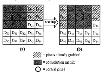
2.2.1 Convolution between image pixel and
coefficient kernel.
The value of D11 after convolution is
D11=(D00xW11)+(D01xW12)+(D02xW13)+(D10xW21)
+(D11xW)+(D12xW23)+(D20xW31)+(D21xW32)+(D22
xW3). (1)
D12=(D01xW11)+(D02xW12)+(D03xW13)+(D11xW21)
+(D12xW22)+(D13xW23)
+(D21xW31)+(D22xW32)+(D23xW33). (2)
The sequence of next pixel to be convolute is shown.
Fig2.2. 1: Method of convolution
KERNAL VALUE
Table 2.2.1:kernel values for gabor filter
G(X,Y
)
1 2 3
1 0.006737943 1.29E-05 -4E-08
2 2.35672E-07 4.14E-08 1.45E-12
3 -1.35859E-11 2.65E-14 8.53E-17
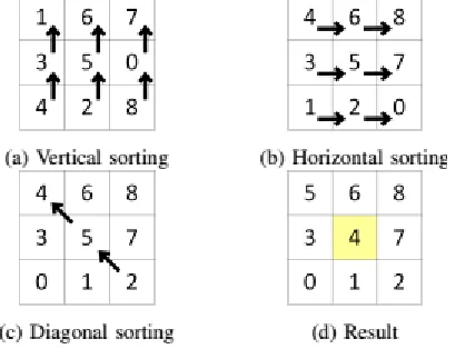
2.3 Median filter
representative of their surroundings; in other words the impulses. Thus, median filter is very helpful in filtering out missing or damaged pixels of the image.
Fig 2.3.1 :Example of median filtering using optimized method.
3. Image processing
Segmentation and classification are important steps in the medical image analyses for radiological evaluation or (CAD). One of the early steps in this stage is image enhancement. The purpose of image enhancement methods is to process a picked image for better contrast and visibility of features of interest for visual examination as well as subsequent computer-aided analysis and diagnoses[1].
3.1 Histogram equalization
This application describes a method of imaging processing that allows medical images to have better contrst.This method usually increases the global contrast of many images, especially when the usable data of the image is represented by close contrast values. Through this adjustment, the intensities can be better distributed on the histogram. This allows for areas of lower local contrast to gain a higher contrast. Histogram equalization accomplishes this by effectively spreading out the most frequent intensity values.
General histogram equalization formula is
(3)
4 Image segmentation
Segmentation is one of the most difficult tasks in image processing. Image segmentation methods can be broadly classified into three categories: 1. Edge-based methods, 2.
Pixel-based direct classification methods, 3. Region-based methods.The edge based method is used for segmentation.
4.1 Edge detection operation
Edge detection is important step in digital image processing for image segmentation. it is process of locating an edge of an image .detection of edge in an image is very important step to words understanding the image features. Edge consists of meaning full region features and contained significant information .it reduces significantly the amount of the image size and preserving the important structural properties images. Since edge occur at image location representing object boundaries, edge detection is extensively used in image segmentation when images are divided into areas corresponding to different objects. The gradient magnitude and directional information from the Sobel horizontal and vertical direction masks can be obtained by convolving the respective x and y masks with the image as in equations
Kernel value for sobel filter
While equation represents the magnitude of the gradient that can be approximated as the sum of the absolute values of the horizontal and vertical gradient images obtained by convolving the image with the horizontal and vertical masks G x and Gy .
Table 4.1.1 : convolution kernel in X and Y direction
-1 0 1 -2 0 2 -1 0 1
X direction Y direction
5. Support vector machine based classification
The SVM classifier is widely used in bioinformatics, due to its high accuracy, ability to deal with high-dimensional data such as genetic factor expression, and edibility in this special context diverse sources of data[1]. The SVM is
1 2 1
0 0 0
used in the classification of histopathology images which is often the final goal in image analysis, particularly in cancer applications.
Different features are extracted from image such as mean, various, skewness, energy, entropy, contrast, etc.. Support vector machines (SVM’s) are currently a hot topic in machine learning community and are becoming popular in a wide variety of biological applications. A support vector machine is a computer algorithm that learns by example to assign labels to objects.
The advantage of SVM for its generalization capability. SVM utilizes procedure for reducing the training data points which participates in defining discriminant function. Thus, the participating data points, which are called support vectors, are minimized. Data points which do not participate in defining a classifier are ignored. This can reduce noise and, consequently, can improve the generalization capability. SVM has proved mostly good performance for classification in varied applications, both for real and artificial standard benchmarking data, including applications in medical fields.
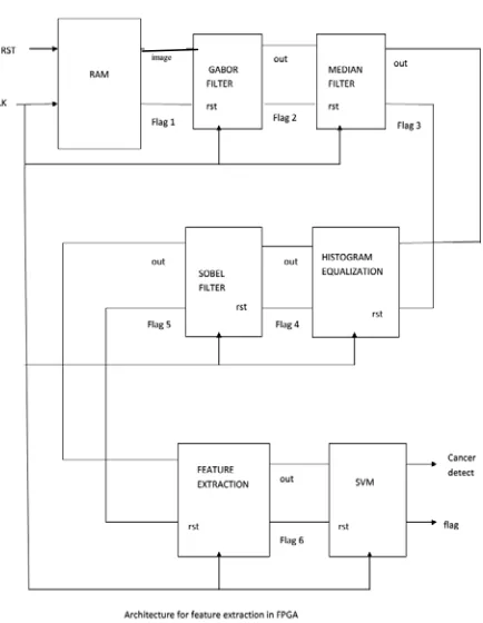
6. HARDWARE IMPLEMENTATION
Fig 6.1: proposed Hardware architecture for feature extraction on FPGA.
Figure showing proposed hardware implementation of feature extraction on FPGA .here the RAM memory is used for storing image,from memory text value of image is fed into filtering section .the gabor filter will remove
Gaussian noise and median filter will eliminate impulse or salt and pepper noise.Output of filtering section is fed into histogram to improve the pixcel contrast,then it send to sobel filter for edge detection operation and output of sobel filter is used for feature extraction. we are extracting different features and also we have range of value for each features, the reference value is different for cancer cell and normal cell. By analyzing the extracted feature is matching with one of the range ,similarly each feature is analyzed finally we can conclude the image have cancer cell or not.By increasing extracted features we can improve the accuracy. The simulation of project is done under Xilinx 14.2(Verilog).and FPGA implementation is down under Xilinx Virtex 7 XC7VX980 FPGA kit.
7.SIMULATION RESULT
7.1 median filter
Fig7.1.1 median filter output
Noise image output image
7.2 gabor filter
Fig7.2.1 output of gabor filter
7.3 Histogram equalization
Fig7.3.1 histogram output
Input image histogram output
Fig7.3.2 Tested image for histogram equalization
7.4 Sobel filter
Fig7.4.1 output of sobel filter
7.5 Final out (cancer detection stage)
Fig7.5.1 Final output
8. CONCLUSION
conducted on Xilinx vertex 7 XC7VX980 FPGA.The features were carried out to generate training and testing of the proposed SVM. This project is a new application based on histo-pathological images of skin lesions that required finding out new features and the correlation of the reduced numbers of features and getting better accuracy. It was required to modify many of the mentioned techniques to make them work for such an application. Concludes that there are some possible factors to improve the accuracy of detecting malignant melanoma by having a higher number of images for training of the SVM network. Future work directions will be to use a hybrid approach of genetic algorithms and Particle Swarm Optimization to improve feature extraction and feature selection.
References
[1] Mohamed Khalad Abu Mahmoud and Adel AI-Jumaily “Novel feature extraction methodology based on histopathalogical images and subsequent classification by Support Vector Machine”2014 IEEE.
[2] A. H. A. Razak and R. H. Taharim “Implementing Gabor Filter for Fingerprint Recognition Using Verilog HDL” 2009 5th International Colloquium on Signal Processing & Its Applications (CSPA).
[3] Prashant L. Paikrao and Swapnil G. Kavitkar “FPGA based Image Feature Extraction Using Xilinx System Generator” International Journal of Computer Science and Information Technologies.
[4] Tarek M. Bittibssi 1 , Gouda I. Salama 2 , Yehia Z. Mehaseb 3 and Adel E. Henawy “Image Enhancement Algorithms using FPGA”. Tarek M Bittibssi et al , International Journal of Computer Science & Communication Networks,Vol 2(4), 536-542