0022-538X/90/115284-06$02.00/0
Copyright © 1990,American Society for Microbiology
Morphogenesis of Hepatitis
A
Virus: Isolation
and
Characterization
of
Subviral Particles
DAVID A. ANDERSON*AND BRUCE C. ROSS
MacfarlaneBurnet Centrefor Medical Research,Fairfield Hospital, YarraBend Road, Fairfield, Victoria3078, Australia Received 2April 1990/Accepted 28 July 1990
The morphogenesis of hepatitis A virus (HAV) in BS-C-1 cells was examined by immunoblotting with
antisera tocapsid proteins and labeling ofvirus-specific proteinswith L-[35S]methionine. Antiserum to VP2
detected twovirus-specific proteinswithapparentmolecular massesof 30.6 and30kDa, representing VP0and
VP2, while antiserum toVP1 detectedproteinswith molecular masses of 33 and 40kDa, representingVP1 and a virus-specific protein which we designated PX, respectively. Sedimentation of cell lysates revealed the
presence of virions, procapsids, and pentamers, but particles analogoustotheprotomers ofother picornavi-ruses were not detected. Although provirions and virions were notfound as discretespecies inour gradient system,itwas evidentthat therateof sedimentationwasproportionaltotherelative amounts ofVP0 and VP2
in particles, with slower-sedimenting particles (provirions) containing predominately VP0 rather than VP2.
Procapsids contained VPO in addition toVP1 and VP3. Pentamers alsocontainedVPO, but PX waspresent ratherthan VP1. These results suggest thatPXis aprecursorto VP1 and is mostlikely1D2A.Primary cleavage oftheviralpolyproteinalsooccursatthe 2A-2Bjunctionincardiovirusesandaphthoviruses,butassemblyof
pentamers containing 1D2A has not been reported for those viruses. The absence of detectable levels of
protomers suggestsahigh efficiencyofpentamer formation,whichmayberelated to thehighefficiencyofviral
RNAencapsidation for HAV(D. A. Anderson, B. C. Ross,andS.A.Locarnini, J. Virol.62:4201-4206,1988). Theresults ofthisstudy reveal further unusual aspectsof the HAV replicative cycle whichdistinguish it from
otherpicornaviruses andmay contributetoits restrictedreplication incell culture.
Picornaviruses are among the most extensively studied
animal viruses. They are often characterized by rapid and
efficient replicative cycles in permissive cells, with produc-tion of 105 particles per cell within 8 h of infection being
common (15). Hepatitis A virus (HAV) has a number of
characteristics which distinguish it from other
picornavi-ruses, such as extreme thermal stability (1, 18), a tendency
to establishpersistent infections of cell cultures (6, 8, 9, 13, 17, 20, 21), and a protracted replicative cycle which pro-duces relatively little virus. In addition, while its physical
characteristics are most like those of enteroviruses, its
greatestnucleotide sequence homology appears to be shared with the cardioviruses (12). Studies of the replicative cycle of HAV (2, 3, 6, 9, 21) haveprovidedfurther evidence for the
divergence ofHAVfrom otherpicornaviruses.
We have previously used nucleic acid probes to detect
HAV-specificRNAincells (2, 3), and those studied showed thatmost of the viral RNA is present within mature virions
throughoutthereplicative cycle. On the basis of this finding,
we have proposed a model for the restriction of HAV
replicationwhereby progeny positive-strand RNA molecules
are preferentially encapsidated rather than being used as
templatesfor further RNA synthesis throughout the
replica-tivecycle. Webelieve that this may be related to the thermal
stability of the virus, reflecting a high affinity of the viral
capsid proteins for one another and/or viral RNA. This
hypothesisalso predicts an excess of capsid protein
precur-sorsin the cell to allow sequestration of the RNA into virions and that capsid precursors have a high affinity for one anotherand/orviral RNA.
To test these predictions, we used immunoblot analysis
and metabolic labeling to examine the morphogenesis of
* Correspondingauthor.
HAV in infected cells. The results are consistent with our
hypothesis but also suggest that the proteolytic processing
pathway of HAV is similartothat of the cardioviruses (10)
andaphthoviruses (19), in whichprimarycleavageoccurs at
thejunction of2A and 2B (by the L434 nomenclature of
Rueckert and Wimmer [16]).
MATERIALSANDMETHODS
Cellsand viruses. CytopathicHAV strain HM175A.2 and
poliovirustype 1(PV; Mahoney)werepropagated in BS-C-1
cellsasdescribedpreviously (1), exceptthat the incubation temperature was 37°C. Stocks of infectious virus were
prepared from cells as follows. Infected cell monolayers were dispersed with a mixture of 0.125% trypsin-0.02%
EDTA, washed with Eagle minimum essential medium
(MEM) containing 5% fetal bovine serum, and pelleted by
centrifugationfor 1 minat6,500 rpm in anMSE
microcen-trifuge. The cell pellet was suspended in 5 volumesofNT
(100mMNaCl,10mMTris[pH 7.4]), and10%Nonidet P-40
(SigmaChemical Co., St. Louis, Mo.)wasadded to afinal
concentration of1%tolysethe cell membranes. Afterbrief
mixingat24°C, the nuclei were removed by centrifugationat
13,000 rpm in the MSE microcentrifuge for 2 min and 10%
N-lauroylsarcosine, sodium salt (Sarkosyl; Sigma), was
addedtothecytoplasmic extract to afinalconcentration of 1%. Six-milliliter celllysate samples were layered on top of discontinuous sucrose-glycerolgradients in Beckman SW41 tubesprepared by layering0.5 mlof80%(vol/vol) glycerolin 100 mM Tris (pH 7.4), 2 ml each of 30 and 20% (wt/vol) sucroseinNT, and 2 ml of10%sucroseinNTcontaining1%
sodiumdodecylsulfate(SDS).Aftercentrifugationat37,000 rpm (170,000 x g) for 5 h at 18°C, virus in the glycerol cushion(total volume,1ml)wascollectedbypuncturingthe bottom of the tube and this purified virus preparation was
stored in aliquots at -70°C. 5284
on November 10, 2019 by guest
http://jvi.asm.org/
Infectious virus was quantitated by plaque assay as de-scribed previously (3). Virus inocula were prepared by extraction of purified virus with an equal volume of chloro-form, followed by dilution in MEM containing 1% fetal bovine serum. Pools of HAV prepared as described typically contained 6 x
109
PFU/ml.Single-cycle infection of cells. Monolayers of BS-C-1 cells were inoculated with 10 PFU ofHM175A.2or PV per cell for 1 h at 37°C, washed with 0.85% NaCl, and maintained in MEM at37°C.The cells were harvested by addition of 0.5 ml ofNPT (100 mM NaCl, 0.5% Nonidet P-40, 10 mM Tris [pH 7.4]) per 75 cm2 of cell monolayer (approximately 2 x
107
cells). Nuclei were removed by centrifugation as described above, and SDS was added to the cytoplasmic extract to a final concentration of 1%.
Labeling of proteins with [35SJmethionine. Cells were fed methionine-free MEM (Flow Laboratories, Inc., McLean, Va.) for 3 h and then similar medium containing 150 ,uCi of L-[35S]methionine (>1,000 Ci/mmol; Amersham Interna-tional plc, Amersham, England) per ml for a further 3 h. Incorporation was stopped by addition of MEM containing 2 mM methionine. All cultures were harvested and processed as described above.
Sucrose densitygradientultracentrifugation. Ultracentrifu-gation was performed in a Beckman SW41 rotor at 4°C with 0.5-ml samples. Gradients were either 5 to 30% (wt/vol) sucrose in NT, which were centrifuged for 3 h at 35,000 rpm (150,000 x g) for isolation of virions and procapsids, or 5 to 20% (wt/vol) sucrose in NT, which were centrifuged for 18 h at 37,000 rpm (170,000 x g) for isolation of smallersubviral particles. Fractions of 0.5 ml werecollected from the bottom of each tube and analyzed for viral RNA and proteins.
Hybridization of viral RNA. Positive-strand HAV RNA was detected in gradient fraction samples by hybridization with 32P-labeled RNA probes as described previously (3). Probes were transcribed by using the T7promoter of plasmid pGEMHAV digested with HindIII, and they represented sequences from 2.0 to 5.0 kb of the HAV genome.
Electrophoresis. Gradient fraction samples (typically, 0.4 ml) were mixed with 20
RI
of 0.05% bovine serum albumin (Commonwealth Serum Laboratories, Melbourne, Austra-lia) and 9 volumes of methanol at -20°C. After incubation overnight at -20°C, the proteinprecipitate was collected by centrifugation at 2,500 rpm in an MSE Mistral 4Lcentrifuge at - 10°C and dried under vacuum. The pellet was dissolved in 30pRI
of LB (2% SDS, 8%glycerol,
0.01% bromophenol blue, 5% 2-mercaptoethanol, 50 mM Tris [pH 6.8]). Crude cell lysates were mixed with 0.25 volume of Sx LB, and samples were boiled for 5 min before electrophoresis in SDS-polyacrylamide gels containing 12% acrylamide with the Laemmli buffer system (11). Resolution of HAV-specific proteins wasfacilitated by inclusion of urea in the gels at a finalconcentration of 3.5 M (submitted for publication), but urea was omitted from gels of PV proteins, as it caused compression of some bands. Electrophoresis and electro-phoretic transfer ofproteins were performed with the Mini Protean system (Bio-Rad Laboratories, Richmond, Calif.). After electrophoresis, gels containing radiolabeled proteins were fixed in 10% acetic acid-20% methanol and dried for autoradiography.Immunoblot analysis of HAV proteins. Proteins for immu-noblot analysis were transferred to nitrocellulose (HyBond C-extra; Amersham) filters for 2 h at 60 V in a buffer of20% methanol-0.15 M glycine-25 mM Tris (pH 7.5). The filters were dried at roomtemperature and incubated for 1 h at 37°C with shaking in 0.1 M Tris (pH 7.5) containing 3% (wt/vol)
A1 2 3 4 5
B
92--_
69-46-
_
30-14-
--.I Px
* * VP1
1 2 3 4 5
92-
_69-46-
130-
*--.vPO
tVP2
14-FIG. 1. Accumulation ofHAV-specific proteinsinBS-C-1 cells. Cells wereinfected with 10 PFU ofHM175A.2 andharvestedafter 24 (lane 3), 48 (lane 4), or 72 (lane 5) h or mock infected and harvestedafter 72 h(lane 2).Afterelectrophoresisin the presenceof 3.5 Murea, proteins were transferred to nitrocellulose and immu-noblottedwithantiserumspecificto VP1(A) or VP2(B)asdescribed in Materials and Methods. Two cellular proteins which were de-tected by anti-VP2 serum in infected and uninfected cells are marked with circles. The molecular massesofmarkerproteins(lane
1)are indicated in kilodaltons.
casein. Rabbit antisera raised to recombinant proteins con-taining sequences ofVP1 and VP2ofHAV(14)were diluted
in TBT(0.05% Tween 20, 0.1MTris[pH7.5])containing 1% casein and incubated with the filters for 1 h at 37°C. The
filters werethen washed sixtimesfor5min eachtime in TBT
withgentle agitation. Antiserumtorabbitimmunoglobulin G
(Bio-Rad) was iodinated and diluted to 106 cpm/ml in TBT
containing 1% casein and incubated with the filters for 1 h.
Afterbeing washed six times withTBT,the filtersweredried and exposed topreflashed X-Omat RP film(Eastman Kodak
Co., Rochester, N.Y.)at -70°C between Cronex
Lightning-Plus intensifying screens (E. I. du Pont de Nemours &
Co.,
Inc., Wilmington, Del.) or at room temperature without screens.
RESULTS
Detection ofviralproteinsininfectedcells.Accumulationof
HAV-specific proteins in infected cells
during
asingle
growth cycle was examined by immunoblot
analysis
using
antisera raised to recombinant HAV proteins (14) todetect VP1 and VP2 (Fig. 1). Antisera to VP2
(Fig.
1B) detected two virus-specific proteins (lanes 3 to 5)migrating
as adoublet with apparent molecular massesof 30.6 and 30kDa.
The slower-migrating protein, which we believe to be
VPO,
was present in considerable excess throughout the
3-day
study, while VP2 (30 kDa) was first detected at 48 h
postinfection and was the minor band at all times. Two
additional proteins were detected
by
this antiserum in bothuninfected (lane 2) and infected cells, and they
presumably
represent cross-reactivity with the large excess of cellular protein presentonthefilter. This
reactivity
was notremoved by absorption of antisera with acetone-fixed BS-C-1 cells (data not shown).Antiserum to VP1 (Fig. 1A) detected two
virus-specific
proteins (lanes 3 to 5). Thefaster-migrating
protein
with amolecular mass of 33 kDaprobablyrepresents
VP1,
but theslower-migrating protein with a molecular mass of 40 kDa
was not identified inaprevious study
using
these antiserato characterize maturevirions(submitted
forpublication).
Thison November 10, 2019 by guest
http://jvi.asm.org/
[image:2.612.311.554.79.226.2]A
92- 69- 46-VPO
P2-VP3
14-M 5 10 15
M 5 10~~~~~~~l 15 20
B
IA^
92-.f
g~m.i.
I 4VPI
VP] - 4
VP2-E
XP2-~
...3
r v
FtVPO
V
VP
2
I I
A
11
BWII
M 10 15
fioationnrumber
C
[image:3.612.67.310.77.293.2]20
FIG. 2. Sucrose density gradient ultracentrifugation of intracel-lular viralproteins. Celllysateswerecentrifuged at 150,000 x gfor 3 h at4°C on linear5 to30%sucrosegradients in Beckman SW41 tubes. Fractions of 0.5 ml were collected from the bottom of each tube,andproteinswere precipitatedwithmethanol andseparated by SDS-polyacrylamide gel electrophoresis. (A) PV-infected cells la-beled with [35S]methionine from 3 to 7 h postinfection. (B) HAV-infected cells harvested at 48 h postinfection and virus-specific proteins detected with a mixture of antisera to VP1 and VP2 as described in Materials and Methods. M, Molecular mass markers (sizes are indicated at the left in kilodaltons). Sedimentation was fromright to left.
suggests thatthis protein, whichwehave designated PX, is aprecursorof VP1. The kineticsofaccumulation of PXand
VP1 are also consistent with a precursor-product
relation-ship, as the level of PX did not increase beyond 24 h
postinfection, while the level of VP1 increased
approxi-matelyfivefoldfrom24 to 72hpostinfection,consistent with
the processingof PX to VP1.
Isolation ofvirions and procapsids. To examine the
distri-bution of viral proteins in more detail, lysates of cells
infected with HAV or PV were sedimented over sucrose
density gradients to separate viral and subviral particles.
When the cell lysates were sedimented over 5 to 30%
sucrose gradients for 3 h, four pools of virus-specific parti-cles were detected in PV-infected (Fig. 2A) and
HAV-infected (Fig. 2B) cells. As expected on the basis of
pub-lished studies(reviewed by Rueckert [15]), PV-infected cells
contained virions (VP1, VP2, VP3, and traces of VPO;
fractions 2to 5), provirions (VP1, VPO, VP3, and traces of
VP2; fractions 6 to 9), procapsids (VPO, VP1, and VP3;
fractions 11 to 13), and slower-sedimenting particulate
ma-terial (containing VPO, VP1, and VP3; fractions 18 to 20) which was poorly resolved from nonparticulate proteins under these conditions. Similarly, HAV-infected cells (Fig. 2B) contained virions sedimenting at 160S containing VP1 and a mixture of VPO and VP2 (fractions 2 to 4), together with slower-sedimenting species containing VP1, VPO, and
tracesofVP2(fraction 5) orPXandVPO(fractions 6 and 7). Theseparticles will be referred to as provirions (7)
(contain-ing VP1 and VPO) and preprovirions (containing PX and
VPO).
Itshould be noted that the proportion of VP2 toVPOappearedtobe greatest in the fastest-sedimenting particles.
I I I I
5
10
[image:3.612.328.569.78.335.2]fraction number
FIG. 3. Sucrosedensity gradient ultracentrifugation of intracel-lularinfectiousvirus, viralproteins,and RNA.HAV-infected cells wereharvested at 48 hpostinfection,celllysateswerecentrifuged, and fractions were collected as described inthe legendto Fig. 2. Panels: A, infectious virus; B, viral capsid proteins; C, positive-strand RNA detectedasdescribedin Materialsand Methods. Only thefirst 10 fractions from the bottomofthe tube areshown.
Adiscretepeak ofemptycapsids (procapsids;fractions 11 to
13) sedimenting at 70S and containing VP1 and VP0 was
detected. Afourth species, sedimenting slightlyfaster than the bulk of cellular protein and containing PX, VP0, and
traces of VP1 (fractions 18 to 20), was detected in these
gradients. Note that the antiserum used in these
immuno-blots does notdetect VP3 but VP3 was detected in virions
and procapsidsby labelingin cell culture (seebelow).
Association of RNA and infectivity with particles. The
distribution of viral RNA and infectivity was examined in gradients of cell lysates similartothose showninFig. 2,with
samples of each gradient fraction being assayed for viral
positive-strandRNAbydot blothybridizationand for
infec-tious virusby plaque assay,inaddition toimmunoblottingof
viralproteins. Theparticles sedimentingat70S and
contain-ingVP0and VP1containednoRNAandwere notinfectious,
confirming their identity as procapsids or empty capsids,
while bothvirions(fractions 1, 2, and 3, containingVP1and predominately VP2) and provirions (fractions 4 to 7,
con-taining VP1 and predominately VP0) contained RNA and
were infectious (Fig. 3). Preprovirions (containing PX and VP0) were not detected in thisexperiment, and their
infec-tivityhas yet tobe established.
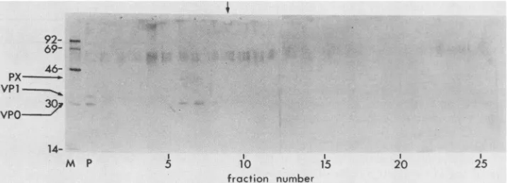
Isolation of pentamers. The material near the tops of
gradients (e.g., Fig. 2) was resolved more effectivelywhen
centrifugation was extended to 18 h over5 to 20%sucrose
gradients (Fig. 4). Under these conditions, virions and
procapsids were recovered as a pellet (P) which contained VP1,VP0, and VP2 (not visible in thisautoradiograph). The presence of an excess of VP0 over VP2 in the pellet was consistent with the sum of these proteins from virions,
I
on November 10, 2019 by guest
http://jvi.asm.org/
92-
69- 46VP1
-30 -.
14-M P 5 10 15 20 25
froction number
FIG. 4. Sucrose density gradientultracentrifugationof subviralHAV-specific structuralproteins. Cell lysate was centrifuged at 170,000 x g for 18 h at4°C on linear 5 to 20%sucrose gradientsin BeckmanSW41 tubes. Fractions were collected and analyzed as described in the legendtoFig.2.M, Molecular mass markers (sizes are given at the left inkilodaltons);P, suspended pellet. The vertical arrow marks the position of catalase(1OS) inaparallel gradient.
provirions, and procapsids of HAV but varied between experiments. Only one other virus-specific species,
contain-ingVPOandPX, was detected. By comparison with catalase
(1OS) in a parallel gradient, this material was estimated to
have a sedimentation coefficient of approximately 14S to
17S, which suggests that theparticles are analagous to the
pentamers described for other picomaviruses (15).
Unex-pectedly, no protomers (5S) or free capsid proteins were
detected'inany of our experiments. Although not visible in
this exposure, tracesof VP1 were sometimes detected in the
pentamericHAV material (for example, fractions 18 to 20 of
Fig. 2B).
Labeling of HAV-specific proteins in cell culture. As the immunoblot analyses do not allow a study of the kinetics of
viral assembly, we investigated the labeling ofHAV-specific
proteins withL-[35S]methioninein infected cells to establish
conditions for sucha study. Monolayers of BS-C-1 cells in
3-cm-diameter petri dishes were infected with 10 PFU of
HAV per celland labeled with 150 ,uCi of [35S]methionine
per ml from 15 to 18 h
postinfection.
Cells were harvestedafter4or24 hofacold chase,andlysateswereanalyzed by
ultracentrifugation and SDS-polyacrylamide gel
electropho-resis (Fig. 5). When cells were harvested after a 4-h chase,
verylittle labeledvirus-specificprotein was detected (Fig. 5, lanes 2 to 7), although most ofthe intracellular
positive-strand
RNA
was presentwithin virionsatthattime (datanotshown), as has been reported previously (3). In contrast,
after a 24-h chase (lanes 8 to 13), significant amounts of
virions and provirions (lanes 8 to 10) andprocapsids(lanes
11 to 13)weredetected. HAVvirions contained VP1, VPO,
andVP2, asshown earlier,as wellas VP3 (27kDa);which
was not detected in our immunoblot system.
Faster-sedi-menting particles also contained higher proportions ofVP2
to VPO (Fig. 5, compare lanes 8 and 10). The relative
intensities of the bands are consistent with the number of
methionine residues
predicted
from the nucleotidesequenceof theparental HM175 strainreported byCohen etal. (5;8
inVP1, 3 inVPO, 2inVP2, and 7inVP3). VP1 migratedas
adoubletinsomeexperiments,but the reason forthisisnot
known. Itis notclearwhether the labeled protein migrating
atapproximately 38 kDais virusspecific.
Samples oflabeled celllysateswerealsocentrifugedfor 18
h to allow isolation of pentamers, but under these
condi-tions, it was not possible to detect labeled
HAV-specific
proteins againstthebackground oflabeled hostcell
proteins
(datanotshown).
DISCUSSION
The detection of viral macromolecules within cells is a
prerequisite foradetailedunderstanding of viral replication,
anddetection ofHAV RNA by hybridization with
radiola-beledprobeshasbeenused extensivelyforthis purpose(2, 3, 6, 9).Studies ofHAV-specific proteinshave beenlimited todetection of viral antigens rather thanindividual proteins
(2, 6, 17, 21), but' with the development of immunoblot assaysfor individual HAV proteins (9, 14; submitted), it is
now possible to detect the capsid proteins irrespective of
theirconformationwithin the cell. Thisreportprovides the
first descriptionof thesynthesisandassembly ofviralcapsid
proteinsin cells infectedwith HAV.
We have previously reported that restriction of HAV
replication is due to inefficient RNA replication (2, 3) and
suggestedthatthiswasaresult of efficientencapsidation of
progenypositive-strand RNA atthe expense of the
replica-tive pool (3). One prediction of this hypothesis, that there
must be a pool of excess capsid proteins within infected
69-46-_
30-
*A,VP1 -VVP0
-VP2 VP3
I , I, I ,I I i I ,I I I
1 2 3 4 5 6 7 8 9 10 11 12 13
FIG. 5. Sucrose density gradient ultracentrifugation of HAV labeled with
[355]methionine.
Cells wereinfected with 10 PFU of HM175A.2 percell andlabeledwith[35S]methioninefrom15to18h postinfection.Incorporationof the labelwasstopped byadditionof excess cold methionine, and cell lysates were centrifuged as de-scribedin thelegendtoFig. 2.Adjacentfractionswerepooledfor analysis, and only the first 12 fractions are shown. Lanes: 1,molecularweight markers (sizesareindicatedtothe left in kilodal-tons);2 to7, fractions 1 plus2to 11plus12,respectively, of cells harvested aftera4-hchase;8to13,fractions 1plus2to11plus 12,
respectively, of cellsharvestedaftera24-hchase.
on November 10, 2019 by guest
http://jvi.asm.org/
[image:4.612.128.486.78.207.2] [image:4.612.314.553.490.625.2]cells, was confirmed in this study (Fig. 2 to 4). However,
detection of virus-specific protein PX (Fig. 1) was
unex-pected.We suggest thatthisproteinisaprecursorof VP1on
thebasis of(i)reactivity with antiserum raised to
recombi-nant VP1 protein (14) (Fig. 1); (ii) the presence of PX in
pentamersand someprovirionsinwhich VP1wasabsentor
presentintrace amounts(Fig.2and4);(iii)thekinetics of its accumulation within thecell, whereit ceasedtoaccumulate
by24 hpostinfection while theamountof VP1 continuedto
increase (Fig. 1).
Withamassof40kDa,PXisunlikelytobe 1CD(16)(VP1
plus VP3, 60 kDa). PX isalso smaller than the sum of1D
plus the 2A predicted for HAV on the basis of sequence
alignments with PV (5;total mass, approximately 53 kDa),
but the limitedhomology between HAV and other
picorna-virusesleaves this prediction open to question. We suggest
instead that HAV 2A has a mass ofapproximately 7 kDa,
with primary cleavage of the polyprotein occurring at the
2A-2Bjunction, ashas been demonstratedforcardioviruses (10)and
aphthoviruses
(19).Although such a 2A would be much smaller than the
corresponding protease in enteroviruses and cardioviruses,
ithas been reportedthat the 16-amino-acid 2Aof
aphthovi-rusesretainsactivity(4). However, whereasaphthovirus2A
ishighly homologousto aregionof cardioviral 2A which has
thereforebeenproposedasthe active motif(4), this motif is
notpresentinthe sequenceofHAV.Asaresult,we cannot
exclude the possibility that cellular proteases or sequences distaltotheputative2AinPX areinvolved in theprocessing
of the polyprotein and/orPX. Definitiveidentificationof PX
may depend on demonstration of self-cleavage activity or
modificationof the1D2Ajunctiontoabolish itsprocessingto
VP1. Itwould be interesting to see whether such a mutant
assembled large amounts of preprovirions
containing
PX ratherthanVP1, likethosedetected in smallamountsin thisstudy (Fig. 2B,fractions 6 and 7).
Therelativeproportions ofVPO and VP2 in HAVvirions
and provirions
(Fig.
2, 3, and 5) may be relevant to theprotracteduncoating which has been observed for HAV(2,
21).It appearsonthebasisoftheincreasedsedimentation of
virionscontaining higher proportions ofVP2thatcleavageof
VPOresults in some conformationalchange,yetall of these
particles appear to be equally infectious and this cleavage
may therefore not be required for receptor binding. How-ever, the role ofVP4 in virion uncoating (15)suggests that
this cleavage is a
prerequisite
fordisassembly, and we arecurrently
examining
thatpossibility.
Wehavenoevidencetosuggestthat the unusual
process-ing
andassemblyobserved forHIAV
inthisstudycontributesto the restriction of its replication, but the absence of5S
promotersinHAV-infected cells lends somesupport to our
hypothesisofahighlyefficient mechanism of RNA
encapsi-dation. In addition, the time required forlabel to flow into
mature virions and procapsids (Fig. 4) demonstrates that
synthesis of proteinis not ratelimiting, althoughwe cannot
exclude thepossibilitythatprocessing of the viral
polypro-teinisa limiting factor.
Themorphogenicpathway ofHAV outlined in this report
may prove to be a useful model for studying picornavirus
assembly
despitethe smallyield ofvirus-specificproducts in cells. In particular, the disparity between the protein con-tents ofprocapsids(VPO,
VP1, and VP3) and pentamers(VPO,
PX, and presumably VP3) should allow the true precursor of virions to be determined, as procapsids and pentamers cannot be in equilibrium in this system. The presenceofasmall quantityofpreprovirions containingPX1A
v vl1B
11C
1D
i
2B
protomer
(VPO0VP3PPX)1
pentamer
(VPO,VP3,PX)5
RNA
(VPO,VP3VP
1
)60
procapsid
pre-provirion
(VPO,VP3,PX)-
RNAVS
2A
provirlon
(VPO,VP3,VPl)
RNA
VPO
K
VP2,
?VP4
virlon
(VP2,VP3,VP
1).oRNA
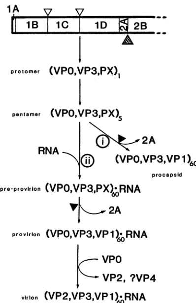
FIG. 6. Model ofprocessingandassemblyof HAV capsid pro-teins.Thepolyproteinisinitiallycleavedatthe 2A-2Bjunction byan unknownprotease
(A
),yieldingP12A.CleavagebetweenlAB-1C (VPO-VP3) and iC-iD (VP3-VP1) is probablycatalyzed by 3CPrO (A), yielding protomers containing VPO, VP3, and PX (1D2A).Thesearerapidlyassembledintopentamers,whichmaybe assem-bled into procapsids with the loss of 2A (i) or with RNA into
preprovirions (ii)containing PX, VPO,and VP3.Cleavageof PXby
anunknownprotease(A) yieldsprovirions,andautocatalytic cleav-ageof VPOtoVP2and VP4generatesthematurevirioncontaining VP1, VP2, andVP3;VP4 has notbeen detectedinthevirionand maybe lost from theparticle.
rather than VP1 (Fig. 2B, fractions 6 and 7) suggests that
pentamers are the precursor for virion assembly in HAV-infected cells.
On the basis oftheseobservations, we propose amodel for themorphogenesisof HAV(Fig.6).Primarycleavageof
thepolyproteinoccursatthejunctionof 2A and2B,yielding
1ABCD2A,
and3C then cleavesbetween lB-1C andiC-iD,yielding 1AB (VPO), 1C (VP3), and 1D2A (PX), which
remain associated as a protomer. These protomers are
rapidly assembled into pentamers, which may then be
as-sembled in either oftwomutuallyexclusivepathways, i.e.,
(i)
into emptycapsids
with loss of2Ayielding 1D (VP1)or (ii)with viral RNAintopreprovirions containing
RNA and60copieseach ofPX, VPO,andVP3,followedbyloss of 2A
to
yield
provirions andcleavage
of VPOtoVP2 and VP4to form mature virions containing RNA and VP1, VP2, andVP3. Furtherstudiesare
required
todetermine the fate and function of the small VP4peptide
liberated fromVPO,
theprotease(s) responsible
forthecleavages
of thepolyprotein,and the
identity
andfunctionof theproposed2Aresidues inPX.
on November 10, 2019 by guest
http://jvi.asm.org/
[image:5.612.342.541.71.379.2]ACKNOWLEDGMENTS
We aregrateful to S. A. Locarnini,N. E. Bishop, D. S. Bowden, and I. D.Gust for helpful discussions.
This investigation received financial support from the World Health Organization Programme for Vaccine Development and the Research Fund ofthe Macfarlane Burnet Centre for Medical Re-search.
LITERATURE CITED
1. Anderson, D. A. 1987. Cytopathology, plaque assay, and heat inactivation of hepatitis Avirus strain HM175. J. Med. Virol. 22:35-44.
2. Anderson, D. A., S. A. Locarnini, B. C. Ross, A. G. Coulepis, B.N. Anderson, and I. D. Gust. 1987. Single-cycle growth kinetics of hepatitis A virus in BSC-1 cells, p. 497-507. In M. A. Brinton and R. R. Rueckert (ed.), Positive strand RNA viruses. Alan R. Liss, New York.
3. Anderson, D. A., B. C. Ross, and S. A. Locarnini. 1988. Restricted replication of hepatitis A virus in cell culture: encap-sidation of viralRNA depletes the pool ofRNA available for replication. J.Virol.62:4201-4206.
4. Clarke, B. E., and D. V. Sangar.1988.Processingand assembly of foot-and-mouth disease virus proteins using subgenomic RNA. J.Gen.Virol. 69:2313-2325.
5. Cohen, J. I., J. R. Ticehurst, R. H. Purcell, A. Buckler-White, and B. M. Baroudy. 1987. Complete nucleotide sequence of wild-type hepatitis A virus: comparison with different strains of hepatitisAvirus and otherpicornaviruses. J. Virol.61:50-59. 6. de Chastonay, J., and G. Siegl. 1987. Replicative events in
hepatitisAvirus-infectedMRC-5cells. Virology 157:268-275. 7. Fernandez-Tomas, C. B., and D. Baltimore. 1973.
Morphogen-esis ofpoliovirus.II. Demonstration ofa newintermediate,the provirion. J. Virol. 12:1122-1130.
8. Gauss-Muller, V., and F.Deinhardt. 1984. Effectofhepatitis A virus infection on cell metabolism in vitro. Proc. Soc. Exp. Biol. Med. 175:10-15.
9. Harmon, S. A., D. F.Summers, and E. Ehrenfeld.1989. Detec-tion ofhepatitis Avirus RNA andcapsid antigen inindividual cells.VirusRes. 12:361-370.
10. Jackson, R. J. 1986. A detailed kinetic analysis of the in vitro synthesis andprocessing of encephalomyocarditis virus prod-ucts. Virology 149:114-127.
11. Laemmli, U. K. 1977.Cleavageof structuralproteinsduring the assembly ofthe head of bacteriophage T4. Nature (London) 227:680-685.
12. Palmenberg, A. C. 1987.Comparativeorganizationand genome structure in picornaviruses, p. 25-34. In M. A. Brinton and R. R. Rueckert (ed.), Positive strand RNA viruses. Alan R. Liss, NewYork.
13. Provost, P. J., and M.R.Hilleman.1979.Propagationof human hepatitis Avirus in cellculture in vitro. Proc. Soc. Exp. Biol. Med. 160:213-221.
14. Ross, B. C.,B.N.Anderson, and I.D.Gust. 1988.Expressionof thehepatitis A virus genome as P-galactosidasefusionproteins in Escherichia coli, p. 62-64. In A. J. Zuckerman (ed.), Viral hepatitis and liverdisease.Alan R. Liss, NewYork.
15. Rueckert, R. R. 1985. Picornaviruses andtheirreplication, p. 705-738. In B. N. Fields, D. M. Knipe,R. M. Chanock,J. L. Melnick, B. Roizman, and R. E.Shope (ed.),Virology. Raven Press, NewYork.
16. Rueckert, R. R., and E. Wimmer. 1984. Systematic nomencla-ture ofpicornavirus proteins. J. Virol. 50:957-959.
17. Siegl, G., J.deChastonay, and G.Kronauer. 1984.Propagation and assay of hepatitis A virus in vitro. J. Virol. Methods 9:53-67.
18. Siegl,G., M. Weitz, andG. Kronauer. 1984.Stability ofhepatitis Avirus. Intervirology22:218-226.
19. Vakharia,V.N., M.A.Devaney, D.M. Moore,J. J.Dunn, and M.J.Grubman.1987.Proteolyticprocessingoffoot-and-mouth diseaseviruspolyproteins expressedin acell-freesystemfrom clone-derivedtranscripts.J. Virol. 61:3199-3207.
20. Vallbracht, A., L. Hofmann, K. G. Wurster, and B. Flehmig. 1984. Persistent infection ofhuman fibroblasts by hepatitis A virus. J. Gen. Virol. 65:609-615.
21. Wheeler, C. M.,H.A.Fields, C. A.Schable,W.J.Meinke,and J. E. Maynard. 1986. Adsorption, purification, and growth characteristicsofhepatitisAvirusstrainHAS-15propagatedin fetal rhesus monkey kidney cells. J. Clin. Microbiol. 23:434-440.