0022-538X/90/062796-06$02.00/0
Copyright C) 1990, American SocietyforMicrobiology
Sequence-Specific Binding
of
DNA
by
the
Moloney
Murine
Leukemia Virus Integrase Protein
PAUL A. KROGSTAD' ANDJAMES J. CHAMPOUX2*
Departmentsof Pediatrics' and
Microbiology,
University of Washington, Seattle, Washington98195Received 14August 1989/Accepted 2 March 1990
Genetic studies have indicated that integration of retroviral DNA into the host genome depends on the
presenceoftheinvertedrepeatsatthefree termini of thelongterminal repeatsontheunintegratedDNAand
on the product of the 3' end of thepolgene (the integrase [IN] protein). While the precisefunction ofthe
Moloney murineleukemia virus INproteinisuncertain,others have shownthat it isaDNA-binding protein
andfunctionsin theprocessingof the inverted repeatspriortointegration. By usingsite-directedmutagenesis,
we cloned and expressed the IN protein in Escherichia coli. Crude extracts of total cellular protein were
fractionatedbysodiumdodecylsulfate-polyacrylamide gel electrophoresis, transferredtonitrocellulosefilters,
denatured inguanidine, renatured, and incubated witholigonucleotide probes. Single- anddouble-stranded
oligonucleotides correspondingtothe termini ofunintegratedlinear viral DNA werespecificallybound bythe
IN protein in this assay. These data suggest that the role of the Moloney IN protein in the early steps of
integration involvessequence-specific recognition ofthe DNAsequencesfound at the ends of thelongterminal
repeats.
During retroviral replication, the linear double-stranded
DNA(dsDNA) productof reverse transcription is integrated
into the host cell genome (2, 10, 11, 22). The biochemical
mechanism of this illegitimate recombination event is
un-known, but it characteristically involves a loss of base pairs
fromthe terminiof the long terminal repeats (LTRs) of the
viral DNA and the generation of short direct repeats (4 to 6
basepairs) in the host cell DNA at the site of integration (33).
Genetic studies haveindicatedthat integrationis dependent
on the product of the 3' end of the pol gene (encoding the
integrase[IN] protein) (7, 16, 28); cells infected with certain polmutants of Moloney murine leukemia virus (M-MuLV) contain the usual linear and circular DNA intermediates in the cytoplasm and nucleus, but few, generally aberrant,
integrants are observed (6, 7). In recent studies, the DNA
intermediates ofintegration have beenisolated and charac-terized (3, 12, 27). Extracts of cells infected with wild-type
M-MuLV are enriched for a linear viral DNA species in
which both 3' ends are shortened by two bases. In contrast, viral DNA isolated from cells infected with M-MuLV pol
mutantsdefectiveinthe IN domain are predominantly blunt ended and presumably represent full-length reverse
tran-scriptionproducts (3, 27). While the precise function of the
M-MuLVINprotein is uncertain, others have shown that it
hasnonspecific DNA binding activity with a preference for
single-stranded(ss) overdsDNA (26).
We have cloned and expressed the IN protein in Esche-richia coli and probed protein blots with synthetic oligonu-cleotides corresponding to the inverted repeats (IRs) found
at the termini of the viral LTRs. The results provide
evi-dence for sequence-specific DNA binding by the M-MuLV
IN protein and suggest an additional role for
oligonucleo-tides in the study of DNA-protein interactions.
MATERIALSAND METHODS
General methods and materials. Restriction
endonu-cleases, Klenow fragment, T4 DNA polymerase, and T4
* Correspondingauthor.
polynucleotide kinase were purchased from Bethesda
Re-search Laboratories, Inc., U.S. Biochemicals Corp., and
NewEngland BioLabs,Inc., andwereusedaccordingtothe specifications of the manufacturers. Indoleacrylic acid,
iso-propyl-p-D-thiogalactopyranoside,
HEPES(N-2-hydroxy-ethylpiperazine-N'-2-ethanesulfonic acid), and
guanidine-HCl were purchased fromSigmaChemical Co. Radioactive
nucleotides were purchased from Dupont, NEN Research
Products. Plasmid DNAwasisolatedbystandard techniques
(1) from transformed E. coli DH5cc. Single-stranded DNA
templates for site-directed mutagenesis and DNA
sequenc-ingwereextracted fromphageparticles produced byE. coli
strainscontainingM13-oriplasmids upon infection with the
helper phage M13K07. Oligonucleotides were end labeled with [_y-32P]ATP (3,000 Ci/mmol) and T4 polynucleotide
kinase in 100mMTrishydrochloride(pH7.5)-S5mM
MgCl2-1 mM dithiothreitol. The kinase reactions were generally
carriedout at0°Cfor 2 h. Unincorporated nucleotideswere
removed by performing two successive ethanol
precipita-tionsin the presence of 2.5 M ammonium acetate.
Oligonucleotide preparation. Oligonucleotides were
syn-thesizedby usingaBiosearchModel 8600synthesizer. They
were purified by electrophoresis through 20%
polyacryl-amide-8 M ureagels containing 89 mM Tris base-89 mM
borate-2.5mM EDTA(TBE).Thefull-length productswere eluted from the gel, ethanol precipitated, and quantified
by UV absorbance. Oligonucleotides were prepared
corre-sponding to both the plus (same polarity as the RNA genome) and minus strands of the inverted repeats found
atthe terminiof the M-MuLV right (R) and left (L) LTRs.
L- is 5'-AATTCGTGGGGTCTTTCATT-3'; L+ is 5'-AAT
GAAAGACCCCACG-3';R+ is5'-AATTCCGGGGGTCTT
TCATT-3';R-is 5'-AATGAAAGACCCCCGG-3'. dsDNA
probes were produced by mixing 5 ng of a 5'-end-labeled
oligonucleotide with asixfold excessof thecomplementary
oligonucleotide in a 15-,lI annealing mixture containing 50
mMTrishydrochloride(pH7.5), 50mMNaCl, 5 mM MgCl2,
and 0.1 mg of bovine serum albumin per ml, followed by
successive 15-min incubations at 65, 37, and 20°C.
5'-2796
on November 10, 2019 by guest
http://jvi.asm.org/
M-MuLVZi
Sol1 BomHl
IN
pTZ19R c z
903k /
...CTTTGTCGATACTGGTAC TATCTTTTAAGTAGTGGGATGTGGAGT...
5'-ACAGCTATGACCATG ATAGAAAATTCATCACCC-3'
Met Thr Met Ile Gtu Asn SerSer,,,
IN
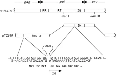
FIG. 1. Cloning ofthe M-MuLV IN protein coding domain. A
2.8-kilobaseSalI-BamHIfragment of p8.8 (a full-length clone of the M-MuLV genome) was ligated into the polylinker of plasmid
pTZ19R (Pharmacia). A total of 903 bases (903b) of vector and
reverse transcriptase sequences were deleted by
oligonucleotide-directed mutagenesis, resulting inaconstructin which theINcoding domain is preceded by the first three codons of lacZasshown. The sequence ofthe 33-base oligonucleotide used to loop out the 903
bases isshown base pairedwith the ssformof the initialconstruct.
The predicted amino acid sequence of the resulting protein is
indicated.PR and RTindicatetheproteaseandreversetranscriptase
coding domains of thepolgene, respectively.
End-labeled L- was annealed with a sixfold excess of
unlabeled L+ to produce a duplex oligonucleotide, IRL,
which corresponds to the left LTR IR. 5'-End-labeled R+
and a sixfold excess of unlabeled R- were annealed to
yield IRR, which corresponds to the right LTR IR. 5'-End-labeled oligonucleotide SL- (5'-AATTCGTGGGGTCTTT CA-3') was similarly annealed with an excess of L+ to
produce IRL*, which corresponds to the IR ofthe left LTR lacking the terminal two 3' thymidine residues from the proviral minus strand. Each ofthese dsDNA probes has a
four-base overhang which represents an EcoRI endonucle-ase cleavage product. A dsDNA control probe (C) with a
similar four-base overhang was produced by annealing the
5'-end-labeled oligonucleotide Ti (5'-CGATAACTGGGC CCAGCTGT-3') with a sixfold excess of T2 (5'-ACAGC
TGGGCCCAGTT-3').Toproduce ssDNAprobesof specific activityequaltoIRRand C(referredtoas ssIRRandssC), a
sixfoldexcess of unlabeledR+ andTi oligonucleotides was
substitutedfor the complementary strandsand the annealing procedure was carried out as before. The products of the
annealing procedure were analyzed by electrophoresis in
20% nondenaturing polyacrylamide gels (19:1 acrylamide-bis) containing xO.5 TBE.
Synthetic oligonucleotides were also produced for use in
site-directed mutagenesis (5'-ACAGCTATGACCATGATA GAAAATTCATCACCC-3') and for sequencing of the
re-sulting product(5'-TCATTAGGCACCCCAGGC-3').
Plasmid construction. By comparison of the published
nucleotide sequence of the M-MuLV genome (29) with the
results of carboxypeptidase Y degradation ofreverse
tran-scriptase (5), the IN coding domain was inferred to begin
with the Ile codonbeginningatnucleotide4611. Afull-length clone of theM-MuLV genome (p8.8) (30) wasdigestedwith
Salland BamHI. A 2.8-kilobase fragmentcontaining the IN coding domain flanked by part of the reverse transcriptase
and envelope coding sequences was ligated into the
poly-linker of pTZ19R (Pharmacia) (Fig. 1).
Oligonucleotide-directed mutagenesis employingasstemplateproduced ina
dutung mutant E.coli strain(21)was usedtodelete903base
pairs ofvectorand reversetranscriptase sequence resulting
in a construct, pIF, inwhich the first three codonsof lacZ
arefollowed bythe M-MuLV INcodingdomain. Toconfirm
the identity ofthe deletion mutant, the resulting DNA was
sequenced by using the commercial Sequenase kit (U.S.
Biochemicals Corp.).
Asecondplasmid,pJC99, was constructedby ligatingthe
trpE leader sequence ofpATH3 (obtained from T. J.
Ko-erner) tothe 3' endofthe INcoding domainbeginningatthe
HindIll siteatnucleotide4894. Thefusion proteinencoded
by pJC99 (TRPE-IN) was eluted from a sodium dodecyl
sulfate (SDS)-polyacrylamide gel and used to produce
IN-specific antibody ina New Zealand White rabbit(19).
Proteinanalysis: Westernblots (immunoblots) and
South-western (DNA-protein) blots. To prepare crude bacterial
extracts, pelleted cells from late-logarithmic-phase cultures
were boiled for 3 min in cracking buffer (10 mM sodium
phosphate [pH 7.2], 6 M urea, 1%SDS, 1%
2-mercaptoeth-anol) and werefrozen ondryice. Theseextractswerestored
at-70°C until used. They were subjectedto
electrophoresis
inSDS-12.5% polyacrylamide gels (8) and either stained with
Coomassie blue or transferred electrophoretically to nitro-cellulose (1). Westernblotanalysis wasperformedby
block-ing the filters with 2% nonfat dry milk, followed by
incuba-tion with the rabbit antibody described above, and
developed with an alkaline phosphatase-conjugated goat
anti-rabbit antibody kit purchased from Bio-Rad
Laborato-ries.
Southwestern analysis of DNA
binding
wasperformed
essentially as described by Roth et al. (27), but labeled
synthetic oligonucleotides were used as probes instead of
radiolabeled plasmid DNA. Extracts of
DH5a(pIF)
andDH5a(pTZ19R) were subjected to electrophoresis in
alter-nate lanes of an SDS-polyacrylamide gel and were
trans-ferredelectrophoretically to anitrocellulose filter. The filter
was soaked for 1 h at 20°C in 50 mlofdenaturing solution
(7
Mguanidine-HCl, 35 mM dithiothreitol, 50 mMTris
hydro-chloride [pH 8.3], 2 mM EDTA) and washed for 1 hat
20°C
in 60to 70 ml ofdilution buffer(glycerol
[10%
vol/vol],
0.1%Nonidet P-40, 0.5 M NaCl, 50 mM Tris
hydrochloride
[pH
7.5], 2 mM EDTA, 2 mM dithiothreitol). The filter was
transferred to a fresh tray containing another 50 to70 ml of
dilution buffer and was stored at4°Cfor aminimum of24 h
(equivalent results were seen with filters stored in this
fashion for up to 8 days). The filterswere
placed
inblocking
solution (30 mM HEPES-NaOH [pH 7.6], 0.2% nonfat
dry
milk) for 1 h at20°C and thenwere washedforaminimum of
30min in 50 ml of incubation buffer
(50
mMNaCl,
30 mMHEPES-NaOH [pH 7.6],5mM MgCl2, 1mM
dithiothreitol).
Duplicate segments of the filter (each with a control lane
next to a lanecontaining the IN
protein)
were cut outwitharazor bladeandplaced in sealed plastic
bags
containing
4mlofincubation buffer and radiolabeled
probes
ofequal
specific
activity, as determined by Cerenkov
counting (1
to 3 ng ofDNA with aspecific activity of
approximately
108cpm/,Lg).
Afterincubation with mild agitationat
20°C
for 1 to3h,
thefiltersegments were washed separately for 30 min in 50 ml of
incubation buffer, dried, and subjected to
autoradiography.
In some cases, the filters were subjected to Western blot
analysis as described above to
verify
thatcomparable
amounts of theIN protein were presenton each ofthe filter
segments. In other experiments, the amount of DNA bound
-1-909 -1- Pot env
on November 10, 2019 by guest
http://jvi.asm.org/
[image:2.612.66.300.76.227.2]U3 R U5
IR AATGAAAGACCCCACG
L TTACTTTCTGGGGTGCTTAA-5'
IR AATGAAAGACCCCACG
LX ACTTTCTGGGGTGCTTAA-5'
201 en v
pot nv U3 R U5
5'-AATTCCGGGGGTCTTTCAT
GGCCCCCAGAAAGTAAIRR
[image:3.612.319.552.72.172.2]-NV;
-31FIG. 2. Expression of the M-MuLVINcodingdomain in E.coli DH5a. Cultures of DH5atransformedwith theplasmid encodingthe
M-MuLV IN protein, pIF, or the parent vector, pTZ19R, were grown to mid-log phase. Crude extracts of bacterial cells were
prepared and subjectedtoelectrophoresis in SDS-12.5% polyacryl-amide gels. Lane 1, DH5a(pTZ19R); lane 2, DH5a(pIF) (culture
grownwithoutisopropyl-,-D-thiogalactopyranoside induction);lane
3, DH5a(pIF) (culturegrownwith 2 mM
isopropyl-P-D-thiogalacto-pyranoside induction); lane 4, sucrose gradient-purified M-MuLV particles disrupted in loading buffer. (A) Coomassie blue-stained gel; (B) Western blot analysis.Proteins inagel identicaltothat shown in panelAweretransferredelectrophoreticallytoanitrocellulosefilterand subjected to Western blot analysis with rabbit antiserum di-rected against the carboxyportion of the M-MuLV IN protein. The positions of molecular sizestandards migrating in adjacent lanesare
indicated.
to the filterfragments wasmeasured by scintillation
count-ing.
RESULTS
Expression oftheM-MuLVIN protein in E.coli.
Oligonu-cleotide-directed mutagenesis was used to construct the
recombinantplasmid pIFinwhich the first three codons of
lacZare followed by the IN protein coding sequence (see
Materials andMethods; Fig. 1). Direct sequenceanalysis of
the recombinant plasmid DNA by the dideoxy method (1)
confirmed that the construct had the correct nucleotide sequence (datanotshown).
Crude extracts of cultures of E. coli DH5a containing
eitherpIF ortheparent vector, pTZ19R, weresubjected to
SDS-polyacrylamide gel electrophoresis. Extracts of
DH5a(pIF) grown withorwithout
isopropyl-p-D-thiogalac-topyranoside induction (Fig. 2A, lanes 2 and 3) contained
increased amounts ofa protein with a molecular weight of
approximately 43,000 compared with control extracts of
DH5oa(pTZ19R) (Fig. 2A, lane 1). Immunoblots with rabbit
antiserumelicited againstaTRPE-INfusion protein showed
that the 43-kilodalton protein is the IN protein and that it
migrateswiththesamemobilityastheINproteinpresentin
sucrosegradient-purified M-MuLVparticles (Fig. 2B). The Western blots also revealed that a smaller amount of a
slightly lower-molecular-weight immunoreactive species
was presentin the bacterial extracts (Fig. 2B; seeFig. SB).
Inspection of the published nucleotide sequenceofM-MuLV
(29) reveals that the first methionine codon of the IN coding
region (nucleotides4770to4772)is preceded byafortuitous
Shine-Dalgarno sequence that could direct translation at a
site downstream from the lac initiation site. This second
band may therefore represent the product oftranslational
initiation at this downstream methionine codon or
alterna-c ACAGCTGGGCCCAGTT
TGTCGACCCGGGTCAATAGC-5'
FIG. 3. Oligonucleotides usedtoprobe protein blots. Synthetic DNAs were annealed to produce duplex oligonucleotides corre-spondingtothe IRs foundatthe termini of the LTRs of viral DNA
(IRL and IRR). IRL* correspondstothe DNAsequencefoundat the
tip of the left LTR oftheputativeimmediateprecursorto integra-tion. Oligonucleotides lacking similaritytothetipsof the M-MuLV
IRswereannealedtoproduceC.
tivelymayresult fromproteolytic cleavageofthefull-length
productencoded by pIF. Aprecedentexists for the former
possibility; Hizi and Hughes (15) found that translation
directed by a similar fortuitous Shine-Dalgarno sequence
present in the human immunodeficiency virus IN coding
domaincomplicatedtheirinitial effortstoclone andexpress
the human immunodeficiency virus INproteinin E. coli. The M-MuLV IN protein expressed in E. coli exhibits
sequence-specificbinding of DNA. We looked forevidence of
sequence-specific binding of DNA by the M-MuLV IN
protein by using a Southwestern assay similar to that
de-scribedbyRothetal. (26)butdiffering principallyin theuse
ofradioactivelylabeledsynthetic oligonucleotides (Fig. 3)as
probes instead ofplasmid DNA. dsDNA probeswere
pro-ducedby annealingthe 5'-end-labeledoligonucleotidewitha sixfold excess of the complementary oligonucleotide; this ratiowaschosentoensurethat all of the labeled oligonucle-otide would be base paired to its complement after the
annealing procedure. Electrophoresisof the annealed
oligo-nucleotides inanondenaturing polyacrylamide gelwasused toexamine thecompletenessof theannealingreaction(Fig.
[image:3.612.98.268.76.214.2]1
2
34
5 6
78
9101112
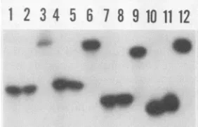
FIG. 4. Electrophoretic analysis of oligonucleotide probes. Oli-gonucleotides L-, SL-, R+, and Ti were radiolabeled with T4
kinase and[_y-32P]ATPandannealed withasixfoldexcessofeither the same or a complementary unlabeled oligonucleotide to yield
ssDNA and dsDNAprobes of equal specific activity.Theseprobes
were analyzed by electrophoresis through a 20% nondenaturing
polyacrylamide gel.Lanes 1, 4, 7,and10,radiolabeled
oligonucle-otidesR+, L-, SL-,andTi (unannealed controls),respectively; lanes 2, 5, 8, and 11, ss probes ssIRR, ssIRL, ssIRL-, and ssC,
respectively; lanes 3, 6, 9, and 12, ds probes IRR, IRL, IRL-, and C, respectively. Overexposure of thisandotherautoradiograms
dem-onstrated that after incubation with its complementary strand, essentially all of theradiolabeled oligonucleotidewaspresentas a
slowermigrating,presumably duplex, species.
A
1 2 34
B
1
234
on November 10, 2019 by guest
http://jvi.asm.org/
[image:3.612.368.509.501.592.2]DS
IR
C
SS
IR
C
IRL
IRL.
IRR
1 2 3 4 5 6
C
7 8
1 2 3 4 5 6 7 8
_m
1
-amfFIG. 6. DNA binding specificity of the M-MuLV IN protein expressed inE. coli.Proteinblotswere prepared as described in the legendtoFig.5. Lanes1, 3, 5, and 7,DH5a(pIF)extracts;lanes2, 4, 6,and 8,DH5a(pTZ19R)extracts. Separatesegmentsoftheblot were incubated with 32P-labeled ds oligonucleotide probes as fol-lows. Lanes1 and2, IRL; lanes 3 and 4, IRL-; lanes 5 and6,IRR; lanes7and 8,C. After beingwashed to remove unboundprobe, the filtersweresubjectedtoautoradiography.
FIG. 5. Sequence-specificDNAbinding activity of the M-MuLV INprotein expressed in E. coli. (A) Proteins in crude extracts of DH5a(pTZ19R) and DH5a(pIF) were subjected to electrophoresis in SDS-polyacrylamide gels, transferred to nitrocellulose,
rena-tured, and probed with 32P-labeled single-stranded (SS)or double-stranded(DS)oligonucleotidescorrespondingtothe M-MuLVLTR
right IRortoC. After being washedtoremoveunbound probe, the filters were subjected to autoradiography. Lanes 1, 3, 5, and 7,
extracts of DH5a(pIF); lanes 2, 4, 6, and 8, extracts of DH5ox(pTZ19R). (B) After autoradiography, the filtersegmentswere
subjectedtoWestern blot analysis.
4). The shift in themobility of each of the labeled
oligonu-cleotides after annealing with the complementary
oligonu-cleotide is consistent with theformation ofa duplex struc-ture. The lack of residual materialatthe position ofthe ss
oligonucleotide indicates that the annealing had gone to
completion.
ExtractsofDH5a(pIF)andDH5a(pTZ19R)were runside
by side in an SDS-polyacrylamide gel, transferred to
nitro-cellulose, denatured in guanidine, andrenatured. Duplicate
portions ofa single filter, each containing the side-by-side
extracts described above, were used in each experiment.
Theywereincubatedwithssordssyntheticoligonucleotides
correspondingtotheIRRof the M-MuLVrightLTRortothe
unrelated control oligonucleotide (C) (Fig. 3). After
incuba-tion, the filters were washed briefly to remove unbound
DNA and were subjected to autoradiography. In some
experiments, autoradiography of the filterswasfollowedby
immunoblot analysis to verify that lanes on the blot
con-tainedcomparable amountsofINprotein. No bindingof the
oligonucleotide probes to proteins in the control extracts
from DH5a(pTZ19R) was observed under the conditions
used(even-numbered lanes, Fig. 5and 6).
Autoradiographs of the blots consistently revealed much
greater binding of the dsIRR probe to the IN protein as
compared with the binding of the control probe (Fig. 5A,
lanes 1 and3). This resultwasconfirmed by cuttingoutthe bandsandquantifyingthebound labelbyscintillation count-ing. The Western blot analysis (Fig. SB, lanes 1 and 3)
confirmed thatcomparable amountsof the IN proteinwere present in each of the lanes containing the DH5a(pIF) extract. ThebindingofIRRdoesnotdependonthepresence
ofMgCl2; comparable bindingwasobserved inexperiments
in whichthe incubation buffer contained 2 mM EDTA(data
not shown). Sequence-specific DNA bindingwas also seen
when filters were incubated with ss oligonucleotides; the
ssIRR probewasboundtoagreaterextentthanwasthe ssC
probe(Fig. SA, lanes 5 and 7). With thessoligonucleotides,
thedifference between the IR and control probes wasnotas
striking as that seen with ds probes but was consistently
observedbybothautoradiography andscintillationcounting
of the filters. A similar preference forthe ssform ofthe IR
oligonucleotidewasobserved when thebinding of ssIRRwas
comparedwith that of otherssoligonucleotides lackingany
sequence similarity to the M-MuLV LTR IRs (data not shown).
Comparison of binding of full-length and processed IR
oligonucleotides bythe M-MuLVprotein.Within the first 24h
ofinfection, theretroviral reversetranscriptase synthesizes
a linear dsDNA intermediate with the RNA genome as a
template. Recent data suggest that this intermediate is
sub-sequently processed for integration by the removal oftwo
bases(thymidineresidues)from the 3' ends of the IRs atthe
termini of the LTRs (3, 12, 27). Southwestern blots,
pre-paredasdescribed above, wereincubated with the dsDNA
oligonucleotide probes correspondingtoIRLandIRR and, in
addition, to a probe lacking the two 3' thymidine kinase
residues (IRL*). IRRandIRLwereboundtoa muchgreater extentthanwasC (Fig. 6). In the experiment shown, IRL*
was bound poorly compared with the full-length inverted
repeatprobes, with thebindingbeing comparabletothatof
C.However,inotherexperiments, IRL*wasboundtonearly
thesame extentas were IRRandIRL. This variationinthe
bindingofIRL*didnotappeartobe duetodifferencesinthe
amountof the cloned INproteintransferredtothe nitrocel-lulose filtersorin thespecific activity of theprobesused.It
is possible that slight variations in the extent of protein
renaturation on the filter could accountfor the variationin
bindingfromexperiment toexperiment (seeDiscussion).
DISCUSSION
During retroviral replication, reverse transcriptase uses
the RNAgenomeas atemplatetosynthesizealineardsDNA intermediate that is subsequently integrated into the host
genomeinaprocessinvolvingthe viral INprotein.Theviral
coding regions of this DNA intermediate are flanked by
LTRs and thetipsofthe LTRs contain IR sequences vitalto
theintegration process (4, 27).
A
B
on November 10, 2019 by guest
http://jvi.asm.org/
In this study, we havepresented evidence for
sequence-specific DNA binding bythe M-MuLV INprotein by using
oligonucleotide probesin a Southwesternassay. While
syn-theticoligonucleotides have been extensivelyused to study
DNA-protein binding by gel mobilityretardation(32)andto
purify DNA-binding proteins by affinity chromatography
(17),weknow ofnoprecedentfor theiruseinthisfashion;in
previous descriptions of the so-called Southwestern
tech-nique,radiolabeledplasmidDNAwasusedtoprobe protein
blots (23, 26). Southwestern analysis with oligonucleotide
probes could be very useful in studying the DNA binding
activity ofproteins present in crude extracts or when the
physical properties of a protein preclude other means of
analysis.
Roth et al.(26)studied thebindingofplasmidDNAprobes by a series of TRPE-IN fusion proteins by using a similar Southwesternassay. Theyfound noevidence for
preferen-tialbindingoflargeDNAfragments containingthe
palindro-mic LTR-LTR circlejunction byaprotein composedof the
complete IN amino acid sequence preceded by 26 foreign
amino acids. Thereare severalpossible explanationsfor the differences between theirfindings andthe resultspresented
here. First, the foreignamino acid residues added to the N
terminus of the IN protein may have altered its tertiary
structure andtherebyitsbinding specificity. Incontrast,we
cloned the M-MuLV IN coding domain as a fusion gene
preceded by onlythree codons of lacZ. In E. coli,theinitial
N-formylmethionine of nascent proteins is frequently
re-moved after translation if followed by a threonine residue
(31). Thus, the bacterial extracts used in the Western and
Southwestern assays described here probably contain a
proteininwhich the IN amino acidsequenceispreceded by
atmosttwo additionalamino acids(Thr-Met, Fig. 1),a more
authentic representation of the viralprotein than that used
byRoth et al.(26). Second, intheirexperiments,Roth et al. (26) examined the binding of the viral LTR-LTR circle
junctionsequenceclonedas apartofamuchlarger plasmid.
It islikely that,inaddition tobindingwithahigh specificity
to a relatively short target sequence, the IN protein also
binds nonspecificallyto DNA withaloweraffinity.Ifthis is
the case, then the vast excess of vector sequences over
specific target sequences in the probe may haveprecluded
detection of thespecific componentof thebindingreaction. Thus, a control plasmid containing no viral nucleotide se-quencesmightbebound toasimilarextentinthe Southwest-ern assay as a plasmid containing the LTR-LTR circle
junction. Similarphenomenon arewelldescribed;
bacterio-phage lambdaintegrase (18), E. coli RNApolymerase (14),
and a number ofrepressor proteins all bind specifically to
theirrespective targetsequencesyetexhibitsignificant
non-specificDNAbindingaswell.Third,recent datasuggestthat
the circularized viral DNA containing the LTR-LTR
junc-tion sequence is inefficiently utilized as a precursor to the
integrated provirus (2, 10, 22). It is thereforepossible that
Roth et al. (26)found noevidenceforspecific bindingof the
circlejunction simplybecausethebinding activityof the IN
protein requiresthatthe IRtargetsequencebepresentatthe
terminus ofalinearDNAmolecule.
Retroviral integration depends onthe presence of the IN
protein (6, 7, 27, 28)and theIRspresentatthetermini of the
LTRs(4).Ourresultsshowing bindingof the IRsequenceby
the IN protein demonstrate a specific interaction between
these elements. Recent data suggest that the initial step of
integrationinvolves theprocessing of thelinear viral DNA
byremovalof theterminaltwo basesfrom the3' endsof the
IR (3, 12, 28). There is certainlyreasonto suspect that the
M-MuLV INprotein itselfmight beanendonuclease and be
responsible for this processing. An endonuclease
activity
has been found in purified viral cores of Rauscher MuLV
(20). The correspondingpol gene product, pp32,from avian
sarcoma and leukemia viruses has been shownto bind the
viral LTRs and cleave viral DNA near the site ofintegration
(9, 13, 24). However, Panet and Baltimore (25) have
pre-sented evidence that themajor endonuclease activity found
in M-MuLV virions is probably notthe product of the IN
codingregion.
In related studies, the IR oligonucleotides used in this
study have been incubated with detergent-disrupted
M-MuLV virions and the 3' ends corresponding to the tips of
the LTRs have been foundtobe shortenedbytwobases(L.
Ishimoto, M. Halperin, and J. Champoux; unpublished
data). This reaction appears to mimic the processing of viral
DNA, which occurspriortointegration. However,wehave
been unsuccessful in attempts to demonstrate any
endonu-clease activity associated with cloned M-MuLV IN protein
that had been eluted from an SDS-polyacrylamide gel and
renatured in vitro. Roth et al. (26) were also unable to
demonstrate endonuclease activity associated with their
TRPE-INfusion proteins. It is possible that the M-MuLV IN
protein actually has an endonucleaseactivity which hasnot
yetbeendemonstrated for technical reasons. However, the
processing of LTR ends prior to integration might instead
involve a cellular endonuclease; the enzyme detected by
Panet and Baltimore (25) in M-MuLV IN-deficient viral
particles is a possible candidate. Ifacellular endonuclease is
infactinvolved, thenperhaps the IN protein acts as a kind
of template for a cellular nuclease that removes the two
basesfrom the 3' ends of the viral DNA.
Wesoughttodetermine whether IRL*, an oligonucleotide
probe correspondingtothe termini of theputative immediate
precursortotheintegrated provirus (i.e., lackingthetwo 3'
terminal T residues), would be bound by the IN protein.
Thus far, these experiments have yielded variable results.
Sincewe neverobserved anyvariations fromexperimentto
experiment with the full-length oligonucleotide probes, we
conclude that the interaction of the IN protein with the
shortened probe in a Southwestern assay is somehow
dif-ferent from the interaction with the full-length duplex
oligo-nucleotide. This result might be explained by assuming that
the binding of the shortened oligonucleotide is slightly
weaker than the binding of the full-length probe and that
there issomevariation fromexperiment to experiment in the
extentof IN protein renaturation during the preparation of
the filters for the Southwestern assays. Accordingly,
par-tiallyrenaturedaswellasfully renatured IN protein may be equally capable of specifically binding the full-length probe,
whereas any partially renatured protein, when present,
would be unable to bind the shortened oligonucleotide.
Furtherexperiments will be required to elucidate the basis
for these observations.
ACKNOWLEDGMENTS
This workwassupported bygrantsfromthe National Institute of Health (CA51605) and the American Cancer Society (MV-232C). P.A.K. was supported by training grant T32-HD07233 from the National Institutes of Health.
The authors wishtothankAlisonRattray for advice and technical assistance and Lance Ishimoto for helpful suggestions throughout the work.
on November 10, 2019 by guest
http://jvi.asm.org/
LITERATURE CITED
1. Ausubel, F., R. Brent, R. Kingston, D. Moore, J. Seidman, J. Smith, and K. Struhl (ed.). 1987. Current protocols in molecular biology, chapters 1 and 10. John Wiley & Sons, Inc., New York. 2. Brown, P., B. Bowerman, H. Varmus,and J. M. Bishop. 1987. Correct integration of retroviral DNA in vitro. Cell 49:347-356. 3. Brown, P., B. Bowerman, H. Varmus, and J. M. Bishop. 1989. Retroviral integration: structure of the initial covalent product and its precursor, and a rolefor the IN protein. Proc. Natl. Acad. Sci. USA 86:2525-2529.
4. Coliceili,J., and S. Goff. 1988. Sequence and spacing require-mentsofaretrovirus integration site. J. Mol. Biol. 199:47-59. 5. Copeland, T. D., G. F. Gerard, C. W.Hixson, and S. Oroszlan.
1985. Amino- and carboxy-terminus sequence of Moloney mu-rineleukemiavirus reverse transcriptase. Virology 143:676-679. 6. Donehower, L. 1989. Analysis of mutant Moloney murine leu-kemia viruses containing linker insertion mutations in the 3' region ofpol.J. Virol. 62:3958-3964.
7. Donehower, L., and H. Varmus. 1984. A mutant murine leuke-mia virus withasingle missensecodon inpolis defective in a function affecting integration. Proc. Natl. Acad. Sci. USA 81:6461-6465.
8. Dreyfuss, G., S. Adam, and Y. D. Choi. 1984. Physical changes in cytoplasmic messenger ribonucleoproteins in cells treated with inhibitors of mRNA transcription. Mol. Cell. Biol. 4: 415-423.
9. Duyk, G., J. Leis, M. Longiaru, and A. Skalka. 1983.Selective cleavageintheavian retrovirallong terminalrepeat sequence by the endonuclease associatedwith thecx3form of avian reverse transcriptase. Proc. Natl. Acad. Sci. USA80:6745-6749. 10. Ellis, J., and A. Bernstein. 1989. Retrovirus vectors containing
aninternalattachmentsite: evidencethatcircles are not inter-mediates to murine retrovirus integration. J. Virol. 63:2844-2846.
11. Fujiwara, T., and R. Craigie.1989. Integrationof mini-retroviral DNA: acell-free reaction for biochemical analysis of integra-tion. Proc. Natl. Acad. Sci. USA86:3065-3069.
12. Fujiwara, T., and K. Mizuuchi. 1988.Retroviral DNA integra-tion: structure ofanintegrationintermediate. Cell54:497-504. 13. Grandgenett, D., A. C. Vora, and R. Schiff. 1978. A
32,000-dalton nucleic acid-binding protein from avian retroviruscores possesses DNAendonucleaseactivity. Virology 89:119-132. 14. Hinkle, D. C., and M. J. Chamberlin. 1972. Studies on the
binding of Escherichia coli RNApolymerase toDNA. I. The role ofsigma subunitsinsite selection.J. Mol. Biol. 70:157-185. 15. Hizi, A., and S. Hughes. 1988. Expression of the Moloney murine leukemia virus and human immunodeficiency virus integration proteinsinEscherichiacoli.Virology 167:634-638. 16. Hu, S., D. Court, M. Zweig, and J. Levin. 1986. Murine
leukemia viruspolgeneproducts:analysis with antisera gener-ated against reverse transcriptase and endonuclease fusion proteins expressed in Escherichia coli.J. Virol.60:267-274. 17. Kadonga, J. T., and R. Tjian. 1986. Affinity purification of
sequence-specificDNAbinding proteins.Proc.Natl.Acad.Sci. USA83:5889-5893.
18. Kikuchi, Y., and H. A. Nash. 1979. Nicking-closing activity associated withbacteriophage lambdaintgene product. Proc. Natl. Acad.Sci. USA76:3760-3764.
19. Konopka, J. B., R. L. Davis, S. M. Watanabe, A. S. Ponticelli, L. Schiff-Maker, N. Rosenberg, and 0. N. Witte. 1984. Only site-directedantibodies reactive with the highly conserved src-homologous region of the v-abl protein neutralize kinase activ-ity. J. Virol. 51:223-232.
20. Kopchick, J., J.Harless, B. Geisser, R. Killam, R. Hewitt, and R. Arlinghaus. 1981. Endodeoxynuclease activity associated with Rauschermurine leukemia virus. J. Virol. 37:274-283. 21. Kunkel, T., J. D. Roberts, and R. A. Zankour. 1987.Rapidand
efficientsite-specific mutagenesis without phenotypicselection. MethodsEnzymol. 154:367-382.
22. Lobel, L., J. Murphy, and S. Goff. 1989. The palindromic LTR-LTRjunction of Moloney murine leukemia virus is not an efficient substrate for proviral integration. J. Virol. 63:2629-2637.
23. Miskimins, W. K., M. Roberts, A. McClelland, and F. Ruddle. 1985. Use of protein-blotting procedure and a specific DNA probetoidentifynuclearproteinsthatrecognizethe promoter of the transferrin receptor gene. Proc. Natl. Acad. Sci. USA 82:6741-6744.
24. Misra, T. K., D. P. Grandgenett, and J. T. Parsons. 1982. Avian retrovirus pp32DNA-binding protein.I.Recognition of specific sequences in retrovirus DNA terminal repeats. J. Virol. 44: 330-343.
25. Panet, A., and D. Baltimore. 1987.Characterization of endode-oxynucleaseactivities inMoloneymurineleukemiavirus and its replication deficientmutants.J. Virol. 61:1756-1760.
26. Roth, M., N. Tanese, and S. Goff. 1988. Gene product of Moloneymurineleukemia virusrequired forproviral integration is aDNA-binding protein.J. Mol. Biol.203:131-139.
27. Roth, M. J.,P.Schwartzberg, and S. Goff. 1989. Structure of the termini ofDNA intermediates in the integration of retroviral DNA:dependenceonINfunction and terminal DNA sequence. Cell 58:47-54.
28. Schwartzberg, P.,J. Colicelli, and S. Goff. 1984. Construction andanalysis of deletion mutations in thepolgeneofMoloney murine leukemia virus: a new viral functionrequiredfor pro-ductiveinfection. Cell37:1043-1052.
29. Shinnick, T., R. Lerner, and J. Sutcliffe. 1981. Nucleotide sequenceof Moloney murineleukemia virus. Nature(London) 293:543-548.
30. Shoemaker, C.S.,S.Goff,E.Gilboa,M.Pashind,S.W.Mitra, and D. Baltimore. 1980.Structure ofacloned circularMoloney murine leukemia virus DNA molecule containing an inverted segment: implications for retrovirus integration. Proc. Natl. Acad. Sci. USA 77:3932-3936.
31. Tsunasawa,J., J.W.Stewart,and F.S. Sherman. 1985. Amino terminalprocessing ofmutantforms ofyeastiso-1-cytochrome
c: the specificities of methionine aminopeptidase and acetyl-transferase. J.Biol. Chem. 260:5382-5391.
32. Turner,R.,and R.Tjian.1989.Leucinerepeatsandanadjacent DNAbinding domainmediate theformation functioncFosand cJunheterodimers. Science 243:1689-1694.
33. Varmus, H., and R. Swanstrom. 1982. Replicationof
retrovi-ruses. p. 369-512. In R. Weiss, N. Teich, H. Varmus, and J. Coffin (ed.), Molecularbiology oftumorviruses: RNA tumor
viruses, 2nd ed. Cold Spring HarborLaboratory, ColdSpring
Harbor,N.Y.