Copyright © 1997, American Society for Microbiology
Genotypic and Phenotypic Characterization of Long Terminal
Repeat Sequences from Long-Term Survivors of Human
Immunodeficiency Virus Type 1 Infection
LINQI ZHANG,1YAOXING HUANG,1HANNAH YUAN,1BENJAMIN K. CHEN,2JAMES IP,1
ANDDAVID D. HO1*
The Aaron Diamond AIDS Research Center, The Rockefeller University, New York, New York 10016,1and
Department of Biology, Massachusetts Institute of Technology, Cambridge, Massachusetts 021392 Received 4 December 1996/Accepted 2 April 1997
Human immunodeficiency virus type 1 (HIV-1)-infected individuals who remain asymptomatic despite prolonged infection present a unique opportunity to understand virologic and immunologic factors involved in the pathogenesis of AIDS. We have previously identified a group of long-term survivors (LTS) who are clinically healthy and immunologically normal despite 13 to 15 years of HIV-1 infection. In this study, we examined the 5* long terminal repeat (5* LTR) sequences in eight of these LTS. A total of 29 nucleotide sequences were obtained from their peripheral blood mononuclear cells (PBMC). Analysis of these sequences revealed no gross deletions within the 5*LTR. Seven of the eight subjects shared nearly identical consensus sequences in the binding sites for NF-kB, Sp1, and the viral trans-activator Tat. In multiple samples from one individual (Pt 5), however, G-to-A hypermutations were found throughout the entire region, suggesting a genetically defective 5*LTR. The effects of the observed genetic variations on LTR transcription were studied by transient transfection of an LTR-driven luciferase reporter gene and by infection with a full-length recombinant HIV-1 containing a luciferase reporter (HIVHXBLTRluc). A wide range of basal and Tat-induced transcriptional activities was found among the 5*LTR from seven of the eight LTS in both transfected 293 cells and donor PBMC, suggesting a functionally intact 5*LTR in these individuals. It is therefore unlikely that defects in the 5*LTR are the underlying explanation for the benign clinical course associated with these seven individuals. However, functional abnormalities were found in the LTR from Pt 5 in directing both heterologous and viral gene expression, providing a possible genetic explanation for the low viral load and prolonged asymptomatic state of this individual. Last, a similar overall degree of genetic diversity was found among viruses from the LTS compared to those from patients with AIDS, reinforcing the notion that a strong correlation between the degree of genetic diversity and the rate of disease progression is unlikely.
Human immunodeficiency virus type 1 (HIV-1) infection leads to generalized dysfunction of the immune system and development of AIDS in the majority of HIV-1-infected indi-viduals. There is a wide spectrum of progression rates from the initial infection to full-blown AIDS among infected individu-als; some develop AIDS within months of infection (18, 31), while others remain clinically healthy for more than 10 years (1, 3, 4, 25, 28, 29, 37, 42). Currently, it is not clear whether all HIV-infected individuals will ultimately develop AIDS. The incubation time from the onset of infection to the development of complications is generally long, as in many lentivirus infec-tions (1, 3, 4, 25, 28, 29, 37, 42). It has become evident recently from several cohort studies that a small proportion (approxi-mately 5 to 8%) of infected individuals has remained clinically healthy and without evidence of immunodeficiency despite prolonged HIV-1 infection (1, 3, 4, 6, 25, 30, 37, 42). These individuals are, therefore, referred to as long-term survivors (LTS) or long-term nonprogressors. The explanation for the benign course of HIV-1 infection in these LTS may be related to viral or host factors or both (1, 3, 4, 6, 16, 17, 19, 23, 25, 28, 29, 33, 37, 42). Considerable research efforts have been made to elucidate the possible factors that may contribute to this attenuated clinical course (1, 3, 4, 6, 16, 17, 19, 23, 25, 28, 29, 33, 37, 42).
We and others have previously identified several groups of LTS (1, 3, 4, 6, 16, 17, 19, 23, 25, 28, 29, 33, 37, 42). Virologic and immunologic characterization of these individuals re-vealed several key findings. Levels of HIV-1 RNA in plasma and the viral burden in peripheral blood mononuclear cells (PBMC) were significantly lower in LTS than in progressors (1, 3, 4, 6, 16, 17, 19, 23, 25, 28, 29, 33, 37, 42). In many cases, infectious HIV-1 could not be isolated from PBMC and plasma samples (4, 6, 19, 33, 37), raising the possibility of an attenu-ated virus. In addition, broadly reactive neutralizing antibodies and potent cytotoxic T-cell activity against HIV-1 were de-tected in the majority of the study subjects (4, 6, 19, 23, 40). Also, the architecture of the lymph nodes of LTS appeared to remain relatively intact (37).
Efforts have also been made to characterize the genetic features of the viruses present in LTS. Sequence analyses of viral genomes in LTS have revealed genetic defects in multiple regions, including vif, vpr, vpu, tat, rev, and nef (6, 19, 23, 33). Perhaps the strongest report comes from the recent study of an Australian cohort of LTS infected with HIV-1 through blood transfusion from a single donor (6, 25). Multiple deletions in nef as well as in the U3 region of the 39 long terminal repeat (LTR) are found in all members of this cohort (6, 25). The lack of disease in these individuals raises the possibility that the specific genetic defect in HIV-1 accounts for the prolonged asymptomatic state. This finding is remarkably similar to those in macaques that remain asymptomatic for a prolonged period of time after infection with the nef-deleted simian
immunode-* Corresponding author. Mailing address: Aaron Diamond AIDS Research Center, 455 First Ave., 7th Floor, New York, NY 10016. Phone: (212) 725-0018. Fax: (212) 725-1126. E-mail: [email protected].
5608
on November 9, 2019 by guest
http://jvi.asm.org/
ficiency virus strain SIVmac239 (21). In our cohort of LTS, however, nef was found to be both genetically and functionally intact (16, 17). In addition, detailed sequence analyses of vif, vpr, and vpu from our cohort of LTS revealed the integrity of these genes (44), indicating that a single common genetic de-terminant responsible for the well-being of LTS is unlikely. The functional properties of the env genes from our cohort of LTS were also studied. Envelope glycoproteins from four members of the cohort have been shown to be poorly pro-cessed and failed to mediate infection in pseudotyped HIV-1 virions, suggesting functional abnormalities in these Env gly-coproteins (5).
In the present study, we have examined the genotypic and phenotypic properties of the HIV-1 LTR of the same cohort of LTS to further explore the possibility that specific viral defects account for the long-term survival of these individuals. Like all retroviruses, the HIV-1 provirus contains two identical LTRs, flanking the three structural genes, gag, pol, and env, that are essential for viral replication. Each LTR consists of three re-gions, namely U3, R, and U5 (28). HIV-1 transcription is regulated by multiple viral and cellular transcription factors that bind to distinct sequence elements in the 59LTR (8–10, 12, 15, 20, 39). For example, efficient transcription of HIV-1 in T lymphocytes or in macrophages requires the presence of the TATA box, the binding sites for NF-kB and Sp1, and the Tat-response region (TAR) (8–10, 12, 20, 27, 35, 36, 38, 41). Mutagenesis of these sequence elements either individually or collectively inhibits or inactivates HIV-1 replication (8–10, 12, 20, 27, 35, 36, 38, 41). It is generally accepted that the initiation of HIV-1 transcription is under the control of cellular factors, whereas the subsequent activation of HIV-1 transcription is largely mediated by virus-encoded trans-activators such as the Tat protein (8–10, 28, 36, 39).
To study the genotypic and phenotypic features of the HIV-1 LTR, PCR amplification, DNA sequencing and se-quence analyses, and in vitro functional analyses of the 59
LTRs were performed on the samples collected from eight of our LTS. Our results suggest the following conclusions. First, there are no gross sequence deletions within 59LTR. Seven of the eight study subjects harbor 59 LTR sequences that are genotypically and phenotypically intact, although among the different LTS a wide range of basal and Tat-induced transcrip-tional activities were found in both transfected 293 cells and donor PBMC. This observation indicates that defects in the 59
LTR are unlikely to be the underlying explanation for the well-being of most of our LTS. Second, a high degree of G-to-A mutations were found throughout the entire 59 LTR
re-gion in multiple samples collected from one individual (Pt 5). The genetic and functional defects of these 59LTR sequences may explain this individual’s low viral load and prolonged asymptomatic state. Third, similar degrees of overall genetic diversity were found among the viruses from these eight LTS compared to those from patients with AIDS, reinforcing the notion that there is no correlation between the degree of ge-netic diversity and the rate of disease progression. Factors other than genetic divergence, such as viral load and pheno-type, probably have a greater impact on disease status.
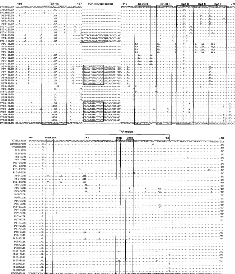
Length polymorphism and nucleotide sequence variation in LTRs. DNA was extracted from PBMC obtained from the eight LTS (Table 1), and sequences spanning the entire 59LTR were amplified by PCR. All eight subjects yielded positive signals. The lengths of the amplified PCR products were ap-proximately the same as that of HIVHXB (data not shown), suggesting that there was no significant degree of gross dele-tions or inserdele-tions in any of the study subjects. Within each sample, the PCR products appeared homogeneous in length as reflected by a single, sharp band on the polyacrylamide gel (data not shown). In multiple, independent PCR amplifica-tions these findings remained consistent. Initially, efforts had also been made to use reverse transcription-PCR to amplify viral RNA sequences from the collected plasma samples. But due to the low viral loads in these individuals, the attempts failed despite repeated efforts. However, 39 LTR sequences were successfully amplified and sequenced from the culture supernatants of three individuals (Pt 7, Pt 8, and Pt 10) (Fig. 1). The homogeneity in lengths of the 59LTRs was also con-firmed by sequence analyses. A total of 29 LTR DNA and 9 LTR RNA sequences were obtained. No intrasample length polymorphism was observed in any of the LTS, although a limited degree of intersample length variations was found up-stream of the NF-kB binding sites (Fig. 1). When compared to the HIVHXB LTR, Pt 4, Pt 7, and Pt 10 exhibited a 22- to 26-nucleotide insertion. Part of these inserted nucleotides was, in fact, a duplication of the upstream regions, previously des-ignated as TCF-1asites (8, 10) (Fig. 1). Duplication of TCF-1a
[image:2.612.61.557.578.684.2]in the HIV-1 LTR is not an uncommon finding, as similar observations have been made in both asymptomatic and symp-tomatic patients (7, 24, 34). Similar patterns of TCF-1a dupli-cation were also found in 39 LTR RNA sequences obtained from tissue culture supernatants of Pt 7 and Pt 10. Interest-ingly, no sequence variation was found in 39 LTR RNA se-quences within each individual (Pt 7, Pt 8, and Pt 10) and obtained sequences represent only a minor portion of corre-sponding proviral LTR DNA sequences (Fig. 1).
TABLE 1. Characteristics of LTS of HIV-1 infectiona
Subject Age (yr)/sexd infectionRoute ofe infection (yr)Duration of CD4 cell count(no./mm3)
Isolation of HIV-1 from: Plasma viral loadc
PBMC Plasma
Pt 2 38/M IV drug use $12 500–700 2 2 ,630
Pt 3 46/M Homosexual $13 560–740 2 2 ,630
Pt 4 40/M Homosexual 14 500–1,200 2 2 6,393
Pt 5 41/M IV drug use $12 800–1,000 2 2 ,630
Pt 6 38/M Homosexual 12 560–860 2 2 5,180
Pt 7 42/F Heterosexual 13 500–850 1b 2 ,630
Pt 8 44/M Homosexual $15 400–800 1 2 839
Pt 10 47/M Homosexual $14 550–850 1 2 11,549
aThis table has been presented previously elsewhere (16), and is included here solely for clarity, with permission of the publisher. bBy CD8 depletion only.
cRNA copies/milliliter measured by bDNA assay (Chiron). dM, male; F, female.
eIV, intravenous.
on November 9, 2019 by guest
http://jvi.asm.org/
Some degree of sequence variation was found within previ-ously identified binding sites for both NF-kB and Sp1 (Fig. 1). When compared to the HIVHXB LTR, sequences from Pt 5, Pt 6, Pt 8, and Pt 10 bear multiple mutations in the NF-kB
[image:3.612.69.549.67.627.2]binding sites. A single-base deletion in the first NF-kB binding site (NF-kB I) was found in Pt 4 (Fig. 1). Variations in the Sp1 binding sites were also frequent in all subjects (Fig. 1). How-ever, the presence of G-to-A hypermutations within both the FIG. 1. Alignment of the 59LTR sequences derived from the LTS against HIVHXB LTR. Dots represent identical nucleotides, while dashes represent gaps introduced to preserve alignment. Locations of the binding sites for TCF-1a, NF-kB and Sp1, the TATA box motif, and bulge and loop regions of TAR are shown in boxes. Nucleotide sequences are numbered according to the method of Gaynor (10).
on November 9, 2019 by guest
http://jvi.asm.org/
NF-kB and Sp1 binding sites is most striking in Pt 5. Such hypermutations are expected to have a significant impact on the function of the LTR since they are within the critical NF-kB and Sp1 binding motifs (2, 10, 11) (see below). The degree of sequence variation in the TATA box and the TAR element is much less compared to that in the NF-kB and Sp1 binding sites. Two of the three clones from Pt 4 shared an A-to-T transition in the TATA box. Sequences in Pt 5, Pt 8, and Pt 10 also displayed mutations in the bulge or loop region of the TAR element (Fig. 1). Again, hypermutations, which may disrupt the proper function of the LTR, were found in the TAR element of sequences obtained from Pt 5. This high degree of G-to-A mutations was unlikely the result of errors introduced during the process of PCR amplification, since se-quencing the PCR products directly from another PBMC sam-ple revealed identical results (data not shown).
To delineate the genetic features of the 59LTR sequences from our cohort of LTS, the degree of sequence diversity was also quantitatively analyzed and compared to sequences (HIVMN, HIVJRCSF, HIVJRFL, HIVCAM1, HIVSF2, HIVYU2, and HIVHXB) from patients with AIDS, using Kimura’s two-parameter model (22). The degree of intrasub-ject sequence diversity within each LTS varied substantially from case to case, ranging from 0.89% in Pt 4 to 3.49% in Pt 8. The mean intersubject diversity among the LTS (7.40%), however, is not significantly different from that determined from AIDS patients (5.58%) (P.0.01). Therefore, the degree of sequence variation in the LTR is not associated with the rate of disease progression, a conclusion consistent with previous reports (7, 16, 26, 32, 44).
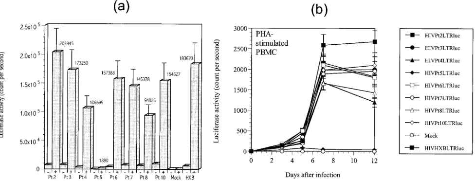
Analysis of transcriptional activity of the LTR by the lucif-erase reporter gene system.The impact of the observed mu-tations in the LTS LTRs on the level of transcriptional activ-ities was evaluated in the context of a luciferase reporter gene system (43). Cloned LTR fragments (Pt 2-4, Pt 3-13, Pt 4-1, Pt 5-5, Pt 6-1, Pt 7-3, Pt 8-1, and Pt 10-3) were subcloned into the upstream region of the luciferase reporter gene in plasmid pGL-2 (Promega) (43). The LTR sequence from HIVHXB was also cloned into the same reporter vector for comparison. The results of transient transfections of these constructs into 293 cells are shown in Fig. 2a.
It is evident that the different LTRs displayed various levels
of basal as well as Tat-induced transcription. At basal levels of transcription, the LTS LTR sequences from seven (Pt 2, Pt 3, Pt 4, Pt 6, Pt 7, Pt 8, and Pt 10) of the eight patients yielded expression levels indistinguishable from that of the positive control (HIVHXB LTR) (Fig. 2a). However, the most remark-able finding is the severely blunted activities of the LTR se-quences from Pt 5 (Fig. 2a). The high degree of G-to-A mu-tations in these LTR sequences presents a likely explanation, since such mutations are expected to disrupt the NF-kB and Sp1 consensus binding sites as well as to destabilize the sec-ondary structure of the TAR element (2, 8–10, 12–14, 39).
A wide range of Tat-induced transcriptional activities was found among the LTR sequences from LTS (Fig. 2a). The LTR sequences from Pt 2 displayed the highest level of activity, although no discernible differences could be detected from those of Pt 3, Pt 6, Pt 7, Pt 10, and the positive control HIVHXB LTR (P.0.01) (Fig. 2a). Significantly lower levels (P,0.05) of activities were found for LTR sequences from Pt 4 and Pt 8. However, the genetic basis for the reduced level of transcription is currently unclear. As expected, LTR sequences from Pt 5 exhibited marginal levels of transcription that were almost 100-fold less than that of the positive control HIVHXB LTR (Fig. 2a). Furthermore, the transcriptional level of Pt 5’s LTR in the presence of Tat is below the basal activities of HIVHXB LTR or of LTR sequences from the other LTS such as Pt 2 or Pt 3. This highlights the effect of G-to-A hypermu-tations on LTR functional abnormalities (Fig. 1). In multiple, independent experiments, these findings remained consistent.
[image:4.612.64.552.72.258.2]Analysis of transcriptional activity of the LTR in the context of the whole infectious virus.LTR sequences (Pt 2-4, Pt 3-13, Pt 4-1, Pt 5-5, Pt 6-1, Pt 7-3, Pt 8-1, and Pt 10-3) were also introduced into the infectious provirus HIVHXBLTRluc to assess the effect of the observed mutations on the level of viral transcription. We replaced the 39 LTR of HIVHXBLTRluc with each LTS’s 59LTR in light of the unique process by which the new 59LTR forms upon subsequent infection (43). Twenty micrograms of each proviral construct was used to transfect 293 cells. Virus concentration in the culture supernatant was standardized by measuring the p24 concentration (Abbott Lab-oratories) prior to use in the infection assay. Five million phytohemagglutinin-stimulated HIV-1-negative donor PBMC were inoculated with 5 ng of HIV-1 p24gag. Luciferase activities
FIG. 2. Comparison of transcriptional activities of the LTS 59LTR and the HIVHXB LTR in the presence (1) and absence (2) of trans-activator Tat and in the context of the whole infectious virus. Luciferase activities were measured 48 h posttransfection of 293 cells or after the infection of 53106
phytohemagglutinin-stimulated donor PBMC on days 0, 2, 4, 6, 8, 10, and 12. The actual readings of luciferase activity from the transfections in the presence of Tat are indicated.
on November 9, 2019 by guest
http://jvi.asm.org/
in infected PBMC measured at postinfection are summarized in Fig. 2b.
Figure 2b shows the temporal level of luciferase activity detected in PBMC on days 0, 2, 3, 5, 7, and 12. For all proviral constructs except HIVPt5LTRluc, peak levels of transcription were recorded approximately 7 days postinfection, with a mod-erate decline or increase in the ensuing period (Fig. 2b). In the context of the whole infectious virus, the transcriptional activ-ities of LTR sequences from Pt 2, Pt 3, Pt 6, Pt 7, and Pt 10 were comparable to that of HIVHXBLTRluc (P.0.01). Sig-nificantly lower levels (P , 0.05) of viral transcription were found under the direction of LTR sequences from Pt 4 and Pt 8, which was remarkably consistent with results obtained using the luciferase reporter gene alone (see above). No detectable level of viral transcription was found in PBMC infected by HIVPt5LTRluc (Fig. 2b), despite using twice as much virus in the infection (data not shown). The lack of transcriptional activity by Pt 5’s LTR in both assay systems shows the detri-mental effect of G-to-A hypermutations on LTR function.
In the present study, we chose to investigate the genotypic and phenotypic properties of the HIV-1 59 LTR region from eight previously identified LTS. The genetic and functional analyses of nucleotide sequences amplified directly from the PBMC of these LTS revealed a prominent degree of sequence diversity associated with a wide range of basal and Tat-induced transcriptional activity. The observed sequence variations in-cluded both point mutations as well as length polymorphisms. Most of the length polymorphisms were associated with a du-plication of the TCF-1afragment, although this duplication is neither complete nor perfect in any of these cases (except Pt 4). Functionally intact 59 LTRs, in terms of directing both heterologous and viral gene expression, were found in seven of the eight LTS, suggesting that genetic and functional defects in the 59 LTR are unlikely to be the underlying explanation for the well-being of these LTS. However, more thorough future studies are needed to evaluate the biological impact of the observed mutations and length polymorphisms on the level of LTR transcriptional activity.
The LTR sequences from Pt 5, however, stood out both in the genetic and transcriptional studies. A high degree of G-to-A mutations was found in multiple samples throughout the regions important for both NF-kB and Sp1 binding and viral trans-activator Tat function. Functional studies further dem-onstrated the deficiency of Pt 5’s LTR in directing gene tran-scription within the context of both the luciferase reporter gene and the whole infectious virus. In our previous study, no detectable level of viral RNA could be found in the plasma samples from Pt 5 (4). Attempts to culture infectious virus from both PBMC and plasma samples were uniformly unsuc-cessful, raising the possibility that viruses in Pt 5 may be some-what attenuated (4). In addition, Pt 5 is able to mount vigorous humoral and cytotoxic immune responses against a diverse panel of laboratory-adapted as well as primary HIV-1 isolates (4). A robust immune response may, therefore, have shifted the balance toward the host in the face of Pt 5’s sequentially and functionally defective LTR, providing some explanations for his well-being over a prolonged period of time.
This work was supported by grants from the National Institutes of Health (AI35168, AI45218, and AI40387) and the Aaron Diamond Foundation.
REFERENCES
1. Bacchetti, P., and A. R. Moss. 1989. Incubation period of AIDS in San Francisco. Nature 338:251–253.
2. Baeuerle, P. A. 1991. The inducible transcription activator NF-kB: regulation by distinct protein subunits. Biochim. Biophys. Acta 1072:63–80.
3. Buchbinder, S. P., D. Mann, L. Louie, F. Katz, and S. D. Holmberg. 1993. Genetic cofactors for delayed HIV disease progression. In Proceedings of the IXth International Conference on AIDS, p. 46. Berlin, Germany. 4. Cao, Y., L. Qin, L. Zhang, J. Safrit, and D. D. Ho. 1995. Virologic and
immunologic characterization of long-term survivors of HIV-1 infection. N. Engl. J. Med. 332:201–208.
5. Connor, R., K. E. Sheridan, C. Lai, L. Zhang, and D. D. Ho. 1996. Charac-terization of the functional properties of env genes from long-term survivors of human immunodeficiency virus type 1 infection. J. Virol. 70:5306–5311. 6. Deacon, N. J., A. Tsykin, A. Solomon, K. Smith, M. Ludford-Menting, D. J.
Hooker, D. A. McPhee, A. L. Greenway, A. Ellett, C. Chatfield, V. A. Lawson, S. Crowe, A. Maerz, S. Sonza, J. Learmont, J. S. Sullivan, A. Cunningham, D. Dwyer, D. Dowton, and J. Mills.1995. Genomic structure of an attenuated quasispecies of HIV-1 from a blood transfusion donor and recipients. Sci-ence 270:988–991.
7. Estable, M. C., B. Bell, A. Merzouki, J. S. G. Montaner, M. V. O’Shaughnessy, and I. J. Sadowski.1996. Human immunodeficiency virus type 1 long terminal repeat variants from 42 patients representing all stages of infection display a wide range of sequence polymorphism and transcrip-tion activity. J. Virol. 70:4053–4062.
8. Garcia, J. A., and R. B. Gaynor. 1994. Regulatory mechanisms involved in the control of HIV-1 gene expression. AIDS 8(Suppl. 1):s3–s17. 9. Garcia, J. A., F. K. Wu, R. Mitsuyasu, and R. B. Gaynor. 1987. Interactions
of cellular proteins involved in the transcriptional regulation of the human immunodeficiency virus. EMBO J. 6:3761–3770.
10. Gaynor, R. 1992. Cellular transcription factors involved in the regulation of HIV-1 gene expression. AIDS 6:347–363.
11. Gilboa, E., S. W. Mitra, S. Goff, and D. Baltimore. 1979. A detailed model of reverse transcription and tests of crucial aspects. Cell 18:93–100. 12. Harrich, D., J. Garcia, F. Wu, R. Mitsuyasu, J. Gonzalez, and R. B. Gaynor.
1989. Role of Sp1-binding domains in in vivo transcriptional regulation of the human immunodeficiency virus type 1 long terminal repeat. J. Virol. 63:2585–2591.
13. Harrich, D., C. Hsu, E. Race, and R. B. Gaynor. 1994. Differential growth kinetics are exhibited by human immunodeficiency virus type 1 TAR mu-tants. J. Virol. 68:5899–5910.
14. Harrich, D., G. Mavankai, A. Mette-Snider, and R. B. Gaynor. 1995. Human immunodeficiency virus type 1 TAR element revertant viruses define RNA structures required for efficient viral gene expression and replication. J. Vi-rol. 69:4906–4913.
15. Henderson, A. J., X. Zou, and K. L. Calame. 1995. C/EBP proteins activate transcription from the human immunodeficiency virus type 1 long terminal repeat in macrophages/monocytes. J. Virol. 69:5337–5344.
16. Huang, Y., L. Zhang, and D. D. Ho. 1995. Characterization of nef sequences in long-term survivors of human immunodeficiency virus type 1 infection. J. Virol. 69:93–100.
17. Huang, Y., L. Zhang, and D. D. Ho. 1995. Functional analysis of nef alleles derived from long-term survivors. J. Virol. 69:8142–8146.
18. Isaksson, B., J. Albert, F. Chiodi, A. Furucrona, A. Krook, and P. Putkonen. 1988. AIDS two months after primary human immunodeficiency virus infec-tion. J. Infect. Dis. 158:866–868.
19. Iversen, A. K. N., E. G. Shpaer, A. G. Rodrigo, M. S. Hirsch, B. D. Walker, H. W. Sheppard, R. C. Merigan, and J. I. Mullins.1995. Persistence of attenuated rev genes in a human immunodeficiency virus type 1-infected asymptomatic individual. J. Virol. 69:5743–5753.
20. Jones, K. A., J. T. Kadonaga, P. A. Luciw, and R. Tjian. 1986. Activation of the AIDS retrovirus promoter by the cellular transcription factor, Sp1. Sci-ence 232:755–759.
21. Kestler, H. W., D. J. Ringler, K. Mori, D. Panicali, P. K. Sehgal, M. D. Daniel, and R. D. Desrosiers.1991. Importance of the nef gene for mainte-nance of high virus loads and/or development of AIDS. Cell 65:651–662. 22. Kimura, M. 1988. A simple method for estimating evolutionary rates of base
substitutions through comparative studies of nucleotide sequences. J. Mol. Evol. 16:111–120.
23. Kirchhoff, F., T. C. Greenough, D. B. Brettler, J. L. Sullivan, and R. C. Desrosiers.1995. Absence of intact nef sequences in a long-term survivors with nonprogressive HIV-1 infection. N. Engl. J. Med. 332:228–232. 24. Koken, S. E. C., J. L. B. van Wamel, J. Goudsmit, B. Berkhout, and
J. L. M. C. Geelen.1992. Natural variants of the HIV-1 long terminal repeat: analysis of promoters with duplicated DNA regulatory motifs. Virology 191: 968–972.
25. Learmont, J., B. Tindall, L. Evans, A. Cunningham, P. Cunningham, J. Wells, R. Penny, J. Kaldor, and D. A. Cooper.1992. Long-term symptom-less HIV-1 infection in recipients of blood products from a single donor. Lancet 340:863–867.
26. Leigh Brown, A. J. 1991. Sequence variability in human immunodeficiency viruses: pattern and process in viral evolution. AIDS 5(Suppl. 2):s35–s42. 27. Leonard, J., C. Parrott, A. J. Buckler-White, W. Turner, E. K. Ross, M. A.
Martin, and A. B. Rabson.1989. The NF-kB binding sites in the human immunodeficiency virus type 1 long terminal repeat are not required for virus infectivity. J. Virol. 63:4919–4924.
on November 9, 2019 by guest
http://jvi.asm.org/
28. Levy, J. 1993. Pathogenesis of human immunodeficiency virus infection. Microbiol. Rev. 57:183–289.
29. Levy, J. A. 1993. HIV pathogenesis and long-term survival. AIDS 7:1401– 1410.
30. Lifson, A. R., S. P. Buchbinder, and H. W. Sheppard. 1991. Long-term human immunodeficiency virus infection in asymptomatic homosexual and bisexual men with normal CD41lymphocyte counts: immunologic and vi-rologic characteristics. J. Infect. Dis. 163:959–965.
31. McLean, K. A., D. A. Holmes, and B. A. Evans. 1990. Rapid clinical and laboratory progression of HIV infection. AIDS 4:369–371.
32. McNeary, T., Z. Hornickova, R. Markham, A. Birdwell, M. Arens, A. Saah, and L. Ratner.1992. Relationship of human immunodeficiency virus type 1 sequence heterogeneity to stage disease. Proc. Natl. Acad. Sci. USA 89: 10247–10251.
33. Michael, N. L., G. Chang, L. A. d’Arcy, P. K. Ehrenberg, R. Mariani, M. P. Busch, D. L. Birx, and D. H. Schwartz.1995. Defective accessory genes in a human immunodeficiency virus type 1-infected long-term survivor lacking recoverable virus. J. Virol. 69:4228–4236.
34. Michael, N. L., L. d’Arcy, P. K. Ehrenberg, and R. R. Redfield. 1994. Nat-urally occurring genotypes of the human immunodeficiency virus type 1 long terminal repeat display a wide range of basal and Tat-induced transcriptional activities. J. Virol. 68:3163–3174.
35. Moses, A. V., C. Ibanez, R. Gaynor, P. Ghazal, and J. A. Nelson. 1994. Differential role of long terminal repeat control elements for the regulation of basal and Tat-mediated transcription of the human immunodeficiency virus in stimulated and unstimulated primary human macrophages. J. Virol. 68:298–307.
36. Olsen, H. S., and C. A. Rosen. 1992. Contribution of the TATA motif to Tat-mediated transcriptional activation of human immunodeficiency virus gene expression. J. Virol. 66:5594–5597.
37. Pantaleo, G., S. Menzo, M. Vaccarezza, C. Graziosi, O. J. Cohen, J. F.
Demarest, D. Montefiori, J. M. Orenstein, C. Fox, L. K. Schrager, J. B. Margolick, S. Buchbinder, J. V. Giorgi, and A. S. Fauci.1995. Studies in subjects with long-term nonprogressive human immunodeficiency virus in-fection. N. Engl. J. Med. 332:209–216.
38. Parrott, C., T. Seidner, E. Duh, J. Leonard, T. S. Theodore, A. Buckler-White, M. A. Martin, and A. B. Rabson.1991. Variable role of the long terminal repeat Sp1-binding sites in human immunodeficiency virus replica-tion in T lymphocytes. J. Virol. 65:1414–1419.
39. Perkins, N. D., N. L. Edwards, C. S. Duckett, A. B. Agranoff, R. M. Schmin, and G. J. Nabel.1993. A cooperative interaction between NF-kB and Sp1 is required for HIV-1 enhancer activation. EMBO J. 12:3551–3558. 40. Rinaldo, C., X. Huang, Z. Fan, M. Ding, L. Beltz, A. Logar, D. Panicali, G.
Mazzara, J. Liebmann, M. Cottrill, and P. Gupta.1995. High level of anti-human immunodeficiency virus type 1 (HIV-1) memory cytotoxic T-lymphocyte activity and low viral load are associated with lack of disease in HIV-1-infected long-term nonprogressors. J. Virol. 69:5838–5842. 41. Ross, E. K., A. J. Buckler-White, A. B. Rabson, G. Englund, and M. A.
Martin.1991. Contribution of NF-kB and Sp1 binding motifs to the repli-cative capacity of human immunodeficiency virus type 1: distinct patterns of viral growth are determined by T-cell types. J. Virol. 65:4350–4358. 42. Schrager, L. K., J. M. Young, M. G. Fowler, B. J. Mathieson, and S. H.
Vermund.1994. Long-term survivors of HIV-1 infection: definitions and research challenges. AIDS 8(Suppl. 1):s95–s108.
43. Zhang, L., Y. Huang, H. Yuan, B. K. Chen, J. Ip, and D. D. Ho. 1997. Identification of a replication-competent pathogenic human immunodefi-ciency virus type 1 with a duplication in the TCF-1aregion but lacking NF-kB binding sites. J. Virol. 71:1651–1656.
44. Zhang, L., Y. Huang, H. Yuan, S. Tuttleton, and D. D. Ho. 1997. Charac-terization of vif, vpr and vpu sequences from long-term survivors of human immunodeficiency virus type 1 infection. Virology 228:340–349.