Copyright © 1997, American Society for Microbiology
The Herpes Simplex Virus Virulence Factor ICP34.5 and the
Cellular Protein MyD116 Complex with Proliferating Cell
Nuclear Antigen through the 63-Amino-Acid Domain
Conserved in ICP34.5, MyD116, and GADD34
S. MOIRA BROWN,1* ALASDAIR R. MACLEAN,2ELIZABETH A. MCKIE,1ANDJUNE HARLAND1
Glasgow University Neurovirology Research Laboratories, Institute of Neurological Sciences, Southern General Hospital, Glasgow G51 4TF,1and Institute of Virology, Glasgow G11 5JR,2United Kingdom
Received 2 July 1997/Accepted 3 September 1997
The herpes simplex virus (HSV) virulence factor ICP34.5, the mouse myeloid differentiation protein MyD116, and the hamster growth arrest and DNA damage protein GADD34 share a 63-amino-acid carboxyl domain which has significant homologies to otherwise divergent proteins. Here we report that both ICP34.5 and its cellular homolog MyD116 complex through the conserved domain with proliferating cell nuclear antigen. In addition, HSV infection induces a novel 70-kDa cellular protein detectable by antisera to both ICP34.5 and GADD34, demonstrating that this novel protein possesses homology with the 63-amino-acid conserved domain.
The existence of a virulence-determining locus in the long repeat region of the herpes simplex virus (HSV) genome is well documented (1, 8, 24, 41, 42). The virulence phenotype has been specifically assigned to the RL1 gene (8, 13) and its encoded protein ICP34.5 (1, 32). Null mutants in ICP34.5 are totally avirulent in mice (9, 24, 41, 42), and the function of the protein in vitro has previously been shown to be cell type and cell state specific, depending on the stage in the cell cycle and the differentiation state (4). In mouse 3T6 cells, the lack of ICP34.5 results in a defect in maturation and the egress of virus from infected cells (5). In the human neuroblastoma SK-N-SH cell line, infection with ICP34.5-negative HSV results in pre-clusion of host cell protein synthesis via the protein kinase PKR pathway (10, 12). The precise molecular function(s) of ICP34.5 remains unknown.
A 63-amino-acid carboxyl-terminal domain of ICP34.5 has previously been shown to have significant homologies with the carboxyl domains of the mouse myeloid differentiation protein MyD116 (23) and the hamster growth arrest and DNA damage protein GADD34 (16), although the amino-terminal parts of these proteins are quite diverse. The role of both MyD116 and GADD34 in the cell appears to be in blocking growth and DNA replication after damage, and the genes may act as tumor suppressor genes. The HSV type 1 (HSV-1) (strain 171)
ICP34.5 protein comprises 248 amino acids, whereas MyD116 and GADD34 consist of 657 and 590 amino acids, respectively. It has previously been demonstrated (11) that the carboxyl-terminal 63 amino acids are essential but not necessarily suf-ficient for the prevention of host cell shutoff by ICP34.5 and can be replaced by the homologous domain of MyD116 (18). By determining which cellular protein(s) interacts specifi-cally with ICP34.5 and the conserved region of MyD116 and GADD34, we wished to know whether the three proteins tar-get the same cellular proteins. In addition, as the requirement for ICP34.5 in the virus replication cycle is cell type and state dependent, we wished to know whether MyD116 and GADD34 can compensate for at least the carboxyl-terminal
portion of HSV. Therefore, we wished to determine whether the expression pattern of MyD116 and GADD34 correlates with the permissivity of different cell types and tissues for ICP34.5-negative viruses.
Here we report the following. (i) HSV ICP34.5 and the 63-amino-acid conserved portion of the cellular protein MyD116 specifically complex with proliferating cell nuclear antigen (PCNA), a cell cycle control protein. (ii) MyD116 is expressed in a range of cell types of different species, and its expression is not dependent on the differentiation state. (iii) MyD116 and GADD34 are distinguishable and are expressed in the same cell types. (iv) The expression of MyD116 or GADD34 does not determine permissivity for ICP34.5-nega-tive HSV. (v) ProducICP34.5-nega-tive HSV infection correlates with the detection of a novel 70-kDa cellular protein by both GADD34 and ICP34.5 antisera.
MATERIALS AND METHODS
Cells.The following cell lines were used in this study: baby hamster kidney clone 13 cells (BHK-21/C13) (26); mouse embryo fibroblasts (3T6 and 3T3); mouse myeloblast M1 cells (23), which were differentiated by the addition of 10 pg of interleukin 6 (IL-6) per ml as previously described (23); human glioblas-toma multiform cell line U373 MG (European Tissue Culture Collection); pri-mary human anaplastic astrocytoma BG557 cells (33); the SK-N-SH human neuroblastoma line (American Type Culture Collection); and HEp-2 cells.
Viruses.Parental HSV-1 strain 171(3) and its RL1(ICP34.5) null mutant 1716 (24) were used throughout this study. HSV-1 strain F, HSV-2 strain HG52 (43), and its RL1-negative mutant 2604 (17) were also used.
Northern blotting.Total cellular RNA was prepared essentially as previously described (7), and the poly(A)1fraction was purified by using the poly(A) Tract mRNA isolation system (Promega). Two micrograms of each poly(A)1RNA species was separated on a 1% agarose formaldehyde gel, Northern blotted, and hybridized to strand-specific riboprobes or a random-primed DNA probe under highly stringent conditions (50% formamide at 65°C) as previously described (27).
Extraction of RNA from tissues and cultured cells.Mouse footpads (skin and muscle), brains (central nervous system), and pooled lumbar, sacral, and thoracic dorsal root ganglia (DRG; peripheral nervous system) tissues were obtained from 4-week-old BALB/c mice and frozen at270°C. Tissue samples were dis-rupted with a Dounce homogenizer and poly(A)1RNA was extracted as de-scribed above.
Reverse transcription reactions.Reverse transcription was performed with poly(A)1RNA, avian myeloblastosis virus reverse transcriptase (RT; Promega), and random hexanucleotide primers under the recommended conditions.
PCR.PCRs were carried out with Vent polymerase (Promega) and 1 to 5ml of the RT reaction mix in a 100-ml reaction volume. The samples were initially
* Corresponding author. E-mail: [email protected].
9442
on November 9, 2019 by guest
http://jvi.asm.org/
denatured for 5 min at 95°C, followed by 30 cycles of denaturation at 95°C for 1 min, renaturation at 57°C for 2 min, and extension at 72°C for 2 min, with final extension at 72°C for 7 min. The primers used for the amplification of the conserved domain of MyD116 were (i) 59GCTGAGAAAGTCACAGTCCAT 39 and (ii) 59CCATGCTCTGGCCCTGGAATC 39.
Cloning and sequencing of PCR products.The PCR products were cloned into
SmaI-digested pGEM3zf(2), and the EcoRI/XbaI fragments from pGEM3zf(2) containing the cloned PCR fragments were ligated into EcoRI/XbaI-cut M13mp18 phage DNA and sequenced with a Sequenase kit (U.S. Biochemicals). Expression of both HSV-1 ICP34.5 and MyD116 as GST fusion proteins and generation of antisera. (i) HSV-1 ICP34.5.HSV-1 ICP34.5 was expressed as a fusion protein by using the pGEX–glutathione S-transferase (GST) system (39). A 780-bp NcoI/BamHI fragment comprising the entire coding region of ICP34.5 from the initiating ATG (31) was cloned in frame into the NcoI/BamHI sites of pGEX2TNMCR (34).
(ii) MyD116. The previously cloned and sequenced 3T6 cell 175-bp PCR fragment encoding the amino acids conserved between ICP34.5 and MyD116 was used to generate a GST fusion protein. The PCR fragment which had been cloned into the SmaI site of pGEM3zf(2) was excised from the multicloning region with BamHI/EcoRI and inserted into the BamHI/EcoRI sites of GST vector pGEX1 (Amrad). Subcloning into pGEM3zf(2) led to the insertion of 2 nucleotides to retain the MyD116 open reading frame in frame with the 39end of GST.
Both GST fusion proteins were expressed in protease-deficient Escherichia coli BL21 (Stratagene), and GST-ICP34.5 was purified as previously described (39). The expression of GST-ICP34.5 was confirmed by Western blotting with an available antipeptide serum (30). With the GST-MyD116 fusion protein, only a small proportion bound to glutathione agarose beads; in addition, the bound material could not be eluted from the beads. To purify the fusion protein, protein extracts from induced bacteria were run on acrylamide gels (approximately 60), the acrylamide band containing the fusion protein was cut from the gel and minced in a Dounce homogenizer, and the fusion protein was eluted with 50 mM ammonium bicarbonate (pH 7.8)–0.1% sodium dodecyl sulfate (SDS), dried, and washed with 80% acetone.
For each immunogen, two New Zealand White rabbits were injected with 1 mg of fusion protein in complete Freund’s adjuvant, followed by three booster injections in incomplete Freund’s adjuvant at 14-day intervals, and subsequently bled.
Labelling of cellular proteins.Cells were labelled for 7 h with 100mCi of [35S]methionine per ml in Eagle’s medium containing one-fifth the normal con-centration of methionine and 2% calf serum. Cell extracts for GST pulldown experiments were prepared by resuspending cells (1 ml per 33107cells) in 50 mM HEPES (pH 7.5)–50 mM NaCl–0.5% Nonidet P-40 (NP-40)–10% glycerol– protease inhibitor cocktail (Boehringer Mannheim), sonicating in a sonibath, and pelleting the debris.
Analysis of cellular proteins bound to GST fusions (pulldown).Freshly pre-pared glutathione agarose beads with bound GST fusion protein (50ml of a 50% slurry) were mixed with 300ml of labelled cell protein extract and incubated for 1 h at 4°C with continuous mixing. Beads were harvested by brief centrifugation and washed three times in a buffer containing 50 mM Tris-HCl (pH 8.0), 0.5 mM NaCl, 1 mM EDTA, 0.5% NP-40, and protease inhibitors (Boehringer Mann-heim). Beads were mixed with SDS-polyacrylamide gel loading buffer, boiled, analyzed by SDS-polyacrylamide gel electrophoresis (PAGE) (29), and either fixed, treated with En3Hance, dried, and autoradiographed or used for Western blotting.
Western blotting.Western blotting was carried out as previously described (31). The following antisera were used for Western blotting and/or immunoflu-orescence: anti-GST-ICP34.5 rabbit polyclonal sera, anti-GST-MyD116 rabbit polyclonal sera, anti-GADD34 (Santa Cruz) rabbit polyclonal immunoglobulin G (IgG), anti-PCNA mouse monoclonal IgG2a (Novocastra), protein A-horse-radish peroxidase (HRP) conjugate (Sigma), and goat anti-mouse IgG-HRP conjugate (Santa Cruz).
Antibodies against GST were removed from GST fusion protein antisera by passing through a column containing pGEX protein bound to glutathione aga-rose beads.
Immunoprecipitation.Infected and uninfected cells (33107) were harvested into 1 ml of the same buffer used for cell extractions in GST pulldown experi-ments. Cell extract (250ml) was incubated with 10ml of the appropriate antibody overnight at 4°C. One hundred microliters of 50% (vol/vol) protein A-Sepharose equilibrated in extraction buffer (0.1 M Tris [pH 8.0], 10% glycerol, 0.5% NP-40, and 0.5% deoxycholic acid sodium salt) was added and mixed end over end for 1 h at 4°C. Protein A-Sepharose was washed four times in the same buffer used for equilibration. Proteins were eluted with 200ml of polyacrylamide gel loading buffer. Precipitates (50ml) were run on SDS–10% PAGE gels and Western blotted. This is a modification of a previously described method (25).
RESULTS
Transcription of MyD116 RNA in a range of cell types and tissues.To detect the expression of MyD116 transcripts, a pair of primers was chosen within the MyD116 and ICP34.5 homol-ogous domain of MyD116, and RT-PCR was carried out with
poly(A)1 RNAs from a range of cell types and tissues. A
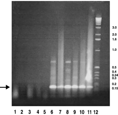
product of the expected size (175 bp), based on the published sequence of MyD116 (23), was detected in resting and expo-nentially growing 3T6 cells and BHK-21/C13 cells. In addition, this product was detected from brains and footpads of BALB/c mice and independently from the DRG of rats by Janice Pater-son (MRC Brain Metabolism Unit, Edinburgh, United King-dom) (Fig. 1, lanes 6 to 11). Control reactions in the absence of RT failed to detect any products (Fig. 1, lanes 1 through 5). We were unable to detect products from poly(A)1RNAs
ex-tracted from a number of human cell lines, possibly due to sequence differences that affected the primer homology. The RT-PCR products from BHK-21/C13 and 3T6 cells were cloned into the SmaI site of pGEM3zf(2) and sequenced. The sequences of both the 3T6 product and the BHK-21/C13 prod-uct were identical to the published sequence of MyD116 (23). The sequence of rat DRG cDNA previously cloned by Janice Patterson with the same PCR primers showed five separate base pair substitutions, leading to a small number of amino acid alterations.
Levels of MyD116 RNA in M1, BHK-21/C13, and 3T6 cells.
To determine the relative levels of MyD116 transcripts in two cell lines in which we previously characterized the phenotype of ICP34.5-negative HSV (permissive BHK-21/C13 and non-permissive 3T6 cells), poly(A)1RNA was extracted from cells
and Northern blotted with the cloned and sequenced 3T6 PCR product as the probe. M1 cells were used as a control (23). In M1 cells, a band of the expected size (2.3 kb), which appeared to be upregulated after differentiation with IL-6, was detected (Fig. 2A). In both BHK-21/C13 and 3T6 cells, the 2.3-kb band was detected; however, in 3T6 cells, two other major bands (4 and 0.6 kb) were detected (Fig. 2B). In all cell types tested, an approximately equal amount of the cellularb-actin gene was expressed (data not shown).
[image:2.612.309.545.68.298.2]Generation of an antiserum against MyD116. In order to analyze the expression of the MyD116 protein in the various FIG. 1. Analysis of expression of the MyD116 conserved domain in a range of tissues and cell types. Lanes 1 through 5, control PCR in the absence of RT; lanes 6 through 11, RT-PCR. Lanes 1 and 6, confluent 3T6 RNA; lanes 2 and 7, dividing 3T6 RNA; lanes 3 and 8, BHK-21/C13 RNA; lanes 4 and 9, mouse brain RNA; lanes 5 and 10, mouse footpad RNA; lane 11, rat DRG RNA; lane 12, molecular size (in kilobase pairs) markers.
on November 9, 2019 by guest
http://jvi.asm.org/
cell types where transcripts had been detected, we generated a polyclonal antiserum to the conserved domain of MyD116. The conserved-domain PCR product was cloned into a pGEX vector to generate a GST fusion protein. After induction with IPTG (isopropyl-b-D-thiogalactopyranoside), the 26-kDa GST band disappeared to be replaced by a 33-kDa band, corre-sponding to the expected size of the fusion protein. Unfortu-nately, only a small proportion of this fusion protein bound to glutathione agarose beads and the bound material could not be eluted from the beads. To purify the fusion protein, extracts from induced bacteria were run on acrylamide gels and the fusion protein was extracted. This purified protein was used to immunize two New Zealand White rabbits. Based on our Northern blot data, we expected a basal level of MyD116 in BHK-21/C13 cells; therefore, to monitor the reactivities of both sera in positive and negative situations, we constructed a plasmid with the MyD116 PCR product with an in-frame me-thionine at the 59 end expressed under the control of the cytomegalovirus immediate-early promoter. This plasmid and a control plasmid were lipofected into BHK-21/C13 cells, and immunofluorescence assays were carried out. All cells fluo-resced weakly, but a small proportion (corresponding to the numbers expected from lipofection) showed strong fluores-cence, indicating that an antibody had been raised against the MyD116-GST fusion protein (data not shown). In addition, both sera detected bands of 72 and 68 kDa in BHK-21/C13, 3T6, and M1 cells (see below).
Expression of MyD116 protein in M1, BHK-21/C13, 3T6, and U373 MG cells. We first analyzed the expression of MyD116 in mouse myeloid leukemic M1 cells before and after induction with IL-6 and after an actinomycin block to allow the detection of immediate-early response proteins. Both before (Fig. 3A) and after differentiation (data not shown), we were able to detect bands of 68 and 72 kDa, either of which would correspond to the expected size of MyD116. Corresponding to upregulation of the MyD116 transcript level upon differentia-tion (Fig. 2A), we also saw an increase in the level of MyD116 protein (data not shown). The specificities of these bands were demonstrated by our ability to block their detection by prein-cubation of the antiserum with the MyD116-GST fusion pro-tein but not by GST alone or an unrelated GST fusion propro-tein (data not shown).
Based on the Northern blot data, we anticipated that BHK-21/C13 and 3T6 cells would express MyD116 or related pro-teins. To analyze the pattern of expression, Western blots were carried out with human U373 MG, mouse M1 and 3T6, and hamster BHK-21/C13 cell extracts plus an extract of HSV-1 strain 171-infected BHK cells. In all the cell lines tested
(in-cluding infected cells), polypeptides of 72 and 68 kDa were detected (Fig. 3A), demonstrating that MyD116 is synthesized in different cell types of diverse species.
Construction of a GST-ICP34.5 fusion protein and genera-tion of an antiserum.To construct a fusion protein, the entire ICP34.5 open reading frame from the initiating ATG was cloned into an appropriate pGEX vector in frame with the 39
end of GST. After induction with IPTG, the 26-kDa GST product disappeared and was replaced by several products, the largest of which corresponded to the expected size (65 kDa) of the fusion protein. The products with lower Mrs were assumed
to be the result of proteolytic degradation. All of these proteins bound efficiently to glutathione agarose beads and were readily eluted with reduced glutathione.
Further proof that the fusion protein was correct was pro-vided by positive staining of the three largest products with an antipeptide serum raised against the PAT trimer in ICP34.5 (31). The GST fusion protein was used to immunize two New Zealand White rabbits. Both antisera specifically detected ICP34.5 in strain 171-infected cell extracts up to a dilution of
1:32,000, an affinity considerably higher than that of any other available ICP34.5 antiserum. With this antibody at a dilution of 1:800, the protein was not detectable beyond a 1:4 dilution of the HSV-1-infected cell extract, confirming our previous con-clusions that ICP34.5 is a low-abundance protein. We were unable to detect ICP34.5 in purified HSV-1 virions (data not shown).
Comparison of proteins detected by GST-MyD116 antibody, GST-ICP34.5 antibody, and an antibody to GADD34.By using the ICP34.5 fusion protein antibody in Western blotting of uninfected extracts of U373 MG, M1, 3T6, and BHK-21/C13 cells plus HSV-1 strain 171-infected BHK-21/C13 cell extracts,
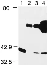
[image:3.612.109.226.68.255.2]the antibody clearly detected the 37-kDa ICP34.5 protein (Fig. 3B, lane 5) which was absent in uninfected BHK-21/C13 cells (Fig. 3B, lane 4). In addition, the antibody detected in both uninfected and infected cells a protein of 68 kDa which cor-responded to the lower-molecular-mass species detected by the FIG. 2. Northern blots of MyD116 RNA in M1 (A) and 3T6 and BHK-21/
C13 (B) cells. (A) Lane 1, M1 cells; lane 2, differentiated (Diff) M1 cells. (B) Lane 1, 3T6 cells; lane 2, BHK-21/C13 (BHK) cells. Molecular size (in kilobases) markers are indicated on the right.
FIG. 3. Extracts from a range of cell types were run on one SDS-PAGE gel in triplicate, transferred to nitrocellulose, separated into three, and probed with anti-GST-MyD116 serum (A), anti-GST-ICP34.5 serum (B), and anti-GADD34 serum (C). Lanes 1, U373 MG; lanes 2, M1; lanes 3, 3T6; lanes 4, BHK-21/C13; lanes 5, HSV-1 strain 171-infected BHK-21/C13 cell extracts. The asterisk marks the position of ICP34.5, and the arrow marks the position of the HSV-induced 70-kDa band recognized by anti-ICP34.5 and -GADD34 sera. Molecular mass (in kilodaltons) markers are shown on the left.
on November 9, 2019 by guest
http://jvi.asm.org/
[image:3.612.311.549.512.659.2]GST-MyD116 antibody (Fig. 3B; compare with Fig. 3A). In HSV-1-infected BHK-21/C13 cells, a strong band of 70 kDa was detected (Fig. 3B, lane 5); it was clearly absent from uninfected cell extracts. In addition, an antipeptide serum raised against the N-terminal 12 amino acids from the 63-amino-acid conserved domain detected the 70-kDa band in infected, but not uninfected, BHK-21/C13 cells. Interestingly, this antipeptide serum failed to detect ICP34.5 (24a).
MyD116 and GADD34 have.80% homology in the 63-amino-acid region conserved with ICP34.5. The published lit-erature states that GADD34 is the hamster homolog of mouse MyD116 (46). To determine whether the antibody to MyD116 identified GADD34 and vice versa, Western blotting was car-ried out with uninfected U373 MG, M1, 3T6, and BHK-21/C13 cell extracts and extracts from BHK-21/C13 cells infected with HSV-1. In Fig. 3C, it can be seen that the protein identified by the GADD34 antibody is clearly distinguishable by size from those identified by the MyD116 antibody (Fig. 3A). The anti-body to GADD34 recognized a 74-kDa species in all cell ex-tracts (Fig. 3C), although in this experiment it was not detected in the M1 extract. In subsequent experiments, it was detectable at a consistently lower level in M1 cells than in other cell types. The GADD34 antiserum also detected a band of 70 kDa in the extract from HSV-1-infected BHK-21/C13 cells (Fig. 3C, lane 5) which comigrated with the 70-kDa species detected by the antibody to ICP34.5 (Fig. 3B, lane 5). It appears that human, mouse, and hamster cells express both MyD116 and GADD34.
Correlation of induction of the novel 70-kDa protein with virus infection in different cell types.To determine the nature of the 70-kDa protein, the following experiments were carried out. Extracts were made from three cell types that demonstrate different responses to infection with RL1-negative HSV. BHK-21/C13 cells are fully permissive, 3T6 cells are nonpermissive due to a block in virus maturation, and SK-N-SH cells are nonpermissive due to inhibition of host cell protein synthesis. In BHK-21/C13 cells, the 70-kDa protein was not detected in the absence of virus infection (Fig. 4, lane 3) or when cells were either stressed with heat shock (lane 1) or subjected to UV light (lane 2). However, the 70-kDa protein was detected after infection with HSV-1 strains 171and F (Fig. 4, lanes 4 and 6,
respectively) and HSV-2 strain HG52 (lane 7). In addition, the 70-kDa protein was detected in extracts from cells infected with HSV-1 strain 171RL1 mutant 1716 (Fig. 4, lane 5) and
HSV-2 strain HG52 RL1 mutant 2604 (lane 8).
To determine whether productive infection was required for the induction of this cellular protein, a number of experiments were carried out. In the presence of phosphonoacetic acid (PAA), which blocks viral DNA replication and true late virus gene expression, the 70-kDa protein was not detected (Fig. 4, lane 9). Confirmation of the requirement for productive infec-tion was provided by the absence of inducinfec-tion of the protein when infection was carried out at the nonpermissive tempera-ture with a temperatempera-ture-sensitive IE175 mutant (data not shown). In 3T6 cells, the 70-kDa protein was not detected in either mock-infected (Fig. 4, lane 10) or 1716-infected (lane 12) cells but was detected in HSV-1 strain 171-infected cells (lane 11).
In SK-N-SH cells, the 70-kDa protein was clearly seen in cells infected with both HSV-1 and -2 wild-type strains (lanes 14 and 16), but in cells with RL1-negative virus infections (lanes 15 and 17) and mock-infected cells (lane 13), there was either no or minimal expression of the novel protein. Most importantly, the levels of expression in mock-infected and RL1 mutant-infected cells were indistinguishable, demonstrating that the mutant failed to induce the 70-kDa protein. Consistent with the observation that HSV-2 strain HG52 has a weaker host cell shutoff phenotype (14) than do HSV-1 strains 171and F, the
induced 70-kDa protein was more strongly expressed after HSV-2 (Fig. 4, lanes 7, 8, and 16) than after HSV-1 infection. It was also obvious that productive infection by HSV-1 but not by HSV-2 induced expression of a 38-kDa protein in all three cell types (Fig. 4, lanes 4, 5, 6, 11, and 14).
As expected with the anti-ICP34.5 serum, the 37-kDa ICP34.5 protein was detected in HSV-1 strain 171-infected
BHK-21/C13, 3T6, and SK-N-SH cells (Fig. 4, lanes 4, 11, and 14, respectively). In the presence of PAA, HSV-1 strain 171
-infected BHK-21/C13 cells also expressed ICP34.5 but at a level lower than when PAA was absent during the infection (Fig. 4, lane 9). Strain F of HSV-1 induced a 39-kDa ICP34.5 protein (Fig. 4, lane 6), but the equivalent HSV-2 protein was not recognized by this antiserum.
Analysis of cellular proteins interacting with ICP34.5 and MyD116.To analyze protein interactions with either ICP34.5 or the conserved domain of MyD116, GST pulldown experi-ments were carried out with [35S]methionine-labelled BHK-21/
C13 cell extracts. In Fig. 5A, the results of a pulldown exper-iment with the GST-ICP34.5 fusion protein can be seen. A strong band of 36 kDa was detected in the GST-ICP34.5 pull-down (Fig. 5A, lane 2) but was absent in the pullpull-down with GST alone (lane 1). When a similar experiment was carried out with extracts from human BG557 and HEp-2 cells as well as with BHK-21/C13 cell extracts, a specific band of 36 kDa was seen in the GST-ICP34.5 pulldown with each extract but not with the control GST (Fig. 5B).
The results of a pulldown experiment with the GST-MyD116 fusion protein are shown in Fig. 5C. A band of 36 kDa was present in the GST-MyD116 pulldown but not in the control GST. Thus, it appears that both ICP34.5 and the conserved domain of MyD116 interact specifically with a 36-kDa protein.
[image:4.612.314.547.69.214.2]Identification of interacting cellular proteins.Previous stud-ies have shown that the role of ICP34.5 is cell type and cell state dependent; therefore, we decided that proteins involved in growth control and cell cycle regulation may be candidates for interaction with ICP34.5. To identify interacting proteins, we analyzed pulldown proteins by Western blotting with a number of antibodies against growth control and cell cycle proteins with molecular masses of approximate 36 kDa. One of these antibodies was against PCNA. This monoclonal antibody FIG. 4. Western blot with the anti-GST-ICP34.5 serum and BHK-21/C13 (lanes 1 through 9), 3T6 (lanes 10 through 12), and SK-N-SH (lanes 13 through 17) cell extracts. Lanes: 1, heat (42°C)-shocked cells; 2, UV-treated cells; 3, 10, and 13, mock-infected cells; 4, 11, and 14, 171-infected cells; 5, 12, and 15, 1716-infected cells; 6, F-infected cells; 7 and 16, HG52-infected cells; 8 and 17, 2604-infected cells; 9, cells infected with 171in the presence of PAA. A solid square (lane 3) marks the position of ICP34.5 of HSV-1 strain 171, and an open square (lane 5) marks the position of HSV-1 strain F ICP34.5. The arrow marks the position of the HSV-induced 70-kDa band. Molecular mass (in kilodaltons) markers are shown on the right.
on November 9, 2019 by guest
http://jvi.asm.org/
specifically recognized a product of 36 kDa in control extracts of U373 MG and BHK-21/C13 cells (Fig. 6, lanes 1 and 2, respectively). It also specifically recognized the 36-kDa protein pulled down by both ICP34.5 (Fig. 6, lane 5) and the MyD116 conserved domain (lane 4) but not pulled down by the GST protein alone (lane 3), confirming the common identity of the interacting protein. This result demonstrates that ICP34.5 of HSV and the cellular protein MyD116 complex with PCNA through their shared 63-amino-acid domain.
In vivo complexing of ICP34.5-MyD116 with PCNA.To con-firm the biological relevance of PCNA complexing with ICP34.5-MyD116, coimmunoprecipitation experiments were carried out with cellular extracts. Firstly, it was demonstrated that both PCNA and MyD116 were precipitated by their re-spective antisera (data not shown). Thereafter, immunopre-cipitation experiments were carried out with uninfected 3T6 cell extracts and with the PCNA and MyD116 antisera as well as a nonspecific (goat anti-mouse) antiserum. Immunoprecipi-tated products and nonprecipiImmunoprecipi-tated 3T6 cell extract were run on an SDS-PAGE gel and Western blotted with the antiserum to PCNA. A 36-kDa product corresponding to PCNA was detected in the total cell extract (Fig. 7, lane 1) and in immu-noprecipitates with the PCNA (lane 3) and MyD116 (lane 4) antisera. Interestingly, the 36-kDa band was weaker when pre-cipitation was carried out with the PCNA antibody than with the MyD116 antiserum, indicating that PCNA is only weakly immunoprecipitated by its specific antiserum. No product was detected with the control goat anti-mouse serum (Fig. 7, lane 2). A 50-kDa band corresponding to the heavy chain of IgG was detected in immunoprecipitates by the protein A-HRP (Fig. 7, lanes 2 through 4) used in the assay. Similar results were obtained with 171- and 1716-infected 3T6 cells and
mock-, 171-, and 1716-infected BHK-21/C13 cell extracts (data
not shown).
Unfortunately, ICP34.5 is not precipitated by its antiserum and is an extremely low-abundance, infected-cell product. Im-munoprecipitations with mock-, 171-, and 1716-infected
BHK-21/C13 cells were carried out with the weakly precipitating PCNA antiserum, run on SDS-PAGE gels, and Western blot-ted with the antisera to both PCNA and ICP34.5. As in the previous experiment, when the PCNA antiserum was used, a 36-kDa band was detected (data not shown). When the anti-serum to ICP34.5 was used in a Western blot, a weak 36-kDa band was seen in 171-infected BHK-21/C13 cells but not in
mock- or 1716-infected BHK-21/C13 cells (data not shown), demonstrating that ICP34.5 and PCNA also form a complex within cells.
DISCUSSION
ICP34.5 null mutants of HSV are selectively replication competent both in vivo and in vitro. After intracerebral inoc-ulation, these mutants display a 50% lethal dose that is at least 106-fold higher than that of the wild-type virus (24, 41, 42).
They replicate inefficiently in peripheral tissues and fail to replicate in DRG neurons but establish latent infections (37, FIG. 5. [35S]methionine profile of GST pulldown experiments. (A) BHK-21/
[image:5.612.363.497.66.299.2]C13 (BHK) cell extract pulled down by GST (lane 1) and GST-ICP34.5 (GST/ 34.5; lane 2). (B) BHK-21/C13 (lanes 1 and 2), BG557 (lanes 3 and 4), and HEp-2 (lanes 5 and 6) cell extracts pulled down by GST (lanes 1, 3, and 5) and GST-ICP34.5 (lanes 2, 4, and 6). (C) BHK-21/C13 cell extract pulled down by GST (lane 1) and GST-MyD116 (lane 2). Asterisks indicate the position of the 36-kDa pulldown protein. Molecular mass (in kilodaltons) markers are shown on the left.
[image:5.612.53.289.67.277.2]FIG. 6. Western blot of a GST pulldown experiment with an anti-PCNA antibody. Lanes 1 and 2, control cell extracts to identify PCNA; lanes 3 through 5, pulldown proteins. Lanes: 1, U373 MG cell extract; 2, BHK-21/C13 (BHK) cell extract; 3 through 5, BHK-21/C13 cell extract pulled down by GST, GST-MyD116, and GST-ICP34.5 (GST/34.5), respectively. The 30-kDa band in lane 3 is due to the nonspecific reaction of high-concentration GST with the ECL detection reagent; this is also seen in lane 4 with the breakdown product from the GST-MyD116 fusion protein. Molecular mass (in kilodaltons) markers are shown on the left.
FIG. 7. Western blot of 3T6 cell extracts with the PCNA antibody. Lanes: 1, whole-cell extract; 2, an immunoprecipitate with nonspecific mouse IgG; 3, an immunoprecipitate with PCNA serum; 4, an immunoprecipitate with anti-MyD116 serum. Molecular mass (in kilodaltons) markers are shown on the left.
on November 9, 2019 by guest
http://jvi.asm.org/
[image:5.612.389.468.583.691.2]40). Stereotactic injection of ICP34.5-negative HSV into xeno-graft tumors results in preferential replication within the tumor with the surrounding tissue excluded (19, 20, 36). In tissue culture, the replication of ICP34.5-negative HSV is cell type and state dependent. In BHK cells, mutant and wild-type vi-ruses grow equally well (4). In human SK-N-SH cells, the block in replication of ICP34.5-negative virus is due to premature host cell protein synthesis shutoff via the PKR pathway (10, 12). In mouse embryo fibroblast 3T6 cells, there is a defect in virus maturation but no evidence of premature cell protein synthesis shutoff (5). In human glioblastoma and anaplastic astrocytoma cells, the mutants range from fully replication competent to totally incompetent (33).
The ICP34.5 protein has a 63-amino-acid carboxy-terminal domain which has significant homologies with the mouse my-eloid differentiation protein MyD116 and the hamster growth arrest and DNA damage protein GADD34 (12, 30). The con-served domain of the MyD116 gene can substitute for the carboxyl terminus of ICP34.5 to restore preclusion of the host cell protein synthesis shutoff phenotype in SK-N-SH cells but not the in vivo function of ICP34.5 (18, 28). It has been sug-gested that HSV has evolved by adopting the cellular sequence to guarantee survival by escaping from cellular growth arrest after HSV infection. If this is correct, it contrasts with the growth arrest phenotype induced by MyD116 and GADD34, suggesting that ICP34.5 and its cellular homologs interact with cell cycle proteins through common and diverse domains and thus casting doubt on whether the expression of MyD116 or GADD34 can complement the lack of ICP34.5 expression and hence determine the fate of ICP34.5-negative HSV in specific cell types. To resolve this anomaly, we analyzed MyD116 and GADD34 expression in different tissues and cell types in the presence and absence of HSV infection. In addition, we looked for cellular protein(s) which complexes specifically with the conserved domain of ICP34.5 and MyD116.
The MyD116 gene has previously been described as a my-eloid differentiation primary response gene induced by IL-6. The gene was originally shown to be transcribed in M1 myelo-blastic leukemic cells and in bone marrow but not in nonmy-eloid tissue (23). MyD116 has been further described as the murine homolog of the hamster GADD34 gene and has been shown to be a DNA damage-induced growth arrest gene (46). Here we have shown that MyD116 is expressed in nonmyeloid central nervous system and peripheral nervous system tissues and in mouse, hamster, and human cells. Sequence analysis of the conserved region derived from cDNAs generated from both 3T6 and BHK-21/C13 cell poly(A)1RNAs demonstrated
complete homology to MyD116. In Northern blot experiments with the conserved region as the probe, a 2.3-kb transcript was detected from poly(A)1RNA of M1 cells, with IL-6 treatment
of cells resulting in upregulation of this transcript. An equiv-alent-sized band was detected in both BHK-21/C13 and 3T6 cells. In 3T6 cells, two additional transcripts (0.6 and 4.0 kb) were detected, indicating expression of a family of related genes or alternatively spliced products which share the con-served domain. We are trying to isolate cDNAs corresponding to these transcripts to determine their relationship to MyD116. To our knowledge, the MyD116 protein has not previously been identified. The conserved part of MyD116 was expressed as a GST fusion protein and used to produce a polyclonal antiserum. In M1 cells, the antiserum recognized two proteins, a predominant one of 72 kDa and a second one of 68 kDa, either of which would be compatible with the 675 amino acids predicted from the published sequence. The relationship be-tween these two proteins has not yet been determined but may indicate one of the following: alternative start sites using
down-stream ATGs, alternative splicing, or alternative processing. Both proteins were present in M1 cell extracts both before and after differentiation. The specificity of the antiserum was con-firmed by blocking with the MyD116-GST fusion protein. As expected from the Northern blot data, the proteins were also detected in hamster BHK-21/C13 cells and mouse 3T6 cells. In addition, we detected the MyD116 products in human U373 MG tumor cells. It is clear from these results that MyD116 is synthesized in different cell types, different differentiation states, and diverse species. Our data therefore contradict the conclusion that MyD116 is exclusively expressed in myeloid tissues of mice (23).
MyD116 and GADD34 have more than 80% homology in the 63-amino-acid region conserved with ICP34.5. It was pos-sible therefore that the proteins detected by the MyD116 an-tiserum were in fact GADD34. This possibility was negated with a commercial rabbit polyclonal antiserum to GADD34 in Western blotting; a protein of 74 kDa, which is distinguishable from both proteins identified by the MyD116 antiserum, was detected. The GADD34 protein was identified in hamster, mouse, and human cells, although the abundance in M1 cells was lower than those in the other cell types. These findings demonstrate that MyD116 and GADD34 can be expressed in the same cells and that their expression is neither cell type nor species dependent.
The rabbit polyclonal antibody obtained after immunization with the GST-ICP34.5 fusion protein detected ICP34.5 at an antiserum dilution of 1:32,000, an affinity significantly higher than that of any other available antibody. The antibody clearly detected ICP34.5 from both HSV-1 strain 171- and strain
F-infected BHK-21/C13 cell extracts, with the protein being absent from 1716-infected extracts. It failed to detect ICP34.5 from HSV-2 strain HG52; ICP34.5 has not yet been detected in any HSV-2 strain. Of particular interest is the finding that the antibody to ICP34.5 also detected the 68-kDa MyD116 species and identified a novel band of 70 kDa which was present in HSV-infected but not uninfected BHK-21/C13 cells. As there was no evidence from the sequence analysis of HSV-1 strain 171that the antiserum to ICP34.5 should identify a viral
pro-tein other than ICP34.5, it seemed likely that this 70-kDa product was a cellular protein induced upon infection with HSV. In addition, this 70-kDa band was detected by an unre-lated antiserum to ICP34.5 (unpublished observations). The band was detected in BHK-21/C13 cells infected with HSV-1 and -2 and with ICP34.5 null mutants of both serotypes (dem-onstrating that it was not an alternatively spliced product from the RL1 gene). The 70-kDa protein was never seen in unin-fected BHK-21/C13 cells, and stressing cells either by UV damage or by heat shock failed to induce it, indicating that it was expressed as a specific response to infection with HSV rather than the result of a nonspecific insult to cells.
Experiments with extracts from 3T6 and SK-N-SH cells and BHK-21/C13 cells in which virus replication was blocked with PAA demonstrated that induction of the 70-kDa protein was seen only after permissive HSV infection. The antibody to GADD34 not only detected the 74-kDa GADD34 protein but also weakly detected the 70-kDa protein detected by the anti-body to ICP34.5. As (i) the antianti-body to GADD34 is raised against an epitope from the conserved region and (ii) the only conserved part of ICP34.5 and GADD34 is the 63-amino-acid region, it is highly likely that the 70-kDa species also contains this motif. Like the 74-kDa GADD34 product, the 70-kDa species was not detected by the antibody directed against the conserved domain of MyD116, suggesting homology to GADD34 rather than to MyD116.
We conclude the following. (i) In BHK-21/C13 cells after
on November 9, 2019 by guest
http://jvi.asm.org/
HSV infection, a 70-kDa protein recognized by both GADD34 and ICP34.5 antisera was induced. Its induction was dependent on productive virus infection and was independent of ICP34.5 expression. The phenotype of ICP34.5 null virus in BHK-21/ C13 cells was indistinguishable from that of the wild type, suggesting that the induced cellular protein either allows virus replication to proceed or is a consequence of virus replication. (ii) In nonpermissive 3T6 and SK-N-SH cells, the novel protein was induced only after wild-type HSV infection, not by ICP34.5-negative virus infection, substantiating the conclusion that it is induced only when productive infection occurs. The 70-kDa species was not detected until 8 h postinfection and was not induced in the presence of PAA, suggesting that it is a late consequence of virus infection. Consistent with the 70-kDa species being a cellular protein, the induced protein was iden-tical in size after infection by several strains of both HSV-1 and HSV-2 (data not shown). In addition, the level of expression of the 70-kDa protein in cells infected with wild-type HSV-2 strain HG52 is consistently greater than that after HSV-2 strain G or HSV-1 strain 171infection, possibly because the
virion host shutoff function of strain HG52 is highly inefficient compared to those of strains G and 171 (14). The 38-kDa
protein recognized by the antiserum to ICP34.5 has the same pattern of expression as does the 70-kDa protein but is present only in HSV-1-infected cells, not HSV-2-infected cells. Exper-iments are in progress to determine the natures of both the 70-and 38-kDa proteins.
The role of ICP34.5 in HSV replication must be dependent on the expression of one or more cellular proteins which in-teract either synergistically or antagonistically with ICP34.5 (with the possible involvement of other viral proteins). As we had shown that the expression of a cellular protein with ho-mology to ICP34.5 occurred only during productive infection, it became important to identify proteins which complexed with not only ICP34.5 but also the MyD116 and GADD34 family of proteins through their conserved domain. Our initial approach in identifying complexing proteins has been to carry out GST pulldown experiments. When BHK-21/C13 proteins complex-ing with the GST-ICP34.5 fusion protein were pulled down and compared to those complexing with GST alone, a number of proteins bound specifically to the fusion protein, with the pre-dominant one being a 36-kDa protein. A protein of the same size also complexed when mouse 3T6, human BG557, and HEp-2 cell extracts were used in the assay. When the GST-MyD116 fusion protein was used in similar pulldown experi-ments with BHK-21/C13 extracts, a predominant band of 36 kDa was clearly identified. These results suggested that ICP34.5 and MyD116 complexed with the same cellular pro-tein. As the MyD116 fusion protein had only the conserved domain, complexing must be through this motif. It has been clear for some time that the requirement for ICP34.5 is de-pendent on the cell type, the stage in the cell cycle, and the differentiation state of infected cells (4). It seemed likely there-fore that proteins involved in cell cycle regulation and growth control could be candidates for interaction with ICP34.5. An analysis of pulldown proteins by Western blotting with a num-ber of antibodies against growth control and cell cycle proteins with molecular masses of approximately 36 kDa showed that an antibody to PCNA specifically recognized the protein inter-acting with both ICP34.5 and MyD116. We subsequently dem-onstrated an in vivo interaction for both MyD116 and ICP34.5 with PCNA by coimmunoprecipitation. The fact that both viral and cellular proteins complex in vitro and in vivo with PCNA provides strong evidence for the importance and biological relevance of these interactions.
The GADD34 and MyD116, GADD45, MyD118, GADD153,
and mdm2 genes have multiple properties in common, such as roles in growth control, unusual charge characteristics, and patterns of expression and regulation. It has been suggested that they participate in a variety of growth control responses, with the MyD response dealing primarily with triggering dif-ferentiation processes and the GADD responses primarily in-volving apoptosis; however, GADD proteins must be involved in other pathways, as they are induced by treatments which do not affect cell viability (46). GADD45 and p21 both directly complex and interact with PCNA in competition with each other (6). The site of interaction between p21 and PCNA has been defined by using small peptides to inhibit DNA replica-tion (45). Although they are still being elucidated, the func-tions of PCNA include acting as a processivity factor for DNA polymerase gamma (2, 35) and the recruitment of replication factors to DNA replication initiation sites (21). The interaction of p21 and PCNA blocks the ability of PCNA to support simian virus 40 DNA replication in vitro without apparently interfer-ing with the repair activity of PCNA (15, 22, 38, 44). It is clear therefore that there are complex interactions among the vari-ous proteins involved in cell cycle checkpoints, differentiation, apoptosis, necrosis, and tumor suppression. Our findings add to the list of proteins which complex with PCNA. It must be deduced that as GADD45 does not have homology in the 63-amino-acid conserved domain, the site of interaction with PCNA through this conserved domain must be different from that involved in PCNA-GADD45 complexing. The effect of the interaction of PCNA with MyD116 and/or ICP34.5 on the other protein complexes involving PCNA is unknown but of obvious interest in elucidating the roles of ICP34.5 and GADD34.
From our findings, we propose the following working hy-pothesis. After HSV infection, ICP34.5 complexes with PCNA either directly or indirectly, and this complex allows cellular DNA replication to continue. When ICP34.5 is not present, PCNA complexing does not take place and the cell goes into a growth arrest state (induced by the insult of virus infection) and does not provide sufficient machinery for the virus to go through the replication cycle. In some cell types and some cell states, ICP34.5 is not required. We postulate that it can be compensated for by a 70-kDa protein which has homology with GADD34 in the conserved region. This GADD34 homolog, which can replace ICP34.5 in complexing with PCNA, is in-duced by HSV infection; therefore, ICP34.5 is no longer nec-essary. In specific situations, it may be that this novel protein can compensate for ICP34.5, thus allowing virus replication to proceed. It is also clear that as MyD116 is expressed in a variety of cells which are both permissive and nonpermissive for ICP34.5 null mutants, MyD116 cannot take the place of the GADD34 homolog, pointing to diverse roles for MyD116 and the GADD34 family of proteins.
ACKNOWLEDGMENTS
We are grateful to Janice Paterson (MRC Brain Metabolism Unit) and Christine MacLean (Q1 Biotech, Glasgow, United Kingdom) for willingly providing unpublished data. The primers for PCRs were sug-gested by Duncan McGeoch. We are indebted to David Latchman (University College, London, United Kingdom) for critically reading the manuscript and to Brendan Kirk (Medical Illustration, S.G.H.) for producing the figures for the manuscript.
This work was supported by the MRC.
REFERENCES
1. Ackermann, M., J. Chou, M. Sarmiento, R. A. Lerner, and B. Roizman. 1986. Identification by antibody to a synthetic peptide of a protein specified by a diploid gene located in the terminal repeats of the L component of the herpes simplex virus genome. J. Virol. 58:843–850.
on November 9, 2019 by guest
http://jvi.asm.org/
2. Bravo, R., R. Frank, P. A. Blundell, and H. Macdonald-Bravo. 1987. Cyclin/ PCNA is the auxiliary protein of DNA polymerase gamma. Nature 326:515– 517.
3. Brown, S. M., D. A. Ritchie, and J. H. Subak-Sharpe. 1973. Genetic studies with herpes simplex virus type 1. The isolation of temperature sensitive mutants, their arrangement into complementation groups and recombina-tion analysis leading to a linkage map. J. Gen. Virol. 18:329–346. 4. Brown, S. M., J. Harland, A. R. MacLean, J. Podlech, and J. B. Clements.
1994. Cell type and cell state determine differential in vitro growth of non-neurovirulent ICP34.5-negative herpes simplex virus. J. Gen. Virol. 75:2367– 2377.
5. Brown, S. M., A. R. MacLean, J. D. Aitken, and J. Harland. 1994. ICP34.5 influences herpes simplex virus type 1 maturation and egress from infected cells in vitro. J. Gen. Virol. 75:3679–3686.
6. Chen, I.-T., M. L. Smith, P. M. O’Connor, and A. Fornace. 1995. Direct interaction of GADD45 with PCNA and evidence of competitive interaction of GADD45 and p21Waf1/Cip1with PCNA. Oncogene 11:1931–1937. 7. Chomczynski, P., and N. Sacchi. 1987. Single-step method of RNA isolation
by acid guanidium thiocyanate-phenol-chloroform extraction. Anal. Bio-chem. 162:156–159.
8. Chou, J., and B. Roizman. 1986. The terminalasequence of the herpes simplex virus genome contains the promoter of a gene located in the repeat sequences of the L component. J. Virol. 57:629–637.
9. Chou, J., E. R. Kern, R. J. Whitely, and B. Roizman. 1990. Mapping of herpes simplex virus-1 neurovirulence to gamma 1 34.5, a gene nonessential for growth in culture. Science 250:1262–1266.
10. Chou, J., and B. Roizman. 1992. The gamma 34.5 gene of herpes simplex virus 1 precludes neuroblastoma from triggering total shutoff of protein synthesis characteristic of programmed cell death in neuronal cells. Proc. Natl. Acad. Sci. USA 89:3266–3270.
11. Chou, J., and B. Roizman. 1994. Herpes simplex gamma 34.5 gene function which blocks the host response to infection maps in the homologous domain of the genes expressed during growth arrest and DNA damage. Proc. Natl. Acad. Sci. USA 91:5247–5251.
12. Chou, J., J.-J. Chen, M. Gross, and B. Roizman. 1995. Association of a Mr 90,000 phosphoprotein with protein kinase PKR in cells exhibiting enhanced phosphorylation of translation initiation factor eIF-2 alpha and premature shutoff of protein synthesis after infection with gamma 1 34.5 negative mutants of herpes simplex virus 1. Proc. Natl. Acad. Sci. USA 92:10516– 10520.
13. Dolan, A., E. M. McKie, A. R. MacLean, and D. J. McGeoch. 1992. Status of the ICP34.5 gene in herpes simplex virus type 1 strain 17. J. Gen. Virol. 73:971–973.
14. Fenwick, M. L., and R. D. Everett. 1990. Inactivation of the shutoff gene (UL41) of herpes simplex virus types 1 and 2. J. Gen. Virol. 71:2961–2967. 15. Flores-Rozas, H., Z. Kelman, F. B. Dean, Z.-Q. Pan, J. W. Harper, and S. J. Elledge.1994. Cdk interacting protein 1 directly binds with proliferating cell nuclear antigen and inhibits DNA replication catalysed by the DNA poly-merase holoenzyme. Proc. Natl. Acad. Sci. USA 91:8655–8659.
16. Fornace, A. J., D. W. Nebert, M. C. Hollander, J. D. Luethy, M. Papatha-nasiou, J. Fragnoli, and N. J. Holbrook.1989. Mammalian genes coordi-nately regulated by growth arrest signals and DNA-damaging agents. Mol. Cell. Biol. 9:4196–4203.
17. Harland, J., and S. M. Brown. 1985. Isolation and characterisation of dele-tion mutants of herpes simplex virus type 2 (strain HG52). J. Gen. Virol. 66:1305–1321.
18. He, B., J. Chou, D. A. Liebermann, B. Hoffman, and B. Roizman. 1996. The carboxyl terminus of the murine MyD116 gene substitutes for the corre-sponding domain of the gamma 34.5 gene of herpes simplex virus to preclude the premature shutoff of total protein synthesis in infected human cells. J. Virol. 70:84–90.
19. Kesari, S., B. P. Randazzo, T. Valyi-Nagy, Q. S. Huang, S. M. Brown, A. R. Maclean, V. M.-Y. Lee, J. Q. Trojanowski, and T. MacKie.1995. Therapy of experimental human brain tumours using a neuroattenuated herpes simplex virus mutant. Lab. Invest. 73:636–648.
20. Kesari, S., V. M.-Y. Lee, S. M. Brown, J. Q. Trojanowski, and N. W. Fraser. 1996. Selective vulnerability of mouse CNS neurons to latent infection with a neuroattenuated herpes simplex virus 1. J. Neurosci. 16:5644–5653. 21. Kill, I. R., J. M. Bridger, K. H. Cambell, G. Maldona-Codina, and C. J.
Huchison. 1991. The timing of the formation and usage of replicase clusters in S phase nuclei of human diploid fibroblasts. J. Cell Sci. 100: 869–876.
22. Li, R., S. Waga, G. I. Hannon, D. Beach, and B. Stillman. 1994. Differential effects by the p21CDK inhibitor on PCNA-dependent DNA replication and repair. Nature 371:534–537.
23. Lord, K. A., B. Hoffman-Liebermann, and D. A. Liebermann. 1990. Com-plexity of the immediate early response of myeloid cells to terminal differ-entiation and growth arrest includes ICAM-1, Jun-B and histone variants. Oncogene 5:387–396.
24. MacLean, A. R., M. U. Fareed, L. Robertson, J. Harland, and S. M. Brown.
1991. Herpes simplex virus type 1 deletion variants 1714 and 1716 pinpoint neurovirulence related sequences in Glasgow strain 171between immediate early gene 1 and the ‘a’ sequence. J. Gen. Virol. 72:631–639.
24a.MacLean, C. A. Personal communication.
25. MacLean, C. A., F. E. Jamieson, and D. G. McGeoch. 1991. Investigation of herpes simplex virus type 1 genes encoding multiply inserted membrane proteins. J. Gen. Virol. 72:897–906.
26. MacPherson, I., and M. G. Stoker. 1962. Polyoma transformation of hamster cell clones—an investigation of genetic factors affecting cell competence. Virology 16:147–151.
27. Maniatis, T., E. F. Fritsch, and J. Sambrook. 1982. Molecular cloning: a laboratory manual. Cold Spring Harbor Laboratory, Cold Spring Harbor, N.Y.
28. Markovitz, N. S., D. Baunoch, and B. Roizman. 1997. The range and distri-bution of murine central nervous system cells infected with the g134.52 mutant of herpes simplex virus 1. J. Virol. 71:5560–5569.
29. Marsden, H. S., I. K. Crombie, and J. H. Subak-Sharpe. 1976. Control of protein synthesis in herpes virus infected cells: analysis of the polypeptides induced by wild type and 16 temperature sensitive mutants. J. Gen. Virol. 31:347–372.
30. McGeoch, D. J., and B. C. Barnett. 1991. Neurovirulence factor. Nature 353:609.
31. McKay, E. M., G. McVey, H. S. Marsden, S. M. Brown, and A. R. MacLean. 1993. The herpes virus type 1 strain 171open reading frame RL1 encodes a polypeptide of apparent Mr 37k, equivalent to ICP34.5 of herpes simplex virus type 1 strain F. J. Gen. Virol. 74:2493–2497.
32. McKie, E. A., R. G. Hope, S. M. Brown, and A. R. MacLean. 1994. Charac-terisation of the herpes simplex virus type 1 strain 171neurovirulence gene RL1 and its expression in a bacterial system. J. Gen. Virol. 75:733– 741.
33. McKie, E. A., A. R. MacLean, A. D. Lewis, G. Cruikshank, R. Rampling, S. C. Barnett, P. G. E. Kennedy, and S. M. Brown.1996. Selective in vitro replication of herpes simplex virus type 1 (HSV1) ICP34.5 null mutants in primary human CNS tumours—evaluation of a potentially effective clinical therapy. Br. J. Cancer 74:745–752.
34. Meredith, M., A. Orr, and R. D. Everett. 1994. Herpes simplex virus type 1 immediate early protein Vmw 110 binds strongly and specifically to a 135 kd cellular protein. Virology 200:457–469.
35. Prelich, G., C. K. Tan, M. Kostura, M. B. Mathews, A. G. So, K. M. Downey, and B. Stillman.1987. Functional identity of proliferating cell nuclear antigen and a DNA polymerase-delta auxiliary protein. Nature 326:517– 520.
36. Randazzo, R. B. P., S. Kesari, R. M. Gesser, D. Alsop, J. C. Ford, S. M. Brown, A. R. MacLean, and N. W. Fraser.1995. Treatment of experimental intracranial murine melanoma with a neuroattenuated herpes simplex virus 1 mutant. Virology 211:94–101.
37. Robertson, L. M., A. R. MacLean, and S. M. Brown. 1992. Peripheral rep-lication and latency reactivation kinetics of the non-neurovirulent herpes simplex virus type 1 variant 1716. J. Gen. Virol. 73:967–970.
38. Shivji, M. K., M. K. Kenny, and R. D. Wood. 1992. Proliferating cell nuclear antigen is required for DNA excision repair. Cell 69:367–374.
39. Smith, D. S., and K. S. Johnson. 1988. Single step purification of polypep-tides expressed in Escherichia coli as fusions with glutathione S transferase. Gene 67:31–40.
40. Spivack, J. G., M. U. Fareed, T. Valyi-Nagy, T. C. Nash, J. S. O’Keefe, R. M. Gesser, E. A. McKie, A. R. MacLean, N. W. Fraser, and S. M. Brown.1995. Replication, establishment of latent infection, expression of the latency-associated transcripts and explant reactivation of herpes simplex virus type 1 gamma 34.5 mutants in a mouse eye model. J. Gen. Virol. 76:321– 332.
41. Taha, M. Y., G. B. Clements, and S. M. Brown. 1989. A variant of herpes simplex virus type 2 strain HG52 with a 1.5 kb deletion in RL between 0 to 0.02 and 0.81 to 0.83 map units is nonneurovirulent for mice. J. Gen. Virol. 70:705–716.
42. Taha, M. Y., G. B. Clements, and S. M. Brown. 1989. The herpes simplex virus type 2 (HG52) variant JH2604 has a 1488 bp deletion which eliminates neurovirulence in mice. J. Gen. Virol. 70:3073–3078.
43. Timbury, M. C. 1971. Temperature sensitive mutants of herpes simplex virus type 2. J. Gen. Virol. 13:373–376.
44. Waga, S., G. J. Hannon, D. Beach, and B. Stillman. 1994. The p21 inhibitor of cyclin dependent kinases controls DNA replication by interaction with PCNA. Nature 369:574–578.
45. Warbrick, E., D. P. Lane, D. M. Glover, and L. S. Cox. 1995. A small peptide inhibitor of DNA replication defines the site of interaction between the cyclin dependent kinase inhibitor p21Waf1 and proliferating cell nuclear antigen. Curr. Biol. 5:275–282.
46. Zhan, Q., K. A. Lord, I. Alamo, M. C. Hollander, F. Carrier, D. Ron, K. W. Kohn, B. Hoffman, D. A. Liebermann, and A. J. Fornace.1994. The gadd and
MyD genes define a novel set of mammalian genes encoding acidic proteins
that synergistically suppress cell growth. Mol. Cell. Biol. 14:2361–2371.