1
A Study of
THYROID MALIGNANCIES
Dissertation submitted to
THE TAMILNADU
DR. M.G.R. MEDICAL UNIVERSITY CHENNAI – 600 032
with fulfillment of the Regulations for the Award of the Degree of
M.S. GENERAL SURGERY
(BRANCH – I)
DEPARTMENT OF SURGERY KILPAUK MEDICAL COLLEGE
CHENNAI – 600 010
2
CERTIFICATE
This is to certify that this dissertation in “A STUDY OF THYROID MALIGNANCIES” is a work done by DR. BALA NATARAJAN, under my guidance during the period 2005-2007. This has been submitted in partial fulfillment of the award of M.S. Degree in
General Surgery (Branch – I) by the Tamilnadu Dr. M.G.R. Medical
University, Chennai – 32.
Prof. Dr. R.N.M. FRANCIS, M.S., Professor and Head of the Department, Department of Surgery,
Government Kilpauk Medical College and Hospital, Chennai.
Prof. Dr.G.GUNASEELAN, M.S., Professor and Unit Chief,
Department of Surgery,
Government Kilpauk Medical College and Hospital, Chennai.
THE DEAN
Prof. Dr. M. DHANAPAL, M.D., D.M., Government Kilpauk Medical College and Hospital,
3
ACKNOWLEDGEMENT
I thank the DEAN of Kilpauk Medical College and Hospital,
Prof. Dr. M. DHANAPAL, M.D., D.M., (Cardiology) for permitting me to conduct this study in the Department of General Surgery of the
Government Kilpauk Medical College and Hospital, Chennai.
I thank Prof. Dr. R.N.M. FRANCIS, M.S., Head of Department of General Surgery, for helping and guiding me during the study.
I am grateful to Prof. G. GUNASEELAN, M.S., for his valuable suggestions and guidance.
I thank Prof. P. KULOTHUNGAN, M.S., who has been my teacher and mentor and under whose guidance I have had the privilege of
working as a postgraduate student.
I am greatly indebted to the Assistant Professor of my Surgical
Unit Dr. V. RUKMAANGADAN, M.S., Dr. M. ALLI, D.G.O., M.S.,
4
I thank the Department of Surgical Oncology, Government
Royapettah Hospital, the Department of Pathology, Government Kilpauk
Medical College and Government Royapettah Hospital and the
Department of Medical Oncology and Radiation Oncology, Government
Royapettah Hospital for their valuable help.
Finally, my heartfelt thanks to my patients who have been the
5
CONTENTS
Chapter Title Page No.
1 INTRODUCTION 1
2 AIM OF STUDY 3
3 REVIEW OF LITERATURE
Historical Background 4
Embryology 5
Surgical Anatomy 6
Physiology 10
Pathology 13
Epidemiology 21
Investigations 25
Staging 31
Treatment 37
Recent Literature 44
4 MATERIALS AND METHODS 50
5 OBSERVATIONS AND DISCUSSION 51
6 CONCLUSIONS 64
PROFORMA
BIBLIOGRAPHY
6
INTRODUCTION
Thyroid carcinoma is a fascinating tumor because of the diversity in the
tumor presentation and behaviour. It is a relatively rare tumor, though it is the
most common endocrine malignancy. The incidence of thyroid malignancy is
rising rapidly. This may be due to the large number of incidentalomas found
during routine head and neck evaluations and better investigatory procedures
available now.
Thyroid cancer is a heterogenous disease that affects all age groups.
The tumors are found to be more aggressive in the elderly. Controversy still
exists over the treatment of this cancer because of the long term survival of
patients with differentiated thyroid cancers irrespective of the type or extent of
treatment. A high index of suspicion is required for the diagnosis of these
cancers.
Undifferentiated thyroid cancers continue to have a dismal prognosis.
Anaplastic thyroid carcinoma remains one of the most difficult human
7
Medullary thyroid cancer is one of the best characterized solid
malignancies. The genetic abnormalities in these patients can be diagnosed
accurately. This can be used to detect and treat patients with familial gene
mutations at an earlier stage.
The knowledge base about thyroid malignancies is continuously
8
AIM OF STUDY
1. To study the epidemiology of thyroid cancers.
2. To study the prevalence of the different types of thyroid cancers
3. To analyse the clinical presentations of malignant thyroid neoplasms
4. To evaluate the various modalities for treatment of thyroid cancer
5. To study the role of neck dissections in thyroid cancers.
6. To analyse the role of TSH suppressive treatment with thyroxine for
malignancies of thyroid.
9
REVIEW OF LITERATURE
HISTORICAL BACKGROUND
¾ The word Goitre is derived from the Latin word ‘Guttur’ which means
throat. Goitres have been recognized since 2700 BC (14).
¾ The name thyroid is derived from the Greek word thyreoeides meaning
shield shaped. This name was given by Thomas Wharton in his work
‘Adenographia’ in 1656 (14).
¾ Iodine – rich seaweed was used for treatment of goiter.
¾ First account of thyroid surgery was by Roger Frugardi in 1170 (14).
¾ Two surgeons contributed greatly to the advancement of thyroid surgeries.
They were Emil Theoder Kocher (1841-1917) and C.A. Theodor Billroth
(1829-1894).
¾ In recognition ‘For his work on physiology, pathology and surgery of the
thyroid gland’ Kocher was awarded the Nobel Prize in 1909.
¾ Felix Simon recognized that myxedema is due to loss of thyroid function.
¾ Myxedema was effectively treated first by George Murray in 1891.
10
¾ Edward Kendall isolated the bioactive material from the thyroid in 1914.
¾ William Halstead revolutionized surgical treatment and education and
contributed greatly to the operative treatments of the thyroid gland.
EMBRYOLOGY
The thyroid gland arises as a midline diverticulum from the floor of the
primitive foregut around the 3rd week of gestation (14). Endodermal cells in
this region thicken to form the medial thyroid anlage. This descends in the
neck anterior to the hyoid bone and larynx. The original attachment to the
pharynx is in the oral cavity at the foramen cecum. The anlage remains
connected to the foramen cecum by an epithelial lined tube known as the
thyroglossal duct. The epithelium of the anlage gives rise to the thyroid
follicular cells. Lateral anlages arise on both sides from the fourth branchial
pouch and fuse with the median anlage at fifth week of gestation. The lateral
anlages are neuroectodermal in origin. They form the parafollicular or ‘C’
cells. Thyroid follicles appear at 8 weeks of gestation. Colloid formation
12
SURGICAL ANATOMY
The normal thyroid gland consists of right and left lobes that are joined
to each other by the isthmus. A pyramidal lobe which is found in about 50% of
people represents the distal end of the thyroglossal duct. The gland is brown in
colour and firm in consistency. In weighs approximately 20 g (14). The
thyroid lobe extends from mid-thyroid cartilage superiorly and lies adjacent to
the carotid sheath and sternomastoid muscles laterally. The isthmus lies in
front of the second and third tracheal rings. The thyroid gland has two
capsules; a true capsule which is the peripheral condensation of the connective
tissue of the gland and a false capsule which is formed by the pretracheal layer
of deep cervical fascia. This false capsule is thickened posteriorly where it
forms the suspensory ligament of berry which is attached to the cricoid
cartilage (7).
ARTERIAL SUPPLY
The thyroid is a highly vascular organ. It is supplied by four main
arteries two superior and two inferior thyroid arteries.
SUPERIOR THYROID ARTERIES
The superior thyroid artery is the first branch of the external carotid
13
and enters the superior pole of the thyroid gland before which it divides into
anterior and posterior branches.
INFERIOR THYROID ARTERIES
The inferior thyroid artery is a branch of the thyroicervical trunk. It
passes behind the carotid sheath and in front of the vertebral vessels as it
travels upwards. It then turns medially and enters the thyroid gland posteriorly.
THYROIDEA IMA ARTERY
It arises directly from the aorta or innominate artery in 1-4% of
individuals (14). It enters the lower part of the isthmus.
ACCESSORY THYROID ARTERIES
These arise from the tracheal and esophageal arteries and supply the
thyroid.
VENOUS DRAINAGE
The venous drainage of the thyroid gland is via multiple small surface
veins which coalesce to form 3 sets of veins: the superior, middle and inferior
thyroid veins. The superior thyroid veins accompany the superior thyroid
arteries and drain into the internal jugular vein. The middle thyroid veins are
least consistent-they course immediately laterally into the internal jugular vein.
14
branchiocephalic veins. A fourth thyroid vein (of Kocher) may emerge
between the middle and inferior veins and drain into the internal jugular vein.
LYMPHATIC SYSTEM
Lymphatic channels are present beneath the capsule of the gland and
communicate between the lobes through the isthmus. The lymphatics drain
into the regional lymph nodes which are the pretracheal, paratracheal,
tracheobronchial nodes, mediastinal nodes in anterior and superior position,
upper, middle and lower jugular nodes, retropharyngeal and esophageal nodes.
NERVES
The sympathetic innervation is from the superior and middle cervical
sympathetic ganglia. The sympathetics are vasomotor in action.
Parasympathetics are derived from the vagus nerve and reach the gland via
branches of the laryngeal nerves.
RELATIONSHIP OF RECURRENT LARYNGEAL NERVE
The left recurrent laryngeal nerve arises from the vagus where it croses
the aortic arch, loops around the ligamentum arteriosum and ascends medially
in the neck within the tracheoesophageal groove. On the right side, the nerve
arises from the vagus at its crossing with the right subclavian artery. Its course
15
The nerve may be non recurrent in 0.5-1% of patients. This is more
common on the right (14).
The recurrent laryngeal nerves may branch or pass anterior or posterior
or interdigitate with branches of inferior thyroid artery. They enter the larynx
on the caudal border of the cricothyroid muscle. Here the nerves are in close
proximity to the superior parathyroid, the inferior thyroid artery and the most
posterior aspect of the thyroid. It is essential to identify the entire course of the
nerves in the neck during thyroid surgeries.
The recurrent laryngeal nerve supplies all the intrinsic muscles of the
larynx except the cricothyroid. Damage to the nerve causes ipsilateral vocal
cord paralysis.
RELATIONSHIP OF THE SUPERIOR LARYNGEAL NERVE
The superior laryngeal nerves arise from the vagus nerves. They
originate at the base of the skull and travel along the internal carotid artery.
They divide into two branches at the level of the hyoid bone the internal and
external laryngeal nerve. The external laryngeal nerve descends alongside the
superior thyroid vessels before supplying the cricothyroid muscle. Injury to the
16
PHYSIOLOGY OF THE THYROID GLAND
Thyroid hormones affect almost every system in the body. They
increase oxygen consumption, basal metabolic rate and heat production in
various tissues. They help to regulate the lipid and carbohydrate metabolism.
They are necessary for normal growth and maturation.
The main hormones secreted by the thyroid are thyroxine (T4) and
triiodothyronine (T3). T3 is also formed by deiodination of T4 in peripheral
tissues.
IODINE METABOLISM
Iodine is the mineral required for thyroid hormone synthesis. The
average daily requirement is 100-150 micrograms (6). The thyroid is the
storage site of greater than 90% of the body’s iodine content. Iodide is actively
transported into the thyroid follicular cells by an ATP dependent process.
Thyroid Hormone synthesis
The thyroid cells have 3 functions
1. They collect and transport iodine
17
3. They remove the thyroid hormones from thyroglobulin and secrete them
into the circulation
In the thyroid gland, iodide is oxidized to iodine and bound to 3
positions of tyrosine residues that are part of the thyroglobulin molecule in the
colloid. Thyroglobulin is synthesized in the thyroid cells and secreted into the
colloid by exocytosis of granules that also contain thyroid peroxidase. This
enzyme catalyzes the oxidation of iodide and its binding. The thyroid
hormones remain a part of the thyroglobulin till they are secreted. When they
are secreted, colloid is ingested by the thyroid cells, the peptide bonds are
hydrolyzed, and free T4 and T3 are released into the capillaries.
In the process of hormone synthesis, monoiodotyrosine (MIT) is
produced first. It is next iodinated in the fifth position to form diiodotyrosine
(DIT). Two molecules of DIT undergo oxidative condensation to form T4. T3
is formed by condensation of DIT with MIT. The average distribution of these
compounds in the normal thyroid is 23% MIT, 33% DIT, 35% T4 and 7% T3
(6).
SECRETION
The human thyroid secretes 80 μg of T4, 4 μg of T3 and 2 μg of RT3
per day. MIT and DIT are not secreted. The thyroid cells ingest colloid by
endocytosis. In the cells, the globules of colloid merge with lysosomes. The
18
the proteases in the lysosomes. T4 and T3 are liberated and are released into
the circulation. The iodine associated with the MIT and DIT is liberated by the
deiodinase enzyme and is reutilized for thyroid hormone synthesis.
TRANSPORT AND METABOLISM
The normal total plasma T4 level is approximately 8 μg/dl and the
plasma T3 level is approximately 0.15 μg/dl. Large amounts of both are bound
to plasma proteins.
The plasma proteins that bind thyroid hormones are albumin,
transthyretin, and thyroxine – binding globulin (TBG).
99.98% of T4 is plasma is protein bound. The free T4 level is about 2
ng/dl. The half life of T4 is 6-7 days (6).
99.8% of T3 is protein bound in plasma. The free T3 level is 0.3 ng/dl.
It has a shorter half life than T4 (6).
The free thyroid hormones in plasma are in equilibrium with the protein
bound thyroid hormones in plasma and in tissues.
It is the free thyroid hormones in plasma that are physiologically active.
19
METABOLISM OF THYROID HORMONES
T4 and T3 are deiodinated in the liver, the kidneys and other tissues.
One third of circulating T4 is normally converted to T3 and 45% is converted
to RT3. In the liver, T4 & T3 are conjugated to form sulphonates and
glucoronides. These conjugates enter the bile and pass into the intestine. The
thyroid conjugates are hydrolysed, some are resorbed and some are exerted in
the stool.
REGULATION OF THYROID SECRETION
Pituitary TSH is the primary hormone controlling the thyroid gland
function. TRH is secreted by the hypothalamus. This in turn increases
secretion of TSH. Circulating free T3 and T4 inhibit TSH by a negative
feedback mechanism.
PATHOLOGY
Malignant thyroid neoplasms are classified as follows (16):
Primary epithelial tumors:
A) Tumours of follicular cells
Well differentiated
20 Follicular
Poorly Differentiated
Insular
Others
Undifferentiated – Anaplalstic
B) Tumors of C cells
Medullary carcinoma
C) Tumors of follicular and C cells
Mixed medullary follicular carcinoma
Primary Non epithelial Tumors:
Malignant Lymphomas
Sarcomas
Others
21
Thyroid Oncogenesis
The development of thyroid neoplasia includes two important processes:
Mutated protooncogenes which result in altered protein production thus an
acceleration in growth and alternations in tumor suppressor genes that cause
unregulated cell growth (13).
Oncogene Activators
1. ras Gene Family – This gene family encodes signal transduction G
proteins. Mutational activation of this oncogene causes production of
inactive form of enzymes that degrade proteins. Thus continuous
protein accumalation is allowed. 40% of thyroid tumors may have one
of the three ras gene point mutations (H- ras, K-ras or N-ras). K-ras
mutations are more frequent in radiation-induced papillary cancers.
Patients in iodine sufficient areas have a higher incidence of ras
mutations.
2. RET protooncogene – This encodes for a tyrosine kinase receptor on the
cell membrane. It is involved in the differentiation of neuronal cells.
Mutation of this gene is associated with the development of medullary
carcinoma of thyroid, papillary carcinoma of thyroid and predilection of
the cancer for distant metastasis. There is increased prevalence in
22
3. Tyrosine kinase Receptors – Activations of these receptors leads to a
cascade of events which through phosphorylation activate multiple
pathways with a variety of metabolic results. Three different tyrosine
kinase receptor groups (RET, trk, met) have been implicated in the
development of thyroid cancer.
Tumor suppressor Genes: p53
This gene encodes a phosphoprotein that inhibits several genes
responsible for normal cell growth and differentiation. Mutations of p53 are
found in the late stage of tumor growth and spread and in the more poorly
differentiated anaplastic thyroid cancer.
Papillary Carcinoma
It is the most common thyroid malignancy. It accounts for 80-85% of
all thyroid cancers (5). There is a 2:1 female preponderance in the incidence of
papillary carcinoma. Mean age at presentation is 30-40 years (14).
Macroscopic Pathology
Papillary carcinomas may be solitary or multifocal lesions within the
thyroid. Cystic change, calcification and ossification may be found in the
tumor. Tumors may appear well circumscribed and capsulated or may infiltrate
adjacent parenchyma. Microcarcinomas are papillary thyroid carcinomas
24
Microscopic Pathology
Papillary carcinomas may have branching papillae with a fibro vascular
stalk covered by epithelium. The cells are cuboidal with pale, abundant
cytoplasm; crowded nuclei and intranuclear cytoplasmic inclusions. These are
the ‘Orphan Annie nuclei.’ Nuclear pseudo inclusions and nuclear grooves are
characteristic features. Concentrically calcified structures termed psammoma
bodies may be present within the lesion.
Histological variants of papillary carcinoma are: mixed papillary and
follicular, follicular, tall cell, insular, columnar, diffuse sclerosing, clear cell,
trabercular and poorly differentiated types (14).
Follicular Carcinoma:
This constitutes 5-10% of all thyroid malignancies (5). Women have a
higher incidence of this cancer, with female: male ratio of 3:1. Mean age of
presentation is 50 years (14).
Macroscopic Pathology
These are single nodules that may be well circumscribed or infiltrative.
26
Microscopic Pathology
Follicular carcinoma can only be diagnosed in the presence of capsular,
lymphatic or vascular invasion.
Using these criteria 2 types of follicular carcinoma are described (14)
a. Minimally invasive carcinoma where microscopic examination is
required to demonstrate lymphavascular and capsular invasion and
b. widely invasive carcinoma where gross invasion is seen.
Follicular carcinoma cannot be diagnosed by an FNAC.
Hurthle cell carcinoma
It accounts for 3% of the thyroid cancers (14). It is considered a subtype
of follicular thyroid cancer. It cannot be diagnosed by FNAC. 30% of hurtle
cell carcinomas are multifocal and bilateral. Only 5% of these tumors take up
radioactive iodine. 20% of the tumors metastasise in 10 years (14).
Microscopic Pathology
The tumor cells arise from the oxyphillic cells of the thyroid. The
28
Medullary Carcinoma
5% of all thyroid malignancies are medullary carcinomas of thyroid.
They arise from the parafollicular or ‘C’ cells of thyroid.
Microscopic Pathology
They may arise as a solitary nodule which is commonly seen in the
sporadic type or may be multicentric and bilateral which are common in
familial cases.
Microscopic Pathology
They are composed of polygonal to spindle shaped cells which may
form nests, trabeculae and even follicles. Acellular amyloid deposits derived
from altered calcitonin molecules are charactertic findings (17).
Various patterns seen are: glandular, solid, spindle cell, oncocytic, clear
cell, papillary, small cell and giant cell (5).
Types of medullary carcinoma are (5)
1. Sporadic
2. Familial
30
4. MEN 2A with cutaneous lichen amyloidosis
5. MEN 2A or familial medullary thyroid cancers with Hirshprung’s
disease
6. MEN 2B
These tumors secrete calcitonin, carcinoembryonic antigen (CEA),
calcitonin gene-related peptide, histaminadases, prostaglandins E2 and F2 and
serotonin.
Anaplastic carcinoma
It is the most aggressive of thyroid malignancies. This accounts for
< 5% of all thyroid cancers. Women are more commonly affected. Most of the
tumors present in the 7th and 8th decades of life. The mean age of presentation
is 65 years.
Macroscopic Pathology
These tumors are firm and whitish in appearance.
Microscopic Pathology
Three histological patterns are seen (12):
31
2. Spindle cells with a sacromatous appearance
3. Small anaplastic cells resembling those seen in small cell carcinomas
arising in other sites.
Lymphoma
Lymphomas account for less than 1% of thyroid malignancies (15).
They are mostly of the non-Hodgkin’s B cell type. Most lymphomas develop
in patients with Hashimoto’s thyroiditis.
Metastatic Carcinoma
The thyroid gland is a rare site of metastasis. The primary tumors
usually are from the kidney, breast, lung and melanoma.
EPIDEMIOLOGY
Thyroid cancer is the most common endocrine malignancy. It accounts
for 91.2% of the total new endocrine cancers and 56.5% of deaths due to
endocrine cancers in the year 2002 (5). The difference between the total
number of cases of all endocrine cancers arising in the thyroid and total
proportion of endocrine cancer deaths indicates that it is an indolent disease
33
Well differentiated cancer has a 2.5:1 female preponderance (4). The
median age at diagnosis is earlier in women than men for both papillary and
follicular cancer.
ETIOLOGY AND RISK FACTORS
1. Radiation
Low dose therapeutic radiation of about 6.5 cGy – 1500 cGy was used
to treat conditions like tinea capitis, thymic enlargement, enlarged tonsils and
adenoids, acne vulgaris, hemangioma and scrofula in the past. Radiation of
about 4000 cGy is used to treat Hodgkin’s disease. It is now known that
exposure to low dose ionizing radiation to the thyroid gland causes increased
risk of development of thyroid malignancy. Most of these are papillary
carcinoma of thyroid. The risk is maximum 20-30 years after exposure, but
patients require lifelong monitoring. Over 9000 cGy the risk declines as the
gland is sterilized.
2. Iodine
Papillary carcinoma of thyroid may be common in areas with high
iodine content in diet. Follicular neoplasia is seen in regions having high
34
3. Heredity
Medullary carcinoma is inherited in an autosomal dominant form in
20% of cases. Other conditions associated with thyroid cancers are Gardner’s
syndrome, Familial Polyposis and Cowden’s syndrome.
Other risks factors are:
1. Hashimoto’s thyroiditis
2. Familial forms of papillary and follicular carcinoma
Factors predisposing to thyroid cancer and types associated are:
TSH - Papillary
Low dose radiation - Papillary
Iodine deficiency - Follicular
Iodine abundance - Papillary
Genetic - Medullary
Thyroiditis - Lymphoma
Preexisting goiter / - Anaplastic
36
Clinical Features (15)
Histological features that suggest malignancy are:
1. Young (<20 years) or old (>70 years) age
2. Male sex
3. A history of external neck radiation during childhood or adolescence
4. Recent changes in speaking, breathing or swallowing.
5. Family history of thyroid cancer or multiple endocrine neoplasia (MEN)
type 2
6. Rapid expansion of an existing nodule
7. History of pain in a thyroid nodule
The findings on physical examination which may indicate malignancy
are
1. Firm consistency of nodule
2. Irregular shape
3. Fixity to underlying or overlying tissues
37 5. Features of Horner’s syndrome
6. Vocal cord paralysis seen on indirect laryngoscopy.
The above clinical features should increase the suspicion of a thyroid
malignancy. Confirmation of the diagnosis requires investigations.
INVESTIGATIONS
1. Fine Needle Aspiration Cytology (FNAC)
FNAC was popularized in the 1960s by Einhorn and Frazen at the
Karolinsta Institute, Stockholm. It is an extremely sensitive and cost effective
method of detecting thyroid malignancies. The accuracy of FNAC ranges from
70% to 97% (5).
A variety of molecular markers have been tried in FNAC specimens to
improve the yield of malignancy. These include telomerase activity, presence
of loss of hetero zygosity by polymerase chain reaction based microsatellite
analysis, patterns of protein expression by immunocytochemical analysis.
False positive results for malignancy occur in 3-6% of FNAC specimens
(5).
39
The results of FNAC can be classified as follows (5):
Result : Percentage of samples
Benign : 53-90%
Malignant : 1-10%
Suspicious or indeterminate : 5-23%
Insufficient sampling : 15-20%
Advantages of FNAC
1. Preoperative diagnosis of malignancy is obtained which determines the
type of surgery to be performed.
2. It reduces the number of patients with benign nodules subjected to
diagnostic thyroid lobectomies
3. It is possible to diagnose lymphoma and anaplastic carcinoma by
FNAC. Unnecessary surgery is avoided in these patients.
4. It has a high degree of sensitivity, specificity and accuracy at a low cost
and can be done with minimum morbidity
5. A diagnosis of malignancy on FNAC will prompt immediate treatment
40
Disadvantages of FNAC
1. Highly experienced pathologist is required for interpretation of the
FNAC specimen.
2. The malignant potential of follicular neoplasms cannot be determined on
FNAC as it requires the demonstration of capsular and vascular
invasion.
3. It is less reliable in patients with history of head and neck irradiation or
a family history of thyroid cancer because of higher likelihood of
multifocal lesions
2. Ultrasound of the neck
This is used to distinguish solid from cystic nodules, for detecting
non-palpable thyroid nodules, for identifying adjacent lymphadenopathy. It proves
as a noninvasive and inexpensive method of following the size of benign
nodules. USG guided FNAC can be done which may increase yield of FNAC
as it enables the radiologist to take the sample from the solid areas of the
nodule. Pure cystic lesions which are 4 cm in size or larger have a 7% chance
of malignancy. Mixed solid and cystic lesions are malignant in 12% of cases
42
3. Core Needle Biopsy / Trucut Biopsy
The indications for this are:
1. Suspected anaplastic carcinoma of thyroid or lymphoma of thyroid
2. Inoperable lesions
4. Thyroid Scanning
The agents used for thyroid scanning are 123I, 131I and Technitium 99 m
per technetate.
The thyroid scan can be used to differentiate between functioning and
non functioning thyroid nodules. 15-20% of patients with cold nodules and
< 5% of patients with hot or warm nodules have thyroid cancer (14). Thyroid
scanning is currently recommended in the assessment of thyroid nodules only
in patients who have follicular thyroid nodules on FNAC and a suppressed
TSH (14).
5. CT scan & MRI
These are unnecessary for routine evaluation of thyroid tumors.
Indications for use are: large lesions, fixed lesions or substernal tumors.
44
6. Blood Tests
a. Thyroid function tests (T3, T4, TSH): This is used to determine the
functional status of the thyroid gland.
b. Thyroglobulin – It is used for monitoring patients with differentiated thyroid
cancer for recurrence, especially after total thyroidectomy and radioactive
iodine ablation.
It can be elevated in various benign diseases of the thyroid like thyroiditis,
Grave’s disease, toxic multinodular goiter
c. Serum calcitonin levels can be measured in medullary carcinoma thyroid
7. Position Emission Tomography (PET) Scan
18-FDG-PET Scan is used for localizing recurrent differentiated and
poorly differentiated thyroid cancer especially if serum thyroglobulin is
elevated and 131I whole body scan is negative.
PET identifies the source of thyroglobulin production in 50-80% of
45
Management of Nodular Goitre (16)
Thyroid Swelling
Ultrasound Neck
Multiple nodules Single nodule
T3, T4, TSH Solid Cystic
Subtotal/Near total FNAC
Thyroidectomy
Insufficient Benign Malignant Suspicious
Sample (15%) (75%) (5%) (20%)
Repeat FNAC Local Symptoms Total Operative
thyroidectomy Exploration
Diagnostic Nondiagnostic Clinical Consider
(50%) (50%) followup hemithyroidectomy Frozen Section
Ultrasound Diagnostic for cancer
guided FNAC
Diagnostic Nondiagnostic Stop with Total
(90%) (10%) hemithyroidectomy thyroidectomy
Low risk High risk
Follow up Surgery
Single Multiple
>2 cm/complex/ If asymptomatic, simple,
symptomatic none > 2 cm
FNAC Observation
Clear, Negative cytology If bloody/
LDH level increased/
cyst recurs/residual mass/
Observation cell cytology positive & repeat USG
after 6 months
Surgery
No Yes
No
46
STAGING OF THYROID CANCER
There are many scoring and staging systems for thyroid cancers.
TNM clinical classification is the universally accepted system (5)
Tx - Primary tumor cannot be assessed
To - No evidence of primary tumor
T1 - Tumor ≤ 2 cm confined to the thyroid
T2 - Tumor > 2 cm and < 4 cm confined to the thyroid
T3 - Tumor > 4 cm confined to the thyroid or
Tumor of any size with minimal extrathyroid extension
T4a - Tumor of any size with extra thyroid extension to subcutaneous
soft tissues, larynx, trachea, esophagus or recurrent larygreal
nerve or Intra thyroidal anaplastic carcinoma
T4b - Tumor invading prevertebral fascia or encasing carotid artery or
47
Regional lymph nodes (N) (Central compartment, lateral cervical and upper
mediastinal)
Nx - Regional lymph nodes cannot be assessed
No - No regional lymph node metastasis
N1 - Regional lymph node metastasis
N1a - Metastasis to level VI (pretracheal, paratracheal and
prelaryngeal) lymph nodes.
N1b - Metastasis to unilateral, bilateral or contralateral cervical or
superior mediatinal lymph nodes
Distant Metastasis (M)
Mx - Distant metastasis cannot be assessed
Mo - No distant metastasis
48 Stage Groupings
Papillary and follicular carcinoma
Under 45 years of age
Stage I Any T Any N Mo
Stage II Any T Any N M1
45 years of age and over
Stage I T1NoMo
Stage II T2NoMo
Stage III T3NoMo
Stage III T1N1aMo
T2N1aMo
T3N1aMo
Stage IV T4aNoMo
T4aN1aMo
49 T3N1bMo
T4aN1bMo
Stage IVB T4b any N Mo
Stage IVC anyT any N M1
Medullary Carcinoma
Stage I T1No Mo
Stage II T2No Mo
T3No Mo
Stage III T1No Mo
T2N1aMo
T3N1aMo
Stage IVA T4aNoMo
T4aN1aMo
T1N1bMo
50 T3N1bMo
T4aN1bMo
Stage IVB T4b any N Mo
Stage IVC any T any N M1
Anaplastic Carcinoma
Stage IVA T4a any N Mo
Stage IVB T4b any N Mo
Stage IVC any T any N M1
The other prognostic classification systems used for well differentiated thyroid
cancers are described below (5):
AGES Scale: The criteria included are:
Age, Grade of tumor, Extent, Size of tumor
AMES Scale: The following factors are included here:
51
EORTC criteria: European organization for Research and treatment of cancer
published the first prognostic scoring system for thyroid malignancies. It
includes the following:
Age, Sex, Cell type, Extrathyroidal invasion, Metastases
DAMES Criteria are: DNA ploidy, Age, Metastases, Size.
MACIS SCALE: This takes into account the following factors:
Metastases, Age, Completeness of resection, Invasion, Size.
Ohio State Criteria – This includes the following:
Size, cervical metastases, multiplicity, invasion, distant metastases.
NTCTS Criteria (National Thyroid Cancer Treatment Cooperative Study) –
The factors taken into account are: Size, multifocality, invasion, differentiation,
cervical metastases, extracervical metastases.
Sloan – Kettering Criteria – These are:
Age, histology, size, extension, metastases.
Degroot and associates have classified thyroid cancers as follows to
determine prognosis: class I (intrathyroidal) Class II (certival nodal) Class III
52
These prognosic classification systems are used to divide patients with
thyroid cancer into the low-risk and high-risk groups. Such a division makes it
possible to counsel patients and help guide decision making about the intensity
of postoperative tumor surveillance and management.
TREATMENT
A. Surgical Treatment of thyroid carcinoma
The goal of surgery is to remove all the malignant neoplastic tissue in
the neck. The extent of surgery appropriate for thyroid malignancy depends on
the histologic diagnosis, the size of the original lesion, the presence of distant
metastasis, the patient’s age and the risk group category.
The advantages of total thyroidectomy for well differentiated thyroid
carcinoma are (5):
1. Higher survival rate for lesions > 1.5 cm in diameter
2. Lowest recurrence rate in all patients
3. Prevention of recurrence in the contralateral lobe
4. Reduction of the risk of developing pulmonary metastasis
5. Can be performed with the same morbidity and mortality as
53
6. Improved sensitivity of serum thyroglobulin as a marker for persistent or
recurrent disease
7. Radioactive iodine can be used to detect and treat persistent or recurrent
disease
8. Reduces possibility of residual tumor in contralateral lobe undergoing
transformation to anaplastic carcinoma
MANAGEMENT OF THE PRIMARY TUMOR
Papillary Carcinoma
When patients are found to have micro papillary thyroid carcinoma in a
specimen removed for other reasons, unilateral hemi thyroidectomy is
sufficient as long as there is no extra thyroidal invasion.
In all other cases, total or near total thyroidecomy is the procedure of
choice.
Follicular Carcinoma
Patients who have FNAC diagnosis of follicular neoplasm should
54
For extremely low risk patients ie < 2 cm lesion with only capsular
invasion ipsilateral hemithyroidectomy may be sufficient (16). For all other
cases, total thyroidectomy should be performed.
Hurthle Cell Carcinoma
Total thyroidectomy with central compartment dissection is the
procedure of choice for these patients
Medullary Carcinoma
The recommended surgery for medullary carcinoma is total
thyroidectomy with central compartment dissection. Pheochromecytoma
should be operated on first if present.
Anaplastic Carcinoma
Prognosis for this cancer is very poor. If the carcinoma presents as a
resectable mass, total thyroidectomy may lead to a small improvement in
survival especially in younger patients. For patients with impending airway
obstruction, isthmectomy should be done. In the presence of an airway
obstruction – tracheostomy may be the treatment of choice. The surgical
treatment is followed by a combination of external irradiation and
55
MANAGEMENT OF NECK NODES
In the presence of palpable metastatic cervical lymph nodes – Ipsilateral
modified radical neck dissection type III (functional neck dissection) is the
treatment of choice for differentiated thyroid cancers and medullary carcinoma.
Prophylactic central compartment nodal dissection is done for hurtle cell
carciroma and medullary carcinoma.
The presence of metastatic disease to lymph nodes does not alter the
prognosis for thyroid cancers.
MANAGEMENT OF LOCALLY ADVANCED THYROID CARCINOMA
Patients with extra thyroidal extension require en-bloc resection of the
invaded structures.
If tumor is on the anterior thyroid – resection of overlying strap muscles
is done. This causes minimal morbidity.
If tumor is posterior – the margins of resection are either the trachea or
esophagus. For well differentiated thyroid cancers, tracheal or esophageal
resections are not indicated. If gross involvement of trachea or esophagus is
56
B. POSTOPERATIVE MANAGEMENT
1. Iodine 131 Therapy (Radioiodine Therapy)
Indications for post operative radioiodine therapy are
1. All papillary and follicular carcinomas larger than 1.0 to 1.5 cm
2. Locally invasive well differentiation thyroid cancers
Postoperative radioiodine therapy reduces recurrence, development of
distant metastases and causes a improvement in survival (5). Metastatic
differentiated thyroid carcinoma can be detected and treated by 131I in 75%
patients.
T4 therapy should be discontinued for approximately 6 weeks before 131I
scanning. Patients should receive T3 during this time to decrease the period of
hypothyroidism – it is discontinued for 2 weeks to allow TSH levels to rise
prior to treatment. Low iodine diet is recommended during this 2 week period.
Doses for treatment of metastic disease are as follows:
Screening Dose : 2 mCi of 131I
Therapeutic Dose : 30-100 mCi in low risk patient
57
2. Thyroid Hormone
The growth of thyroid tumor cells is controlled by TSH and inhibition of
TSH secretion with levothyroxine decreases recurrence and improves survival
rates. All patients with well differentiated thyroid cancer should take thyroxine
postoperatively.
Thyroxine should be administered to ensure that the patient remains
enthyroid with circulating TSH levels at about 0.1 μU/L in low risk patients or
less than 0.1 μU/L in high risk patients (14).
In patients with anaplastic thyroid carcinoma, medullary carcinoma or
thyroid lymphoma replacement dose of levothyroxine is given with the aim of
obtaining a serum TSH level in the normal range.
3. Thyroglobulin Measurement
Thyroglobulin levels should be below 2 ng/ml when patient is taking
thyroxine and below 5 ng/ml when patient is not taking thyroxine (14). A high
thyroglobulin level is highly suggestive of metastatic disease or persistent
58
4. External Beam radiotherapy
1. It is used to control unresectable, locally invasive or recurrent disease.
2. It is used to treat metastasis in support bones to decrease the risk of fractures.
3. It can be used for treatment and control of pain from bony metastases when
there is minimal or no radio iodine uptake.
5. Chemotherapy
Indications for chemotherapy are: inoperable and 131I resistant tumors
Adriamycin and Taxol are the most frequently used agents.
OUTCOME PREDICTION
The overall 10 year survival for patients with well differentiated
papillary thyroid carcinoma ranges between 74% and 93% (5). Patients with
follicular cancer have a 10 year survival of 43% to 94% (5).
For patients with anaplastic thyroid carcinoma, the median survival is 3 to 4
months from the time of diagnosis (5).
59
RECENT LITERATURE
Predicting Outcome and directing therapy for papillary thyroid
carcinoma (10). Kim S et al., concluded in this study that papillary thyroid
carcinomas in low risk patients had a favourable prognosis regardless of
treatment. Older high risk patients had a survival benefit with total
thyroidectomy and lymph node dissection. Radioactive iodine did not affect 20
year survival in any of the risk groups.
Predictive factors in mortality and morbidity in patients with
differentiated thyroid cancer. The conclusions drawn from this study were.
The mortality rate is high for those who are 45 years and older. Patients with
tumor size less than 4 cm had significantly lower mortality and recurrence.
Other significant risk factors for death were male sex and follicular thyroid
cancer (as opposed to papillary thyroid cancer).
Haigh PI et al., (8) in their study showed that the extent of
thyroidectomy is not a major determinant of survival in low or high risk
papillary thyroid cancer.
Another study by Bilimoria KY et al., (4) however, demonstrated that
total thyroidectomy results in lower recurrence rate and improved survival for
60
Thus controversy still exists over the extent of surgery for differentiated
thyroid cancers.
rhTSH – aided radioiodine ablation and treatment of differentiated
thyroid carcinoma (11): Luster M et al found that rh TSH aided treatment may
be preferred in patients who are at greater risk of hypothyroid complications
from withdrawal of thyroid hormone or are unable to produce sufficient
endogenous TSH.
Shaha AR et al (15) have made the following observations and
recommendations for management of thyroid cancer. The incidence of thyroid
cancer is rapidly increasing. A large number of incidentalomas are found
during routine head and neck evaluations. The diagnostic workup revolves
around fine needle aspiration biopsy. Ultrasound guided fine needle aspiration
biopsy is likely to yield the best results. Surgical resection offers the best
treatment choice. Controversy continues in relation to total versus less than
total thyroidectomy. The incidence of complications is inversely proportional
to the extent of surgery. The decision regarding the extent of thyroidectomy
should be based on prognostic factors and risk groups. Prognostic factors are
age, grade of tumor, extrathyroidal extension, size, distant metastasis and
histology. Nodal metastasis does not alter prognosis. Based on these
prognostic factors, thyroid cancers can be divided into low, intermediate and
high risk groups. In the high risk group and in selected intermediate risk
61
total thyroidectomy. PET scanning and the use of recombinant TSH have been
major advances in follow up care for patients with thyroid cancer.
Thyroglobulin appears to be a very good tumor marker for follow up. No
major breakthrough is noted in the management of anaplastic thyroid cancer.
Identification of RET mutation has been extremely helpful in evaluating the
family members of the patient with medullary thyroid cancer with strong
consideration given to total thyroidectomy.
Clinicopathological characteristics and longterm outcome in patients
with distant metastasis from differentiated thyroid cancer (3): Benbassat. CA
et al., concluded from their study that complete resection of the thyroid gland at
diagnosis and high dose adjuvant radioactive iodine are associated with
improved survival in patients with metastatic DTC.
Role of radioactive iodine for adjuvant therapy and treatment of
metastases (9). The article by Jonklass J. showed that radioiodine
administration is a unique and powerful means of treating differentiated thyroid
cancer because of the ability of thyroid cancer cells to concentrate beta emitting
radiolabeled iodine. Several manipulations such as iodine depletion and
thyroid hormone stimulating hormone elevation are used to enhance uptake of
radiolabeled iodine by tumor cells. When radioactive iodine therapy is used in
patients with residual or metastatic disease it clearly improves outcomes. It
63
TECHNIQUE OF TOTAL THYROIDECTOMY
Preoperative preparation
The main aim of preoperative preparation is to make the patient
euthyroid at operation
Anesthesia
General anesthesia with nitrous oxide and oxygen is usually preferred.
Position
The neck of the patient is extended by placing a pillow below the
shoulders.
Technique (2)
Incision: A curved incision is made about 2 finger breaths above the
suprasternal notch extending from the lateral border of one sternomastoid
muscle to the corresponding point on the other sternomastoid muscle. Skin and
superficial fascia are incised. The playsma is divided at a slightly higher level
to produce a fine scar. The upper flap containing skin, subcutaneous tissue and
platysma is reflected upwards to the level of the thyroid cartilage. The lower
flap is reflected downwards to the sternum. The anterior jugular veins may
64
Exposure of the goiter
The investing layer of the deep fascia is incised vertically in the midline.
The infrahyoid muscles are retracted. The veins, which come in the way are
divided between ligatures. The anterior surface of the gland covered by
pretracheal fascia is exposed. This fascia is incised and a finger is insinuated to
know the whole extent of the thyroid. If retraction of the infrahyoid muscles
does not provide adequate exposure of the gland the infrahyoid strap muscles
can be divided as high as possible to avoid damage to the Ansa cervicalis.
Ligature of pedicles
The lateral surface of the thyroid lobe is cleared by finger dissection.
The upper pole of the gland is delivered into the wound. The superior thyroid
vessels will be seen forming the vascular pedicle. These vessels are clamped,
cut and ligated as close to the gland as possible to avoid injury to the external
laryngeal nerve. The middle thyroid vein is the identified by retracting the
lateral lobe forward. This vein is ligated and divided. This step may be done
before ligation of the superior pole. The recurrent laryngeal nerve is identified
all along its course. The inferior thyroid artery is then identified and ligated in
continuity taking care not to damage the nerve. The same procedure is
65
Resection of the goitre
The thyroid gland is mobilized off the trachea by applying serial
hemostats to the capsule of the lobe on its posterolateral aspect. The lobe is
then sectioned from lateral to medial side, in a plane towards the front of the
trachea. The wound is closed in layers. A suction drain may be kept for a
66
MATERIALS AND METHODS
This is a combined retrospective and prospective study of all patients
with thyroid cancers admitted in the Departments of General Surgery and
Surgical Oncology at the Government Kilpauk Medical College and Hospital
and the Government Royapettah Hospital from January, 2005 to July, 2007.
Patients who had thyroid carcinoma on histopathologic examination were
included in the study.
All the patients with thyroid disease underwent a detailed clinical
examination and indirect laryngoscopy (done by E.N.T. Surgeons) for
evaluation of vocal cord status. All patients with an obvious thyroid swelling
were subjected to an ultrasound of the neck and a Fine Needle Aspiration
Cytology (FNAC). Patients who were symptomatic or had a positive or
indeterminate FNAC were subjected to surgery. After surgery, all patients with
differentiated thyroid cancers received suppressive dose of thyroxine. The
patients were then followed up regularly after discharge or till the time of
death.
The relevant information was tabulated and used for analysis. The
observations were compared with the current literature on thyroid cancers and
67
OBSERVATIONS AND DISCUSSION
All patients with thyroid cancer proven by histopathology admitted
between January, 2005 and July, 2007 were included in the study. The study
group consisted of 72 patients. A proforma was used to record the pertinent
information about each patient. This was summarized into a master chart. The
statistical inferences drawn are presented below.
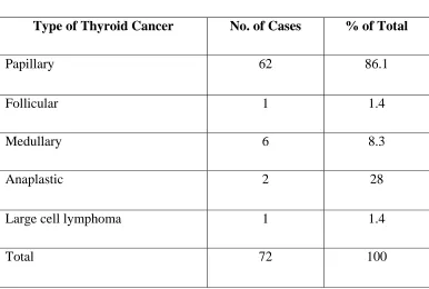
1. Prevalence of the different types of Thyroid Cancer
The prevalence of the different types of thyroid cancers in the study group is as
[image:67.612.116.502.427.687.2]follows:
Table - 1
Type of Thyroid Cancer No. of Cases % of Total
Papillary 62 86.1
Follicular 1 1.4
Medullary 6 8.3
Anaplastic 2 28
Large cell lymphoma 1 1.4
68
Diagram - 1
86.10% 1.40%
8.30% 2.80%
1.40%
Papillary Ca Follicular Medullary Anaplastic
Large cell lymphoma
Papillary Carcinoma (86.1%) is found to be the commonest type of thyroid
69
2. Sex Distribution
[image:69.612.137.509.193.619.2]The sex distribution of thyroid cancers is tabulated below
Table - 2
Sex No. of Cases % of Total
Males 18 25
Females 54 75
Total 72 100
Diagram - 2
25%
75%
Male Females
There is a female preponderance seen in thyroid cancers
70
Table - 3
The sex incidence in different types of thyroid cancers is as follows:
Males Females Type of
thyroid cancer
Number of cases
Percentage Number of cases
Percentage
Total No. of cases
Papillary 16 25.8 46 74.2 62
Follicular - - 1 100 1
Medullary 1 16.67 5 83.33 6
Anaplastic 1 50 1 50 2
Large cell lymphoma
- - 1 100 1
71
Diagram - 3
16
0 1 1 0
46 1 5 1 1 0 5 10 15 20 25 30 35 40 45 50
Papillary Follicular Medullary Anaplastic Lymphoma
N u m b er o f P a ti en ts Males Females
Papillary carcinoma is the commonest type of thyroid cancer seen in teeth
72
3. Age Distribution
[image:72.612.115.521.180.354.2]The age distribution of thyroid cancers is as documented below
Table - 4
Age in years Males Females Total
<10 - - -
11-20 1 7 8
21-30 4 19 23 31-40 1 13 14
41-50 7 6 13
51-60 3 7 10
>60 2 2 4
18 54 72
Diagram - 4
73
The peak incidence of thyroid cancers is between 21-30 years of age.
The peak incidence of thyroid malignancies in males is between 41-50 years of
age and in females is between 21-30 years of age. The average age of
presentation of thyroid cancer is 38.3 years.
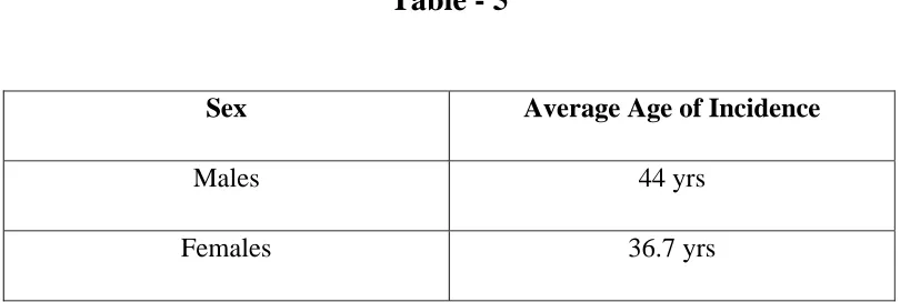
The average age of incidence of thyroid cancers in males and females is as
[image:73.612.114.519.341.477.2]follows
Table - 5
Sex Average Age of Incidence
Males 44 yrs
74
4. Distribution of patients according to age and peak incidence for the different
[image:74.612.118.517.188.406.2]types of thyroid cancers is as follows
Table - 6
Type of cancer Age Range (in years)
Average age of incidence (in years)
Peak incidence (in years)
Papillary 13-78 36.5 21-30
Follicular 55 55 51-60
Medullary 27-60 41.5 31-40
Anaplastic 60-65 62.5 ≥60
Large cell lymphoma
55 55 51-60
Papillary & Medullary carcinomas are seen in the younger age group than the
other types of thyroid cancers
5. Symptomatology
69 cases out of the 72 cases in the study group presented with thyroid
swelling. Regional lymph node metastasis was seen in 25 patients and distant
metastasis in 2 patients. 1 patient had an occult papillary carcinoma (with no
thyroid swelling). 2 patients with papillary carcinoma who had undergone
thyroidectomy previously presented during the study period with cervical nodal
75 The findings noted are tabulated below:
Table - 7
Symptoms Papillary Follicular Medullary Anaplastic Large cell lymphoma
Total Percentage
Thyroid swelling
MNG 31 - 2 2 1 36 50
SNT 28 1 4 - - 33 38.9
Absent 3 - - - - 3 4.2
Dysphagia 2 2 - 2 - 6 8.3
Dyspnea 4 - - 2 - 6 8.3
Hoarseness 2 - - 2 - 4 5.5 Regional
lymph node metastasis
24 - 1 - - 25 34.7
Vocal cord palsy
1 - - 2 - 3 4.2
Distant metastasis
76
Diagram - 5
36 33 3 6 6 4 25 3 2 0 4 8 12 16 20 24 28 32 36 40
MNG SNT No thyroid
swelling
Dysphagia Dyspnea Hoarseness Regional lymph
node metastasis Vocal cord palsy Distant metastasis Symptoms Num b er of Patient s
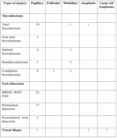
6. Treatment of Thyroid Malignancy
The commonest surgery done for the primary cancer is total
thyroidectomy. 22 out of 25 (88%) patients with palpable cervical nodes were
subjected to Modified radical neck dissection on the involved side (ie the side
with palpable nodes). Bilateral paratracheal dissection was also done in all
these cases. 17 patients out of the 47 patients (36%) without regional lymph
nodes underwent a bilateral paratracheal dissection. 3 patients underwent
trucut biopsy for histopathological confirmation of diagnosis and were treated
by non surgical modalities - the patient with papillary carcinoma was treated
with external beam radiotherapy. The patients with undifferentiated cancer and
lymphoma were treated with radiotherapy and chemotherapy. The observations
77
Table - 8
Types of surgery Papillary Follicular Medullary Anaplastic Large cell lymphoma Thyroidectomy
Total
thyroidectomy
39 1 1
Near total thyroidectomy
2
Subtotal thyroidectomy
4 1
Hemithyroidectomy 7 2
Completion thyroidectomy
9 1 2
Neck Dissection MRND / RND / FND
22
Paratracheal dissection
17
Posterolateral neck dissection
2
78
Diagram - 6
39 2 4 7 9 0 5 10 15 20 25 30 35 40 45
Total Near Total Sub total Hemi Completion
Thyroidectomy
Number
of
Pati
ents
Distribution of patients undergoing thyroidectomies
Diagram - 7
22 17 2 0 5 10 15 20 25
MRND/FND/RND Paratracheal dissection Posterolateral neck dissections
Num
b
e
r of P
a
tien
ts
79
All patients with differentiated thyroid cancers were followed postoperatively
with suppressive doses of thyroxine. The other patients were given replacement
doses of thyroxine.
7. Outcomes of thyroid cancer in the study group
Life expectancy following surgery could not be determined because of
the short duration of the study. 2 patients died during the follow up period-both
80
CONCLUSION
1. Incidence of various types of thyroid cancers correlates approximately
with the world literature except for follicular carcinoma which has a
lower incidence in the study group.
Type of carcinoma % In the study group World literature (%)
Papillary 86.1 80-85
Follicular 1.4 5-10
Medullary 8.3 < 10
Anaplastic 2.8 5
Lymphoma 1.4 < 2
Papillary carcinoma of thyroid is the commonest thyroid malignancy in the
study group. This correlates with the world literature (5). The incidence of
follicular carcinoma is lower than the world literature.
2. Male: Female ratio in the study group is 1:3. This is comparable to the
world literature where male: female ratio is 1:2.5 (5)
3. In the study group, median age at diagnosis for females is 30 years and
81
median age at diagnosis is earlier in females than in males for both
papillary and follicular subtypes (5).
4. Usually 2/3 rd (67%) of patients with differentiated thyroid carcinoma
have disease localized to thyroid at presentation (5). In the study group,
60.3% patients have disease localized to the thyroid.
5. According to known data, 33-61% of patients with papillary thyroid
cancer have metastatic cervical lymphadenopathy at diagnosis (5). In
this study group, 38.7% patients (ie 24 patients out of 62) with papillary
thyroid cancers had metastatic cervical lymph nodes at presentation.
6. 1-2% of patients with papillary thyroid cancer have distant metastasis at
diagnosis (5). In the study group 1.6% of patients (ie 1 patient out of 64
patients) had distant metastasis. This is associated with a very poor
prognosis.
7. The most common symptom at presentation for all thyroid cancers is a
thyroid swelling. 26.3% of patients presented with symptoms of locally
advanced disease.
8. Total thyroidectomy is now advocated for all well differentiated thyroid
cancers and medullary thyroid cancer. Central compartmental dissection
is to be done for all medullary thyroid cancers. Modified Radical Neck
82
lymph nodes. In the study group, total thyroidectomy was the
commonest surgery performed. 22 out of 25 patients (88%) with
palpable cervical lymph node underwent modified radical neck
dissection on the involved side.
9. A combination of radiotherapy and chemotherapy (with adriamycin) is
the most appropriate therapy for non resectable undifferentiated
carcinomas (5). In the study group, one patient with anaplastic
carcinoma underwent a total thyroidectomy supplemented with
postoperative radiotherapy and chemotherapy. The other patient was
treated with radiotherapy and chemotherapy alone.
10.TSH suppressive doses of levothyroxine is recommended for all patients
with papillary and follicular thyroid cancers. This was followed for all
patients in the study group with differentiated thyroid cancers. The goal
of this therapy is serum TSH concentration of 0.1 mU/L or less.
11.Life expectancy of patients following surgery could not be ascertained
due to the short duration of the study. 2 patients died during the follow
up period – both were diagnosed to have anaplastic carcinoma of
83
PROFORMA
Name: Age: Sex:
Inpatient No.: Ward:
I PRESENTING FEATURES
1. Swelling : Side Duration Progress
Any sudden change in size
2. Pressure effects : Dysphagia Dyspnoea
3. Pain
4. Hoarseness of Voice
5. Toxic symptoms
6. Loss of Weight
7. Hypo or Hyper Thyroid Symptoms
8. Any other neck Swelling
II. H/O PREVIOUS SURGERY / IRRADIATION
III. MENSTRUAL AND OBSTETRIC HISTORY
IV FAMILY H/O. DEATH FROM THYROID MALIGNANCY
Pheochromocytoma
Diabetes
Hypertension
84 V. GEN. EXAMINATION:
Pulse BP Anaemia
VI. EXAMINATION OF LUMP
1. Site 2. Size 3. Shape 4. Surface 5. Extent 6. Margins
7. Skin over the swelling 8. Consistency
9. Movement with Deglutition 10.Intrinsic Mobility
11.Fixity
12.Tracheal Position 13.Carotids
14.Mediastinal Extension 15.Auscultation for Bruit 16.Neck Nodes
85 VII. EXAM OF OTHER SYSTEMS
VIII. Laboratory Post operative complication Chest X-Ray Hemorrhage
X-Ray Neck Hypoclacemia
IDL Rec. Laryngeal Nerve Palsy Thyroid scan Respiratory Obstruction FNAC Follow up
USG After every 3 months
Treatment Surgery
86
BIBLIOGRAPHY
1. Bailey and Love’s Short Practice of Surgery, 24th Edition, 2004, 776-804.
2. Baker RJ, Fischer JE, Mastery of Surgery, 4th Edition, 2001, 500-511.
3. Benbassat CA, Mechlis – Frish S. Hirsch D., Clinicopathological characteristics and long term outcome in patients with distant metastasis from differentiated thyroid cancer, World J Surg. 2006 June, 30 (6) : 1088-95.
4. Bilimoria KY, Bentram DJ, Ko CY, Stewart AK, Winchester DP, Talamonti MS, Stugeon C, Extent of Surgery affects survival for Papillary thyroid cancer, Ann Surg. 2007 Sep: 246 (3): 375-384.
5. Devita VT, Cancer – Principles and Practice of Oncology, 7th edition, 2005, 1502-1519.
6. Ganong WF, Review of Medical Physiology, 22nd Edition, 2005, 317-332.
7. Gray’s Anatomy, 39th edition, 2005, 1891-1897.
87
9. Jonklass J., Role of Radioactive Iodine for adjuvant therapy and treatment of metastases J. Natt. Compr Canc. Netw. 2007 Jul; 5 (6): 631-40.
10.Kim S, Wei JP, Braveman JM, Brams DM, Predicting outcome and directing therapy for papillary thyroid carcinoma Arch Surg. 2004 Apr; 139 (4): 390-4, discussion 393-4.
11.Lauster M, Lippi F, Jarzab B, Perros P, Lassmann M, Reiners C, Pacini F, rh TSH aided radioiodine ablation and treatment of differentiated thyroid carcinoma a comprehensive review, Endoer Relat. Cancer 2005 Mar; 12 (1): 49-64.
12.Robbins and Cotran Pathologic Basis of Disease, 7th Edition 2004, 1175-1180.
13.Sabiston Textbook of Surgery, 17th Edition, 2005, 947-986.
14.Schwartz’s Principles of Surgery, 8th Edition, 2005, 1395-1470.
15.Shaha AR, Advances in the management of thyroid cancer. Int. J. Surg 2005; 3(3): 213-20.
16.Williams Textbook of Endocrinology, 10th Edition, 2003, 465-484.