RISK FACTOR ANALYSIS, CLINICAL AND
MICROBIOLOGICAL
PROFILE OF CHILDREN WITH SYMPTOMATIC
OTITIS MEDIA IN A TERTIARY CARE CENTRE
Dissertation submitted to
THE TAMIL NADU DR.M.G.R. MEDICAL UNIVERSITY In partial fulfillment of the regulations for the award of the degree of
M.D. BRANCH – VII PAEDIATRICS
GOVT. STANLEY MEDICAL COLLEGE & HOSPITAL THE TAMIL NADU DR.M.G.R. MEDICAL UNIVERSITY
CHENNAI, INDIA .
CERTIFICATE
This is to certify that the dissertation entitled “RISK FACTOR
ANALYSIS, CLINICAL AND MICROBIOLOGICAL PROFILE OF CHILDREN WITH SYMPTOMATIC OTITIS MEDIA IN A TERTIARY CARE CENTRE” is the bonafide work of
DR.K.INDU PRIYA in partial fulfillment of the requirements for M.D. (PAEDIATRICS) Branch – VII Examination of the Tamilnadu
Dr.M.G.R. Medical University to be held in April 2013.
DEAN DIRECTOR
Govt. Stanley Medical College Institute of Social Paediatrics,
& Hospital, Govt. Stanley Medical College
Chennai – 600 001. & Hospital,
DECLARATION
I, DR.K.INDU PRIYA, solemnly declare that the dissertation
titled, “RISK FACTOR ANALYSIS, CLINICAL AND
MICROBIOLOGICAL PROFILE OF CHILDREN WITH
SYMPTOMATIC OTITIS MEDIA IN A TERTIARY CARE CENTRE” is a bonafide work done by me at Institute of Social
Paediatrics, Govt. Stanley Medical College & Hospital during 2010 -2013
under the guidance and supervision of PROF.DR.G.KARUNAKARAN,
M.D., D.C.H, Director, Institute of Social Paediatrics, Govt. Stanley
Medical College & Hospital, Chennai – 600 001.
The Dissertation is submitted to The Tamilnadu Dr.M.G.R.
Medical University, towards partial fulfillment of requirement for the
award of M.D. Degree (Branch – VII) in Paediatrics.
Place :
ACKNOWLEDGEMENTS
I owe my sincere thanks to the Dean, Prof. Dr. S. Geethalakshmi
M.D.,PhD., Govt. Stanley Medical College & Hospital, for granting me
permission to conduct this study at Institute of Social Paediatrics, Govt.
Stanley Hospital.
I thank our beloved Director, Prof.Dr.G.Karunakaran
M.D.,D.C.H., for guiding me throughout the study.
My sincere thanks to Prof.Dr.Amudha Rajeshwari M.D.,D.C.H.,
Prof.Dr.P.Ambikapathy M.D.,D.C.H. and Prof.Dr.Sujatha Sridharan M.D.,D.C.H. for their immense support for this study.
I also thank Dr.J.Ganesh M.D.,D.C.H. and Dr.M.A.Aravind
M.D. for their valuable guidance and without whose help my study
would not have been possible.
I also thank our Assistant Professors Dr. Rathinavelu
M.D.,D.C.H., Dr.K.Elango M.D.,D.C.H., Dr.T.S.Ekambaranath M.D., Dr.Raja M.D., Dr.Kumar D.C.H., Dr.V.Radhika M.D., Dr.Venkatesan M.D., and Dr.Ezhil Srinivasan M.D.,D.C.H. for their
critical reviews and suggestions.
I offer my special thanks to Mr.Ravanan, Associate professor of
statistics, Presidency college for helping me in statistical analysis.
I also thank our ENT department and Microbiology department
I am greatly indebted to all my friends, Post Graduate colleagues
who have been the greatest source of encouragement, support, enthusiasm,
friendly concern and timely help.
Last but not the least I owe my sincere thanks and gratitude to all
the children and their parents without whom this study would not have
been possible.
CONTENTS
Serial No.
INTRODUCTION
Otitis media is a common childhood infection especially in
developing countries. Otitis media refers to the inflammation of the
middle ear mucosa. About 80% of children experience at least one
1. Introduction 1
2. Review of Literature 2
3. Aims of the study 32
4. Materials & Methods 33
5. Observations & Results 35
6. Discussion & Analysis 76
7. Limitations 81
8. Conclusion 82
9. Bibliography -
10. Annexures
Profoma
Master Chart
Key to Master Chart
Abbreviations
-
-
-
-
episode of otitis media within their first 3 years of life (2,3) . It is the reason for every third hospital visit in a pediatric practice. Serious complications
can occur from otitis media. It is the commonest cause of preventable and
treatable hearing loss. Long term hearing loss can have serious impact on
language and communication, psychosocial and cognitive development
and academic performance of the child.
Identifying the predictors and avoiding them and treating the
infection with appropriate antibiotics prevent complications and results in
good outcome. Good knowledge and understanding of the host and the
environmental factors for development of otitis media is important in
identifying a child at risk of recurrent and persistent otitis media. This
helps in primary and secondary prevention of otitis media and decreasing
its complications and sequelae.
This study is done to analyse the
risk factors associated with acute and chronic otitis media and to
find the spectrum of organisms causing otitis media to enable
prevention and appropriate treatment respectively.
REVIEW OF LITERATURE
RISK FACTORS FOR OTITIS MEDIA 1 ) Age :
The incidence of otitis media is highest between 6 – 20 months of
age (1,4). 63% – 85% develop at least one episode of otitis media by 1 year and 66% - 99% by 2 years. Poor immunity, structural and functional
deficiencies of the Eustachian tube results in increased occurrence of
otitis media in infancy and young children. Earlier the age of onset of
otitis media, greater is the risk for the occurrence of recurrent and chronic
otitis media.
2 ) Gender :
The incidence is more in boys compared to girls. Surgeries like
tympanostomy tube insertion, tympanoplasty and adenoidectomy are
more in boys suggesting a greater severity in boys.
3) Socioeconomic class:
Otitis media is common among children belonging to low
socioeconomic class(5). Overcrowding, poor hygienic facilities, suboptimal nutritional status, limited access to medical care and limited
resources for complying with prescribed medications makes the children
in low socioeconomic class susceptible to otitis media.
4 ) Breast milk Vs Formula feeds(6,7,8):
Exclusive breast feeding for 6 months provides protection against
than to the mechanics of breast feeding. Nursing in incorrect positions
like supine nursing increases the risk of otitis media(9,10). There is some controversy regarding the exact duration of breast feeding that provides
protection against otitis media. One study showed after breast feeding was
stopped, there was decreased risk for occurrence of otitis media for upto 4
months. After about 12 months of stopping breast feeding, the odds of
occurrence of otitis media was same between the children who were
breast fed and the children who were not breast fed(11).
5 ) Use of Pacifiers :
Pacifier usage had an influence on otitis media and affected the
number of episodes of occurrence acute otitis media. A meta - analysis
linked pacifier use with 24% increased risk for acute otitis media(12).
6 ) Passive Smoking :
Many studies were conducted to find out the association between
passive smoking and occurrence of otitis media. Some studies assessed
exposure to tobacco smoking using urinary cotinine and some using
salivary cotinine levels. Two meta - analysis showed that passive smoking
increased the risk for recurrent acute otitis media and chronic otitis media
with effusion. There was not a significant increase in risk for non
recurrent acute otitis media with passive smoking(12,13).
7 ) Exposure to other children :
There was an increase in occurrence of acute otitis media and
chronic otitis media with effusion in children belonging to large families.
Order of birth had an influence on otitis media(14). Children who were first
born had lower incidence of acute otitis media compared to children who
had elder siblings. There is an increased nasopharyngeal colonization
with repeated exposure to other children.
8) Upper airway infections :
Upper airway infection plays a significant role in the etiology of
otitis media. Upper airway infection causes inflammation and damage to
the mucociliary epithelial lining of the Eustachian tube predisposing the
child to otitis media(15).
9) Seasonal influence :
The incidence of otitis media is more during the autumn and winter
months and less in summer in both hemispheres(16). This parallels with the occurrence of the upper airway infection. This evidence also supports the
fact that upper airway infection predisposes otitis media.
Congenital anomalies like unrepaired cleft palate, submucosal cleft
palate, other craniofacial anomalies and Downs syndrome have deficiency
in functioning of Eustachian tube and predispose to OM.
ACUTE OTITIS MEDIA
According to Nelson Textbook of Pediatrics,19th edition, “ The diagnosis of acute otitis media requires
1) Acute onset of signs and symptoms
2) Presence of middle ear effusion
3) Signs and symptoms of middle ear inflammation
Definition of acute otitis media includes
- Recent, usually abrupt onset of signs and symptoms of middle ear
inflammation and middle ear effusion
- Presence of middle ear effusion, indicated by any of the following
- Bulging tympanic membrane
- Limited or absent mobility of tympanic membrane
- Air fluid level behind the tympanic membrane
- Otorrhoea
- Signs and symptoms of middle ear inflammation indicated by either
- Distinct erythema of tympanic membrane
- Distinct otalgia ( discomfort clearly referable to the ears
that results in interference with or precludes normal activity
ETIOLOGY :
Bacteria are isolated in 65 – 75 % of cases from the middle ear
fluid. Numerous studies show that there are 3 common organisms causing
acute otitis media namely Streptococcus pneumoniae, non typable
Hemophilus influenza and Moraxella catarrhalis(17,18). Streptococcus pnuemoniae has been isolated from the middle ear fluid in 25% - 50% of
the cases with acute otitis media, Hemophilus influenza in 15% - 30% and
Moraxella catarrhalis in 3% - 20%. Staphylococcus aureus, group A
Streptococcus gram negative organisms have also been implicated as the
causative agent in about 5% of the cases.
There is a change in the microbiological flora with the increased
pneumococcal vaccinations. Block et al study shows that “the incidence of
Hemophilus influenza has increased from 39% to 52% and the incidence
of Streptococcus pneumonia has decreased from 49% to 34% in the
isolates in children between 7 to 24 months with acute otitis media
between 1992 – 1998 and 2000-2003”(19).
Viruses like respiratory syncytial virus, rhinovirus, coronavirus,
Para influenza, adenovirus and enterovirus have been isolated from
respiratory secretions and middle ear effusion from about 405 to 75%
cases of acute otitis media and about 5% to 22% of cases without bacteria
in middle ear effusion. This might be responsible for the failure of
is limited to favoring bacterial invasion, amplifying inflammatory process
and interfering with resolution of bacterial infection. In about 16% to
25% no bacteria or virus has been isolated (20,21).
PATHOGENESIS :
Eustachian tube, child’s immune system, risk factor profile and
host pathogen interaction play a role in pathogenesis (22). Anatomical factors :
Eustachian tube is closed passively and opened by the contraction
of tensor veli palatii. It has 3 main functions – ventilation, protection and
clearance of middle ear. Tubal obstruction elicits a complex inflammatory
response - secretory metaplasia, mucociliary transport system
compromise and effusion into tympanic cavity. Eustachian tube
obstruction can occur extraluminally ( from hypertrophied
nasopharyngeal adenoid tissue or tumor ) or intraluminally ( from
inflammatory edema of tubal mucosa ).
Progressive reduction in compliance as the child grows is the
reason for the decrease in occurrence of OM as age advances. Patulous or
excessively compliant eustachian tube does not provide protection to the
tympanic cavity from the spread of infection from the nasopharynx. The
shorter or more horizontal orientation of the Eustachian tube in infants
and children increases the reflux from nasopharynx and reduces the
In children with craniofacial anomalies like cleft palate and
children with Downs syndrome, there is an increased occurrence of otitis
media. This is also attributed to the Eustachian tube dysfunction.
Host factors :
The level of immunity of the child plays an important role in the
occurrence otitis media. As the child grows, the immune system matures
and hence there is a decrease in the incidence of otitis media in older
children. IgA deficiency has been noted in some children with recurrent
otitis media. But its role is doubtful since IgA deficiency is also found in
children without recurrent otitis media. Selective IgG subclass deficiency
may be found in children with recurrent AOM in association with
recurrent sinopulmonary infections.
Allergy:
Respiratory allergy as an etiology is not definite. It is possible that
otitis media may be exaggerated by allergy(23). Alteration in mucociliary clearance by repeated viral exposure or tobacco smoke slide the balance
of pathogenesis in favour of pathogens.
CLINICAL FEATURES :
Acute otitis media is associated with abrupt onset of signs and
pulling of the ear, incessant crying and altered sleep habits. Otorrhoea and
fever can occur. The specificity and sensitivity for pulling at the ear is
low. Hearing loss may be present which may manifest as change in
speech pattern. These findings except for otorrhoea are not specific and
usually overlaps with that of uncomplicated upper respiratory
infections(24).
Otoscopy:
Visualization of the tympanic membrane with identification of an
middle ear effusion and inflammatory changes is necessary to establish
the diagnosis with certainty. For pneumatic otoscopy, a speculum of
proper shape and diameter must be selected to permit a seal in the
external auditory canal. Appropriate restraint of the child to provide
adequate examination is necessary.
The findings on otoscopy indicating the presence of middle ear
effusion and inflammation associated with acute otitis media has been
well defined. Fullness or bulging of the tympanic membrane is often
present and has the highest predictive value for the presence of middle ear
effusion. When combined with colour and mobility, bulging is also the
best predictor of acute otitis media(25,26).
Reduced or absent mobility of the tympanic membrane during the
performance of pneumatic otoscopy is additional evidence of fluid in the
is also a consistent finding and is caused by the edema of the tympanic
membrane. Redness of the tympanic membrane caused by inflammation
may be present and must be differentiated from the pink erythematous
flushing evoked by crying or high fever, which is usually less intense and
remits as the child calms down. In bullous myringitis, blisters may be
seen on the tympanic membrane(27). When the presence of middle ear fluid is difficult to determine, the use of tympanometry or acoustic
reflectometry can be helpful in establishing a diagnosis.
TREATMENT
Management of otalgia
Many episodes of acute otitis media are associated with pain(28).The management of pain especially during the first 24 hours of an episode of
acute otitis media, should be addressed regardless of the use of
antibacterial agents.
Acetaminophen and ibuprofen are effective drugs in cases of mild
to moderate pain(29). In children with moderate to severe pain, narcotic analgesics with codeine or analogs can be used. Distraction and external
application of heat or cold have also been tried for pain relief.
Tympanostomy and myringotomy procedures are used in cases of severe
pain and they require a lot of technical expertise.
Antimicrobial treatment :
Though previously acute otitis media was routinely treated with
antibiotics, the emergence of bacterial resistance to these drugs has
prompted withholding antibiotics in some cases.The three main reasons
which were cited as favouring the use of antimicrobials are majority of
otitis media are caused by bacteria; a faster resolution of infection in
those treated with antibiotics and early treatment with antibiotics prevents
suppurative complications.
The likelihood of recovery without antibacterial therapy differs
depending on the severity of signs and symptoms at the initial
examination. Kaleida et al(30) “ divided patients into severe and non-severe
groups based on the degree of fever , a scoring system based on duration
and severity of pain and apparent discomfort and estimated parental
anxiety. In the non-severe group of children, the initial failure rate on
placebo plus myringotomy was 23.5% versus an initial failure rate of
9.6% on patients with amoxicillin alone”. Palva et al cited “Routine
antibacterial therapy for acute otitis media is often cited as the main
reason for the decrease in the incidence of mastoiditis in the antibacterial
era”(31). By 1950s, mastoiditis has decreased dramatically.
The AHRQ evidence report on acute otitis media concluded that
mastoiditis is not increased with initial observation, provided the children
are followed closely and antibiotics are started in those who do not
showed comparable rates of mastoiditis in children who received initial
antibacterial treatment and children who received placebo or observation.
External validity may be limited because some trials excluded very young
children and those with severe disease(32). Thus current evidence does not suggest a clinically increased risk of mastoiditis in children when acute
otitis media are managed only with initial symptomatic treatment without
antibacterial agents, Clinicians should be aware that antibacterial
treatment might mask the symptoms and signs of mastoiditis producing a
subtle presentation that can delay diagnosis.
Bacterial Resistance :
Children < 2 years of age, who are constantly exposed to other
children and who have received antibiotics recently are more prone for
development of antibacterial resistance. The degree of resistance varies
from place to place. Approximately 40% of Hemophilus influenza and all
strains of Moraxella catarrhalis are resistant to aminopenicillins. The
resistance is mainly due to the production of β lactamases. It can be
overcome by using a β lactamase inhibitor. Some strains confer resistance
not by the production of β lactamases but by the alteration of penicillin
binding proteins.
About 50% of Streptococcus pneumonia are resistant to penicillins.
Nearly half show intermediate resistance and the remaining half are
due to the alteration in penicillin binding proteins, There are about 6
penicillin binding proteins. More the number of proteins are altered, the
higher the degree of resistance. This resistance can be overcome by the
use of higher concentration of β lactam antibiotics for sufficient periods.
Penicillin resistant Streptococcus are mostly resistant to other classes of
antibiotics also.
Resistance to macrolides occurs by two mechanisms. One is
mediated by mef (A) gene which is due to an efflux pump. It decreases
the intracellular concentration of macrolides. It confers low level
resistance. The other mechanism is due to the erm (B) gene which
produces ribosomal methylases and modifies ribosomal RNA. This
mechanism is also responsible for the resistance against clindamycin.
Unlike β lactam antibiotics, resistance to macrolides cannot be overcome
by increasing the concentration of the drug.
Guidelines for treatment :
A consensus guidelines has been published by the American
Academy of Pediatrics as to who should have a period of “ watchful
waiting “ or observation and who should be treated. It is essential to
ensure the follow up of patients to assess for non - resolution or
worsening of the problem. The consensus takes into consideration three
factors – age of the patient, the certainty of diagnosis and the severity of
2 years and ≥ 2 years group. The presence of the three criteria i) rapid
onset, ii) signs of middle ear effusion and iii) symptoms and signs of
middle ear inflammation makes the diagnosis of acute otitis media certain.
Patients with temperature of > 102 F ( > 39 C ), severe otalgia or toxic
appearance are considered to have severe disease.
In children < 6 months of age, irrespective of the certainty of
diagnosis or the severity of the disease, all should be treated with
appropriate antibiotics because of the increased risk of morbidities due to
complications. In children between 6 months to 2 years, antibiotics are
started in cases of certain diagnosis or when there is severe disease. The
child is observed for 48 to 72 hours in cases of non-severe disease. In
children ≥ 2 years, all episodes are treated with a period of observation
except in confirmed cases of acute otitis media with severe disease.
Table 1: Treatment based on age and certainty of diagnosis
AGE CERTAIN DIAGNOSIS UNCERTAIN DIAGNOSIS
< 6 months Antibacterial treatment Antibacterial treatment
6 moon – 2 yrs. Antibacterial treatment Antibacterial treatment of severe
Observation if non severe illness
≥ 2 yrs. Antibacterial treatment of severe illness
Observation if non severe illness
Observation
First line drugs:
Amoxicillin is usually considered as the first choice drug due to its
good efficacy and safety, low cost and palatability (33). Both penicillin susceptible and non-susceptible Streptococcus pneumonia respond to
Amoxicillin. This is achieved by increasing the traditional dose of
Amoxicillin from 40 – 45 mg/kg/day to 80 – 90 mg/kg/day(34). One
limitation of use of Amoxicillin is that it is not efficient against β
lactamase producing strains of non typable Hemophilus influenza and
Moraxella catarrhalis. With the widespread coverage pneumococcal
vaccine and decline in S.pneumoniae and rise of H.influenzae, the above
factor has become important.
Allergic reactions can occur to penicillin which can be either type 1
with urticarial and angioedema or non-type 1 with rashes. For non-type 1
reactions, where there is no cross reaction with cephalosporins, cefdinir is
the antibiotic of choice(35). For patients with type 1 reaction, azithromycin is the drug of choice.
Duration of treatment :
Treatment for atleast 10 days is essential especially in children less
than 2 years of age. In older children with mild episodes and who show
rapid improvement, a 3 to 5 day course may be sufficient. Longer than 10
days treatment may be needed in very young children, in severe disease
and in children with previous problematic episode of otitis media.
Unsatisfactory response :
Children who are prescribed antibiotics should improve within 48
to 72 hours. If there is no improvement, it is possible that the treatment
was not adequate or there is a different diagnosis. Poor compliance to
treatment, ineffective antibiotics, concurrent or intercurrent viral
infections, poor host immunity, Eustachian tube dysfunction or
reinfection from other sites are some of the causes for failure to respond
to treatment.
Middle ear effusion can persist even after improvement of acute
symptoms. 60 – 70 % have middle ear effusion after 2 weeks of acute
otitis media, 40 % at 1 month and 10 – 25 % at 3 months(36). It needs further follow up and observation. The presence of middle ear effusion
following acute otitis media is not an indication for addition of antibiotics
or changing to second line of drugs.
Second line drugs:
The second line of drugs must be effective against β lactamase
susceptible and non-susceptible strains of S.pneumoniae. Amoxicillin
clavulanate, cefdinir, cefuroxime axetil and intramuscular ceftriaxone are
the drugs which meet the above requirements. With the addition of
clavulanate component, the spectrum of amoxicillin is extended to β
lactamase producing organisms and forms an ideal second line drug.
Cefdinir can be give as an once daily regimen and is well tolerated. One
limitation to the use of cefuroxime is that it is not palatable. Intramuscular
ceftriaxone is used when oral therapy is not possible, when there is no
response to oral second line antibiotics and when highly resistant strains
of Streptococcus are isolated from the aspirate from diagnostic
tympanocentesis(37).
Azithromycin and clarithromycin have only limited action against β
lactamase strains of H.influenzae and non-susceptible strains of
S.pneumoniae . Macrolide use is more a problem of resistance than
beneficial effects. Clindamycin is active against most of the strains of
S.pneumoniae but they are not active against H.influenzae and
M.catarrhalis.
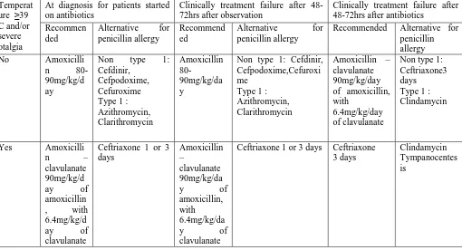
Table 2 : Recommendations for antibacterial treatment
Temperat ure ≥39 C and/or severe otalgia
At diagnosis for patients started on antibiotics
Clinically treatment failure after 48-72hrs after observation
Clinically treatment failure after 48-72hrs after antibiotics
Recommen ded
Alternative for penicillin allergy
Recommend ed
Alternative for penicillin allergy
Recommended Alternative for penicillin allergy No Amoxicilli
n 80-90mg/kg/d ay
Non type 1: Cefdinir,
Cefpodoxime, Cefuroxime Type 1 : Azithromycin, Clarithromycin Amoxicillin 80-90mg/kg/da y
Non type 1: Cefdinir, Cefpodoxime,Cefuroxi me
Type 1 : Azithromycin, Clarithromycin
Amoxicillin – clavulanate 90mg/kg/day of amoxicillin, with
6.4mg/kg/day of clavulanate
Non type 1: Ceftriaxone3 days
Type 1 : Clindamycin
Yes Amoxicilli n – clavulanate 90mg/kg/d ay of amoxicillin , with 6.4mg/kg/d ay of clavulanate
Ceftriaxone 1 or 3 days
Amoxicillin –
clavulanate 90mg/kg/da y of amoxicillin, with 6.4mg/kg/da y of clavulanate
Ceftriaxone 1 or 3 days Ceftriaxone 3 days
Clindamycin Tympanocentes is
Figure 1 : Algorithm for management of Acute otitis media
Uncomplicated AOM in children 2 mon – 12 yrs
Is pain present ?
Myringotomy and Tympanocentesis :
Indications :
1) Severe refractory pain
2) Hyperpyrexia Analgesics
Start
Amoxicillin 80-90mg/kg/day Is fever ≥39 C and/or
moderate or severe otalgia present ?
Is observation appropriate ?
Start appropriate antibiotics Observation for 48 – 72
hrs and follow up
Is there response to initial treatment ( either antibiotics or observation )
Reassess and confirm the diagnosis of AOM Follow up
Is diagnosis of AOM
confirmed ? Assess for other
causes of illness & manage
Start antibiotics for children managed initially with observation and change antibiotics if already started on antibiotics Yes
No No
Yes
Yes
No
Yes
3) Complications of AOM like labyrinthitis, facial palsy, mastoiditis
and intracranial infection
4) Immunological compromise from any source
In patients with no response to second line drugs or in very young patients
as a part of sepsis work up, diagnostic tympanocentesis or myringotomy
can help in identifying the causative organism.
CHRONIC SUPPURATIVE OTITIS MEDIA
Chronic suppurative otitis media is a long standing infection of a
a permanent perforation.It usually begins in childhood as acute otitis
media. When squamous epithelium lines the edges of the perforation, it
does not spontaneously close and becomes permanent. Children with
CSOM have ear discharge from 6 weeks to 3 months or more inspite of
medications. WHO definition requires only 2 weeks of otorrhoea. but
otolaryngologists usually adopt a longer period.
CAUSATIVE ORGANISMS :
Chronic suppurative otitis media is mostly caused by Pseudomonas
aeruguinosa, Proteus mirabilis, Klebsiella species , Escherichia coli and
anaerobes like Bacteriodes, Peptostreptococcus and Propionibacterium(38).
A chronic perforation paves way for the entry of bacteria into the middle
ear. Pseudomonas is particularly very notorious resulting in progressive
destruction by the proteolytic enzymes.
TYPES OF CSOM :
It is clinically divided into 2 types.
1) Tubotympanic type :
It is also called the safe ear. It involves the anteroinferior part of the
middle ear cleft and causes central perforation.
2) Atticoantral type :
It is also called the unsafe ear. It involves the posterosuperior part
Features Tubotympanic type Atticoantral type
Discharge Profuse, mucoid, odourless Scanty, purulent , foul
smelling
Perforation Central Attic or marginal
Granulation Uncommon Common
Polyp Pale Red or fleshy
Cholesteatoma Absent Present
Complications Rare Common
Audiogram Mild to moderate
conductive deafness
Conductive or mixed
deafness
ETIOLOGY :
1) Sequelae to Acute otitis media :
Acute otitis media can result in a large central perforation. The
perforation may become permanent and results in recurrent infection from
the external ear. When the middle ear mucosa is exposed to the external
environment, it gets sensitized to dust, pollen, etc and results in persistent
otorrhoea.
Infection from the infected sinuses, tonsils and adenoids can spread
to the middle ear cleft through the Eustachian tube and result in otitis
media.
HEARING LOSS:
Tubotympanic disease usually results in conductive hearing loss.
Hearing loss ranges from none to 50dB. Sometimes a paradoxical effect
of better hearing in the presence of discharge than when the ear is dry. It
is due to the round window shielding effect by the discharge. In a dry ear
with perforation, when sound waves strike the round window and oval
window at the same time, they get cancelled.
When CSOM is present for longer periods, toxins are absorbed
through the round window and oval window and causes cochlear damage.
This can result in mixed type of hearing loss.
Assessment of hearing :
a) Clinical tests :
i) Watch test :
It was used as a screening test in the pre audiometric era. A
clicking watch is brought from a distance towards the ear. The distance
when the clicking of the clock is heard is measured.
ii) Speech test :
Normally a person can hear a whispered voice at a distance of 6
metres and a conversation at a distance of 12 metres. In this test, the
patient is made to stand at a distance of 6 metres with his ear to be tested
facing the examiner. The patient is blind folded to avoid lip reading and
the other ear is blocked by intermittent tragal pressure. The examiner
recites words and starts walking towards the patient. The distance at
which the patient hears a whisper and normal conversational voice is
noted. The intensity and pitch of voice and the surrounding noise cannot
be standardized in this test.
iii) Tuning fork tests :
Tuning fork of 512 Hz is ideal for hearing assessment. Tuning
forks having lesser frequencies gives a sense of bone vibration and those
with greater frequencies have a short decay time. These tests are based on
the fact that in a normal ear, air conduction is better than bone conduction.
Rinne test :
Here air conduction is compared with bone conduction. A tuning
fork is made to vibrate and placed on the mastoid. When the patient stops
hearing, the tuning fork is placed adjacent to the external auditory meatus.
If the patient is able to hear, it is implied that air conduction is better than
bone conduction and Rinne test is positive. When the bone conduction is
better than air conduction, it is a negative Rinne test. A negative Rinne is
Table 3: Assessment of hearing
RESULT INFERENCE
Positive Rinne AC > BC Normal ear
Sensorineural hearing loss
Negative Rinne BC > AC Conductive hearing loss
False negative Rinne BC > AC Unilateral severe
sensorineural hearing loss
Weber test:
In this test, a tuning fork is vibrated and placed in the center of the
forehead. Here there is direct stimulation of the cochlea through the bone.
The patient is asked to tell in which ear he hears the sound better. In a
normal ear, it is heard equally in both ears. When there is conductive
hearing loss, it is lateralized to the diseased ear and in sensorineural
hearing loss; the test is lateralized to the normal ear. Lateralization
indicates that there is either a conductive hearing loss of 15 to 20 dB in
the ipsilateral ear or a sensorineural hearing loss of 15 to 20 dB in the
contralateral ear.
Absolute bone conduction test :
In this test, the bone conduction of the patient is compared with that
of the examiner. The meatus of both the patient and examiner are
occluded. When the patient hears the tuning fork for the same duration as
the examiner, then there is conductive hearing loss. When the patient
hears the tuning fork for less duration than the examiner, it implies that
there is sensorineural hearing loss.
b) Audiometry
i) Pure tone audiometry;
This device produces pure tones. The intensity of the pure tone
can be increased or decreased by 5 dB. The amount of intensity that has
to be increased higher than the normal indicates the degree of
hearing impairment at that frequency. The air bone gap is a measure of
the amount of conductive hearing loss.
ii) Impedance audiometry:
This test is an objective test. It includes tympanometry and acoustic
reflex measurements. It is based on the fact that when a sound wave falls
on the tympanic membrane, a part of it gets absorbed and the remaining is
reflected. A tympanic membrane which is stiff can reflect more sound
waves when compared to a more compliant tympanic membrane. By
sealing the external ear and adjusting the pressures and measuring the
sound that is reflected, the compliance of the tympanic membrane can be
against the pressures and a tympanogram with different types of graph is
obtained. It helps in diagnosing some middle ear pathologies.
Types of tympanogram
Type A - Normal
Type As - Reduced compliance at ambient pressures ( osteosclerosis )
Type Ad - Increased compliance at ambient pressures ( ossicular
discontinuity )
Type B - Flat or dome shaped ( fluid in middle ear )
Type C - Maximum compliance at pressures more than 100 mmH2O
( negative pressure in middle ear )
INVESTIGATIONS :
1)Audiogram
2) Pus for culture and sensitivity
3) X ray of the mastoids
TREATMENT :
Aims of treatment :
1) To control the infection and make the ear dry
2) Complete eradication of the disease
3) Restoration of hearing
Aural toileting can be either done by dry mopping or with the help
of absorbent cotton buds. It can be done with suction clearance using a
microsope or irrigation. Removal of the discharge can help by removing
the infected material and also improves the efficacy of topical antibiotics
(39)
. A Cochrane review from studies in Solomon Islands(40) and Kenya(41) showed that there was no significant benefits with aural toilet alone
compared to no treatment.
ii) Topical antiseptics:
A trial by Eaton et al (40) showed that use of topical antiseptics was more effective than aural toilet alone. Zinc peroxide, boric acid, iodine
powder and dilute acetic acid are some of the topical antiseptics which are
reported in literature.
iii) Antibiotics:
According to a Cochrane review, combining antibiotics with aural
toilet is more efficient than aural toilet alone(42). It is a question of debate whether to use topical antibiotics or to use systemic antibiotics.
Ludman(39) and Nelson(43) preferred oral antibiotics. They cited the
potential ototoxic effects due to the use of topical antibiotics as a major
concern.
With the above stand by most pediatricians, most otolaryngologists
prefer topical antibiotics due to the poor penetration of the systemic drugs
found that topical drugs were better than systemic drugs in decreasing the
otorrhoea and clearing the middle ear disease(45). Some of the topical antibiotics which are used are gentamycin, tobramycin, chloramphenicol,
ciprofloxacin, ofloxacin and polymyxin B. Cephalexin, amoxicillin,
coamoxiclav, erythromycin, ciprofloxacin, ofloxacin and trimethoprim
sulfamethoxazole are some of the systemic drugs used. There are studies
to support that combined topical and systemic antibiotics did not produce
better results than topical antibiotics alone(40). But the risk of ototoxicity is still a concern. Much of what is known about the ototoxicity of topical
drugs is mostly based on animal studies (46).
Administration of parenteral antibiotics had good results than aural
toilet alone(47). Parenteral antibiotics used for CSOM are(48) penicillins ( ampicillin, penicillin G, piperacillin, ticarcillin ), cephalosporins
( cefotaxime, cefuroxime, cefoperazone, ceftazidime, cefazolin ),
aminoglycosides ( gentamycin, amikacin ) , clindamycin, vancomycin and
aztreonam.
iv) Treatment of contributing conditions:
In order to eliminate the trigger and source of infection, conditions
like allergies, sinusitis, adenoiditis and tonsillitis must be treated.
a ) Myringoplasty :
Closure of the perforation of the pars tensa of the tympanic
membrane is called myringoplasty. It helps in restoring hearing and
prevents re infection from the external auditory canal. It also prevents the
aeroallergens from the external environment from reaching the middle ear
mucosa.
b) Tympanoplasty:
When myringoplasty is combined with ossicular reconstruction, it
is called tympanoplasty. Depending on the amount of damage to the
ossicular chain, specific type of tympanoplasty is done. Sequential
destruction of malleus, incus and stapes results in progressively medially
placed graft.
c) Mastoidectomy:
1) Cortical Mastoidectomy:
It is also called Intact canal wall mastoidectomy. Here the posterior
meatal wall which separates the middle ear and the mastoid cavity is
preserved. An opening is made through the posterior canal wall for entry
into the middle ear cavity, In this procedure, anatomy of the middle ear is
conserved and restoration of hearing is possible through tympanoplasty.
But this technique needs expertise and there is the risk of recurrent or
residual disease due to limited access to middle ear .
2) Radical Mastoidectomy :
It is also called Canal wall down procedure. Here the posterior
meatal wall is removed and middle ear, attic, antrum and mastoid is made
into a single cavity and exteriorized. This procedure aims to eradicate the
disease from the middle ear and mastoid without attempts at preserving
middle ear anatomy for reconstruction of hearing.
3) Modified Radical Mastoidectomy:
Here as much of the hearing mechanism as possible is preserved. It
is done when the cholesteatoma is confined to the attic and antrum and in
localized chronic otitis media.
COMPLICATIONS:
They can be classified into intratemporal complications and
intracranial complications.
Intratemporal Complications
1) Mastoiditis
2) Petrositis
3) Facial paralysis
4) Labyrinthitis
Intracranial Complications
1) Extradural abscess
2) Subdural abscess
3) Meningitis
4) Brain abscess
5) Lateral sinus thrombhophlebitis
6) Otitic hydrocephalus
Features indicating complications:
1) Pain – It is usually not seen in uncomplicated cases. When present, it
may indicate extradural, perisinus or brain abscess. It can also be due to
otitis externa.
2) Vertigo – It is because of the erosion of lateral semicircular canal. It
can lead to labyrinthitis or meningitis. Fistula test should be done.
3) Persistent headache – Occurs when there is intracranial complications.
4) Facial weakness – It due to the erosion of the facial canal.
5) Fever, nausea and vomiting – It occurs when there is intracranial
spread of infection.
6) Irritability and neck rigidity is a feature of meningitis.
7) Diplopia – Feature of Gradinego syndrome.
8) Ataxia – It is suggestive of labyrinthitis or cerebellar abscess.
9) Abscess around the ear – It is a feature of mastoiditis.
AIMS OF THE STUDY
1) To analyse the risk factors in children < 12 years with symptomatic
otitis media in a tertiary care centre
2) To study the clinical profile of children < 12 years with
symptomatic otitis media in a tertiary care centre
3) To identify the organisms causing otitis media and their sensitivity
patterns in a tertiary care centre
MATERIALS AND METHODS
Type of study:
Prospective descriptive study with Case control analysis of risk
factors
Place of study:
Tertiary care hospital
Period of study:
October, 2011 to September, 2012
Inclusion criteria for acute otitis media:
1) Acute onset of signs and symptoms
2) Presence of middle ear effusion
(Indicated by any of the following )
- Bulging TM
- Air fluid level behind the TM
- Otorrhoea
3) Signs and symptoms of middle ear inflammation
- Distinct erythema of TM
- Distinct otalgia ( discomfort clearly referable to the ears that
results in interference with or preclude normal activity or sleep )
Inclusion criteria for chronic otitis media:
Persistent ear discharge for > 3 weeks
Method of study:
Children fulfilling the inclusion criteria are selected. Informed
written consent is obtained from the parents and they are included in the
study. Detailed history using the questionnaire and clinical examination
was carried out. Otological examination was done using an otoscope.
Hearing assessment was done using Rinnes test and Weber test. Aural
swab is taken and sent for culture and sensitivity. Cultures were done for
bacteria – both aerobes and anaerobes and fungus. Aerobes were cultured
using blood agar, chocolate agar and Mac Conkey’s agar. Anaerobes were
cultured with Robertson cooked meat media. Sabourad’s dextrose agar
was used for isolation of fungus. X ray of the paranasal sinuses and
mastoid were taken for children with CSOM with clinical suspicion of
sinusitis or mastoiditis.
Age and sex matched controls for the cases were randomly chosen
from the children brought for immunization and other complaints
excluding respiratory symptoms. The same questionnaire pertaining to the
risk factors was asked to the care givers of the controls. The data were
entered and analyzed using
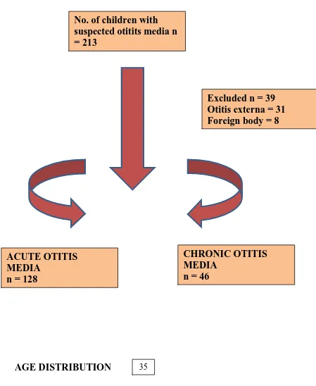
About 213 children were suspected to have otitis media. Of them,
39 cases were excluded – 31 cases had otitis externa and 8 cases had
foreign body. A total of 128 cases of Acute suppurative otitis media and
[image:43.595.87.536.230.778.2]46 cases of Chronic suppurative otitis media were included in the study.
Figure 2: Inclusion of cases for AOM and CSOM
AGE DISTRIBUTION
No. of children with suspected otitits media n = 213
CHRONIC OTITIS MEDIA
n = 46
Excluded n = 39 Otitis externa = 31 Foreign body = 8
ACUTE OTITIS MEDIA
n = 128
Of the 128 cases of ASOM, 11 cases were < 1 year (8.6%), 88 were
between 1 – 5 years of age (68.8%) and 29 were > 5 years (22.7%). Of the
46 cases of CSOM, 11 cases were between 1 – 5 years of age (23.9%) and
35 were > 5 years of age (76.1%)
Table 4: Age Distribution of ASOM and CSOM
Chart 1: Age
Distribution of ASOM and CSOM
AGE ASOM CSOM
NO. % NO. %
< 1 year 11 8.6% 0 0
1 – 5 yrs. 88 68.8% 11 23.9%
> 5 yrs. 29 22.7% 35 76.1%
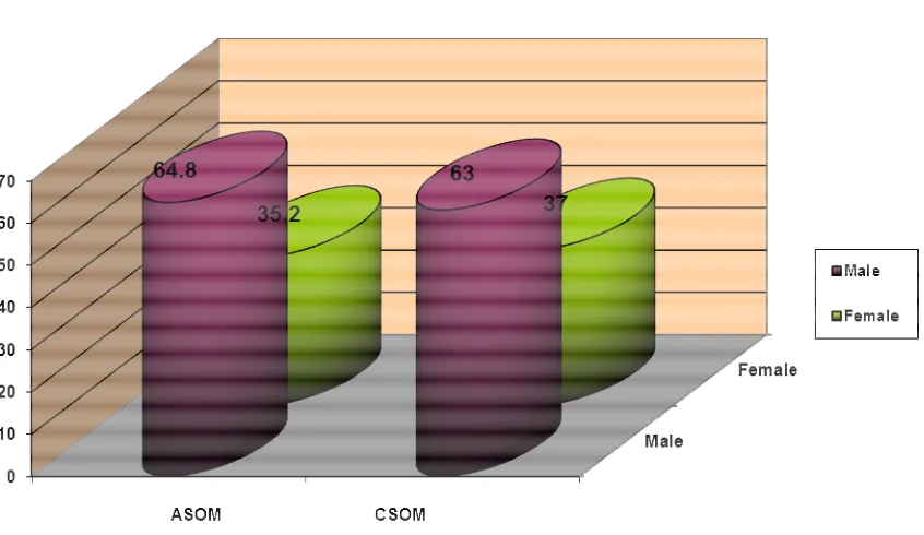
SEX DISTRIBUTION
Of the 128 cases of ASOM, 83 were male (64.8%) and 45 were
female (35.2%). Of the 46 cases of CSOM, 29 cases were male (63%) and
[image:45.595.132.555.450.695.2]17 were female (37%).
Table 5: Sex Distribution
SEX ASOM CSOM
NO. % NO. %
Male 83 64.8% 29 63%
Female 45 35.2% 17 37%
Chart 2: Sex Distribution
ACUTE SUPPURATIVE OTITIS MEDIA
RISK FACTOR ANALYSIS
The following risk factors were analyzed – urban residence,
siblings, passive smoking, low socioeconomic class, bottle feeding, not
exclusively breast feeding for 6 months, supine nursing and bad practices.
Table 6: Risk factor analysis
S.NO. RISK FACTOR NO. PERCENTAGE
1. Urban Residence 103 80.5%
2. Siblings 96 75%
3. Passive Smoking 92 71.9%
4. Low Socioeconomic class 99 77.3%
5. Bottle feeding 88 68.8%
6. Not exclusively breast fed 85 66.4%
7. Supine nursing 79 61.7%
8. Bad practices 99 77.3%
URBAN / RURAL DIVIDE:
Among the cases, 103 hailed from urban area (80.5%) and 25
hailed from rural area (19.5%). Among the controls, 95 hailed from urban
area (74.2%) and 33 hailed from rural area (25.8%).
Table 7: Urban rural distribution
Chart 4: Urban rural distribution
The difference was statistically not significant. CASES
n = 126
CONTROLS
n = 126
SIGNIFICANCE
p Value
URBAN 103 ( 80.5% ) 95 ( 74.2% ) 1.427 0.232
RURAL 25 ( 19.5% ) 33 ( 25.8% )
SIBLINGS:
Among the cases, 96 had siblings (75%) and 32 had no siblings
(25%). Among the controls, 62 had siblings (48.4%) and 66 had no
siblings (51.6%).
Table 8: Siblings
SIBLINGS CASES
n = 126
CONTROLS
n = 126
SIGNIFICANCE
p Value
YES 96 ( 75% ) 62 ( 48.4% ) 19.112 <0.001
NO 32 ( 25% ) 66 ( 51.6% )
Chart 5: Siblings
The difference was statistically significant with a p value of < 0.001.
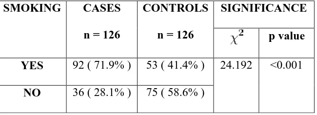
PASSIVE SMOKING:
92 cases were exposed to paternal smoking (71.9%) as against 53
of the controls (41.4%). 36 cases were not exposed paternal smoking
[image:50.595.156.481.243.361.2](28.1%) as against 75 of the controls (58.6%).
Table 9: Passive smoking
Chart 6: Exposure to smoking
The difference was statistically significant with a p value of < 0.001.
SMOKING CASES
n = 126
CONTROLS
n = 126
SIGNIFICANCE
p value
YES 92 ( 71.9% ) 53 ( 41.4% ) 24.192 <0.001
NO 36 ( 28.1% ) 75 ( 58.6% )
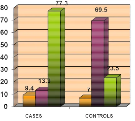
SOCIOECONOMIC CLASS
12 children (9.4%) belonged to class III socioeconomic class as per
modified Kuppuswamy classification among the cases as against 9 ( 7% )
among controls. 17 ( 13.3% ) belonged to class IV SEC among the cases
compared to 89 ( 69.5% ) in the control group. 99 ( 77.3% ) of the cases
[image:51.595.208.425.492.686.2]belonged to class V SEC as against 30 ( 23.5% ) among the controls.
Table 10: Socioeconomic class distribution
Chart 7: Socioeconomic class distribution
The difference was statistically significant with a p value of < 0.001
SEC CASES
n = 126
CONTROLS
n = 126
SIGNIFICANCE
p value
III 12 ( 9.4% ) 9 ( 7% ) 86.241 <0.001
IV 17 ( 13.3% ) 89 ( 69.5% )
V 99 ( 77.3% ) 30 ( 23.5% )
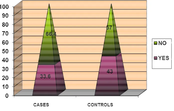
EXCLUSIVE BREAST FEEDING
43 of the cases (33.6%) were exclusively breast fed for 6 months
compared to 55 controls ( 43% ).
[image:52.595.143.492.465.689.2]The difference was not statistically significant.
Table 11: Exclusive breast feeding
Chart 8: Exclusive breast feeding
EBF CASES
n = 126
CONTROLS
n = 126
SIGINIFICANCE
p value
YES 43 ( 33.6% ) 55 (43% )
2.381 0.123
NO 85 ( 66.4% ) 73 (57%)
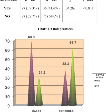
BOTTLE FEEDS
Among the cases 88 children were bottle fed (68.8%) while among
the controls only 49 children were bottle fed (38.3%).
Table 12: Practice of bottle feeding
Chart 9: Practice of bottle feeding
The difference was statistically significant. BOTTLE
FEEDS
CASES
n = 126
CONTROLS
n = 126
SIGNIFICANCE
p value
YES 88 ( 68.8% ) 49( 38.3% ) 23.884 < 0.001
NO 40 ( 31.2% ) 79( 61.7% )
SUPINE NURSING:
The mothers of 79 cases (61.7%) practiced upright nursing as
against the mothers of 48 controls ( 37.5% ).
[image:54.595.143.385.445.619.2]The difference was statistically significant with p value of < 0.001
Table 13: Practice of upright nursing
Chart 10: Practice of upright nursing UPRIGHT
NURSING
CASES
n = 126
CONTROLS
n = 126
SIGNIFICANCE
p value
YES 79 ( 61.7% ) 48 (37.5% ) 15.017 < 0.001
NO 49 ( 38.3% ) 80 ( 62.5% )
BAD PRACTICES
The mothers of 99 cases ( 77.3% ) practiced nose blowing , oil
instillation into the nose, use of pacifiers and other bad practices
compared to the mothers of 55 controls ( 41.4% ) .
[image:55.595.122.494.305.697.2]The difference was statistically significant with p value of < 0.001
Table 14: Bad practices
Chart 11: Bad practices BAD
PRACTICES
CASES
n = 126
CONTROLS
n = 126
SIGNIFICANCE
p value
YES 99 ( 77.3% ) 55 (41.4% ) 34.267 < 0.001
NO 29 ( 22.7% ) 75 ( 58.6% )
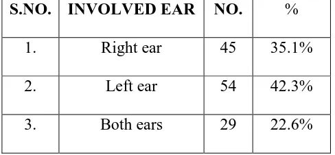
SYMPTOMATOLOGY Laterality
Among the 128 cases, 29 cases (22.6%) had discharge in both ears,
45 cases (35.1%) had discharge in right ear and the remaining 54 cases
[image:56.595.195.439.273.386.2](42.3%) had discharge in left ear.
Table 15: Laterality of ear involvement
Chart 12: Laterality of ear involvement
S.NO. INVOLVED EAR NO. %
1. Right ear 45 35.1%
2. Left ear 54 42.3%
3. Both ears 29 22.6%
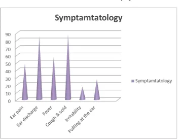
62 cases (48.4%) had complaints of ear pain and 109 cases (85.2%)
had ear discharge. Fever was present in 74 cases ( 57.8% ), cough and
cold in 112 cases ( 87.5% ), irritability in 22 cases ( 17.2% ) and pulling
[image:57.595.108.459.462.736.2]at the ear in 35 cases ( 27.3% ) .
Table 16: Profile of symptoms
Chart 13: Profile of symptoms
S.NO. SYMPTOMS NO. %
1. Ear pain 62 48.4%
2. Ear discharge 109 85.2%
3. Fever 74 57.8%
4. Cough &cold 112 87.5%
5. Irritability 22 17.2%
6. Pulling at the ear 35 27.3%
SIGNS
Erythema of the tympanic membrane was seen in 85 cases
( 66.4% ), otorrhoea was seen in 65 children ( 50.8% ), bulging of the
tympanic membrane was seen in 8 cases ( 6.2% ), perforation was seen in
26 cases ( 20.3% ) and otitis extern in 31 cases ( 24.29% ). All the
[image:58.595.204.430.300.483.2]children with perforation has tubotympanic type of central perforation.
Table 17 : Profile of signs
Chart 14: Profile of signs
S.NO. SIGNS NO. %
1. Erythema –TM 85 66.4%
2. Otorrhoea 65 50.8%
3. Bulging Tm 8 6.2%
4. Perf0ration 26 20.3%
5. Otitis Externa 31 24.29%
ASSOCIATED CONDITIONS
In 66 cases (51.6%) tonsillitis was present. Previous history of
otitis media was seen in 87 cases (68%), history of measles was present in
55 cases (43%) and history of upper respiratory infection was seen in 110
[image:59.595.211.423.263.433.2]cases (85.9%).
Table 18: Profile of associated conditions
Chart 15: Profile of associated conditions ASSOCIATED
CONDITIONS
NO. %
Tonsillitis 66 51.6%
Previous H/O OM 87 68%
H/O Measles 55 43%
H/O URTI 110 85..9%
ORGANISMS
The aural swab from the children with acute otitis media showed no
growth in majority of the cases – 57 children (47.5%). Staphylococcus
aureus growth was seen in 37 cases (28.9%), Coagulase negative
staphylococcus aureus in 19 cases (14.8%), Pseudomonas in 10 children
[image:60.595.215.419.298.468.2](7.8%) and Klebsiella growth in 5 cases (3.9%).
Table 19: Organisms in aural swab
Chart 16: Organisms in aural swab
S.NO. ORGANISMS NO. %
1. No Growth 57 44.5%
2. Staphylococcus 37 28.9%
3. Pseudomonas 10 7.8%
4. Klebsiella 5 3.9%
5. CONS 19 14.8%
CULTURE AND SENSITIVITY PATTERNS STAPHYLOCOCCUS AUREUS
Among the 37 isolates of Staphylococcus aureus, 75.7% were
sensitive to Ampicillin and Amoxicillin, 67.5% to Cefotaxime, 40.5% to
Erythromycin, 32.4% to Amikacin, 27% to Ciprofloxacin and
Norfloxacin, 59.4% to Cotrimoxazole, 8% to Gentamycin, 62.2% to
Cephalexin and 24.3% of the isolates were sensitive to Vancomycin.
Chart 17: Sensitivity pattern of Staphylococcus aureus
COAGULASE NEGATIVE STAPHYLOCOCCUS AUREUS
Of the 19 isolates of Coagulase negative staphylococcus aureus,
100% were sensitive to Ampicillin and Amoxicillin, 78.9% to Cefotaxime,
63.1% to Erythromycin, 42.1% to Amikacin and Cotrimoxazole, 52.6% to
Ciprofloxacin, 31.5% to Norfloxacin, 26.3% to Gentamycin, 31.5% to
Cephalexin and 21% were sensitive to Vancomycin.
Chart 18: Sensitivity pattern of Coagulase negative staphylococcus aureus
PSEUDOMONAS
Of the 10 isolates of Pseudomonas, 60% were sensitive to
Ampicillin, Norfloxacin and Amoxicillin, 30% to Cefotaxime and
Gentamycin, 40% to Amikacin, 80% to Ciprofloxacin and 20% to
Cotrimoxazole.
Chart 19: Sensitivity pattern of Pseudomonas
KLEBSIELLA
Among the 5 isolates of Klebsiella, 40% were sensitive to
Ampicillin, Amoxicillin and Cephalexin, 60% to Cefotaxime,
Ciprofloxacin and Norfloxacin, 100% to Amikacin and 80% were
sensitive to Cotrimoxazole and Gentamycin.
Chart 20: Sensitivity pattern of Coagulase negative staphylococcus aureus
LOGISTIC REGRESSION ANALYSIS
By multivariate logistic regression analysis, low socioeconomic
class, presence of siblings and supine nursing were found to be significant
[image:65.595.141.491.509.613.2]risk factors for acute otitis media.
Table 20: Logistic regression analysis of risk factors for AOM
Table 21: Classification table
Observed Predicted
Group
Percentage Correct
Control Cases
Group Control 99 29 77.3
Cases 27 101 78.9
Overall Percentage 78.1
78.9% of the cases and 77.3% of the controls were correctly classified.
B S.E. Wald df Sig. Exp(B)
95.0% C.I.for EXP(B)
Lower Upper
AGE .101 .303 .110 1 .740 1.106 .611 2.003 SEX .354 .332 1.137 1 .286 1.424 .744 2.728 AREA -.317 .389 .663 1 .415 .729 .340 1.561 SIBLINGS .950 .378 6.311 1 .012 2.586 1.232 5.428 SMOKING .796 .432 3.401 1 .065 2.218 .951 5.170 SEC 1.492 .261 32.728 1 .000 4.447 2.667 7.414 EBF -.429 .324 1.750 1 .186 .651 .345 1.229 BOTTLE FEEDING .710 .411 2.989 1 .084 2.034 .909 4.548 SUPINE NURSING 1.092 .327 11.155 1 .001 2.981 1.570 5.658 BAD PRACTICES .567 .478 1.408 1 .235 1.764 .691 4.501 Constant -6.100 1.282 22.624 1 .000 .002
CHRONIC SUPPURATIVE OTITIS MEDIA
RISK FACTOR ANALYSIS
The following risk factors were analyzed for chronic suppurative
otitis media – urban residence, presence of siblings, passive smoking and
[image:66.595.107.525.463.759.2]socioeconomic class.
Table 22: Risk factor analysis for CSOM
S.NO. RISK FACTOR NO. %
1. Urban residence 40 87%
2. Siblings 32 69.6%
3. Passive smoking 37 80.4%
4. Low socioeconomic class 18 39.1%
Chart 21: Risk factor analysis for CSOM
URBAN / RURAL DIVIDE:
Among the cases, 40 hailed from urban area (87%) and 6 hailed
from rural area (13%). Among the controls, 34 hailed from urban area
(73.9%) and 12 hailed from rural area (26.1%).
[image:67.595.138.437.522.687.2]The difference was not statistically significant.
Table 23: Urban rural distribution in CSOM
Chart 22: Urban rural distribution in CSOM CASES
n = 46
CONTROLS
n = 46
SIGNIFICANCE
p value
URBAN 40 ( 87% ) 34 ( 73.9% ) 2.486 0.115
RURAL 6 ( 13% ) 12 ( 26.1% )
SIBLINGS:
Among the cases, 32 had siblings (69.6%) and 14 had no siblings
(30.4%). Among the controls, 11 had siblings (23.9%) and 35 had no
siblings (76.1%).
[image:68.595.155.482.265.383.2]The difference was statistically significant with a p value < 0.001
Table 24: Sibling
Chart 23: Sibling
SIBLINGS CASES
n = 46
CONTROLS
n = 46
SIGNIFICANCE
p value
YES 32 ( 69.6% ) 11 ( 23.9% ) 19.256 < 0.001
NO 14 ( 30.4% ) 35 ( 76.1% )
PASSIVE SMOKING:
37 cases were exposed to paternal smoking (80.4%) as against 19
of the controls (41.3%). 9 cases were not exposed paternal smoking
(19.6%) as against 27 of the controls (58.7%).
[image:69.595.147.490.265.383.2]The difference was statistically significant with a p value < 0.001
Table 25: Exposure to smoking
Chart 24: Exposure to smoking
SMOKING CASES
n = 46
CONTROLS
n = 46
SIGNIFICANCE
p value
YES 37 ( 80.4% ) 19 ( 41.3% ) 14.786 < 0.001
NO 9 ( 19.6% ) 27 ( 58.7% )
SOCIOECONOMIC CLASS
5 children (10.9%) belonged to class III socioeconomic class as per
modified Kuppuswamy classification among the cases as against 11
( 23.9% ) among controls. 23 ( 50% ) belonged to class IV SEC among
the cases compared to 25( 54.3% ) in the control group. 18 ( 39.1% ) of
the cases belonged to class V SEC as against 10 ( 21.7% ) among the
controls.
[image:70.595.170.462.364.508.2]The difference was statistically not significant
Table 26: Socioeconomic class distribution among CSOM
Chart 25: Socioeconomic class distribution
SEC CASES
n = 46
CONTROLS
n = 46
SIGNIFICANCE
p value
III 5( 10.9% ) 11 ( 23.9% ) 4.619 0.099
IV 23 ( 50% ) 25 ( 54.3% )
V 18 ( 39.1% ) 10 ( 21.7% )
10.9
23.9
50 54.3
39.1
21.7
SEC III SEC IV SEC V
SYMPTAMATOLOGY LATERALITY
There was right ear involvement in 11 cases (23.9%), left ear
involvement in 8 cases (17.5%) and both ears were involved in 27 cases
[image:71.595.188.446.259.374.2]constituting about 58.6%.
Table 27: Laterality of involvement
S.NO. EAR INVOLVED NO. %
1. Right ear 11 23.9%
2. Left ear 8 17.5%
3. Both ears 27 58.6%
Chart 26: Laterality of involvement
Laterality
Right ear
Left ear
Both ears
All the 46 cases (100 %) presented with otorrhoea. 17 patients
( 37 % ) had ear pain, 23 ( 50% ) had fever and 34 cases ( 73.9% ) had
symptoms of upper respiratory infection. 8 children ( 17.3 % ) complained
[image:72.595.108.525.489.723.2]of hard of hearing. 2 patients ( 4.3 % ) had retroauricular pain.
Table 28: Symptoms of Chronic otitis medi
Chart 27: Symptoms of Chronic otitis medi
S.NO. SYMPTOMS NO. %
1. Ear pain 17 37%
2. Ear discharge 46 100%
3. Fever 23 50%
4. Cough & cold 34 73.9%
5. Hard of hearing 8 17.3%
6. Retroauricular pain 2 4.3%
SIGNS
All the 46 cases (100 %) had perforation and otorrhoea. 16 cases
( 34.8% ) had otitis externa, 5 cases ( 10.8% ) conductive hearing loss, 3
( 6.5% ) had sensorineural hearing loss and 2 ( 4.3% ) had mastoid
[image:73.595.114.521.246.751.2]tenderness.
Table 29: Distribution of signs among CSOM
Chart 28: Distribution of signs
S.NO. SIGNS NO. %
1. Otorrhoea 46 100%
2. Perforation 46 100%
3. Otitis externa 16 34.8%
4. Hearing loss 8 17.3%
5. Maxillary sinus tenderness 9 19.5%
6. Mastoid tenderness 2 4.3%
OTORRHOEA
Among the 35 children who had otorrhoea, 5 cases ( 10.9 % ) had
serous discharge, 17 children ( 37% ) had mucoid discharge and 24 cases
[image:74.595.109.527.413.659.2]( 52.1% ) had mucopurulent discharge.
Table 30 : Types of discharge
S.NO. DISCHARGE NO. %
1. Serous 5 10.9%
2. Mucoid 17 37%
3. Mucopurulent 24 52.1%
Chart 29 : Types of discharge
PERFORATION
41 cases with chronic suppurative otitis media had central
perforation which constituted to 89% and the remaining 5 cases had
[image:75.595.108.529.185.607.2]atticoantral perforation constituting about 11%.
Table 31 : Types of perforation
S.NO. PERFORATION NO. %