0022-538X/95/$04.0010
Copyrightq1995, American Society for Microbiology
Replicative Function and Neutralization Sensitivity of Envelope
Glycoproteins from Primary and T-Cell Line-Passaged
Human Immunodeficiency Virus Type 1 Isolates
NANCY SULLIVAN,1,2YING SUN,1JOHN LI,1WOLFGANG HOFMANN,1
ANDJOSEPH SODROSKI1,2*
Division of Human Retrovirology, Dana-Farber Cancer Institute, Department of Pathology, Harvard Medical School,1
and Department of Cancer Biology, Harvard School of Public Health,2Boston, Massachusetts 02115
Received 9 December 1994/Accepted 28 March 1995
The structure, replicative properties, and sensitivity to neutralization by soluble CD4 and monoclonal antibodies were examined for molecularly cloned envelope glycoproteins derived from human immunodefi-ciency virus type 1 (HIV-1) viruses either isolated directly from patients or passaged in T-cell lines. Comple-mentation of virus entry into peripheral blood mononuclear cell targets by primary patient envelope glyco-proteins exhibited efficiencies ranging from that observed for the HXBc2 envelope glycoglyco-proteins, which are derived from a T-cell line-passaged virus, to approximately fivefold-lower values. The ability of the envelope glycoproteins to complement virus entry roughly correlated with sensitivity to neutralization by soluble CD4. Laboratory-adapted viruses were sensitive to neutralization by monoclonal antibodies directed against the CD4-binding site and the third variable (V3) loop of the gp120 glycoprotein. By comparison, viruses with envelope glycoproteins from primary patient isolates exhibited decreased sensitivity to neutralization by these monoclonal antibodies; for these viruses, neutralization sensitivity correlated with replicative ability. Subin-hibitory concentrations of soluble CD4 and a CD4-binding site-directed antibody significantly enhanced the entry of viruses containing envelope glycoproteins from some primary patient isolates. The sensitivity of viruses containing the different envelope glycoproteins to neutralization by soluble CD4 or monoclonal antibodies could be predicted by assays dependent on the binding of the inhibitory molecule to the oligomeric envelope glycoprotein complex but less well by assays measuring binding to the monomeric gp120 glycoprotein. These results indicate that the intrinsic structure of the oligomeric envelope glycoprotein complex of primary HIV-1 isolates, while often less than optimal with respect to the mediation of early events in virus replication, allows a relative degree of resistance to neutralizing antibodies. The interplay of selective forces for higher virus replication efficiency and resistance to neutralizing antibodies could explain the temporal course described for the in vivo emergence of HIV-1 isolates with differing phenotypes.
Human immunodeficiency virus type 1 (HIV-1) is the etio-logic agent of AIDS (8, 25). Following acute HIV-1 infection, in which high levels of viremia are observed, a vigorous anti-viral immune response, including both neutralizing antibodies and cytotoxic T lymphocytes, is generated. HIV-1 loads typi-cally decrease at this time, but virus replication persists (16, 31, 46). Concomitantly, depletion of CD4-positive lymphocytes oc-curs, eventually resulting in immune system compromise (15, 19, 33). During this phase of viral persistence, HIV-1 variants resistant to neutralization by autologous sera emerge (1, 3, 4, 37, 57). Eventually, the level of replicating virus increases, portending the development of a clinically significant immu-nocompromised state.
Primary HIV-1 isolates, which by definition have been de-rived directly from patient tissue or have been propagated only in peripheral blood mononuclear cells (PBMC), have been classified into two groups on the basis of virus replication kinetics (6, 12, 20, 21, 26, 53). During early infection and the long, clinically asymptomatic phase of HIV-1 infection, most isolates exhibit ‘‘slow, low’’ replication kinetics in PBMC. These isolates do not replicate efficiently in established T-cell lines and do not efficiently form syncytia in these cells. During late HIV-1 infection, often immediately prior to or
concomi-tant with clinical immunocompromise, HIV-1 variants with ‘‘rapid, high’’ replication kinetics can be isolated (51). A subset of these rapid, high primary viruses can establish infection and form syncytia in T-cell lines, thus becoming laboratory adapted. Laboratory adaptation is associated with loss of the ability of viruses to replicate in primary monocyte-derived macrophages. The properties of the primary and laboratory-adapted HIV-1 strains are determined by the envelope glyco-proteins and are related to the efficiency with which virus entry into the target cell occurs (11, 22, 42, 59). The interaction of the envelope glycoproteins of different HIV-1 isolates with the CD4 viral receptor appears to differ, since primary viruses are resistant to a soluble form of CD4 (sCD4), in contrast to laboratory-passaged isolates (18, 28). Differences in the affinity of sCD4, apparent only in the context of the oligomeric enve-lope glycoproteins, for primary and laboratory-passaged HIV-1 isolates have been suggested as an explanation of this relative resistance (5, 32, 39, 41, 45, 58, 60).
The relationship of several of the neutralization targets on the HIV-1 envelope glycoproteins to the CD4-binding site (54, 56, 63) suggests the possibility that the resistance of primary isolates to neutralization by sCD4 reflects structural features of these viruses relevant to antibody neutralization. Indeed, pri-mary viruses have been suggested to be less sensitive than laboratory-adapted viruses to neutralization by sera from HIV-1-infected patients or vaccine recipients, but whether assay-specific variables influenced this outcome is unknown (3, 4, 36, 50, 62). Here, we examine the sensitivity of recombinant
vi-* Corresponding author. Mailing address: Jimmy Fund 824, Dana-Farber Cancer Institute, 44 Binney St., Boston, MA 02115. Phone: (617) 632-3371. Fax: (617) 632-4338. Electronic mail address: Joseph_ [email protected].
4413
on November 9, 2019 by guest
http://jvi.asm.org/
ruses that contain the envelope glycoproteins derived from molecular clones of primary and laboratory-adapted HIV-1 isolates to neutralization by sCD4 and monoclonal antibodies. This single-step replication system allows precise quantitation of neutralization sensitivity under conditions where variables unrelated to envelope glycoprotein structure are eliminated.
(This research was conducted by N. Sullivan in partial ful-fillment of the requirements for an S.D. from the Harvard School of Public Health.)
MATERIALS AND METHODS
Antibodies and sCD4.The F105 antibody (47) was provided by Marshall Posner. The Fab b12 antibody fragment (10) was provided by Dennis Burton and Carlos Barbas III. Antibody 1121 and sCD4 were purchased from AGMED, Inc., Bedford, Mass.
Cells and cell lines.Human PBMC were prepared by Ficoll gradient separa-tion, stimulated with phytohemagglutinin for 48 to 72 h, and maintained in RPMI 1640 medium supplemented with 10% fetal bovine serum and 5% interleukin-2. COS-1 cells were grown in Dulbecco’s modified Eagle’s medium containing 10% fetal bovine serum. The T-cell lines Molt 4 clone 8 and SupT1 were maintained in RPMI 1640 medium containing 10% fetal bovine serum.
Envelope expressor plasmids.The 89.6 and YU2 molecular clones were gifts from Ronald Collman and the AIDS Research and Reference Reagent Program (source, Beatrice Hahn), respectively. ADA and MN subclones were provided by Lee Ratner and Philip Berman, respectively. The SV-A-MLV-Env plasmid ex-pressing the amphotropic murine leukemia virus envelope glycoproteins was obtained from Dan Littman (32b). To create the ADA and MN envelope ex-pressor plasmids, the KpnI(6347)-BsmI(8054) fragment of the pSVIIIenv plas-mid (30) was replaced with the equivalent fragments from ADA and MN. YU-2 and 89.6 envelope glycoprotein expressors were made by introducing a BamHI site into each of the envelope genes by PCR and substituting the KpnI(6347)-BamHI(8475) fragment of the pSVIIIenv plasmid with the amplified fragment. Envelope glycoprotein expression and CD4-binding assay.COS-1 cells were transfected by the DEAE-dextran method with pSVIIIenv DNA expressing en-velope glycoproteins as described previously (17, 30). To measure protein ex-pression, the cells were radiolabeled with [35
S]cysteine overnight and precipi-tated with an excess of a mixture of sera derived from patients with AIDS (30). To measure the CD4-binding ability of mutant glycoproteins, supernatants of radiolabeled transfected cells were incubated with an excess of SupT1 lympho-cytes at 378C for 90 min (44). The cells were washed twice with phosphate-buffered saline (PBS), lysed, and immunoprecipitated as described above. The amount of gp120 precipitated from both bound and unbound fractions was measured by densitometric analysis of autoradiograms from sodium dodecyl sulfate (SDS)-polyacrylamide gels.
Determination of envelope glycoprotein concentration on the virion.COS-1 cells cotransfected with the envelope-expressing plasmid and the pHXBDenv CAT plasmid (30) were labeled with [35S]cysteine (70mCi/ml) and [35
S]methi-onine (100mCi/ml) overnight. Supernatants were centrifuged at 8003g for 10 min to remove cell debris, layered over a 20% sucrose cushion, and centrifuged for 90 min at 27,000 rpm in a Beckman SW28 rotor. The supernatants were completely removed, and the viral pellet was lysed in 53RIPA buffer (0.15 M NaCl, 0.05 M Tris-HCl [pH 7.2], 0.1% SDS, 1% Triton X-100, 1% sodium deoxycholate) and immunoprecipitated as described. The ratio of the gp120 glycoprotein to p55gagprotein associated with the pelleted virions was assessed by
densitometry of autoradiograms from SDS-polyacrylamide gels.
Virus neutralization assay.Complementation of a single round of replication of the env-deficient chloramphenicol acetyltransferase (CAT)-expressing provi-rus by the various envelope glycoproteins was performed as described previously (30, 54). To inhibit viral replication, either monoclonal antibody or sCD4 was incubated with recombinant virus for 1 h at 378C before addition of the virus to target lymphocytes. At 3 days after infection, the target cells were lysed and CAT activity was measured as described previously (30). The standard deviation in this assay was experimentally determined and was less than 10% of the mean (data not shown).
For the cocultivation neutralization assay, virus-producing COS-1 cells were treated with mitomycin (100mg/ml) for 30 min at 378C, washed three times with serum-free RPMI 1640, and incubated with monoclonal antibody for 90 min at 378C. Target Molt 4 clone 8 cells were cocultivated with the COS-1 cells for 60 min, separated from the COS-1 cells, and cultured for an additional 10 to 14 days. Molt 4 clone 8 cells were then lysed, and CAT activity was measured as described previously (30). The SV-A-MLV-Env plasmid expressing the ampho-tropic envelope glycoproteins was used to complement the pHXBDenvCAT virus as a control in some of the F105 antibody neutralization experiments.
Binding of antibodies to envelope glycoproteins expressed on the surface of COS-1 cells.The binding of the antibodies to the HIV-1 envelope glycoprotein complex expressed on the surface of transfected COS-1 cells was measured as described previously (48, 63). Briefly, COS-1 cells were transfected with pSVIII-env DNA expressing pSVIII-envelope glycoproteins. The cells were labeled overnight
with [35S]cysteine 2 days after transfection and washed once with PBS containing
2% fetal bovine serum, and monoclonal antibodies were incubated with the cells for 90 min at 378C in PBS. In parallel, cells were incubated with a 1:300 dilution of a mixture of sera from patients with AIDS. After three washes with PBS–2% fetal bovine serum, the cells were lysed in Nonidet P-40 buffer, and bound antibody-envelope glycoprotein complexes were precipitated with protein A-Sepharose (Pharmacia) and analyzed by SDS-polyacrylamide gel electrophoresis. Scanning densitometry was performed to determine antibody binding relative to that measured with a mixture of sera from patients with AIDS.
Binding of antibodies to soluble envelope glycoproteins.To analyze the bind-ing of monoclonal antibodies to monomeric gp120 envelope glycoproteins, COS-1 cells were transfected with envelope glycoprotein expressor plasmids and labeled with [35S]cysteine 2 days after transfection. Radioactively labeled cell
supernatants were harvested 12 to 16 h later. Binding of monoclonal antibodies to soluble gp120 in the supernatants was assessed by immunoprecipitation, and the amount bound was determined by SDS-polyacrylamide gel electrophoresis followed by scanning densitometry. Parallel immunoprecipitations with a mixture of sera from patients with AIDS were performed to control for the amount of the different gp120 glycoproteins present in the COS-1 supernatants.
RESULTS
Virion-associated gp120 glycoproteins.Five envelope glyco-proteins (HXBc2, MN, 89.6, ADA, and YU2) were chosen for this study. The HXBc2 and MN envelope glycoproteins were derived from HIV-1 isolates adapted to replicate on estab-lished T-cell lines (laboratory-adapted viruses) (23, 25, 52). The 89.6, ADA, and YU2 glycoproteins were derived either from viruses that had been passaged only in human PBMC (89.6 and ADA) or by direct cloning of env sequences from the tissue of an HIV-1-infected individual (YU2) (14, 27, 35). The 89.6, ADA, and YU2 viruses thus represent primary isolates. Plasmids expressing the env alleles of these viruses were fected into COS-1 cells. Immunoprecipitation of the trans-fected COS-1 cell lysates with a mixture of sera from patients with AIDS revealed the presence of the gp160 envelope gly-coprotein precursor, which was efficiently processed to the gp120 and gp41 envelope glycoprotein in all five cases (data not shown).
It has been previously shown that the density of envelope glycoproteins present on virion particles is higher for primary HIV-1 isolates than for laboratory-adapted viruses (39, 43, 60, 61). To examine whether the ADA, YU2, and 89.6 envelope glycoproteins produced in COS-1 cells retained this character-istic, the envelope glycoprotein expressor plasmids were
co-transfected into COS-1 cells with the pHXBDenvCAT plasmid.
The pHXBDenvCAT plasmid contains an HIV-1 provirus with
a deletion in the env gene and a gene encoding CAT replacing the nef gene (30). Radiolabeled virion particles produced by the transfected COS-1 cells were pelleted, lysed, and precipi-tated by a mixture of sera from patients with AIDS. The ratio of the gp120 envelope glycoprotein to the p55 gag protein was two to three times higher for the primary virus-derived enve-lope glycoproteins than for HXBc2 or MN enveenve-lope proteins (Fig. 1; Table 1). Thus, primary virus envelope glycoproteins produced in this expression system exhibit higher virion spike density, which is probably related to the affinity of the gp120-gp41 interaction.
Ability of envelope glycoproteins to complement HIV-1 entry into cells. The ability of the envelope glycoproteins derived from the different viruses to complement the entry of the
env-defective virus produced from the pHXBDenvCAT
plas-mid was examined. COS-1 cells were cotransfected with the
pHXBDenvCAT plasmid and the envelope
glycoprotein-ex-pressing plasmids, and an equal number of reverse tran-scriptase units of recombinant viruses in the cell supernatants was incubated with target cells. CAT activity in the target cells was measured 3 days after infection (Table 1). When human PBMC were used as target cells, the HXBc2 and 89.6 envelope glycoproteins exhibited the ability to complement entry
effi-4414 SULLIVAN ET AL. J. VIROL.
on November 9, 2019 by guest
http://jvi.asm.org/
ciently. The ADA and YU2 envelope glycoproteins were less efficient in this assay.
While primary HIV-1 isolates do not necessarily replicate efficiently in established T-cell lines, the T-cell line Molt 4 clone 8 has been reported to support the replication of mac-rophage-tropic and T-cell line-tropic viruses (24). Therefore, the ability of the envelope glycoproteins to mediate entry into the Molt 4 clone 8 cells was examined. Table 1 indicates that all of the envelope glycoproteins tested displayed an ability to complement HIV-1 entry into Molt 4 clone 8 cells and did so with the same rank order of efficiency as that observed with human PBMC as target cells. Since Molt 4 clone 8 cells do not exhibit the variability associated with different preparations of
human PBMC, we used Molt 4 clone 8 lymphocytes as target cells for subsequent experiments in this system.
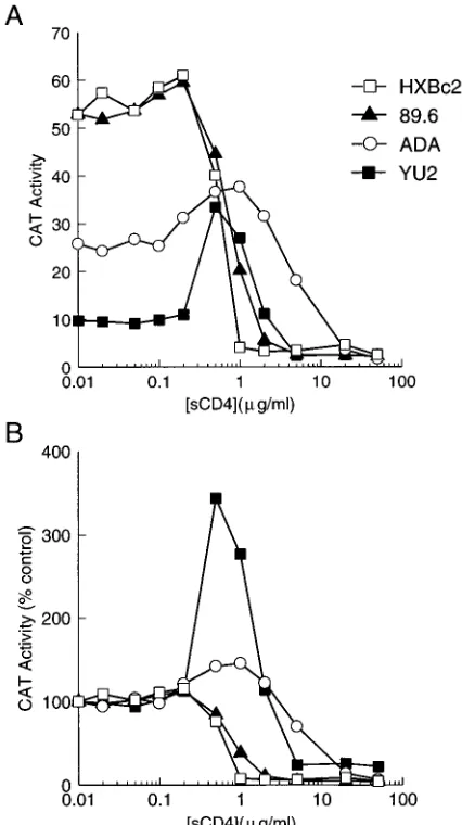
Neutralization by soluble CD4. Some primary HIV-1 iso-lates have been shown to exhibit a decreased sensitivity to sCD4 relative to that exhibited by viruses passaged on T-cell lines (18, 28). To examine the sensitivity of HIV-1 isolates with different envelope glycoproteins to neutralization by sCD4, recombinant virions produced in COS-1 cells were incubated
for 1 h at 378C with sCD4 prior to addition of Molt 4 clone 8
target cells (Fig. 2). Viruses with the HXBc2 envelope glyco-proteins were the most sensitive to neutralization, with a 50%
inhibitory concentration (IC50) of 0.7 mg of sCD4 per ml.
Viruses with the 89.6 envelope glycoproteins were slightly less sensitive to sCD4 neutralization. Viruses with the ADA and YU2 envelope glycoproteins were not only relatively resistant to neutralization by sCD4 but also demonstrated enhanced
[image:3.612.327.540.71.451.2]entry at sCD4 concentrations between 0.1 and 1.0mg/ml. At
FIG. 1. Envelope glycoprotein incorporation into recombinant virions. Ra-diolabeled recombinant viruses bearing different envelope glycoproteins were produced in COS-1 cells, and virion proteins were resolved on SDS–10% poly-acrylamide gels after immunoprecipitation with a mixture of sera from patients with AIDS.
[image:3.612.109.250.73.282.2]FIG. 2. Effect of sCD4 on replication of viruses with different envelope glycoproteins. Recombinant viruses with different envelope glycoproteins were produced in COS-1 cells and incubated with sCD4 at 378C for 1 h prior to exposure to Molt 4 clone 8 target cells. (A) The CAT activity (percent conver-sion) in the Molt 4 clone 8 cells 72 h after incubation with recombinant viruses is plotted against the concentration of sCD4 used. (B) The CAT activity in the Molt 4 clone 8 as a percentage of the CAT activity observed in cells infected with each virus in the absence of sCD4 is shown.
TABLE 1. Properties of envelope glycoproteins
Envelope glycoprotein
Relative CD4-binding ability of monomeric gp120a
Virion gp120/p55
ratiob
Complementation of virus entryc
Human PBMC
Molt 4 clone 8
HXBc2 1.00 0.6 45 46
MN 0.66 0.6 ND 29
89.6 0.33 1.8 77 51
ADA 0.28 1.6 23 26
YU2 0.72 1.9 4 10
aBinding of labeled gp120 glycoprotein derived from transfected COS-1
su-pernatants to SupT1 CD4-positive lymphocytes was determined as described in Materials and Methods and reference 44. Binding was normalized to that seen for the HXBc2 gp120 glycoprotein.
b
The values shown represent the ratios of immunoprecipitated gp120 and p55 proteins on virions produced in transfected COS-1 cells, as described in Mate-rials and Methods. The amount of the p55gag
precursor detected in the virions was proportionate to the amount of the p24 major capsid protein (data not shown).
c
The percentage of conversion of chloramphenicol to acetylated forms ob-served in human PBMC and Molt 4 clone 8 target cells is shown following incubation with equivalent reverse transcriptase of recombinant virions contain-ing the designated envelope glycoproteins. Values represent the average of three experiments.
on November 9, 2019 by guest
http://jvi.asm.org/
[image:3.612.57.298.520.613.2]higher sCD4 concentrations (10mg/ml or greater), viruses with the ADA and YU2 envelope glycoproteins were neutralized.
Previous studies indicate that the susceptibility of patient isolates of HIV-1 to sCD4 neutralization cannot be predicted by the binding affinity of the monomeric gp120 glycoprotein for CD4 (5, 58). Likewise, the binding of monomeric gp120 glycoproteins from the different HIV-1 isolates used in our study did not correlate with sCD4 sensitivity (Table 1). For example, the YU2 gp120 glycoproteins bound CD4-positive cells more efficiently than did the envelope glycoproteins of the MN and 89.6 viruses, yet viruses with the YU2 envelope gly-coproteins were less sensitive to sCD4 neutralization than were viruses with the 89.6 and MN glycoproteins (Fig. 2; data not shown).
The ability of sCD4 to bind the oligomeric envelope glyco-protein complexes and to induce the dissociation of the gp120 glycoprotein from these complexes (29, 38) was examined by incubating radiolabeled COS-1 cells expressing the different envelope glycoproteins with sCD4. The amount of labeled gp120 glycoprotein present in the cell supernatants was deter-mined. The amounts of the gp120 glycoprotein detected in the cell supernatants in the absence of sCD4 were comparable for the HXBc2, 89.6, ADA, and YU2 envelope glycoproteins (data not shown). As increasing concentrations of sCD4 were incu-bated with the envelope glycoprotein-expressing cells, the HXBc2 envelope glycoproteins exhibited the most efficient shedding of the gp120 glycoprotein (Fig. 3). The sCD4-induced dissociation of the 89.6 envelope glycoproteins was slightly less efficient but was comparable to that of the HXBc2 envelope
glycoprotein at sCD4 concentrations above 20 mg/ml. The
ADA and YU2 envelope glycoproteins exhibited sCD4-in-duced gp120 shedding only at the highest sCD4 concentrations
tested (20 to 30mg/ml). There is a rough correlation between
the efficiency of gp120 shedding induced by sCD4 and the sensitivity of viruses containing the different envelope glycop-roteins to neutralization by sCD4. These experiments, along with the structural and replication studies described above, indicate that the cloned envelope glycoproteins of the primary HIV-1 isolates exhibit phenotypes similar to those described for the complete viruses.
Neutralization by monoclonal antibodies.The ability of vi-ruses containing the different envelope glycoproteins to be neutralized by monoclonal antibodies was studied. Antibodies directed against the CD4-binding site and the V3 loop of the gp120 glycoprotein constitute the major component of the neutralizing activity in the sera of HIV-1-infected individuals (40). Therefore, monoclonal antibodies directed against these two regions were used in these experiments.
Two neutralization assays were used. Inhibition of the cell-free infection of Molt 4 clone 8 cells by recombinant viruses produced in COS-1 cells was studied. In addition, because the efficiency of cell-free virus entry was low for viruses containing the YU2 and ADA envelope glycoproteins, a coculture neu-tralization assay was developed. In the latter assay, following treatment with mitomycin, virus-producing COS-1 cells were incubated with the monoclonal antibody and cocultivated with target Molt 4 clone 8 lymphocytes for a brief period. The Molt 4 clone 8 cells were then removed from the COS-1 cells and cultured for 10 to 14 days to allow any contaminating COS-1 cells to die before CAT activity was measured. The absence of contaminating COS-1 cells, which contain CAT activity as a result of the transfection, was confirmed by using a control
plasmid (pSVIIIenvDKS) that does not encode a functional
envelope glycoprotein in the complementation system (51).
When the pSVIIIenvDKS plasmid was used, the CAT activity
in the target cells resulted in less than 3% conversion of chlor-amphenicol to acetylated forms.
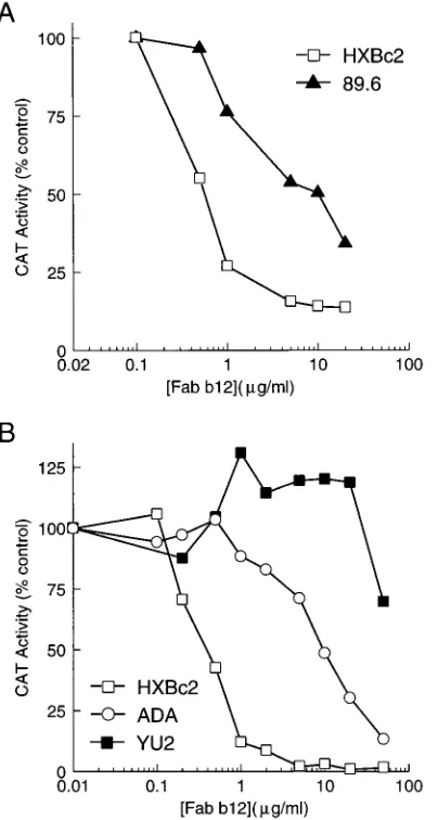
Fab b12 is an Fab fragment derived from a combinatorial phage library prepared from the bone marrow of an HIV-1-infected individual (10). Fab b12 recognizes a gp120 epitope overlapping the CD4-binding site and exhibits particularly po-tent neutralizing activity against a variety of HIV-1 isolates (7, 10a, 48). Figure 4 illustrates the results obtained with Fab b12 in the cell-free (Fig. 4A) and cocultivation (Fig. 4B) neutral-ization assays. Viruses containing the HXBc2 envelope glyco-proteins were neutralized by Fab b12 in the same dose range in
either assay, with an IC50of 0.4 to 0.6mg/ml. Viruses with the
89.6 or ADA envelope glycoproteins required 17 to 25 times more Fab b12 to achieve 50% neutralization. Viruses with the YU2 envelope glycoproteins demonstrated even greater resis-tance to inhibition by Fab b12.
To examine whether the decreased sensitivity of the viruses with the 89.6, ADA, and YU2 envelope glycoproteins to neu-tralization resulted from strain-specific variation in the Fab b12 epitope, the radiolabeled monomeric gp120 glycoproteins of the different strains were precipitated in the absence of deter-gent from transfected COS-1 cell supernatants (Fig. 5A). Com-pared with the recognition of the HXBc2 and 89.6 gp120 gly-coproteins, Fab b12 displayed decreased recognition of the ADA and YU2 gp120 glycoproteins, possibly explaining the decreased sensitivity of viruses with these envelope glycopro-teins to neutralization by Fab b12. However, Fab b12 precip-itated the monomeric 89.6 gp120 glycoprotein almost as effi-ciently as it precipitated the HXBc2 glycoprotein, suggesting that the relative resistance of viruses containing the 89.6 en-velope glycoprotein to Fab b12 neutralization is not due to a loss of the Fab b12 epitope.
Since the binding of Fab b12 to monomeric gp120 glyco-proteins did not completely explain the observed neutraliza-tion results, the ability of Fab b12 to bind the oligomeric envelope glycoprotein complex on the COS-1 cell surface was examined. As shown in Fig. 5B, Fab b12 binding to the differ-ent envelope glycoprotein complexes correlated well with the neutralization of viruses containing these envelope glycopro-teins. The HXBc2 envelope glycoproteins on the COS-1 cell surface were recognized most efficiently, with the 89.6 and
FIG. 3. Shedding of the gp120 glycoprotein induced by sCD4. COS-1 cells expressing the different HIV-1 envelope glycoproteins were incubated at 378C for 1 h, and the amount of the gp120 glycoprotein in the cell supernatants was determined. The amount of the gp120 glycoprotein in the supernatants is ex-pressed in arbitrary densitometric units. In the absence of sCD4, the amounts of gp120 glycoproteins of the different isolates found in the cell supernatant were equivalent (data not shown).
4416 SULLIVAN ET AL. J. VIROL.
on November 9, 2019 by guest
http://jvi.asm.org/
ADA envelope glycoproteins exhibiting intermediate levels of recognition by Fab b12. The recognition of the YU2 envelope glycoproteins on the cell surface was very inefficient, consistent with the observed resistance of viruses with the YU2 envelope glycoproteins to neutralization by Fab b12.
To determine whether the relative neutralization resistance of viruses with envelope glycoproteins from primary HIV-1 isolates applied to other, less potent antibodies directed against the CD4-binding site, experiments similar to those de-scribed above were performed with the F105 antibody. Like Fab b12, the F105 antibody recognizes a discontinuous gp120 epitope overlapping the CD4-binding site (47, 56). Figure 6 illustrates the neutralization by the F105 antibody of cell-free infection by viruses containing the HXBc2, MN, 89.6, ADA, and YU2 envelope glycoproteins. While the F105 antibody
neutralized viruses with the HXBc2 and MN envelope glyco-proteins, little or no neutralization was observed for viruses containing envelope glycoproteins from primary isolates (Fig. 6A). In fact, the addition of the F105 antibody to the viruses with the ADA and YU2 envelope glycoproteins resulted in an enhancement of infectivity, which was particularly pronounced for the viruses with YU2 envelope glycoproteins (Fig. 6). Treatment of recombinant HIV-1 isolates containing the am-photropic murine leukemia virus envelope glycoproteins with the identical preparation of the F105 antibody resulted in no positive or negative effect on virus entry (Fig. 6B).
[image:5.612.81.283.72.450.2]The recognition of the monomeric gp120 glycoproteins from the different strains by the F105 antibody is shown in Fig. 7A. The YU2 monomeric gp120 glycoprotein was recognized more efficiently than the HXBc2, ADA, and 89.6 gp120 glycopro-teins were. Since the viruses containing the YU2 envelope glycoproteins were neutralized the least efficiently by the F105 antibody, recognition of the monomeric gp120 glycoproteins by this antibody does not predict the efficiency of neutralization of viruses bearing the different envelope glycoproteins.
[image:5.612.338.535.72.452.2]FIG. 4. Neutralization of recombinant viruses by Fab b12. (A) Cell-free vi-ruses with the HXBc2 and 89.6 envelope glycoproteins were incubated with Fab b12 at 378C for 1 h prior to incubation with Molt 4 clone 8 cells. The CAT activity measured in the target cells following incubation of each virus with a given amount of Fab b12 as a percentage of the amount of CAT activity observed in the absence of the antibody fragment is shown. (B) Neutralization of recombinant viruses by Fab b12 in the cocultivation assay. Mitomycin-treated COS-1 cells expressing recombinant viruses were incubated with different amounts of Fab b12 prior to cocultivation with Molt 4 clone 8 target cells. The CAT activity measured in the Molt 4 clone 8 cells 10 to 14 days after cocultivation as a percentage of the amount of CAT activity observed in the absence of the Fab b12 antibody is shown.
FIG. 5. Recognition of the different gp120 glycoproteins by Fab b12. (A) Precipitation of monomeric gp120 glycoproteins from COS-1 supernatants by Fab b12, normalized for the amount of gp120 glycoprotein precipitated by an excess of a mixture of sera from patients with AIDS. The values are expressed as arbitrary units. (B) Recognition of different envelope glycoproteins expressed on the surface of COS-1 cells by Fab b12, normalized for the recognition of the cell surface glycoproteins by a mixture of sera from patients with AIDS.
on November 9, 2019 by guest
http://jvi.asm.org/
Incubation of the F105 antibody with COS-1 cells expressing the oligomeric envelope glycoprotein complexes revealed that antibody binding was significantly greater for the HXBc2 en-velope glycoproteins than for the enen-velope glycoproteins of the primary isolates (Fig. 7B). The binding of the F105 antibody to the cell surface envelope glycoproteins correlated better with neutralization sensitivity than did recognition of the mono-meric gp120 glycoproteins.
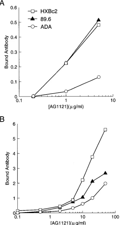
To examine whether some envelope glycoproteins derived from primary patient HIV-1 isolates also exhibit decreased sensitivity to neutralization by antibodies directed against the V3 loop, an antibody, AG1121, that recognized the monomeric gp120 glycoproteins of the HXBc2 and 89.6 strains equiva-lently (Fig. 8A) was selected. Figure 9 illustrates that viruses with the 89.6 envelope glycoproteins were less sensitive to neutralization by the AG1121 antibody than were viruses bear-ing the HXBc2 envelope glycoproteins. This roughly threefold difference in sensitivity to neutralization by the AG1121
anti-body was smaller than that observed for the antibodies di-rected against the CD4-binding site in the same assay. Despite equivalent recognition of the HXBc2 and 89.6 monomeric gp120 envelope glycoproteins, recognition of the HXBc2 oli-gomeric envelope glycoproteins on the cell surface was signif-icantly better than that of the 89.6 envelope glycoproteins (Fig. 8B). As was observed for the antibodies directed against the CD4-binding site, the recognition of the oligomeric cell surface envelope glycoproteins predicted the observed neutralization sensitivity better than did recognition of monomeric gp120 glycoproteins.
DISCUSSION
In this study, we used env complementation assays, which measure the efficiency of the early events in a single round of HIV-1 replication (30), to study the replicative properties and neutralization sensitivity associated with different HIV-1
[image:6.612.78.283.71.455.2]enve-FIG. 6. Neutralization of recombinant viruses by the F105 antibody. (A) Cell-free viruses with the HXBc2, MN, 89.6, or ADA envelope glycoproteins were incubated with the F105 antibody at 378C for 1 h prior to incubation with Molt 4 clone 8 cells. The CAT activity shown is described in the legend to Fig. 4. (B) Neutralization of recombinant viruses bearing the HXBc2, YU2, or SV-A-MLV envelope glycoproteins by the F105 antibody in the cell-free neutraliza-tion assay. The dose-response curve shown for HXBc2 in panel A has been replotted here for comparison.
FIG. 7. Recognition of the different gp120 glycoproteins by the F105 anti-body. (A) Precipitation of monomeric gp120 glycoproteins from COS-1 super-natants by the F105 antibody, normalized for the amount of gp120 glycoprotein precipitated by an excess of a mixture of sera from patients with AIDS. The values are expressed in arbitrary units. (B) Recognition of different envelope glycoproteins expressed on the surface of COS-1 cells by the F105 antibody, normalized for the recognition of the cell surface glycoproteins by a mixture of sera from patients with AIDS.
4418 SULLIVAN ET AL. J. VIROL.
on November 9, 2019 by guest
http://jvi.asm.org/
[image:6.612.331.536.80.449.2]lope glycoproteins. The envelope glycoproteins were derived from HIV-1 isolates that were passaged on T-cell lines or that represent primary, clinical strains. The use of the env comple-mentation system has several advantages for these studies. First, precise measurement of early-phase replicative ability can be made. Second, since all measurements are performed with a single cycle of infection initiated by cloned viral genes, the possibility of changes in viral phenotype during virus prop-agation is eliminated. Third, all of the envelope glycoproteins are synthesized in the identical cell type, in which effects of genotypic changes on envelope glycoprotein processing, sub-unit association, and cell surface expression can be readily determined. Thus, the observed phenotypic differences in rep-licative efficiency and sensitivity to neutralization can be attrib-uted to intrinsic structural features of the envelope glycopro-teins rather than to assay-specific variables.
Our results and those previously reported (30, 55) indicate that the envelope glycoproteins of extensively passaged viruses like HXBc2, derived from the HIV-1 LAI (IIIB) isolate, effi-ciently mediated virus entry into human PBMC and many
T-cell lines. The envelope glycoproteins of the primary HIV-1 isolates exhibited a spectrum of replicative abilities. The enve-lope glycoproteins of the 89.6 isolate, which is a highly cyto-pathic primary virus (14), mediated entry into human PBMC and Molt 4 clone 8 cells with an efficiency comparable to or better than that of the envelope glycoproteins derived from laboratory-adapted viruses. The ADA envelope glycoproteins exhibited lower efficiency in this assay, and the YU2 envelope glycoproteins, cloned directly from patient tissue without prior culture of the virus (35), exhibited the lowest intrinsic replica-tive ability. These results are consistent with those obtained with a larger panel of envelope glycoproteins derived from HIV-1 isolates passaged only on PBMC. In the cell-free complementation assay used here, the majority of these pri-mary virus-derived envelope glycoproteins exhibited efficien-cies lower than that observed for the HXBc2 envelope glyco-proteins (32a).
Even when the target cells were cocultivated with virus-producing COS-1 cells, the ADA and YU2 primary virus en-velope glycoproteins did not support HIV-1 entry into the SupT1, Jurkat, and C8166 established T-cell lines (data not shown). Thus, the relative efficiency of primary virus entry into most established T-cell lines is significantly lower than that in PBMC or Molt 4 clone 8 target cells.
The rank order of efficiency of complementation of virus
entry into PBMC or Molt 4 clone 8 cells (HXBc2589.6.
ADA.YU2) resembles that associated with the sensitivity to
neutralization by sCD4. This suggests that one significant fac-tor contributing to the decreased efficiency of PBMC entry for some of the primary HIV-1 isolates is a quantitative difference in the efficiency with which CD4 binding occurs. These differ-ences in CD4 binding are most apparent in assays in which CD4 binds to the oligomeric as opposed to the monomeric envelope glycoproteins, as has been suggested by previous studies (5, 32, 39, 41, 45, 58, 60).
At subinhibitory concentrations, sCD4 mediated the en-hancement of infection for recombinant viruses with envelope glycoproteins derived from some primary HIV-1 isolates.
En-hancement of HIV-2 and SIVagminfection by sCD4 has been
[image:7.612.343.528.71.243.2]previously observed (2, 13), but enhancement of HIV-1 infec-tion has not been reported. The enhancement effect on YU2 is modest (threefold), and it is possible that other neutralization
[image:7.612.81.278.72.440.2]FIG. 8. Recognition of the different gp120 glycoproteins by the AG1121 antibody. (A) Precipitation of monomeric gp120 glycoproteins from COS-1 su-pernatants by the AG1121 antibody, normalized for the amount of gp120 glyco-protein precipitated by an excess of a mixture of sera from patients with AIDS. (B) Recognition of different envelope glycoproteins expressed on the surface of COS-1 cells by the AG1121 antibody, normalized for the recognition of the cell surface glycoproteins by a mixture of sera from patients with AIDS.
FIG. 9. Neutralization of recombinant viruses by the AG1121 antibodies. Cell-free viruses with the HXBc2 and 89.6 envelope glycoproteins were incu-bated with the AG1121 antibody at 378C for 1 h prior to incubation with Molt 4 clone 8 cells. The CAT activity shown is described in the legend to Fig. 4.
on November 9, 2019 by guest
http://jvi.asm.org/
assay systems lack the sensitivity to detect sCD4-mediated en-hancement of HIV-1. sCD4-mediated enen-hancement was less apparent for viruses with envelope glycoproteins derived from more replication-competent HIV-1 isolates, including the 89.6 primary isolate and the laboratory-passaged isolates. Enhance-ment is probably more apparent when competing reactions like gp120 shedding are less efficient, as is the case for HIV-2,
SIVagm(2, 13), and some primary HIV-1 isolates (Fig. 3) (39,
41, 45, 60). Enhancement by sCD4 may reflect the proposed role of receptor binding in triggering fusion-related conforma-tional changes in the primate immunodeficiency virus envelope glycoproteins (2, 49). It has been suggested by others that CD4 binding to gp120 establishes a metastable intermediate of fu-sion activation (23a).
Enhancement of the infection of viruses with some primary envelope glycoproteins by a neutralizing antibody directed against the CD4-binding site of gp120 was observed. This effect was specific for virions with HIV-1 envelope glycoproteins, implying that antibody binding to the viral envelope glycopro-teins is a prerequisite for enhancement. A low affinity of anti-body binding to some HIV-1 envelope glycoproteins may result in lower occupancy of the four available epitopes within the tetrameric envelope glycoprotein spike, allowing the overlap-ping CD4-binding sites on the unoccupied subunits of the tetramer to bind receptor. It is not yet known whether the CD4-binding site antibody elicits conformational changes in the envelope glycoproteins similar to those induced by CD4 or whether another mechanism accounts for enhancement.
Despite differences in replicative ability, all of the primary virus envelope glycoproteins demonstrated less sensitivity to neutralizing antibodies directed against the CD4-binding site or the V3 loop, relative to that seen for the HXBc2 envelope glycoproteins. A few instances of neutralization resistance re-sulted from natural sequence variation within or near the epitope, which could be detected by examination of the binding of the antibody to the monomeric gp120 glycoprotein. How-ever, the resistance of the primary virus envelope glycoproteins to neutralization correlated best with the ability of the antibody to bind the oligomeric envelope glycoproteins. These results suggest that particular structural features of primary virus en-velope glycoproteins, operative only in the context of the as-sembled, oligomeric envelope glycoprotein complex, decrease the binding ability of neutralizing antibodies. In some cases, these structural features, probably fortuitously, also occlude the binding site on the gp120 glycoprotein for CD4 and de-crease the replication efficiency of the virus. Compared with viruses containing the HXBc2 envelope glycoproteins, viruses with 89.6 envelope glycoproteins exhibited significant resis-tance to CD4-binding site-directed neutralizing antibodies, only minimal resistance to sCD4, and efficient replication ca-pacity. This suggests that masking of the epitopes for the CD4-binding site-directed antibodies can occur without necessarily occluding the binding site for CD4 to the same extent. On the other hand, the more replication-competent configuration of the 89.6 envelope glycoproteins may render viruses with these envelope glycoproteins sensitive to neutralizing antibodies other than those directed against the CD4-binding site. For example, while the viruses with the 89.6 envelope glycoproteins were less sensitive to neutralization by the V3 loop-directed antibody than were viruses with the HXBc2 envelope glyco-proteins, the V3 loop on the viruses with the 89.6 envelope glycoproteins was still accessible. It has recently been sug-gested that the V3 loop of some primary HIV-1 isolates is very poorly exposed to neutralizing antibodies (9). It is likely that different primary virus envelope glycoproteins will have evolved slightly different configurations depending on the
gen-eration and titer of particular subgroups of neutralizing anti-bodies within the individual host. The conformations associ-ated with optimal resistance to the complete spectrum of naturally occurring neutralizing antibodies may necessitate some decrease in the efficiency of CD4 binding and virus entry. Although our sample size is small, among viruses with en-velope glycoproteins derived from primary HIV-1 isolates, sen-sitivity to neutralization by CD4-binding site-directed antibod-ies correlated with replicative ability. The dual selective forces of intrinsic replicative ability and neutralization susceptibility may contribute to the temporal pattern described for the in vivo appearance of HIV-1 phenotypes (51). In immunocom-petent individuals, the presence of high titers of neutralizing antibodies favors viruses that are relatively resistant to such neutralization, even if intrinsic viral replication rates must be decreased to achieve resistance. Later in infection, when im-mune system compromise results in a decreased titer of neu-tralizing antibodies, the appearance of viruses with improved intrinsic replication rates will be favored, since the associated increase in neutralization sensitivity is less of a liability. In this model, the emergence of rapid, high viruses is a consequence of a deteriorating immune response rather than a prerequisite for pathogenesis.
A second feature of the envelope glycoproteins of many primary HIV-1 isolates that has been described previously (39, 43, 60, 61) and that we have confirmed is the higher concen-tration of gp120 glycoprotein associated with the virions. This property presumably reflects a more stable association be-tween the gp120 and gp41 glycoproteins and results in a higher density of envelope glycoprotein spikes on primary HIV-1 viri-ons. In vivo, higher spike density may help to compensate for lower CD4-binding ability of primary virus envelope glycopro-teins and may facilitate entry into target cells, like macro-phages, with low receptor density. Furthermore, the higher density of envelope glycoproteins on viruses bearing the 89.6, ADA, or YU2 envelopes may contribute to the relative neu-tralization resistance of these viruses. Layne et al. (34) showed that HIV-1 infectivity is blocked at lower concentrations of sCD4 in virion preparations containing fewer gp120 molecules per virion.
Further characterization of the envelope glycoproteins of primary HIV-1 viruses will be important in determining the availability of neutralization targets on these clinically relevant isolates.
ACKNOWLEDGMENTS
We thank Beatrice Hahn, George Shaw, Ronald Collman, and Lee Ratner for molecular clones and cell lines. We thank Jan Welch for manuscript preparation and Amy Emmert for artwork.
This work was supported by National Institutes of Health grants AI31783, P30CA06516 (Cancer Center grant to Dana-Farber Cancer Institute), and P30 AI28691 (Center for AIDS Research grant to Dana-Farber Cancer Institute). This work was also supported by gifts from the late William McCarty-Cooper and from the G. Harold and Leila Y. Mathers Charitable Foundation.
REFERENCES
1. Albert, J., B. Abrahamsson, K. Nagy, E. Aurelius, H. Gaines, G. Nystrom,
and E. M. Fenyo¨.1990. Rapid development of isolate-specific neutralizing antibodies after primary HIV-1 infection and consequent emergence of virus variants which resist neutralization by autologous sera. AIDS 4:107–112. 2. Allan, J., J. Strauss, and D. Buck. 1990. Enhancement of SIV infection with
soluble receptor molecules. Science 247:1084–1088.
3. Arendrup, M., C. Nielsen, J.-E. S. Hansen, C. Pedersen, L. Mathiesen, and
J. O. Nielsen.1992. Autologous HIV-1 neutralizing antibodies: emergence of neutralization-resistant escape virus and subsequent development of escape virus neutralizing antibodies. J. Acquired Immune Defic. Syndr. 5:303–307. 4. Arendrup, M., A. So¨nnerborg, B. Svennerholm, L. Åkerblom, C. Nielsen, H.
4420 SULLIVAN ET AL. J. VIROL.
on November 9, 2019 by guest
http://jvi.asm.org/
Clausen, S. Olofsson, J. O. Nielsen, and J.-E. S. Hansen.1993. Neutralizing antibody response during human immunodeficiency virus type 1 infection: type and group specificity and viral escape. J. Gen. Virol. 74:855–863. 5. Ashkenazi, A., D. H. Smith, S. A. Marsters, L. Riddle, T. J. Gregory, D. D.
Ho, and D. J. Capon.1991. Resistance of primary isolates of human immu-nodeficiency virus type 1 to soluble CD4 is independent of CD4-gp120 binding affinity. Proc. Natl. Acad. Sci. USA 88:7056–7060.
6. Åsjo¨, B., L. Mo¨rfeldt-Manson, L. J. Albert, G. Biberfeld, A. Karlson, K. Lidman, and E. M. Fenyo¨.1986. Replicative capacity of human immunode-ficiency virus from patients with varying severity of HIV infection. Lancet
ii:660–662.
7. Barbas, C. F., III, D. Hu, N. Dunlop, L. Sawyer, D. Cabada, R. M. Henry,
P. L. Nara, and D. R. Burton.1994. In vitro evolution of a neutralizing human antibody to human immunodeficiency virus type 1 to enhance affinity and broaden strain cross-reactivity. Proc. Natl. Acad. Sci. USA 91:3809– 3813.
8. Barre´-Sinoussi, F., J. C. Chermann, F. Rey, M. T. Nuge`yre, S. Chamaret, J.
Gruest, C. Dauget, C. Axler-Bin, F. Vezinet-Brun, C. Rouzioux, W. Rozen-baum, and L. Montagnier.1983. Isolation of a T-lymphocyte retrovirus from a patient at risk for acquired immunodeficiency syndrome (AIDS). Science
220:868–871.
9. Bou-Habib, D. C., G. Rodriquez, T. Oravecz, P. W. Berman, P. Lusso, and
M. A. Norcross.1994. Cryptic nature of envelope V3 region epitopes pro-tects primary monocytotropic human immunodeficiency virus type 1 from antibody neutralization. J. Virol. 68:6006–6013.
10. Burton, D. R., C. F. Barbas III, M. A. A. Persson, S. Koenig, R. M. Chanock,
and R. A. Lerner.1991. A large array of human monoclonal antibodies to HIV-1 from combinatorial libraries of asymptomatic seropositive individu-als. Proc. Natl. Acad. Sci. USA 88:10134–10137.
10a.Burton, D. R., J. Pyati, R. Koduri, S. J. Sharp, G. B. Thornton, P. W. H. I.
Parren, L. S. W. Sawyer, R. M. Hendry, N. Dunlop, P. L. Nara, M. Lamac-chia, I. Garratty, E. R. Stiehm, Y. J. Bryson, Y. Cao, J. P. Moore, D. D. Ho, and C. F. Barbas III.1994. Efficient neutralization of primary isolates of HIV-1 by a recombinant human monoclonal antibody. Science 266:1024– 1027.
11. Cheng-Mayer, C., M. Quiroga, J. W. Tung, D. Dina, and J. A. Levy. 1990. Viral determinants of human immunodeficiency virus type 1 T-cell or mac-rophage tropism, cytopathogenicity, and CD4 antigen modulation. J. Virol.
64:4390–4398.
12. Cheng-Mayer, C., D. Seto, M. Tateno, and J. A. Levy. 1988. Biologic features of HIV-1 that correlate with virulence in the host. Science 240:80–82. 13. Clapham, P. R., A. McKnight, and R. A. Weiss. 1992. Human
immunodefi-ciency virus type 2 infection and fusion of CD4-negative human cell lines: induction and enhancement by soluble CD4. J. Virol. 66:3531–3537. 14. Collman, R., J. W. Balliet, S. A. Gregory, H. Friedman, D. L. Kolson, N.
Nathanson, and A. Srinivasan.1992. An infectious molecular clone of an unusual macrophage-tropic and highly cytopathic strain of human immuno-deficiency virus type I. J. Virol. 66:7517–7521.
15. Connor, R. I., H. Mohri, Y. Cao, and D. D. Ho. 1993. Increased viral burden and cytopathicity correlate temporally with CD41T-lymphocyte decline and clinical progression in human immunodeficiency virus type 1-infected indi-viduals. J. Virol. 67:1772–1777.
16. Coombs, R. W., A. C. Collier, J.-P. Allain, B. Nikora, M. Leuther, G. F.
Gjerset, and L. Corey.1989. Plasma viremia in human immunodeficiency virus infection. N. Engl. J. Med. 321:1626–1631.
17. Cullen, B. R. 1989. Use of eukaryotic expression technology in the functional analysis of cloned genes. Methods Enzymol. 152:64–73.
18. Daar, E., X. L. Li, T. Moudgil, and D. D. Ho. 1990. High concentrations of recombinant soluble CD4 are required to neutralize primary HIV-1 isolates. Proc. Natl. Acad. Sci. USA 87:6574–6578.
19. Fauci, A., A. Macher, D. Longo, H. C. Lane, A. Rook, H. Masur, and E.
Gelmann.1984. Acquired immunodeficiency syndrome: epidemiologic, clin-ical, immunologic, and therapeutic considerations. Ann. Intern. Med. 100: 92–106.
20. Fenyo¨, E. M., J. Albert, and B. Åsjo¨.1989. Replicative capacity, cytopathic effect and cell tropism of HIV. AIDS 3(Suppl. 1):S5–S12.
21. Fenyo¨, E. M., L. Mo¨rfeldt-Manson, F. Chiodi, B. Lind, A. Von Gegerfelt, J. Albert, E. Olausson, and B. Åsjo¨.1988. Distinct replicative and cytopathic characteristics of human immunodeficiency virus isolates. J. Virol. 62:4414– 4419.
22. Fernandez-Larsson, R., K. K. Srivastava, S. Lu, and H. L. Robinson. 1992. Replication of patient isolates of human immunodeficiency virus type 1 in T cells: a spectrum of rates and efficiencies of entry. Proc. Natl. Acad. Sci. USA
89:2223–2226.
23. Fisher, A. G., E. Collalti, L. Ratner, R. C. Gallo, and F. Wong-Staal. 1985. A molecular clone of HTLV-III with biologic activity. Nature (London)
316:262–265.
23a.Fu, Y. K., T. K. Hart, Z. L. Jonak, and P. J. Bugelski. 1993. Physicochemical dissociation of CD4-mediated syncytium formation and shedding of human immunodeficiency virus type 1 gp120. J. Virol. 67:3818–3825.
24. Gabuzda, D. H., H. Li, K. Lawrence, B. S. Vadir, K. Crawford, and E.
Langhoff.1994. Essential role of vif in establishing productive HIV-1
infec-tion in peripheral blood T lymphocytes and monocyte-macrophages. J. Ac-quired Immune Defic. Syndr. 7:908–915.
25. Gallo, R., S. Z. Salahuddin, M. Popovic, G. M. Shearer, M. Kaplan, B. F.
Haynes, T. J. Palker, R. Redfield, J. Oleske, B. Safai, G. White, and P. Foster.1984. Frequent detection and isolation of cytopathic retroviruses (HTLV-III) from patients with AIDS and at risk for AIDS. Science 224: 500–503.
26. Gendelman, H. E., L. M. Baca, H. Husayni, J. A. Turpin, D. Skillman, D. C.
Kalter, J. M. Orenstein, D. L. Hoover, and M. S. Meltzer.1990. Macro-phage-HIV interaction: viral isolation and target cell tropism. AIDS 4:221– 228.
27. Gendelman, H. E., J. M. Orenstein, M. A. Martin, C. Ferrva, R. Mitra, T.
Phipps, L. A. Wahl, C. H. Lane, A. S. Fauci, and D. S. Burke.1988. Efficient isolation and propagation of human immunodeficiency virus on recombinant colony-stimulating factor 1-treated monocytes. J. Exp. Med. 167:1428–1441. 28. Gomatos, P. J., N. M. Stamatos, H. E. Gendelman, A. Fowler, D. L. Hoover,
D. C. Kalter, D. S. Burke, E. C. Tramont, and S. Meltzer.1990. Relative inefficiency of soluble recombinant CD4 for inhibition of infection of mono-cyte-tropic HIV in monocytes and T cells. J. Immunol. 144:4183–4188. 29. Hart, T., R. Kirsch, H. Ellens, R. Sweet, D. Lambert, S. Petteway, J. Leary,
and P. Bugelski.1991. Binding of soluble CD4 proteins to HIV-1 and infected cells induces release of envelope glycoprotein gp120. Proc. Natl. Acad. Sci. USA 88:2189–2193.
30. Helseth, E., M. Kowalski, D. Gabuzda, U. Olshevsky, W. Haseltine, and J.
Sodroski.1990. Rapid complementation assays measuring replicative poten-tial of human immunodeficiency virus type 1 envelope glycoprotein mutants. J. Virol. 64:2416–2420.
31. Ho, D. D., T. Moudgil, and M. Alam. 1989. Quantitation of human immu-nodeficiency virus type 1 in the blood of infected persons. N. Engl. J. Med.
321:1621–1625.
32. Kabat, D., S. L. Kozak, K. Wehrly, and B. Chesebro. 1994. Differences in CD4 dependence for infectivity of laboratory-adapted and primary patient isolates of human immunodeficiency virus type 1. J. Virol. 68:2570–2577. 32a.Karlsson, G., F. Gao, B. Hahn, and J. Sodroski. Unpublished observations. 32b.Landau, N. R., K. A. Page, and D. R. Littman. 1991. Pseudotyping with human T-cell leukemia virus type I broadens the human immunodeficiency virus host range. J. Virol. 65:162–169.
33. Lane, M. C., J. Depper, W. Greene, G. Whalen, T. Waldemann, and A. Fauci. 1985. Qualitative analysis of immune functions in patients with the acquired immunodeficiency syndrome. N. Engl. J. Med. 313:79–84.
34. Layne, S. P., M. J. Merges, M. Dembo, J. L. Spouge, S. R. Conley, J. P.
Moore, J. L. Raina, H. Renz, H. R. Gelderblom, and P. L. Nara.1992. Factors underlying spontaneous inactivation and susceptibility to neutraliza-tion of human immunodeficiency virus. Virology 189:695–714.
35. Li, Y., J. C. Kappas, J. A. Conway, R. W. Price, G. M. Shaw, and B. H. Hahn. 1991. Molecular characterization of human immunodeficiency virus type 1 cloned directly from uncultured human brain tissue: identification of repli-cation-competent-defective viral genomes. J. Virol. 65:3973–3985. 36. Mascola, J. R., J. Louwagie, F. E. McCutchan, C. L. Fischer, P. A. Hegerich,
K. F. Wagner, A. K. Fowler, J. G. McNeil, and D. S. Burke.1994. Two antigenically distinct subtypes of human immunodeficiency virus type 1: viral genotype predicts neutralization serotype. J. Infect. Dis. 169:48–54. 37. Montefiori, D. C., J. Zhou, B. Barnes, D. Lake, E. M. Hersh, Y. Masuho, and
L. B. Lefkowitz, Jr.1991. Homotypic antibody responses to fresh clinical isolates of human immunodeficiency virus. Virology 182:635–643. 38. Moore, J., J. McKeating, R. Weiss, and Q. Sattentau. 1990. Dissociation of
gp120 from HIV-1 virions induced by soluble CD4. Science 250:1139–1142. 39. Moore, J. P., L. C. Burkly, R. I. Connor, Y. Cao, R. Tizard, D. D. Ho, and
R. A. Fisher.1993. Adaptation of two primary human immunodeficiency virus type 1 isolates to growth in transformed T-cell lines correlates with alterations in the responses of their envelope glycoproteins to soluble CD4. AIDS Res. Hum. Retroviruses 9:529–539.
40. Moore, J. P., and D. D. Ho. 1993. Antibodies to discontinuous or conforma-tionally sensitive epitopes on the gp120 glycoprotein of human immunode-ficiency virus type 1 are highly prevalent in sera of infected humans. J. Virol.
67:863–975.
41. Moore, J. P., J. A. McKeating, Y. Huang, A. Ashenazi, and D. D. Ho. 1992. Virions of primary human immunodeficiency virus type 1 isolates resistant to soluble CD4 (sCD4) neutralization differ in sCD4 binding and glycoprotein gp120 retention from sCD4-sensitive isolates. J. Virol. 66:235–243. 42. O’Brien, W., Y. Koyanagi, A. Namazie, J.-Q. Zho, A. Diagne, K. Idler, J.
Zack, and I. S. Y. Chen.1990. HIV-1 tropism for mononuclear phagocytes can be determined by regions of gp120 outside the CD4-binding domain. Nature (London) 348:69–73.
43. O’Brien, W. A., S.-H. Mao, Y. Cao, and J. P. Moore. 1994. Macrophage-tropic and T-cell-line-adapted chimeric strains of human immunodeficiency virus type 1 differ in their susceptibilities to neutralization by soluble CD4 at different temperatures. J. Virol. 68:5264–5269.
44. Olshevsky, U., E. Helseth, C. Furman, J. Li, W. A. Haseltine, and J.
So-droski.1990. Identification of individual human immunodeficiency virus type 1 gp120 amino acids important for CD4 receptor binding. J. Virol. 64:5701– 5707.
on November 9, 2019 by guest
http://jvi.asm.org/
45. Orloff, S. L., M. S. Kennedy, A. A. Belperron, P. J. Maddon, and J. S.
McDougal.1993. Two mechanisms of soluble CD4 (sCD4)-mediated inhibi-tion of human immunodeficiency virus type 1 (HIV-1) infectivity and their relation to primary HIV-1 isolates with reduced sensitivity to sCD4. J. Virol.
67:1461–1471.
46. Pan, L. Z., A. Werner, and J. A. Levy. 1993. Detection of plasma viremia in human immunodeficiency virus-infected individuals at all clinical stages. J. Clin. Microbiol. 31:283–288.
47. Posner, M., T. Hideshima, T. Cannon, M. Mukherjee, K. Mayer, and R.
Bryn.1991. An IgG human monoclonal antibody that reacts with HIV-1 gp120, inhibits virus binding to cells, and neutralizes infection. J. Immunol.
146:4325–4332.
48. Roben, P., J. P. Moore, M. Thali, J. Sodroski, C. F. Barbas III, and D.
Burton.1994. Recognition properties of a panel of human recombinant Fab fragments to the CD4 binding site of gp120 that show differing abilities to neutralize human immunodeficiency virus type 1. J. Virol. 68:4821–4828. 49. Sattentau, Q. J., and J. P. Moore. 1991. Conformational changes induced in
the human immunodeficiency virus envelope glycoprotein by soluble CD4 binding. J. Exp. Med. 174:407–415.
50. Sawyer, L. S. W., M. T. Wrin, L. Crawford-Miksza, B. Potts, Y. Wu, P. A.
Weber, R. D. Alfonso, and C. V. Hanson.1994. Neutralization sensitivity of human immunodeficiency virus type 1 is determined in part by the cell in which the virus is propagated. J. Virol. 68:1342–1349.
51. Schuitemaker, H., M. Koot, N. A. Koostra, M. W. Dercksen, R. E. Y. de
Goede, R. P. van Steenwijk, J. M. A. Lange, J. K. M. Eeftink-Schattenkerk, F. Miedema, and M. Tersmette.1992. Biological phenotype of human im-munodeficiency virus type 1 clones at different stages of infection: progres-sion of disease is associated with a shift from monocytotropic to T-cell-tropic virus populations. J. Virol. 65:356–363.
52. Shaw, G. M., B. H. Hahn, S. K. Arya, J. E. Groopman, R. C. Gallo, and F.
Wong-Staal. 1984. Molecular characterization of human T-cell leukemia (lymphotropic) virus type III in the acquired immune deficiency syndrome. Science 226:1165–1171.
53. Tersmette, M., R. E. Y. de Goede, B. J. M. Al, I. N. Winkel, R. A. Gruters,
H. T. Cuypers, H. G. Huisman, and F. Miedema.1988. Differential syncyti-um-inducing capacity of human immunodeficiency virus isolates: frequent detection of syncytium-inducing isolates in patients with acquired immuno-deficiency syndrome (AIDS) and AIDS-related complex. J. Virol. 62:2026– 2032.
54. Thali, M., J. P. Moore, C. Furman, M. Charles, D. D. Ho, J. Robinson, and
J. Sodroski.1993. Characterization of conserved human immunodeficiency virus type 1 gp120 neutralization epitopes exposed upon gp120-CD4 binding. J. Virol. 67:3978–3988.
55. Thali, M., U. Olshevsky, C. Furman, D. Gabuzda, J. Li, and J. Sodroski. 1991. Effects of changes in gp120-CD4 binding affinity on human immuno-deficiency virus type 1 envelope glycoprotein function and soluble CD4 sensitivity. J. Virol. 65:5007–5012.
56. Thali, M., U. Olshevsky, C. Furman, D. Gabuzda, M. Posner, and J.
So-droski.1991. Characterization of a discontinuous human immunodeficiency virus type 1 gp120 epitope recognized by a broadly reactive neutralizing human monoclonal antibody. J. Virol. 65:6188–6193.
57. Tremblay, M., and M. A. Wainberg. 1990. Neutralization of multiple HIV-1 isolates from a single subject by autologous sequential sera. J. Infect. Dis.
162:735–737.
58. Turner, S., R. Tizard, J. DeMarinis, R. B. Pepinsky, J. Zullo, R. Schooley,
and R. Fisher.1992. Resistance of primary isolates of human immunodefi-ciency virus type 1 to neutralization by soluble CD4 is not due to a lower affinity with the viral envelope glycoprotein gp120. Proc. Natl. Acad. Sci. USA 89:1335–1339.
59. Westervelt, P., H. E. Gendelman, and L. Ratner. 1991. Identification of a determinant within the human immunodeficiency virus surface envelope glycoprotein critical for productive infection of primary monocytes. Proc. Natl. Acad. Sci. USA 88:3097–3101.
60. Willey, R. L., M. A. Martin, and K. W. C. Peden. 1994. Increase in soluble CD4 binding to and CD4-induced dissociation of gp120 from virions corre-lates with infectivity of human immunodeficiency virus type 1. J. Virol.
68:1029–1039.
61. Willey, R. L., T. S. Theodore, and M. A. Martin. 1994. Amino acid substi-tutions in the human immunodeficiency virus type 1 gp120 V3 loop that change viral tropism also alter physical and functional properties of the virion envelope. J. Virol. 68:4409–4419.
62. Wrin, T., L. Crawford, L. Sawyer, P. Weber, H. W. Sheppard, and C. V.
Hanson.1994. Neutralizing antibody responses to autologous and heterolo-gous isolates of human immunodeficiency virus. J. Acquired Immune Defic. Syndr. 7:211–219.
63. Wyatt, R., N. Sullivan, M. Thali, H. Repke, D. Ho, J. Robinson, M. Posner,
and J. Sodroski.1993. Functional and immunologic characterization of hu-man immunodeficiency virus type 1 envelope glycoproteins containing dele-tions of the major variable regions. J. Virol. 67:4557–4565.
4422 SULLIVAN ET AL. J. VIROL.